Báo cáo sinh học: "Phylogenetic evidence for the distinction of Saaremaa and Dobrava hantaviruses" pot
Bạn đang xem bản rút gọn của tài liệu. Xem và tải ngay bản đầy đủ của tài liệu tại đây (234.65 KB, 6 trang )
BioMed Central
Page 1 of 6
(page number not for citation purposes)
Virology Journal
Open Access
Research
Phylogenetic evidence for the distinction of Saaremaa and Dobrava
hantaviruses
Tarja Sironen, Antti Vaheri and Alexander Plyusnin*
Address: Department of Virology, Haartman Institute, Haartmaninkatu 3, FIN-00014 University of Helsinki, Finland
Email: Tarja Sironen - ; Antti Vaheri - ; Alexander Plyusnin* -
* Corresponding author
Abstract
Dobrava virus (DOBV) and Saaremaa virus (SAAV) are two closely related hantaviruses carried by
different rodent species. The distinction of these two viruses has been a matter of debate. While
the phylogenies based on the viral M segment sequences were repeatedly showing monophyly of
SAAV strains, some trees based on the S segment sequences were not, thus causing questions on
the demarcation between these two viruses. In order to clarify this issue, the current collection of
the virus S segment sequences was subjected to extensive phylogenetic analysis using maximum
likelihood, maximum parsimony and distant matrix methods. In all inferred phylogenies, the SAAV
sequences were monophyletic and separated from DOBV sequences, thus supporting the view that
SAAV and DOBV are distinct hantavirus species. Since collection of the S segment sequences used
in this study "obeyed" the molecular clock, calculations of the split of DOBV and SAAV were now
repeated resulting in an estimation of 3.0–3.7 MYA that is very close to the values obtained earlier.
Background
Hantaviruses (genus Hantavirus, family Bunyaviridae) are
enveloped viruses with a segmented, single-stranded RNA
genome of negative polarity [1]. The large (L) segment
encodes the viral RNA polymerase, the medium (M) seg-
ment the two surface glycoproteins, and the small (S) seg-
ment the nucleocapsid protein (N). Hantaviruses cause
two human zoonoses, hemorrhagic fever with renal syn-
drome (HFRS) in Eurasia and hantavirus pulmonary syn-
drome (HPS) in the Americas [reviewed in [2]]. DOBV is
carried by yellow-necked mouse (Apodemus flavicollis) and
is associated with severe HFRS in the Balkans (Slovenia,
Albania and Greece). SAAV is carried by striped field
mouse (A. agrarius) [3]. So far, the virus has been found in
Estonia, the European part of Russia, Slovakia, Slovenia,
Hungary, Denmark and Germany [2].
SAAV was initially called an A. agrarius-carried variant of
Dobrava virus [3], but the accumulating data suggest that
the virus should be regarded as a distinct hantavirus spe-
cies. It is carried by a specific rodent host [3], there is a
four-fold difference in two-way cross-neutralization tests
[4], and the coexistence of SAAV and DOBV in the same
geographic region [5,6] indicates reproductive isolation.
They also exhibit 6.1–6.3% difference in the glycoprotein
precursor amino acid sequences. This level is a fraction
lower than the officially accepted 7% cut-off value [1]. It
should be mentioned that some of the officially approved,
distinct hantavirus species show lower than 7% diversity
in their N or GnGc-sequences: Sin Nombre and New York
viruses, Topografov and Khabarovsk viruses, Rio Mamore
and Laguna Negra viruses, and Blood Land Lake and Pros-
pect Hill viruses [7].
Published: 08 December 2005
Virology Journal 2005, 2:90 doi:10.1186/1743-422X-2-90
Received: 27 June 2005
Accepted: 08 December 2005
This article is available from: />© 2005 Sironen et al; licensee BioMed Central Ltd.
This is an Open Access article distributed under the terms of the Creative Commons Attribution License ( />),
which permits unrestricted use, distribution, and reproduction in any medium, provided the original work is properly cited.
Virology Journal 2005, 2:90 />Page 2 of 6
(page number not for citation purposes)
SAAV and DOBV also exhibit only 3% diversity on their N
protein sequences. This unusually low level of diversity is
most probably a reflection of host switching in their evo-
lution [8,9]; this event seems to be historically recent
(2.7–3.4 MYA) and these two viruses are still diverging
[8]. There is another important feature differentiating
DOBV and SAAV, and that is the apparently different
pathogenicity in humans: while DOBV causes severe
Table 1: Sequences used in the analysis
Strain Accession number
Saaremaa virus (SAAV) Saaremaa/160 V AJ009773
90Aa/97 AJ009775
Lolland/Aa1403/2000 AJ616854
Kurkino/44Aa/98 AJ131672
Kurkino/53Aa/98 AJ131673
East Slovakia/856/Aa AJ269549
East Slovakia/862/Aa AJ269550
Dobrava virus (DOBV) Slovenia L41916
East Slovakia/400Af/98 AY168576
Ano-Poroia/9Af/1999 AJ410615
Ano-Poroia/13Af/99 AJ410619
As-1/Goryachiy Klyuch-2000 AF442622
P-s1223/Krasnodar-2000 AF442623
Seoul virus (SEOV) Gou3 AB027522
L99 AF288299
Z37 AF187082
SR11 M34881
Hantaan virus (HTNV) Ah09 AF285264
84Fli AY017064
76–118 M14626
Lr1 AF288294
Andes virus (ANDV) AH-1 AF324902
Topografov virus (TOPV) Ls136V AJ011646
Sin Nombre virus (SNV) NM H10 L25784
El Moro Canyon virus (ELMCV) RM-97 U11427
Puumala virus (PUUV) Sotkamo X61035
Tula virus (TULV) Moravia/5302v/95 Z69991
Table 2: Bootstrap and puzzle support values for DOBV and SAAVclades in phylogenetic trees calculated using different methods.
method outgroup support for: DOBV support for: SAAV
maximum likelihood SEOV 100 70
maximum likelihood collection* 100 49
maximum likelihood no outgroup 100 100
maximum parsimony SEOV 100 75
maximum parsimony collection* 100 75
distance matrix: Neighbor-joining SEOV 100 84
distance matrix: Neighbor-joining collection* 100 91
distance matrix: Fitch-Margoliash SEOV 79 58
distance matrix: Fitch-Margoliash collection* 100 79
distance matrix: Fitch-Margoliash no outgroup 100 99
TreePuzzle** SEOV 99 87
TreePuzzle collection* 99 75
*A collection of hantavirus sequences including SNV, ANDV, ELMCV, TULV, TOPV, PUUV, SEOV strains SR11 and Gou3, HTNV strains 76–118
and 84Fli **Tamura-Nei was used as the nucleotide (nt) substitution model in TreePuzzle, as suggested by Modeltest.
Virology Journal 2005, 2:90 />Page 3 of 6
(page number not for citation purposes)
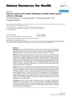
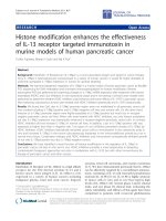
Phylogenetic tree created with TreePuzzle for a smaller data setFigure 1
Phylogenetic tree created with TreePuzzle for a smaller data set. The tree is based on the nt 37–1232 of the S segment
sequences.
0.1
ELMCV
SNV
PUUV
EastSlovakia856
EastSlovakia862
Kurkino44
Kurkino53
Saaremaa160V
Saaremaa90
ESlovakia400
Slovenia
Gou3
L99
Z37
Sr11
AH09
84Fli
76-118
LR1
TULV
99
99
97
100
100
100
99
98
98
98
62
92
HTNV
SEOV
DOBV
SAAV
Virology Journal 2005, 2:90 />Page 4 of 6
(page number not for citation purposes)
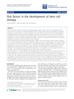
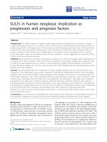
Phylogenetic tree created with TreePuzzle for a more representative data setFigure 2
Phylogenetic tree created with TreePuzzle for a more representative data set. The tree is based on the nt 37–1232 of the S
segment sequences. Two SAAV sequences that are placed differently on the trees shown on Fig. 1 and Fig 2 are underlined.
0.1
97
94
98
97
100
96
99
100
79
90
71
93
100
98
95
87
ELMCV
SNV
PUUV
TULV
Kurkino44
Kurkino53
Slovenia
ESlovakia400
ESlov856
ESlov862
Gou3
L99
Z37
Sr11
AH09
84Fli
76-118
LR1
Saaremaa160V
Saaremaa90
Lolland1403
AnoPoroja9
AnoPoroja13
GorKlyuch1
Krasnodar1223
HTNV
SEOV
DOBV
SAAV
Virology Journal 2005, 2:90 />Page 5 of 6
(page number not for citation purposes)
HFRS in humans, SAAV causes a milder form of the dis-
ease, similar to nephropathia epidemica [2]. This differ-
ence is also reflected in different pathogenicity in suckling
mice: DOBV is lethal to suckling mice, while SAAV is not
[10].
The phylogenetic distinction of SAAV and DOBV was
recently a matter of debate [11,12]. While the phylogenies
based on the M segment/GnGc protein sequences were
repeatedly showing monophyly of SAAV strains, some
trees based on the S segment/N protein sequences were
not [[11,13], and our unpublished observations], thus
causing questions on the demarcation between these two
viruses. In order to clarify this issue, the current collection
of DOBV and SAAV S segment sequences was subjected to
extensive phylogenetic analysis. Especially important
additions to the dataset include an A. agrarius -derived
SAAV strain from Denmark, Saaremaa/Lolland/Aa1403/
2000 [AJ616854), and two DOBV sequences from south-
ern Russia, P-s1223/Krasnodar-2000 (AF442623) and As-
1/Goryachiy Klyuch-2000 (AF442622). Our earlier data
indicated that these sequences could be helpful for resolv-
ing the S phylogeny [14].
Results and discussion
Our analysis was restricted to nt 37–1232 of the S segment
available for all the strains. This part of the S segment
includes almost complete coding region for the N protein.
Accession numbers for the sequences are given in Table 1.
Since recombinant sequences might influence phyloge-
netic reconstructions (e.g. by "breaking" the molecular
clock [15]), we wanted to check whether the sequences
used in this study included any recombinants ones. A sim-
ilarity plot (Stuart Ray's SIMPLOT2.5) was created in
order to visualize the pattern of similarity between the
DOBV and SAAV S segment nucleotide sequences, and
phylogenetic trees were created on partial sequences, that
were possibly of recombinant origin. Although we have
found some indications on a recombinant origin of the
strain Lolland (in particular, nt 200–460 were most simi-
lar to the Estonian SAAV strains, while other regions, espe-
cially nt 1150–1450, were more similar to SAAV strains
from Russia and Slovakia), they were not unequivocal. For
instance, the SIMPLOT data were not mirrowed by a
mosaic-like pattern of the N protein sequence of Lolland
strain. Moreover, the presence of this sequence did not
"break" the molecular clock (see below). The Lolland
sequence was, therefore, not excluded from our data set.
Next, we wanted to study whether the new additional
sequences would have any effect on the clustering of
DOBV and SAAV. A phylogenetic tree was re-calculated
with the same collection of sequences and same parame-
ters as has been done by Klempa et al. [11] (Fig. 1). The
additional DOBV and SAAV sequences were then
included to this set, a new phylogenetic tree was created,
and indeed, a change in the topology was seen. The SAAV
sequences turned monophyletic with a puzzle support of
71% (Fig. 2).
In order to confirm the phylogeny, trees were calculated
using different algorithms listed earlier (Table 2). All
methods agreed on placing DOBV and SAAV sequences
into their own clusters. Placing of the two above men-
tioned DOBV sequences derived from southern Russia
was more variable, but in most cases they were sharing a
common ancestor with the other DOBV strains. The puz-
zle support values and bootstrap support for the DOBV
cluster were in most cases very high (79–100%). For
SAAV, the support was more variable, but only in two out
of 12 phylogenies below the widely accepted confidenti-
ality limit (70%) [16]. The support values were also vary-
ing depending on the phylogenetic algorithm, on the
parameters used, and on the sequences chosen as out-
group. In the case of maximum likelihood trees, the use of
additional hantavirus sequences as outgroup resulted in a
lower bootstrap support for SAAV. In fact, a 100% support
for SAAV monophyly was reached, when no outgroup
sequences were used at all. This algorithm goes through
an exhaustive search of all the possible trees, and it is pos-
sible that additional information creates an interfering
noise to the phylogenetic signal. The opposite was hap-
pening with Fitch-Margoliash distance-matrix method. As
more sequences were added, the bootstrap support for
SAAV was increasing, most probably due to more accurate
distance estimations. Nevertheless, in every tree, all the
SAAV sequences were monophyletic and separated from
DOBV. It should be stressed that bootstrap or puzzle sup-
port values do not estimate accuracy of a tree (i.e. right
topology), but precision (how many trees had to be
rejected) [17]. Phylogenies inferred here with different
algorithms, and by varying the parameters used in the
analyses (Table 2), gave a consensus answer on the mono-
phyly of all SAAV strains, thus suggesting that this tree
topology is most accurate.
Earlier it has been estimated, that the split of DOBV and
SAAV happened 2,7–3.4 million years ago (MYA) (10).
Since the larger collection of the S segment sequences
used in this study "obeyed" the molecular clock, these cal-
culations were now repeated resulting in an estimation of
3.0–3.7 MYA.
Conclusion
In all phylogenies inferred in this study using different
approaches such as maximum likelihood, maximum par-
simony and distant matrices, the SAAV sequences were
monophyletic and separated from DOBV sequences, thus
Publish with BioMed Central and every
scientist can read your work free of charge
"BioMed Central will be the most significant development for
disseminating the results of biomedical research in our lifetime."
Sir Paul Nurse, Cancer Research UK
Your research papers will be:
available free of charge to the entire biomedical community
peer reviewed and published immediately upon acceptance
cited in PubMed and archived on PubMed Central
yours — you keep the copyright
Submit your manuscript here:
/>BioMedcentral
Virology Journal 2005, 2:90 />Page 6 of 6
(page number not for citation purposes)
supporting the view that SAAVand DOBV are distinct
hantavirus species.
Methods
Sequences were handled with BIOEDIT [18], and align-
ments were created using CLUSTALX [19]. The various
methods used for phylogenetic analysis included maxi-
mum likelihood ("classic" maximum likelihood from
PHYLIP [20] and TreePuzzle [21], maximum parsimony
(PHYLIP) and distance matrix methods Neighbor joining
and Fitch-Margoliash (PHYLIP). 500 boostrap replicates
were used in PHYLIP programs and 10000 puzzling steps
in TreePuzzle. MODELTEST and PAUP were used to
check, which DNA substitution model would fit best to
this data set [22,23]. The test for molecular clock and esti-
mation of the time of split of these two viruses was done
with TreePuzzle [21].
Competing interests
The author(s) declare that they have no competing inter-
ests.
Authors' contributions
TS carried out experiments, participated in the analysis of
the results and drafted the manuscript. AV participated in
the analysis of the results and helped to draft the manu-
script. AP designed the study, participated in the analysis
of the results and helped to draft the manuscript.
References
1. Elliott RM, Bouloy M, Calisher CH, Goldbach R, Moyer JT, Nichol ST,
Pettersson R, Plyusnin A, Schmaljohn CS: Family Bunyaviridae. In
Virus taxonomy. VIIth report of the International Committee on Taxonomy
of Viruses Edited by: van Regenmortel MHV, Fauquet CM, Bishop
DHL, Carsten EB, Estes MK, Lemon SM, Maniloff J, Mayo MA, McGe-
och DJ, Pringle CR, Wickner RB. San Diego: Academic Press;
2000:599-621.
2. Vapalahti O, Mustonen J, Lundkvist Å, Henttonen H, Plyusnin A,
Vaheri A: Hantavirus infections in Europe. Lancet 2003,
3:653-661.
3. Nemirov K, Vapalahti O, Lundkvist Å, Vasilenko V, Golovljova I, Ply-
usnina A, Niemimaa J, Laakkonen J, Vaheri A, Plyusnin A: Isolation
and characterization of Dobrava hantavirus carried by the
striped field mouse (Apodemus agrarius) in Estonia. J Gen Virol
1999, 80:371-379.
4. Brus-Sjölander K, Golovljova I, Plyusnin A, Lundkvist Å: Serological
divergence of Dobrava and Saaremaa hantaviruses: evidence
for two distinct serotypes. J Epidemiol Infect 2002, 128:99-103.
5. Avsic-Zupanc T, Nemirov K, Petrovec M, Trilar T, Poljak M, Vaheri
A, Plyusnin A: Genetic analysis of wild-type Dobrava hantavi-
rus in Slovenia: co-existence of two distinct genetic lineages
within the same natural focus. J Gen Virol 2002, 81:1747-1755.
6. Sibold C, Ulrich R, Labuda M, Lundkvist Å, Martens H, Schutt M,
Gerke P, Leitmeyer K, Meisel H, Krüger DH: Dobrava hantavirus
causes hemorrhagic fever with renal syndrome in central
Europe and is carried by two different Apodemus mice spe-
cies. J Med Virol 2001, 63:158-167.
7. Plyusnin A: Genetics of hantaviruses: implications to taxon-
omy (review). Arch Virol 2002, 147:665-682.
8. Nemirov K, Henttonen H, Vaheri A, Plyusnin A: Phylogenetic evi-
dence for host switching in the evolution of hantaviruses car-
ried by Apodemus mice. Virus Res 2002, 90:207-215. Erratum
2003, 92:125–126
9. Wang H, Yoshimatsu K, Ebihara H, Ogino M, Araki K, Kariwa H,
Wang Z, Luo Z, Li D, Hang C, Arikawa J: Genetic diversity of
hantaviruses isolated in china and characterization of novel
hantaviruses isolated from Niviventer confucianus and Rattus
rattus. Virology 2000, 278:332-345.
10. Klingström J, Hardestam J, Lundkvist Å: Dobrava, but not Saare-
maa, hantavirus is lethal and induces nitric oxide production
in suckling mice. Microbes and Infection 2005 in press.
11. Klempa B, Schmidt HA, Ulrich R, Kaluz S, Labuda M, Meisel H, Hjelle
B, Krüger DH: Genetic interaction between distinct Dobrava
hantavirus subtypes in Apodemus agrarius and A. flavicollis in
nature. J Virol 2003, 77:804-809.
12. Plyusnin A, Vaheri A, Lundkvist Å: Genetic interaction between
Dobrava and Saaremaa hantaviruses: now or millions of
years ago? J Virol 2003, 77:7156-7157.
13. Plyusnin A, Krüger DH, Lundkvist Å: Hantavirus infections in
Europe. (Review). Adv Vir Res 2001, 57:105-136.
14. Nemirov K, Andersen HK, Leirs H, Henttonen H, Vaheri A, Lundkvist
Å, Plyusnin A: Saaremaa hantavirus in Denmark. J Clin Virol
2004, 30:254-257.
15. Schierup MH, Hein J: Recombination and the molecular clock.
Mol Biol Evol 2000, 17:1578-1579.
16. Hillis DM, Bull JJ: An empirical test of bootstrapping as a
method for assessing confidence in phylogenetic analysis.
Syst Biol 1993, 42:182-192.
17. Page RDM, Holmes EC: Inferring molecular phylogeny. In Molec-
ular evolution: a phylogenetic approach UK: Blackwell Science Ltd;
1998:216-225.
18. Hall T: BioEdit. Biological sequence alignment editor for
Windows. 1998 [ />].
North Carolina State University, NC, USA
19. Thompson JD, Gibson TJ, Plewniak F, Jeanmougin F, Higgins DG: The
CLUSTAL X windows interface: flexible strategies for multi-
ple sequence alignment aided by quality analysis tools. Nucl
Acids Res 1997, 25:4876-4882.
20. Felsenstein J: PHYLIP – Phylogeny Inference Package (Version
3.2). 1989.
21. Strimmer K, von Haeseler A: Quartet puzzling: A quartet max-
imum likelihood method for reconstructing tree topologies.
Mol Biol Evol 1996, 13:964-969.
22. Posada D, Crandall KA: MODELTEST: testing the model of
DNA substitution. Bioninformatics 1998, 14:817-818.
23. Swofford DL: PAUP*. Phylogenetic Analysis Using Parsimony
(*and Other Methods). Version 4. Sinauer Associates, Sunder-
land, Massachusetts; 2003.