Báo cáo hóa học: " Dipolar cortico-muscular electrical stimulation: a novel method that enhances motor function in both - normal and spinal cord injured mice" pdf
Bạn đang xem bản rút gọn của tài liệu. Xem và tải ngay bản đầy đủ của tài liệu tại đây (1.99 MB, 15 trang )
RESEA R C H Open Access
Dipolar cortico-muscular electrical stimulation:
a novel method that enhances motor function in
both - normal and spinal cord injured mice
Zaghloul Ahmed
Abstract
Background: Electrical stimulation of the central and peripheral nervous systems is a common tool that is used to
improve functional recovery after neuronal injury.
Methods: Here we described a new configuration of electrical stimulation as it was tested in anesthetized control
and spinal cord injury (SCI) mice. Constant voltage output was delivered through two electrodes. While the
negative voltage output (ranging from -1.8 to -2.6 V) was delivered to the muscle via transverse wire electrodes
(diameter, 500 μm) located at opposite ends of the muscle, the positive output (ranging from + 2.4 to +3.2 V) was
delivered to the primary motor cortex (M1) (electrode tip, 100 μm). The configuration was named dipolar cortico-
muscular stimulation (dCMS) and consisted of 100 pulses (1 ms pulse duration, 1 Hz frequency).
Results: In SCI animals, after dCMS, cortically-elicited muscle contraction improved markedly at the contralateral
(456%) and ipsilateral (457%) gastrocnemius muscles. The improvement persisted for the duration of the
experiment (60 min). The enhancement of cortically-elicited muscle contraction was accompanied by the reduction
of M1 maximal threshold and the potentiation of spinal motoneuronal evoked respons es at the contralateral
(313%) and ipsilateral (292%) sides of the spinal cord. Moreover, spontaneous activity recorded from single spinal
motoneurons was substantially increased contralaterally (121%) and ipsilaterally (54%). Interestingly, spinal
motoneuronal responses and muscle twitches evoked by the test stimulation of non-treated M1 (received no
dCMS) were significantly enhanced as well. Similar results obtained from normal animals albeit the changes were
relatively smaller.
Conclusion: These findings demonstrated that dCMS could improve functionality of corticomotoneuronal pathway
and thus it may have therapeutic potential.
Introduction
After a spinal cord injury (SCI), spared regions of the
central nervous syste m are spontaneously capable of
repairing the damaged pathway, although the process is
very limited. Moreover, despite the many promising
treatment strategies to improve connections across the
damaged spinal cord, the strength of connectivity and
functional recovery of the impaired spinal cord is still
unsatisfactory. It is well known that sp ared axons sprout
after an SCI [1-3], but fine-tuning of this process as well
as synapse stabilization m ight be dependent on precise
pathway-selective activity. Electrical stimulation is an
effective method that promotes reactive sprouting
through w hich an increase in the number of functional
connections may be possible [3]. Electrical stimulation
can also improve functional conn ections by stre ngthen-
ing the weak existing synapses and/or by promoting
synaptogenesis. Of relevance, o ne of the e merging con-
cepts is that the nervous system contains latent path-
ways that can be awoken by electrical stimulation or
pharmacological manipulation [3-8].
The majority of the methods employing electrical sti-
mulation use unipolar or bipolar stimuli delivered locally
at one region of the nervous system. The loss of neuro-
muscular activity after SCI leads to inevitable abnormal-
ities that limit the effectiveness of localized stimulation.
Correspondence:
Department of Physical Therapy and Neuroscience Program, The College of
Staten Island/CUNY, 2800 Victory Boulevard, Staten Island, NY 10314, USA
Ahmed Journal of NeuroEngineering and Rehabilitation 2010, 7:46
/>JNER
JOURNAL OF NEUROENGINEERING
AND REHABILITATION
© 2010 Ahmed; licensee BioMed Central Ltd. This is an Open Access article distributed under the terms of the Creative Commons
Attribution License ( which permits unrestricted use, distribu tion, and reproduction in
any medium, provided the original work is prope rly cited.
Some of these abnormalities are muscle atrophy [9-12]
and peripheral nerve inexcitability [13,14]. Furthe rmore,
changes o f the sensorimotor pathway below and above
the lesion may involve s everal different mechanisms;
some of them may be maladaptative [15-17]. This mala-
daptive function w ill bias stimuli toward connections
with better integrity, further limiting the effectiveness of
localized stimulation.
According to the Habbian plasticity principle [18],
physiological processes strengthen synaptic connections
when presynaptic activity correlates with postsynaptic
firing. This phenomenon is known as long term poten-
tiation (LTP) [19]. LTP could be induced by high-fre-
quency presynaptic stimulation or by pairing low-
frequency stimulation with postsynaptic depolarization.
LTP can also be induced if a pre-synaptic input is acti-
vated concurrently with post-synaptic input [20]. In
addition, direct current passed through a neuronal path-
way can mo dulate the excitability of that pathway
depending on t he curr ent polarity and neuronal geome-
try [21,22]. In that, anodal stimulation would excite
while cathodal stimulation inhibits ne uronal activity.
Drawing from these principles and findings, it was pre-
dicted in the present study that encompassing character-
istics of current application like pairing cortical with
muscular stimu lation combined with polarizing current
would initiate ph ysiological processes that strengthen
connections of the corticomotoneuronal pathways wea-
kened by SCI.
In the present study, we asked the question whether
the passage of pul sed direct c urrent across the cortico-
motoneuronal pathway promotes stronger connections
between spinal motor circuits a nd the motor cortex.
Given the electrodes’ location, this configuration was
called dipolar cortico-muscular stimulation (dCMS).
The positive electrode was situated at the motor cortex
and the negative electrode was at the contralateral par-
tially isolated gastrocnemius muscle. Here, it was
demonstrated that dCMS substantially improved corti-
cally-elicited muscle contractions and spinal cord
responses in control and SCI animals.
Methods
Animals
Experiments were carried out on CD-1, male and female
adult mice in accordance with NIH guidelines. All pro-
tocols were approved by the College of Staten Island
IACUC. Animals were housed under a 12 h light-dark
cycle with free access to food and water.
Spinal cord contusion injury
Mice were deeply anaesthetized with ketamine/xylazine
(90/10 mg/kg i.p.). A spinal contusion lesion was pro-
duced (n = 15 mice) at spinal s egment T13 using the
MASCIS/NYU impactor [23]. 1 mm-diameter impact
head rod (5.6 g) was released from a distance of 6.25
mm onto T13 spinal cord level exposed by a T10 lami-
nectomy. After injury, the overlying muscle and skin
was sutured, a nd the ani mals were allowed to recover
under a heating lamp at 30°C. To prevent infection after
the wound was sutured, a layer of ointment contained
gentamicin sulfate was applied. Following surgery, ani-
mals were m aintained under pre-op erative conditions
for 120 days before testing. The time of recovery was
selected to ensure that animals developed a stable
chronic spinal cord injury.
Behavioral testing
Behavioral testing (n = 15 animals with SCI) was per-
formed 120 days post-injury to confirm that animals
developed behavioral signs of locomotor abnormalities,
spasticity syndrome, and sensorimo tor incoordination at
the hindlimbs. We have only used animals that demon-
strated higher (approximately symmetrical in both hin-
dlimbs) behavioral abnormalities. Afte r acclimatization
to the test environment, three different testing p roce-
dures were used to quantify these behavioral problems.
Basso mouse scale (BMS)
Motor ability of the hindlimbs was assessed by the
motor rating of BMS [24]. The rating is as follows: 0, no
ankle movement; 1-2, slight or extensive ankle move-
ment; 3, planter placing or dorsal stepping; 4, occasional
planter stepping; 5, frequent or consistent planter step-
ping;noanimalscoredmorethan5.Eachmousewas
observed for 4 min in an open space, be fore a score was
given.
Abnormal pattern scale (APS)
After SCI, animals usually developed muscle tone
abnormalities that were exaggera ted during l ocom otion
and lifting the animal off the ground (by the tail). We
developed APS to quantify the number of muscle tone
abnormalities demonstrated by animals after SCI in two
situations: on ground and off ground. The rating is as
follows: 0, no abnormalities; 1, for each of the following
abnormalities: limb crossing of midline, abduction, and
extension or flexion of the hip joint, paws curling or
fanning, knee flexion or extension, ankle dorsi or planter
flexion. The total score is the sum of abnormalities from
both hindlimbs. The maximal score in A PS is 12.
Abnormal patterns were usually accompanied by spas-
modic movements of the hindlimbs.
Horizontal ladder scale (HLS)
For accurate placing for the hindlimb, animals have to
have control coordination between sensory and motor
systems. To test for sensorimotor co ordination, we used
a grid with equal spacing (2.5 cm). Animals were placed
on the grid and were allowed to take 20 consecutive
steps. Foot slips were counted as errors.
Ahmed Journal of NeuroEngineering and Rehabilitation 2010, 7:46
/>Page 2 of 15
Electrophysiological procedures
Intact (n = 10) and SCI (n = 21) animals underwent a
terminal electrophysiological experiment. Animals were
anesthetized usi ng ketam ine/xylazine (90/10 mg/kg i.p.),
which was found to preserve corticospinal evoked
potential [3,25-27]. Electrophysiological procedures
started approximately 45 min after the first injection to
maintain anesthesia at light to moderate level, as recom-
mended by Zandieh and colleagues [25]. Anesthesia was
kept at this level using supplemental dosages (~5% of
the original dose).
The entire dorsal side of each animal was shaved. The
skin covering the two hindlimbs, lumbar spine, and the
skull was removed. Both gastrocnemii muscles were
carefully separated from the surrounded tissue preser-
ving blood supply and nerves. The tendon of each of
the muscles was threaded with a hook shaped 0-3 surgi-
cal silk, which wa s connected to the force transducers.
Next, we performed a laminectomy in the 2
nd
,3
rd
,and
4
th
lumbar vertebrae (below the lesion in animals with
SCI); the 13
th
rib was used as a bone land mark to iden-
tify the level of spinal column. Since spinal cord levels
are ~ 3 level displaced upward relative to vertebral
levels, we assumed that recording was performed at
spinal cord levels: 5
th
and 6
th
lumbar and 1
st
sacral. A
craniotomy was made to expose the primary motor cor-
tex (M1) (usually the right M1) of the hindlimb muscles
located between 0 to -1 mm from the Bregma and 0 to
1 mm from midline [28]. The dura was left intact. The
exposed motor cortical area was explo red with a stimu-
lating electrode to locate the motor point from which
the strongest contraction of the contralateral gastrocne-
mius m uscle was obtained using the weakest stimuli. In
experiments aimed to test the effect of dCMS on nonsti-
mulated motor pathway, two craniotomies were m ade
over the right and left hind limb areas of M1.
Both hind and fore limbs and the proximal end of the
tail were rigidly fixed to the base. Both knees were also
fixed into the base to prevent transmitting any move-
ment from stimulated muscles to the body and vice
versa. Muscles were attac hed to force displacement
transducers (FT10, Grass Technologies, RI, and USA.)
and the muscle length was adjusted to obtain the stron-
gest twitch force (optimal length). The head was fixed in
a cust om made clamping system. The whole setup was
placed on an anti-vibration table (WPI, Sarasota, FL,
USA). A nimals were kept warm during the experiment
with radiant heat.
A stainless steel stimulating electrode (500 μmshaft
diameter; 100 μmtip)(FHC,ME,USA)wassetonthe
exposed motor cortex. Paired stainless steel stimulating
electrode (~15 mm spacing; 550 μm diameter) was
placed on the belly of the gastrocnemius muscle, see
Figure 1 (the same electrode was alternated between left
and right muscles according to experimental procedure).
Electrodes were then connected to stimulator outputs
(PowerLab, ADInstruments, Inc, CO, USA). Extracellu-
larrecordingsweremadewithpureiridiummicroelec-
trodes (0.180 shaft diameter; 1-2 μm tip; 5.0 MΩ) (WPI,
Sarasota, FL, USA). Two microelectrodes were inserted
through two sma ll openings that were carefully made
into the spinal dura matter on left and right sides of the
spinal cord. The insertion was made at approximately
the same segmental level of the spinal cord. Reference
electrodes were placed in the tissue slightly rostral to
the recording sites. The ground electrodes were con-
nected to the flap of skin near the abdomen. Motorized
micromanipulators (Piezo-translator, WPI, Sarasota, FL,
USA) were used to advance the microelectrodes into the
ventral horns. The record of extracellular activity was
passed through a standard head stage, amplified, (Neuro
Amp EX, ADInstrument s, Inc, CO, USA) filtered (band-
pass, 100 Hz to 5 KHz), digitized at 4 K Hz, and stored
inthecomputerforfurtherprocessing.Apowerlab
data acquisition system and LabChart 7 software (ADIn-
struments, Inc, CO, USA) were used to acquire and ana-
lyze the data.
Once a single motoneuron was isolated at the left and
right side of the spinal cord, few antidromic pulses
(range, -9 to -10 V) were applied to the homonymous
gastrocnemius muscle. As described by Porter [29], the
presence of antidromical ly-evoked response with a short
latency (3.45 ms) indicated that the recording electrode
was placed in the vicinity of the neuron innervating sti-
mulated muscle. These recordings were also used to cal-
culate the latency of ipsilateral and contralateral spinal
responses to muscle stimulation. A cortical pre-test sti-
mulation of 10 pulses (anodal monopolar) at maximal
stimulus strength (usually +8 to +10 V) was applied to
the primary motor cortex (M1). Maximal stimulus
strength was defined as the strength of stimulation
when no further increase in muscle contraction was
observed. This was also used to calculate t he maximal
threshold of M1 stimulation.
Next, dCMS was applied through two electrodes as
shown in Figure 1. The negative output was connected
to an electro de situated on the gastrocnemius muscle
and the positive electrode was at M1 (Figure 1). The
voltage strength and polarity were computer-controlled
(LabChart, ADInstruments, Inc, CO, USA). Different
combinations of stimulus parameters were tried before
determining the one with the best responses. The
strength o f dCMS stimulation was adju sted so that con-
traction of the ipsilateral muscle (to M1) was at maxi-
mal strength which was reached just before the
appearance of tail contraction (visually observed). This
level of response was achieved by simult aneously apply-
ing a negative output (range, -2.8 to -1.8 V) to the
Ahmed Journal of NeuroEngineering and Rehabilitation 2010, 7:46
/>Page 3 of 15
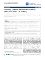
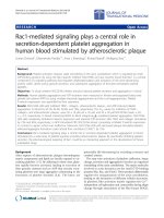
Figure 1 Experimental setup a nd procedure. A: The diagram illustrating the exper imental set-up for dipolar cortico-muscular stimulation
(dCMS). The positive and negative voltage outputs were connected to electrodes situated on the primary motor cortex (M1), and on the
contralateral gastrocnemius muscle, respectively. Both gastrocnemii muscles were attached to force transducers (not shown). Recording from
single motoneuron (Rec) was performed simultaneously on each side of the spinal cord below the lesion, as shown. IGM - ipsilateral
gastrocnemius muscle, CGM - contralateral gastrocnemius muscle. B: The experimental procedure consisted of three phases designed to
stimulate the preparation and to evaluate its reactions to dCMS. The force of muscle contraction and cortically-elicited spinal responses were
evaluated before and after the application of dCMS in Pre-test and Post-test phases by application of ten monopolar pulses. The type of
stimulation and location of the stimulation and recording electrodes was the same in these two phases. During dCMS phase the preparation
was stimulated by application of the positive and negative pulses to the motor cortex (M1) and contralateral gastrocnemius muscle (CGM)
respectively. While the number of pulses delivered during Pre- and Post-test phases was the same (10), the number of pulses delivered during
dCMS was 100. The duration (1 ms) and the frequency of stimulation (1 Hz) were the same in all three phases of the experiment. The shape of
the stimulating current at each phase is shown. There was a continuous recording of ipsilateral and contralateral muscle twitches and evoked
and spontaneous spinal activity during the entire experiment.
Ahmed Journal of NeuroEngineering and Rehabilitation 2010, 7:46
/>Page 4 of 15
muscle and positive output (range, +2.2 to +3.2 V) to
M1. At this maximal strength, dCMS was delivered (100
pulses, 1 ms pulse duration, 1 Hz frequency) , 15 to 20
seconds after the stimulating paradigm was ended, a
post-test (with identical parameters as pre-test) stimuli
were delivered to M1. See Figure 1B for experimental
design. Thereafter, spontaneous activity was followed for
5 min, then t he experiment was ended and a nimals
were injected with a lethal overdose of anesthesia. In a
subgroup of animals, the maximal threshold of M 1 was
re-tested. In addition, in this subgroup, in order to
determine the duration of dCMS effect, the magnitude
of cortically-elicited muscle twitches and spinal
responses were retested every 20 min for 60 min after
dCMS.
White matter staining
At the end of each experiment, animals were injected
with a lethal dose of Ketamine. Two parts of the spinal
column (including vertebrae and spinal cord) were dis-
sected, one part (1.5 cm) included the lesion epicenter
and another part (~0.5 cm) included the recording area
(to confirm the electrodes location). Tissues were kept
overnight (4°C) in 4% paraformaldehyde in 0.1 m PBS
and cryoprotected in 20% sucrose in PBS at 4°C for 24
h. The spinal column was freeze mounted and cut into
30 μm sections and placed on poly-L-lysine-coated glass
slides. The spinal column part including the lesion epi-
center was sequent ially sectioned. Slides were numbered
to identify their locations relative to the lesion epicenter.
4 slides from each SCI animal (n = 6) containing the
lesion epicenter and 2 slides containing no signs of
damaged spinal cord tissue from above and below the
lesion were taken for luxol fast blue (Sigma) staining.
The lesion epicenter was identified as the section con-
taining the least amount of Luxol fast blue. Sections
from control animals (n = 3) at spinal cord T13 level
were stained with luxol fast blue. Sections from the
recording area were stained with cresyl violet.
The amount of spared white matter was measured
using Adobe Photoshop CS4 (Adobe Systems, San Jose,
CA). To assess the extent of the spinal cord damage we
compared the spared white matter at the lesion epicen-
ter with white matter at spinal cord level T13 in control
animals.
Data analysis
To evaluate the latencies, we recorded the time from the
start of the stimulus artifact to the onset of the first
deflection of spinal response. Measurements were made
withacursorandatimemeteronLabChartsoftware.
The amplitude of spinal responses was measured as
peak-to-peak. Analysis of muscle contractions were per-
formed with peak analysis software (ADInstruments, Inc,
CO, USA), as the height of twitch force measured relative
to the baseline. Spike Histogram software was used to
discriminate and analyze extracellular motoneuronal
activity. All data are reported as gr oup means ± standard
deviat ion (SD). Paired student’s t-test was performed for
before-after comparison or two sample student’s t-test to
compare two groups; statistical significance at the 95%
confidence level (p < 0.05). To compare responses from
bot h sides of spinal cords recorded from control animals
andfromanimalswithSCI,weperformedoneway
ANOVA followed with Solm-Sidak post hoc analysis. Sta-
tistical analyses were performed using SigmaPlot ( SPSS,
Chicago, IL), Excel (Microsoft, Redwood, CA), and Lab-
Chart software (ADInstruments, Inc, CO, USA).
Results
Behavioral assessment
A contusion lesion of the spinal cord resulted in the
appearance of signs of spasticity syndrome such as cross -
ing of both limbs and fanning of the paws (compare 2A
and 2C). These postural changes were quantified using
the abnormal pattern scale (APS). APS showed substan-
tial increase fo r both on (APS
on
9.8 ± 0.70) and off (APS-
off
9.8 ± 0.70) ground conditi ons. These postural
abnormalities were also accompanied by reduction in
Basso Mouse Scale (BMS) scores from 9 in control
mouse to 1.2 ± 0.47 and 1.0 ± 0.63 for right and left hin-
dlimb in SCI mouse (n = 15), respectively. In addition,
the number of errors on a horizontal ladder test was
close to maximum (20) for left (19.5 ± 0.50) and right
(18.83 ± 1.16) hindlimb. Collectively, these results indi-
cate that spinal cord injury procedure used in the current
study was reliable in inducing behavioral signs of the
injury. This strengthens the interpretation of our data.
Anatomical assessment
Figure 2 B and 2D show p hotographs of cross-sectional
slices from the thoracic spinal cord region and the lesion
epicenter taken from control and SCI animals, respec-
tively. The lesion size was proximally equal in all injured
animals tested histologically (n = 6). A rim of white mat-
ter was spared on the lateral and ventral side of the spinal
cord. The area of spa red white matte r at the lesion epi-
center (0.06 ± 0.03 mm
2
) was significantly reduced 16
weeks after SCI compared to the area of white matter at
the same spinal level (0.15 ± 0.06 mm
2
) in control ani-
mals (n = 3) (p = 0.04, t-test), Figure 2E. On average, the
total cross-sectional area (white and gray matters) of the
lesion epicenter was 75 ± 14% of the total cross-sectional
area of the same spinal level in control animals.
Spinal motor neuron identification
Spinal motoneur ons innervating the gastrocnemius mus-
cle were at first identified by their large spontaneous
Ahmed Journal of NeuroEngineering and Rehabilitation 2010, 7:46
/>Page 5 of 15
spikes. The motoneuronal spike was also accompanied by
adistinctiveandcrispsoundrecordedwithaloud
speaker. Second criterion used to identify spinal m oto-
neurons was their response to the stimulation of the gas-
trocnemius muscle. Stimulating the g astrocnemius
muscle produced a short latency antidromically-gener-
ated response that was recorded from motoneurons in
the ipsilateral spinal cord. Simultaneously, the microelec-
trode on the contralateral side of t he spinal cord
recorded a response t hat had relatively longer latency
than the one picked up from the ipsilateral side. In Figure
3A, three representative condit ions were seen during the
identification of motoneurons. The left and middle panel
show simultaneous motoneuronal responses to stimu-
lated gastrocnemius muscle. The far left panel shows the
response of the motoneuron in the ipsilateral side. The
middle panel shows the response of the motoneuron in
the contralateral side. The far right panel shows a
situation when the motoneuron was not responding to
the antidromic stimulation of the homonymous gastro-
cnemius muscle. This confirmed that the unit was not
innervating the stimulated gastrocnemiu s muscle. Third,
as depicted in Figure 3B the muscle twitches (lower
panel) were correlated with motoneuron activity (upper
panel). This association between spontaneous spikes and
muscle twitches was used to confirm the connection. In
Figure 3B, the enlarged illustration (right) shows typical
spike generated by motoneuron. Finally, we histologically
confirmed that recording electrodes were localized in the
ventral horn of the spinal cord.
Latencies
Stimulating the gastrocnemius muscle resulted in short
and long latency spinal responses recorded by micro-
electrodes placed in the ipsilateral and contralateral ven-
tral horns of the spinal cord, respectively. Figure 4A
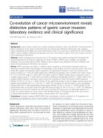
Figure 2 Anatomical assessment of spinal cord injury. A: a photograph of control animal shows the control posture of the hindlimbs. B: a
representative photograph of spinal cord cross-sectional slice taken from the thoracic level of a control animal. WM - white matter, GM - gray
matter. C: a photograph of SCI animal shows the abnormal pattern of the hindlimbs. D: a photograph of a representative slice showing the
lesion epicenter taken from SCI animal. Scale bars: 1 mm. E: quantification of spared white matter at the lesion epicenter (n = 6) and from
control animals (n = 3). Lesion epicenter had significantly less white matter from control (p = 0.04).
Ahmed Journal of NeuroEngineering and Rehabilitation 2010, 7:46
/>Page 6 of 15
shows superimposed traces of 6 antidromically-evoked
responses. While the average latency of antidromically-
evoked responses was 3.45 ± 1.54 ms, the average
latency of the contralateral responses (not shown) was
longer (5.94 ± 1.24 ms) indicating a transynaptic path-
way. The difference between ips ilateral and contralateral
spinal responses was statistically significant (n = 15, p <
0.001, t-test). Stimulating M1 resulted in ipsilateral and
contralateral spinal motoneuronal responses. Figure 4B
shows six superimposed contralateral responses. The
ipsilateral response is not shown in Figure 4. The aver-
age latency of ipsilatera l and contralateral responses was
16.09 ± 1.02 ms and 22.98 ± 1.96 ms, respectively. The
difference in latency between ipsilateral and contralat-
eral responses (6.9 ms) was statistically significant (n =
15, p < 0.001, t-test). The application of dCMS resulted
in successive spinal motoneuronal responses picked up
from the contralateral (to M1) electrode. Figure 4C
shows six superimposed recorded traces. In this illustra-
tion (Figure 4C), three distinctive r esponses are seen,
one with short latency (3.45 ± 1.54 ms), the second with
longer latency (6.02 ± 1.72 ms), a nd a third with much
longer latency (19.21 ± 2.28 ms) ( n = 15). The latency
of the ipsilateral (to M1) spinal motoneuronal responses
(not shown) was 6.02 ± 2.8 ms. Figure 4D summaries
the average latencies collected during muscle, M1, and
dCMS paradigms.
Changes in cortically-elicited muscle contraction and
spinal responses during dipolar cortico-muscular
stimulation (dCMS)
The application of dCMS gradu ally increased the twitch
peak force recorded from the gastrocnemii muscles and
neuronal activity recorded from the spinal cord. Since
the magnitude of these enhancements were similar in
control and injured animals, only d ata obtained from
SCI animals (n = 9) are prese nted. The in crease in the
force of the contralateral cortically-elicited muscle con-
traction is shown in Figure 5 A&5B. While Fi gure 5A
depicts representative recordings, the averaged results
obtained from all 9 SCI animals are shown in Figure 5B.
The increase from an initial twitch peak force of 4.8 ±
1.12 g to a final twitch peak force of 6.1 ± 0.71 g was
statistically significant (percent change = 25.0 ± 3.8%,
p = 0.001, paired t-test). The amplitude of ip silateral
cortically-elicited muscle contraction increased as well.
Representative recordings and averaged results are
showninFigure5C&5D.Thefinaltwitchforce
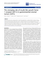
Figure 3 Identification of spinal motor neurons. A: responses to the gastrocnemius muscle stimulation. The far left and middle panels show
the simultaneous responses of spinal motoneurons located ipsilateral and contralateral to the stimulated gastrocnemius muscle, respectively. The
right panel shows recordings from the neuron did not respond to muscle stimulation. B: motoneurons were further identified when their
spontaneous activity (upper panel) was time locked with spontaneous contractions at the ipsilateral muscle (lower panel).
Ahmed Journal of NeuroEngineering and Rehabilitation 2010, 7:46
/>Page 7 of 15
Figure 4 Spinal responses. A: six superimposed spinal responses aft er homonymous gastrocnemius muscle stimulation. T he line marks the
spinal responses. B: six superimposed spinal responses after dipolar cortico-muscular stimulation (dCMS). C: six superimposed spinal responses
after motor cortex (M1) stimulation. The first and second arrows and the line mark the first, second, and third motoneuronal responses to dCMS,
respectively, recorded from the contralateral spinal cord to stimulated M1. D: the average latency of spinal responses after muscle stimulation,
dCMS (second and third responses), and after M1 stimulation. Ipsilateral spinal response to M1 stimulation (Ip) was significantly faster than the
contralateral response (Co) (p < 0.05). Muscle stimulation generated significantly shorter response at ipsilateral motoneuron than the ones at the
contralateral side (p < 0.05).
Figure 5 Muscle contraction during dipolar cortico-muscular stimulation (dCMS) in animals with SCI. A: representative initial and final
muscle twitches demonstrated greater twitch peak force at the end (final) than the beginning (initial) of dCMS on the contralateral muscle to
stimulated M1. B: Bars show averages (n = 9) of initial and final twitch peak force of the contralateral muscle, which was significantly larger at
the end of dCMS. C: representative initial and final muscle twitches of the ipsilateral muscle (to stimulated M1) during dCMS demonstrated an
increase in twitch force in response to dCMS. D: bars show averages (n = 9) of initial and final twitch peak force of the ipsilateral muscle. *p <
0.05. Data show means ± SD.
Ahmed Journal of NeuroEngineering and Rehabilitation 2010, 7:46
/>Page 8 of 15
increased si gnificantly from its initial value of 1.8 ± 0.74
g (percent change = 37.7 ± 1.14%; p = 0.001, paired
t-test).
Similar results were obtained by comparing the first
and the last spinal motoneuronal responses of the 100
pulses of dCMS protocol. On average, the contralateral
(to stimulated M1) spinal motoneuronal responses
showed significant increase (percent change = 49.75 ±
16.9%, p = 0.013, one sample t-test), as did the ipsilat-
eral ( to stimulated M1) spinal motoneuronal r esponses
(percent change = 48.10 ± 19.8%, p = 0.04, one sample
t-test). These findings suggest that physiological pro-
cesses that mediate stronge r connections of the cortico-
motoneuronal pathway were initiated during dCMS
application.
The influence of dCMS application on cortically-elicited
muscle twitches and neuronal activity in SCI animals
We examined cortically-elicited muscle twitches (mea-
sured as peak twitch force) before and after dCMS in
SCI animals. In all animals used in these experiments,
twitch force was remarkably increased after dCMS. An
example of twitches of the contralateral (to stimulated
M1) (Figure 6A) and ipsilateral (to stimulated M1) (Fig-
ure 6C) gastrocnemius muscles before (upper panels)
and after (lower panel) dCMS are shown. We also
examined c ortically-elicite d spinal responses (measured
as peak - to - peak), which was also substantially
increased. Examples of contralateral (Figure 6B) and
ipsilateral (Figure 6D) spinal responses are shown. In
Figure 6E, the twitch peak force of the contralateral
muscle showed significant increase (n = 9; p < 0.001)
(average before = 0.50 ± 0.28 g vs. average after = 2.01
± 0.80 g) (percent change = 456.1 ± 117.5%) after
dCMS , as did the twitch peak force of t he ipsilateral (to
stimulated M1) muscle (average before = 0.21 ± 0.12 vs.
average after = 1.38 ± 0.77, p < 0.001, paired t-test)
(percent change = 457 ± 122.7%). In Figure 6F, spinal
motoneuronal respo nses (n = 9) contralateral (to stimu-
lated M1) showed significant in crease after dCMS (aver-
age before = 347.67 ± 294.68 μV vs. average after =
748.90 ± 380.59 μV, p = 0.027, paired t-test) (increased
by 313 ± 197%), as did ipsilateral (to stimulated M1)
spina l motoneuronal responses (average before = 307.13
± 267.27 μV vs. average after = 630.52 ± 389.57 μV, p =
0.001, paired t-test) (increased by 292 ± 150%). Data are
shown as means ± SD. These results show that dCMS
greatly potentiates the corticomotoneuronal p athway in
injured animals.
The maximal cortical threshold defined as the lowest
electrical sti mulus eliciting the strongest muscle twitch
peak force was reduced from 9. 4 ± 0 .89 V to = 5.7 ±
0.95 V after dCMS application (n = 4, p < 0.001, t-test).
The cortically-elicited muscle twitch force and the
magnitude of spinal motoneuronal responses, evaluated
60 min after dCMS in 5 SCI animals, were still signifi-
cantly elevated on both sides (p < 0.001).
Effects of dCMS on the non-stimulated
corticomotoneuronal pathway in animals with SCI
The test stimulat ion of the other M1, contralateral to
M1 where dCMS had been applied, revealed an increa se
of the contraction force recorded from contralateral and
ipsilateral gastrocnemii muscles. The increase in contral-
ateral (percent change = 182.8 ± 87.18%), and ipsilateral
muscles (percent change = 174.8 ± 138.91%) was statis-
tically significant (n = 6, p < 0.05, t-test).
Contralateral spinal motoneuronal response was
increased significantly (p = 0.006, t-test) (average per-
cent change = 373.8 ± 304.99%), as did ipsilateral (aver-
age percent change = 289.2 ± 289.62%, p = 0.025,
t -test). These results indicate that e ven though dCMS
was unilaterally applied, it affected the corticomotoneur-
onal pathway bilaterally.
The influence of dCMS application on cortically-elicited
muscle twitches and neuronal activity in control animals
The application of dCMS across the corticomotoneuro-
nal pathway in control animals (n = 6) resulted in an
increase in the cortically-elicited muscle contraction
force produced by both gastrocnemii muscles. The
twitch peak force of the contralateral muscle increased
from 1.62 ± 1.0 g before to 5.12 ± 1.67 after dCMS
application (percent change = 250.75 ± 129.35%, p =
0.001, paired t-test, Figure 7A). The twitch peak force of
the muscle on the ipsilateral side increased as well,
although the increase was less pronounced (from 0.16 ±
0.05 g to 0.39 ± 0.08 g), before and after dCMS, respec-
tively (percent change = 166.38 ± 96.56%, p = 0.001,
paired t-test, Figure 7A)
The amplitude of evoked responses recorded from
spinal motoneurons was also enhanced by dCMS appli-
cation. As depicted in Figure 7B, the average amplitude
of these spikes recorded at the contralateral side
increased from 127.83 ± 46.58 μV to 391.17 ± 168.59
μV (perce nt change = 168.83 ± 152.00%, p = 0.009,
paired t-test). The increase at the ipsilateral side was
even greater (percent change = 369.00 ± 474.00%, 77.50
± 24.73 μV before versus 267.00 ± 86.12 μVafter
dCMS, p = 0.007, paired t-test).
Comparison between control and SCI animals
The cortically-elicited muscle twitches of contralateral
muscle, recorded from control animals were stronger
than twitches observed in SCI animals regardle ss of
whether they were recorded before (p = 0.009, t-test), or
after (p = 0.001, t-test) the dCMS procedure. The
response of ipsilateral muscles, however, was more
Ahmed Journal of NeuroEngineering and Rehabilitation 2010, 7:46
/>Page 9 of 15
Figure 6 Dipolar cortico-muscular stimulation (dCMS) augments cortically-elicited muscle contraction and spinal motoneuronal
response in animals with SCI. A: representative gastrocnemius muscle twitches induced by stimulating the contralateral M1, upper and lower
panels show muscle twitches before and after dCMS. B: contralateral cortically-elicited spinal response before (upper panel) and after (lower
panel) dCMS are shown. C: representative muscle twitches recorded from the ipsilateral (to stimulated M1) gastrocnemius muscle. D: the upper
and lower panels show ipsilateral cortically-elicited spinal responses before and after dCMS. E: quantification of results from 9 animals with SCI
revealed that contralateral (Co) (to stimulated M1) muscle twitch force was significantly increased, as did the ipsilateral (Ips) (to stimulated M1)
muscle twitch force. F: similarly, quantification of cortically-elicited spinal responses from the same animals revealed significant increase in both
contralateral and ipsilateral (to stimulated M1) after dCMS. *p < 0.05. Data show means ± SD.
Ahmed Journal of NeuroEngineering and Rehabilitation 2010, 7:46
/>Page 10 of 15
complex. Before dCMS, SCI animals showed higher ipsi-
lateral twitch peak force than control animals, although
the difference was not statistically significant (p = 0.39,
t-test). This dif ference became statistically significant
after dCMS intervention (p = 0.01, t-test).
Similarly, before dCMS, the cortically-elicited
responses recorded from spinal motoneurons were
higher in SCI animals at ipsilateral and contralateral
sides, although the difference did not reach statistical
significance (p = 0.13, t-test). However, following dCMS,
this difference was increased and b ecame statistically
significant (p = 0.009, t-test).
Next we have calculated a relative measure of muscle
performance, which we called “fidelity index” (FI). FI is
the ratio of cortically-elicited spinal motoneuronal
response to the corresponding muscle twitch peak force
(spinal response/muscle twitch ratio). Lower fidelity
index v alue indicates b etter association between spinal
responses and their corresponding muscle twitches. In
other words, it means better ability of a spinal response
to induce muscle contraction. Therefore, changes in this
index may indicate changes in r elation between spinal
and peripheral excitability.
After dCMS, SCI animals showed overall significant
group reduction in FI (F = 3.3, p < 0.033, ANOVA)
(Figure 8). Solm-Sidak post hoc test showed reduction in
contralateral FI (average before = 368.35 ± 342.51 vs.
average after = 246.15 ± 112.24), however, the difference
was not statistically significant (p = 0.46). The ipsilateral
FI was significantly r educed after dCMS (average before
= 704.59 ± 625.7 vs. average after = 247.95 ± 156.27) (p
= 0.011). The effect of dCMS treatment was the oppo-
site in control animals which demonstrated overall
group increase in FI after this procedure (F = 31.51, p <
0.001, ANOVA). FI was significantly increased after
dCMS (Solm-Sidak post hoc, p < 0.001) in the ipsilatera l
side (average before = 328.53 ± 104.83 vs. average after
526.83 ± 169.38). There was also a trend reflecting an
increase in the contralateral side (average before = 48.59
± 17.71 vs. average aft er = 56.15 ± 24.19), but was not
statistically significant (Solm-Sidak post hoc, p = 0.89).
Comparing FI from control animals with FI from SCI
animals showed a statistically signif icant lower index in
Figure 7 Cortically-elicited muscle contraction and spinal responses after dipolar cortico-muscular stimulation (dCMS) in control mice .
A: quantification of results from 6 control animals revealed significant increase in contralateral (CO) and ipsilateral (Ips) (to stimulated M1) muscle
twitch force after dCMS. B: contralateral (to stimulated M1) cortically-elicited spinal responses were significantly increased after dCMS, as did
ipsilateral responses. *p < 0.05. Data show means ± SD.
Figure 8 Fidelity index analysis. Fidelity index (spinal response/
muscle twitch force) was quantified from 6 control and 9 with SCI
animals. In animals with SCI, Contralateral (CO) to stimulated M1
fidelity index shows reduction after dCMS but was not statistically
significant; however, ipsilateral (Ips) fidelity index was significantly
reduced after dCMS. In control animals, after dCMS, Fidelity index
was increased contralateral to stimulated M1 but was not
statistically significant, however, the ipsilateral Fidelity index was
significantly higher after dCMS. Note that the lower the fidelity
indexes the better the correlation between muscle contraction and
spinal response. *p < 0.05, lower from before dCMS; **p < 0.05,
higher from before dCMS. Data show means ± SD.
Ahmed Journal of NeuroEngineering and Rehabilitation 2010, 7:46
/>Page 11 of 15
the contralateral side of control animals (p < 0.001,
ANOVA, Solm-Sidak post hoc) both before and after
dCMS. These results support the findings that periph-
eral nerves are in-excitable or of higher threshold in
subjects with SCI [13].
dCMS increased spinal motoneurons spontaneous activity
Comparing the firing rate of sponta neous activity before
and after dCMS intervention demonstrated significant
increase in both control and SCI animals. In Figure 9
A&9B, a representative spontaneous activity recording
from an SCI animal is shown. In SCI animals, sponta-
neous activity was significantly increased in the contral-
ateral side of the spinal cord (average before = 17.31 ±
13.10 spikes/s vs. average after = 32.13 ± 14.73 spikes/s;
p = 0.001) (121.71 ± 147.35%), as it did in the ipsilateral
side (average before = 18.85 ± 13.64 spikes/s vs. average
after = 26.93 ± 17.25; p = 0.008) (percent change =
54.10 ± 32.29%). In control animals, spontaneous activ-
ity was signific antly increased in the contralateral (to
stimulated M1) side of the spinal cord (average before =
11.40 ± 8.65 spikes/s vs. average after = 20.53 ± 11.82
spikes/s; p = 0.006) (percent change = 90.10 ± 42.53%),
as it did in the ipsilateral side (avera ge before = 11.63 ±
5.34 spikes/s vs. average after = 22.18 ± 10.35 spikes/s;
p = 0.01) (percent change = 99.10 ± 1.10%). One way
ANOVA showed no sign ificant difference between con-
trol and SCI animals in firing rate, although, SCI ani-
mals demonstrated higher firing rate.
Effects of monopolar stimulation of muscle or cortex
In order to determine that the effect was unique to
dCMS, the influence of monopolar stimulation (maximal
sti mulation for100 pulses, 1 Hz frequency) of eithe r the
muscle or the motor cortex on spinal motoneuronal
response and muscle twitch peak force was examined.
As expected, muscle stimulation resulted in significant
reduction in muscle twitch force (-20.28 ± 7.02%, p <
0.001, t-test) (n = 5, 3 SCI and 2 control). It also
resulted in a significant reduction in spinal motoneuro-
nal responses evoked by the contralateral (to stimulated
muscle) M1 test stimulation (average before = 747.50 ±
142.72 μV, vs. average after = 503.14 ± 74.78) (F =
17.11, one way ANOVA, Solm-Sidak post hoc,p<
0.001), however, no significant change was seen in
responses recorded in the ipsilateral (to stimulated mus-
cle) side of the spinal cord (average before 383.33 ±
140.67 μV vs. average after = 371.43 ± 35.61, p = 0.84).
In a separate group of animals (n = 5, 3 SCI and 2
control), we also tested the effect of the monopolar sti-
mulation paradigm applied only at the motor cortex on
contralateral muscle twitch peak force and spinal moto-
neuronal response. Both, the muscle tw itch and moto-
neuron response were significantly reduced by over 50%
(-53.69 ± 4.3%, p = 0.001, t-test) and almost 15% (-14.59
± 9.10%, p = 0.003, t-test), respectively. These results
indicate that monopolar muscle or cortical stimulation
at maximal strength results in fatigue of muscle twitch
force and reduction in spinal responses.
Discussion
The results show remarkable enhancement of the excit-
ability of the corticomotoneuronal pathway induced by
unilateral application of the dCMS. This enhancement
was observed in control animals and in SCI animals that
had severe locomotor impair ment associated with signs
of spastic syndrome. The effect was observed both in
the ipsilateral and contralateral pathways. The maximal
threshold of the ipsilateral cortex was reduced. Improve-
ment in muscle strength was accompanied by an
increase in spontaneous activity and potentia tion of
evoked response s of the spinal motoneurons. The spinal
motoneuronal respon ses and muscle twitches evoked by
the stimulation of the contralateral, non-treated M1
Figure 9 dCMS increased spontaneous activity of spinal
motoneurons (MNs). A: an example of spontaneous activity from
one MN shows the level of activity before (upper panel) and after
(lower panel) dCMS intervention. This example was taken from SCI
animal. B: a representative experiment shows firing rate (spikes/s)
during an entire experiment. Arrows show the start and end of
dCMS application. C: quantification of spontaneous activity before
compared with after dCMS show significant increase in both
contralateral (Co) and ipsilateral (Ips) spinal recordings from control
and with SCI animals. *p < 0.05. Data show means ± SD.
Ahmed Journal of NeuroEngineering and Rehabilitation 2010, 7:46
/>Page 12 of 15
were significantly enhanced as well. The dCMS-induced
effect persisted beyond the phase of stimulation and
extended through the entire period of the experiment
(60 min).
Bilateral responses to cortical stimulation have been
routinely observed [3,6,30-33]. They can be mediated by
interhemispheric connections, ipsilateral cortico-spinal
connections (5-6% of the contralateral projections) [34],
or commissural spinal neurons. As seen in Figure 6F
and 7B, ipsilateral responses to unilateral stimulation of
motor cortex evoked larger responses in SCI animals
compared to controls. These results further support the
idea that ipsilateral corticospinal projections are more
efficient in evoking muscle contraction after SCI [3].
The mechanism of the dCMS induced increase in the
excitability o f the corticomotoneuronal pathway is not
clear and one can only speculate as to what processes
have been modulated. It is obvious that the potentiation
in cortically-el icited muscle contraction during dCMS is
not like the potentiation seen after n euromuscular sti-
mulation [35]. While neuromuscular stimulation leads
to a brief potentiation of muscle force followed by a
steep reduction in force, dCMS leads to a gradually pro-
ceeding increase in the amplitude of cortically-elicited
muscle contraction. Since the enhancement occurred at
contra- and ipsilateral sides, the locus of potentiation is
most likely either spinal or supraspinal. The enhance-
ment of cortically-elicited muscle contraction was
accompanied by a reduction in maximal threshold to
cortical stim ulation, an increase in spinal motoneuronal
responses, and an increase in cortically-elicited spinal
motoneuronal responses. T herefore, one can assume
that improvements occurred simultaneously at several
functional levels of the corticomotoneuronal pathway.
In view of the f act that the current employed in our
stimulation paradigm was always positive at one end
and negative at the other, our stimulation can be con-
sidered in part polarizing. The paradigm of polarizing
current was used to study excitability of different parts
of the nervous system [36-39]. In these studies, polariz-
ing current produced potential membrane changes in
which hyperpolarization occurs at cellular parts near the
positive electrode and depolarization occurs near the
negative elec trode. Complying with this rule, for exam-
ple, the situation of two polarizing electrodes on the
spinal cord (one on the ventral side and the other on
the dorsal side) produced changes in membrane and
spike potentials of primary fibers from muscles [36]. In
our study we suggest that the current is polarizing dur-
ing the brief, steady moment of pulse duration (1 ms).
Given the electrodes placement, in which negative at the
muscle and posit ive at the cortex, the cell body of corti-
cospinal neurons is expected to hyperpolarize and their
nerve terminals depolarize. Moreover, spinal
motoneurons expected to hyperpolarize at the cell body
and dendrites, and depolarize at the neuromuscular
junction. According to cell topography relative to the
applied electrical field, membrane potential changes are
also expected to occur at intervening interneurons.
These membrane changes that occur briefly during each
pulse of dCMS, seem to prime corticomotoneuronal
pathway for potentiation. In addition, the stimulating
pulse has two more periods: rising (0.250 ms) and falling
(0.250 ms). These changing periods caused a flow of
current that exited from one end and entered at the
other end of the corticomotoneuronal pathway. This
idea is supported by the observation of stimulus artifact
picked up by electrodes in the spinal cord. The current
flowed throughout the entire pathway independent from
the factors confounding active exc itability (see introduc-
tion). This might cause activation of the corticomoto-
neuronal pathway at any possible excitable site/s. This
will ensure eliciting spike-timing-dependent plasticity
[40] that might be one of the mechanisms that mediates
the effe ct of the dCMS. In a dditio n, the high frequency
multiple spinal re sponses, evoked during dCM S, can, in
principle, induce long-term potentiation [41]. Because
dCMS can engage a variety of neuronal mechanisms as
well as non-neuronal activity, its effect might be a com-
bination of many changes along the corticomotoneuro-
nal pathway.
The dCMS-induced enhancement of cortically-elicited
muscle contraction has been observed in both - contr ol
and injured animals. The mechanisms responsible for
this amplification in these two groups of animals may
overlap, but they do not have to be identical. Although,
as discussed above the potent iating effect of dCMS
could be mediated by strengthening synaptic responses,
thenatureandsourceofthesechangesmaydiffersub-
stantially in the corticomotoneuronal pathway of control
and injured animals. Axonal sprouting is probably the
primary source of synaptic connections in the damaged
spinal cord [1-3]. However, axonal sprouting does not
grant the formation of functional connections. There-
fore, one of the probable mecha nisms that may mediate
the potentiating effect of dCMS is the refining and
strengthening of the weak synaptic connections that
have resulted from sprouting. Moreover, dormant con-
nections that exist throughout the sensorimotor system
[6] may be activated and become functional after dCMS.
Potentiating the spared normal connections could also
happen after dCMS. On the other hand, in cont rol ani-
mals, potentiating normal connections and facilitating
dormant connections might be the only processes that
mediate the effect of dCMS. The results show that
dCMS stimulation was almost twice as effective in
injured animals compared with controls. This indicates
that injured spinal cord is more prone for dCMS
Ahmed Journal of NeuroEngineering and Rehabilitation 2010, 7:46
/>Page 13 of 15
sti mulat ion and posses extra mechanism s mediating the
dCMS effect.
In SCI animals, even before the application of dCMS,
the spinal motoneurons were responding more aggres-
sively to cortical sti mulation than were controls. Never-
theless, very weak or no muscle contraction was seen
(Figure6).Thismightbeduetooneoftwomechan-
isms. One would be located in the spinal cord caudal to
the lesion and/or the other being, the inexcitable periph-
eral nerves an d/or the irrespon siveness of the muscle.
Caudal to the lesion, the activity of the spinal moto-
neuron pool was probably desynchr onized as a result of
reorganization . Supporting this idea are t he findings b y
Brus-Ramer and colleagues [3]. The authors reported
that chronic stimulation of corticospinal tracts resulted
in preferential axonal outgrowth toward the ventral
horn. This indicates that inter motoneuronal connec-
tions are dynamic processes, which may change by
decentralization. Inexcitable peripheral axons were
found in patients with SCI [13]. Assuming that the
axons in SCI animals are in similar conditions, they
could experience an action potential failure resulting in
reduced muscle contraction. Muscle atrophy is always
seen in animals with SCI [9, 10, and 12] and humans
[11]. This might also be one of the reasons why spinal
motoneurons responses were not translated adequately
into muscle contraction. We quantified the adequacy of
motoneuronal responses by calculating the fidelity
index, which is the ratio of spinal response to muscle
twitch force. The dCMS-induced changes i n the fidelity
index were opposed in control and injured animals.
While this index has been reduced in injured animals,
indicat ing improvement in the effectiveness of the corti-
comotoneuronal pathway, it had increased in control
animals suggesting lowering of the pathway effectiveness
probably due to fatigue interfe rence. Therefore, one can
imply that injury to the spinal cord initiates processes
which favor regeneration of the function. Apparently
our procedure synchronizes and facilitates these pro-
cesses, promoting recovery.
It has been demo nstrated that spontaneous activity in
spinal motoneurons is a significant factor in developing
spinal circuits involved in locomotion [42,43]. It has also
been shown that increasing or decreasing the frequency
of spontaneous activity will disrupt connectivity in a
developing spinal cord [44,45]. In the light of these stu-
dies and our data one can ask what role the changes in
spinal motoneuronal spontaneous activity play in recov-
ery after SCI, and what are the interactions between
interventions, spontaneous activity and functional recov-
ery a fter SCI? These questions await further investiga-
tions which could be guided by our observation that
dCMS increased the tonic activity of spinal motoneur-
ons in animals with SCI as well as in control animals.
Before the dCMS application, the spontaneous activity
of motoneurons in animals with SCI was higher than
that of control animals. This and the exaggerated
evoked spinal responses in animals with SCI, is consis-
tent with the behavioral measurements that show spastic
syndrome-like characteristics. The exaggerated sponta-
neous firing rate of spinal motoneurons is also consis-
tent with data from motor unit firing in humans and
animals after SCI [46,47] and with results from intracel-
lular recordings from sacrocaudal motoneurons that
show a sustained and exaggerated firing rate in animals
with SCI [48]. Minutes after dCMS, motoneuronal spon-
taneous activity was still substantially increased. Some of
these activities were rhythmic, as shown in Figure 3B,
although most of the spontaneous activity was in an un-
modulated pattern of firing as shown in Figure 9A. Vol-
tage-dependent persistent inward currents (PICs) that
strengthen synaptic inputs in normal behavior depend
on descending brain-stem-released serotonin (5-HT) or
noradrenalin [49-51]. Here the increase in the sponta-
neous firing rate and the appearance of modulated activ-
ity in some animals after dCMS may indicate better
connections with brain-stem centers.
In conclusion, the results showed clear evidence that
dCMS is an effective method that enhances the excit-
ability of the corticomotoneuronal connections. This
technique has the potential to be used in humans suffer-
ing after spinal cord injury, stroke, multiple sclerosis,
and others. In practice, it can be employed to strengthen
or awake n any weak or dormant pathway in the nervous
system.
Acknowledgements
This research was supported by NYS/DOH grant # CO23684 and PSCCUNY
grant 60027-37-39.
Competing interests
Currently applying for a patent relating to the content of the manuscript.
Received: 24 March 2010 Accepted: 17 September 2010
Published: 17 September 2010
References
1. Murray M, Goldberger ME: Restitution of function and collateral sprouting
in the cat spinal cord: the partially hemisected animal. J Comp Neurol
1974, 158(1):19-36.
2. Bareyre FM, Kerschensteiner M, Raineteau O, Mettenleiter TC, Weinmann O,
Schwab ME: The injured spinal cord spontaneously forms a new
intraspinal circuit in adult rats. Nat Neurosci 2004, 7:269-77.
3. Brus-Ramer M, Carmel JB, Chakrabarty S, Martin JH: Electrical stimulation of
spared corticospinal axons augments connections with ipsilateral spinal
motor circuits after injury. J Neurosci 2007, 27:13793-13901.
4. Miles R, Wong RK: Latent synaptic pathways revealed after tetanic
stimulation in the hippocampus. Nature 1987, 329:724-6.
5. Fregni F, Boggio PS, Valle AC, Rocha RR, Duarte J, Ferreira MJ, Wagner T,
Fecteau S, Rigonatti SP, Riberto M, Freedman SD, Pascual-Leone A: A sham-
controlled trial of a 5-day course of repetitive transcranial magnetic
stimulation of the unaffected hemisphere in stroke patients. Stroke 2006,
37:2115-22.
Ahmed Journal of NeuroEngineering and Rehabilitation 2010, 7:46
/>Page 14 of 15
6. Brus-Ramer M, Carmel JB, Martin JH: Motor cortex bilateral motor
representation depends on subcortical and interhemispheric
interactions. J Neurosci 2009, 29:6196-206.
7. Ridding MC, Taylor JL: Mechanisms of motor-evoked potential facilitation
following prolonged dual peripheral and central stimulation in humans.
J Physiol 2001, 537:623-31.
8. Huerta PT, Volpe BT: Transcranial magnetic stimulation, synaptic plasticity
and network oscillations. J Neuroeng Rehabil 2009, 2:6-7.
9. Ahmed Z, Wieraszko A: Combined effects of acrobatic exercise and
magnetic stimulation on the functional recovery after spinal cord
lesions. J Neurotrauma 2008, 25:1257-1269.
10. Liu M, Bose P, Walter GA, Thompson FJ, Vandenborne K: A longitudinal
study of skeletal muscle following spinal cord injury and locomotor
training. Spinal Cord 2008, 46:488-93.
11. Shah PK, Stevens JE, Gregory CM, Pathare NC, Jayaraman A, Bickel SC,
Bowden M, Behrman AL, Walter GA, Dudley GA, Vandenborne K: Lower-
extremity muscle cross-sectional area after incomplete spinal cord
injury. Arch Phys Med Rehabil 2006, 87:772-778.
12. Gordon T, Mao J: Muscle atrophy and procedures for training after spinal
cord injury. Phys Ther 1994, 74:50-60.
13. Lin CS, Macefield VG, Elam M, Wallin BG, Engel S, Kiernan MC: Axonal
changes in spinal cord injured patients distal to the site of injury. Brain
2007, 130:985-994.
14. Curt A, dietz V: Electrophysiological recordings in patients with spinal
cord injury: significance for predicting outcome. Spinal Cord 1999,
37:157-65.
15. Tillakaratne NJ, Mouria M, Ziv NB, Roy RR, V Edgerton R, Tobin AJ: Increased
expression of glutamate decarboxylase (GAD (67)) in feline lumbar
spinal cord after complete thoracic spinal cord transection. J Neurosci Re
2000, 60:219-230.
16. Knikou M: Neural control of locomotion and training-induced plasticity
after spinal and cerebral lesions. Clin Neurophysiol 2010, 12:1655-1668.
17. Raineteau O, Schwab ME: Plasticity of motor systems after incomplete
spinal cord injury. Nat Rev Neurosci 2001, 2:263-73.
18. Hebb D: organization of behavior. New York: Wiley 1949.
19. Levy WB, Steward O: Temporal contiguity requirements for long-term
associative potentiation/depression in the hippocampus. Neuroscience
1983, 8:791-797.
20. Bliss TVP, Collingridge GL: A synaptic model of memory: Long-Term
Potentiation in the hippocampus. Nature 1993,
361:31-39.
21. Eccles JC, Kostyuk PG, Schmidt RF: The effect of electric polarizing of the
spinal cord on central afferent fibres and on their excitatory synaptic
action. J Physiol 1962, 162:138-150.
22. Nitsche MA, Paulus W: Excitability changes induced in the human motor
cortex by weak transcranial direct current stimulation. J Physiol 2000,
527:633-639.
23. Gruner JA: A Monitored contusion model of spinal cord injury in the rat.
J Neurotrauma 1992, 9:123-126.
24. Engesser-Cesar C, Anderson AJ, Basso DM, Edgerton VR, Cotman CW:
Voluntary wheel running improves recovery from a moderate spinal
cord injury. J Neurotrauma 2005, 22:157-171.
25. Zandieh S, Hopf R, Redl H, Schlag MG: The effect of ketamine/xylazine
anesthesia on sensory and motor evoked potentials in the rat. Spinal
Cord 2003, 41:16-22.
26. Hicks RG, Woodforth IJ, Crawford MR, Stephen JP, Burke DJ: Some effects
of isoflurane on I waves of the motor evoked potential. Br J Anaesth
1992, 69:130-136.
27. Sloan TB: Anesthetic effects on electrophysiologic recordings. J Clin
Neurophysiol 1998, 15:217-226.
28. Franklin KBJ, Paxinos G: The Mouse Brain in Stereotaxic Coordinates.
Academic Press, 3 2007.
29. Porter R: Early facilitation at corticomotoneuronal neuronal synapses. J
Physiol 1970, 207:733-745.
30. Ghosh A, Sydekum E, Haiss F, Peduzzi S, Zorner B, Schneider R, Baltes C,
Rudin M, Weber B, Schwab ME: Functional and Anatomical
Reorganization of the Sensory-Motor Cortex after Incomplete Spinal
Cord Injury in Adult Rats. J Neurosci 2009, 29:12210-12219.
31. Chakrabarty S, Shulman B, Martin JH: Activity-Dependent Codevelopment
of the Corticospinal System and Target Interneurons in the Cervical
Spinal Cord. J Neurosci 2009, 29:8816-8827.
32. Maier IC, Baumann K, Thallmair M, Weinmann O, Scholl J, Schwab ME:
Constraint-Induced Movement Therapy in the Adult Rat after Unilateral
Corticospinal Tract Injury. J Neurosci 2008, 28:9386-9403.
33. Frigon A, Yakovenko S, Gritsenko V, Tremblay ME, Barriere G: Strengthening
Corticospinal Connections with Chronic Electrical Stimulation after
Injury. J Neurosci 2008, 28:3262-3263.
34. Brosamle C, Schwab ME: Cells of origin, course, and termination patterns
of the ventral, uncrossed component of the mature rat corticospinal
tract. J Comp Neurol 1997, 386:293-303.
35. Luke R, Harris W, Bobet J, Sanelli L, Bennett DJ: Tail Muscles Become Slow
but Fatigable in Chronic Sacral Spinal Rats With Spasticity.
J Neurophysiol
2006, 95:1124-1133.
36. Landau WM, Bishop GH, Clare MH: Analysis of the form and distribution
of cortical potentials under the influence of polarizing currents. J
Neurophysiol 1964, 27:788-813.
37. Gorman ALF: Differential patterns of activation of the pyramidal system
elicited by surface anodal and cathodal cortical stimulation. J
Neurophysiol 1965, 29:547-64.
38. Terzoulo CA, Bullock TH: Measurement of imposed voltage gradient
adequate to modulate neuronal firing. Proc Natl Acad Sci USA 1956,
42:687-694.
39. Bindman LJ, Lippold OCJ, Redfearn JWT: Long-lasting changes in the level
of the electrical activity of the cerebral cortex produced by polarizing
currents. Nature 1962, 196 :584-585.
40. Dan Y, Poo M: Spiking Timing-dependent plasticity: From synapse to
perception. Physiol Rev 2006, 86:1033-1048.
41. Bliss TVP, Collingridge GL: A synaptic model of memory: Long-Term
Potentiation in the hippocampus. Nature 1993, 361:31-39.
42. Landmesser LT, O’Donovan MJ: activation patterns of embryonic chick
hindlimb muscles recorded in-ovo and in an isolated spinal cord
preparation. J Physiol 1984, 347:189-204.
43. Katz LC, Shatz CJ: Synaptic activity and the construction of cortical
circuits. Science 1996, 274:1133-1138.
44. Hanson MG, Landmesser LT: Normal patterns of spontaneous activity are
required for correct motor axon guidance and the expression of specific
guidance molecules. Neuron 2004, 46:687-701.
45. Hanson MG, Landmesser LT: Increasing the frequency of spontaneous
rhythmic activity disrupts pool-specific axon fasciculation and
pathfinding of embryonic spinal motoneurons. J Neurosci 2006,
26:12769-12780.
46. Gorassini M, Bennett DJ, Kiehn O, Eken T, Hultborn H: Activation patterns
of hindlimb motor units in the awake rate and their relation to
motoneuron intrinsic properties. J Neurophysiol 1999, 82:709-717.
47. Thomas CK, Ross BH: Distinct patterns of motor unit behavior during
muscle spasms in spinal cord injured subjects. J Neurophysiol 1997,
77:2847-2850.
48. Harvey JP, Gorassini M, Bennett DJ: The spastic rat with sacral spinal cord
injury. In Animal model of movement disorders. Edited by: Mark LeDoux. El
Sevier Academic Press; 2005:691-697.
49. Hounsgaard J, Hultborn H, Jespersen B, Kiehn O: Bistability of alpha-
motoneurons in the decerebrate cat and in the acute spinal cat after
intravenous 5-hydroxytryptophan. J Physiol 1988, 405:345-367.
50. Hsiao CF, Del Negro CA, Trueblood PR, Chandler SH: Ionic basis for
serotonin-induced bistable membrane properties in guinea pig
trigeminal motoneurons. J Neurophysiol 1998, 79:2847-2856.
51. Lee RH, Heckman CJ: Bistability in spinal motoneurons in vivo: systematic
variations in persistent inward currents. J Neurophysiol 1998, 80:583-593.
doi:10.1186/1743-0003-7-46
Cite this article as: Ahmed: Dipolar cortico-muscular electrical
stimulation: a novel method that enhances motor function in both -
normal and spinal cord injured mice. Journal of NeuroEngineering and
Rehabilitation 2010 7:46.
Ahmed Journal of NeuroEngineering and Rehabilitation 2010, 7:46
/>Page 15 of 15