Báo cáo hóa học: " Reversal of TMS-induced motor twitch by training is associated with a reduction in excitability of the antagonist muscle" ppt
Bạn đang xem bản rút gọn của tài liệu. Xem và tải ngay bản đầy đủ của tài liệu tại đây (624.14 KB, 8 trang )
RESEARC H Open Access
Reversal of TMS-induced motor twitch by training
is associated with a reduction in excitability of
the antagonist muscle
Viola Giacobbe
1*
, Bruce T Volpe
1
, Gary W Thickbroom
2
, Felipe Fregni
3,6
, Alvaro Pascual-Leone
3,5
, Hermano I Krebs
4
and Dylan J Edwards
1,2,3
Abstract
Background: A single session of isolated repetitive movements of the thumb can alter the response to transcranial
magnetic stimulation (TMS), such that the related muscle twitch measure d post-training occurs in the trained
direction. This response is at tributed to transient excitability changes in primary motor cortex (M1) that form the
early part of learning. We investigated; (1) whether this phenomenon might occur for movements at the wrist, and
(2) how specific TMS activation patterns of opposing muscles underlie the practice-induced change in direction.
Methods: We used single-pulse suprathreshold TMS over the M1 forearm ar ea, to evoke wrist movements in 20
healthy subjects. We measured the preferential direction of the TMS-induced twitch in both the sagittal and
coronal plane using an optical goniometer fixed to the dorsum of the wrist, and recorded electromyographic
(EMG) activity from the flexor carpi radialis (FCR) and extensor carpi radialis (ECR) muscles. Subjects performed
gentle voluntary movements, in the direction opposite to the initial twitch for 5 minutes at 0.2 Hz. We collected
motor evoked potentials (MEPs) elicited by TMS at baseline and for 10 minutes after training.
Results: Repetitive motor training was sufficient for TMS to evoke movements in the practiced direction opposite
to the original twitch. For most subjects the effect of the newly-acquired direction was retained for at least 10
minutes before reverting to the original. Importantly, the direction change of the movement was associated with a
significant decrease in MEP amplitude of the antagonist to the trained muscle, rather than an increase in MEP
amplitude of the trained muscle.
Conclusions: These resul ts demonstrate for the first time that a TMS-twitch direction change following a simple
practice paradigm may result from reduced corticospinal drive to muscles antagonizing the trained direction. Such
findings may have implications for training paradigms in neurorehabilitation.
Background
Human motor control of individual joints involves orga-
nized coupling of agonist and antagonist muscles to
achieve a desired movement efficiently. During contrac-
tion of agonist muscles, the antagonists do not behave
passively, but are actively inhibited by central nervous
mechanisms [1]. Reciprocal control of antagonistic mus-
cles is critical for execution of coordinated limb move-
ments, and through a mechanism of reciprocal
inhibition, the central nervous system ensures that
antagonist muscle activity is suppressed during contrac-
tion of an agonist [2].
During motor learning, patterns of motor activation
are encoded in the brain through distributed networks
including motor cortex, deep brain nuclei and the cere-
bellum [3]. In primary motor cortex (M1) these changes
can be probed with mapping techniques showing excit-
ability changes and representational reorganization asso-
ciated with extensive motor training [4-6], depending on
the nature of movements performed during training [7].
These studies have clinical implications since motor
training is known to positively influence motor control
in neurological patients [8-11], and novel interventions
* Correspondence:
1
Burke-Cornell Medical Research Institute, White Plains, NY, USA
Full list of author information is available at the end of the article
Giacobbe et al. Journal of NeuroEngineering and Rehabilitation 2011, 8:46
/>JNER
JOURNAL OF NEUROENGINEERING
AND REHABILITATION
© 2011 Giacobbe et al; licensee BioMed Ce ntral Ltd. This is an Open Access article distributed under the terms of the Creative
Commons Attribution License ( which permits unrestricted use, distribution, and
reproduction in any medium, provided the original work is properly cited.
are emerging that actively alter cortic al excitability and
might interact with training effects [12]. However, the
corticomotor excitability changes associated with well-
defined, simple training paradigms in healthy humans
are poorly understood, particularly those relating to ago-
nist-antagonist muscle pairs.
A single suprathreshold pulse of Transcranial Mag-
netic Stimulation (TMS) over the hand area of M1
results in a balance of inhibitory and excitatory pro-
cesses that leads to an observed twitch of t he thumb in
a consistent direction with each stimulus [13]. Further, a
short period of practice with movements in the opposite
direction can change the direction o f the TMS-induced
twitch to that of the practice direction. It remains to be
investigated how the relationship between agonist and
antagonist muscle activation might lead to this direction
change, or if this phenomenon is peculiar to muscles of
the thumb.
Inthepresentstudyweexaminedinhealthyadults
whether the direction of TMS-induc ed wrist movements
can be modulated or changed by a short period of sim-
ple repetitive wrist training. We proposed to test mus-
cles controlling the wrist that are located in the
proximal forearm area and that have a more defined
functional agonist-antagonist role. We hypothesized that
a short period of repetitive gentle wrist movements in a
direction opposite to the initial TMS-twitch direction,
with only concentric contraction of the agonist (passive
return), would result in a change of twitch direction eli-
citedbyTMS,andacorrespondingreductionindes-
cending drive to the antagonist muscle.
Methods
Subjects
Twenty right-handed healthy volunteers (mean age 28
yrs, range 22-37 yrs) with no history of neurological or
psychiatric illness, and no contraindications to TMS,
were recruited for the experiment. The subjects were
seated comfortably in a chair with their right arm freely
hanging to the side in a relaxed posture. All subjects
were screened for TMS exclusion criteria and gave their
written informed consent before participating. The
study was approved by the Institutional Review Boar d of
Burke Rehabilitation Hospital.
Stimulation set-up
Biphasic single-pulse TMS was delivered through a fig-
ure-of-eight-shaped coil (inner diameter: 35 mm, outer
diameter: 75 mm, MagVenture), using a MagPro x100
stimulator (Mindcare Co.). To identify the area of sti-
mulation, a tight lycra cap was positioned over the head
and the vertex was marked by measuring the mid-point
intersection between the nasion-inion and inter-aural
lines. Potential s timulus sites were marked on the cap
using the vertex as a reference point, in 1-cm steps in
the coronal and sagittal planes, over the region of the
primary motor cortex. Using a supra-threshold stimulus
intensity, the coil was systematically moved over motor
cortex to determine the optimal location for eliciting
isolated wrist movement, and maximal amplitude motor
evoked potentials (MEPs) in both the flexor carpi radia-
lis (FCR) and extensor carpi radialis (ECR) muscles.
MEPs were obtained from the FCR and ECR muscles
simultaneously. Once the optimal position of the coil
was established, it was marked on the cap, to ensure a
constant coil placement throughout the experiment.
During stimulation, the center of the coil was placed
tangentially to the scalp with the handle pointing pos-
terior and laterally rotated at a 45° angle from the mid-
line, in order to induce a posterior-anterior current flow
in the cortical tissue approximately perpendicular to the
line of the central sulcus. Focal TMS was delivered to
the brain with the target muscles at rest, that is, in the
absence of any electromyographic (EMG) activity
exceeding a background noise level of 20 μV.
Recording of EMG and twitch direction
Surface EMG activity was recorded from pre-amplified
electrodes (SX230, fixed electrode distance: 20 mm, Bio-
metrics Ltd.) positioned over the muscle belly of the
right FCR and ECR muscles. EMG signals were ampli-
fied (x1000) at the site and band-pass filtered between
20 and 400 Hz. The signals were collected and digitized
at a frequency of 1000 Hz using a Cambridge Electronic
Design (CED) 1401 A/D converter and a data-collection
program (CED Spike 2), then stored into the computer
for further off-line analysis. EMG activity of the training
muscles was continuously monitored during p ractice to
provide visual feedback during the experiment and
ensure regular contractions during training. In this
study the antagonist muscle was defined as the muscle
opposing the direction of training.
Resting motor threshold (RMT), defined as the mini-
mum TMS intensity that evoked a MEP of at least 50
μV peak-to-peak amplitude in 6 of 10 trials, was mea-
suredfortheFCRandECRinstimulusstepsof1%of
maximum stimulator output (MSO). RMT was deter-
mined with the wrist resting on the subject’slap,start-
ing at a low intensity and using four stimuli for each 1%
increment of stimulator output intensity.
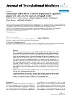
A two degree-of-freedom optical goniometer (SG65,
max stretch length: 65 mm, Biometrics Ltd.) was posi-
tioned on the dorsum of the wrist, aligned in the sagittal
plane (Figure 1a), to quantify joint rotations in both the
sagittal (wrist flexion or extension) and coronal (wrist
ulnar or radial deviation) planes. The output of the
goniometer (Figure 1b), together with the EMG read-
ings, was acquired using CED Spike 2 software.
Giacobbe et al. Journal of NeuroEngineering and Rehabilitation 2011, 8:46
/>Page 2 of 8
Experimental design
The preliminary phase of the experiment lasted about
25 minutes; with t he electrodes and goniometer posi-
tioned as described above, the optimal site for stimula-
tion and RMT were determined. The experimental
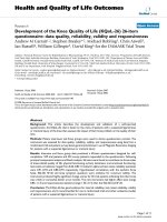
design was structured in 3 phases: b aseline, training,
and post-training measurements (Figure 2). Subjects
were comfortably seated with the right arm and hand
relaxed in a vertical position, to avoid confounding grav-
itational contributions. This position was maintained
throughout the experiment.
Baseline
Before training (at time-point t0), 20 TMS stimuli were
delivere d at 0.2 Hz to the optimal scalp site. Intensity of
stimulation was calculated as the RMT intensity + 30%
of MSO, to ensure large-size and ea sily measurable
MEPs. Subjects usually perceived the twitch in the wrist,
but not its direction, which was t herefore indicated by
the reading of the goniometer. Although the resultant
movement induced by TMS would theoretically yield a
vectorial combination of both sagittal and coronal
deflection, all subjects exhibited a pref erential plane of
movement, thus explaining the choice to consider the
dominant plane only.
Training
Once the baseline twitch direction in the dominant plane
had been identified, subjects were instructed to perform
voluntary phasic wrist movements in a direction opposite
to it for 5 minute s at 0.2 Hz, as displayed on a monitor
in front of the subject. The subjects performed one
dynamic contraction through normal wrist movement
range (extension or flexion) from the neutral position,
followed by immediate relaxation, in which they were
asked to let their wrist slowly and naturally drop, to
allow passive return to neutral position. They were
allowed 10 practice contractions to become familiar with
the experimental setup. After each movement, we were
able to monitor that the wrist returned to the start posi-
tion by natural relaxation through visual feedback of the
goniometrical traces. Accuracy and consistency of the
direction of training exercises were monitored in real-
time by the investigators throughout the experiment.
Figure 1 (a) Two deg ree-of-freedom optical goniometer fixed to the dorsum of the wrist to measure deflection produced by TMS-
induced twitch in the sagittal and coronal plane; (b) An example of goniometer trace as seen in the signal output for sagittal plane.
Figure 2 Schematic summary of the experimental design.
Giacobbe et al. Journal of NeuroEngineering and Rehabilitation 2011, 8:46
/>Page 3 of 8
Post-training
At the e nd of the training period (at time-points t1 to
t5), TMS was reapplied to the optimal site of motor cor-
tex using the same parameters of stimulation, and sub-
jects were tracked for 10 minutes, recei ving 5 sets of 10
stimuli (at ~0.2 Hz), with a 2 minute delay between
each set. Within each set, TMS pulses were separated
by 5 seconds (50 seconds total at each time-point).
Data Analysis
The outcome measures for this experiment were: 1) pre-
dominant direction of the TMS-induced movement
twitch, indicated by the optical goniometer placed on
the wrist; and 2) MEP amplitude for both FCR and ECR
muscles, obtained through surface EMG recording and
characterized during off-line analysis. For the g oniome-
trical measurements of direction, we characterized
changes in direction with a binary response by compar-
ing consecutive pairs of time-points (t1 vs. t0, t2 vs. t1,
etc.). For instance, ‘1’ indicated a change in direction
and sign, while a ‘0’ was indicative of no change in
direction and sign. We performed such comparison
between all pairs of consecutive time-points and then
analyzed whether there was a difference in the propor-
tion of response across time-points. The data was ana-
lyzed using Fisher’s exact test.
For the MEP amplitude, we conducted a mixed
ANOVA model, with MEP amplitude as the dependent
variable, and time-points and subject ID as independent
variables. When appropriate we conducted post-hoc
analysis with correction for multiple comparisons. Ana-
lyses were done with Stata
®
statistical software (version
8.0, College Station, Texas).
Results
Muscle-Twitch Direction Change
Of the 20 subjects, 13 showed an initial and consistent
TMS-twitch into flexion and thus trained into exten-
sion, while 7 sub jects initially twitched into extension
and trained into flexion. For the goniometer measure-
ments treated as categoric al data, the analysis per-
formed across all time-points showed the change in
direction to be maximal at the first time-point post
training t1 compared to pre-training t0 (t1 vs. t0 =
70%, p < 0.01, percentage indicates percentage of sub-
jects who changed direction), while the difference for
each successive comparison was not significant: t2 vs.
t1 = 15%, t3 vs. t2 = 10%, t4 vs. t3 = 10%, t5 vs. t4 =
5%; p > 0.05 (Figure 3). The difference between t1 vs.
t0 remained significant until the last assessment at 10
minutes post intervention (p < 0.05 for the compari-
sons t2 vs. t0, t3 vs. t0, t4 vs. t0 and p = 0.06 for the
comparison t5 vs. t0).
Antagonist Muscle
For the analysis of MEPs in the antagonist muscle, we
observed a significant effect of time (F(5,95); p = 0.038)),
suggesting that the training significantly affected MEP
size in the antagonist muscle over time. Post-hoc analy-
sis showed a significant difference in amplitude between
the first time-point post training t1 and t0 (Figure 4):
MEP amplitudes significantly decreased from 0.28 ±
0.05 mV at t0, to 0.24 ± 0.04 mV at t1 (p < 0.05). An
example of such reduction taken from a single typical
subject is presented in Figure 5, which shows averaged
MEP waveforms collected from the antagonist muscle at
rest (a) and following training (b). All the other compar-
isons were not significant (p > 0.05).
Agonist Muscle
For the analysis of MEP amplitudes in the agonist mus-
cle, the mixed ANOVA showed no significant differ-
ences in MEP for the main effect of time. Indeed,
already at time-point t1 MEP amplitude was non-signifi-
cantly elevated in the trained muscle, compared to t0
(t0 = 0.28 ± 0.07 mV, t1 = 0.29 ± 0.08 mV; F(5,95), p =
0.89), Figure 4.), suggesting that the training had no
effect on the activity of the agonist muscle.
Discussion
The present study demonstrated that five minutes of
periodic, repetitive wrist movements w ere sufficient to
invert the movement direction of the wrist generated by
a TMS-induced muscle twitch. These direction changes
were evident immediately post-training and progres-
sively returned to baseline over the 10 minutes post-
Figure 3 Mean group data for change in twitch direction of
the wrist, showing a significant effect post intervention at
time-point 1, with ~70% of subjects having a reversed
direction from the original twitch. This effect was not sustained
at time-point 2-5, and showed a trend to return to baseline across
subjects by 10 minutes post.
Giacobbe et al. Journal of NeuroEngineering and Rehabilitation 2011, 8:46
/>Page 4 of 8
intervention. The change in twitch direction was asso-
ciated with reduced cortico-motor excitability of the
muscle opposing the trained direction, and did not
depend on increased excitability in the agonist or
trained muscle. Thus, these data suggest t hat early
effects of repetitive non-skilled practice, considered to
involve short-term plasticity in primary motor cortex,
may involve release of constraining antagonist muscle
activation.
It is well known that repetitiv e motor performance
and skill learning result in functional organization of
the human corticomotor system. The primary motor
cortex can reorganize during recovery from lesion and
motor skill acquisition [14-19], through unmasking of
latent synapses [17] and modification of synaptic
strength, including long-term potentiation mechan-
isms [20]. Numerous TMS studies have demonstrated
that motor practice, skill acquisition and learning are
associated with an increase in target muscle cortical
excitability and a modulation of intracortical inhibi-
tion, but the relationship of cortical excitability
changes with specific behavioural outcomes remains
unclear [21].
Classen and colleagues showed that simple voluntary
movements of the thumb repeated for a short time lead
to a transient change in direction of a TMS-evoked
twitch, towards the direction of training [13]. This sug-
gests that the unskilled repetition of movements is suffi-
cient to induce a reorganization of the neural network
in M1 that encodes, at least in the short term, specific
kinematic aspects of the practiced action. This experi-
mental paradigm was also used to investigate use-depen-
dent plasticity in sub jects pre-medicated with drugs that
influence synaptic plasticity [22]. Training was shown to
evoke a relatively specific increase in cortical excitability
for muscles mediating movements in the training direc-
tion, and a decrease in cortical excitability for muscles
mediating movements in the baseline direction. This
effect lasted for at least 30 minutes. Similarly, when
learning-related change s in M1 excitability were studied
with subjects who practiced either a ballistic or a ramp
pinch task, an increase in force and acceleration, asso-
ciated with an increase in MEP amplitude, was observed
in the muscle involved in the training, but not in a mus-
cle unrelated to the task. While MEPs returned to their
baseline amplitude after subjects had acquired the new
skill, no practice-induced changes in MEP amplitude
were observed after subjects had over-learned the task,
or after practicing a different task [23].
The principal difference between our study and the
original work describing changes occurring with ballistic
movements in the thumb, is that movements in the pre-
sent study were ‘ steady and controlled’ rather than
‘ brisk’ , as well as less frequent (0.2 Hz versus 1 Hz).
Brisk movements require more synchronous activation
of motor units to overcome limb inertia and accelerate
the limb. It is interesting to note that both brisk and
slow-to-moderate speed movements appear to yield a
similar effect. Another difference in our study is the use
of a biphasic TMS pulse, which is thought to recruit a
larger population of cortical interneurons and conse-
quently produce a greater MEP response than mono-
phasic stimulation. Both forms of stimulation lead to
multiple I-waves however [24], and our findings support
the original paper by Classen and colleagues using a
monophasic pulse, to suggest that this phenomenon is
robust with both waveforms.
Figure 4 Group MEP ampli tude data (n = 20) recorded at rest
before and immediately after training (t1). MEP amplitude in the
antagonist muscle (to the trained muscle) was significantly reduced
post training relative to pre, while the agonist (trained) muscle MEP
amplitude was non-significantly elevated following the same
training period.
Figure 5 Averaged MEP waveforms of one subject collected
from the antagonist muscle at rest; (a) pre training and (b)
immediately post training (t1), showing decreased amplitude
following 5 minutes of training, associated with wrist
movement, in direction opposite to that of original TMS-
induced twitch.
Giacobbe et al. Journal of NeuroEngineering and Rehabilitation 2011, 8:46
/>Page 5 of 8
The precise mechanism of reduced antagonist muscle
excitability cannot be elucidated from the present
experiment. One possible explanation for the decreased
antagonist excitability could be that M1 map expansion
of the trained muscle could potentially result in cortical
competition with surrounding muscle representations
[25], which might include the antagonist muscle, how-
ever this is more likely to occur with skill training than
simple repetition [3,26,27]. Similarly, the role of local
intraco rtical excitability changes is unclear in relation to
this type of practice. It is plausible that altered intracor-
tical inhibition influences the evoked response ampli-
tude, since this may be im plicated with motor practice
[28-30] but would need to be tested with the present
protocol. The repetitive activation in our study involved
agonist muscle activation only, since gravity returned
the limb to the starting position. The antagonist partici-
pated passively, undergoing repeated passive lengthening
and shortening. Our previous work shows that passive
muscle lengthening alone can profoundly reduce cor-
tico-motor excitability as the muscle undergoes length-
ening, yet these effects are typically not sustained longer
than the movement itself, and thus are unlikely to con-
tribute to these results [31,32]. Furthermore, we might
not consider the antagonist muscle to be purely pas-
sively involved during this protocol (such as when an
external device is responsible for the cyclic back and
forth movement). The precise mechanism of reciprocal
inhibition in spinal circuits controlling wrist muscles is
complex and unclear pertaining to our findings [33],
however we expect that coupled with the repetiti ve des-
cending voluntary drive to the agonist muscle, is local
or descending inhibition to antagonist muscles through
spinal interneurons [1,34,35]. Repeated net inhibitory
activity of the antagonist corticospinal pathway may lead
to a short-term sustained effect such as that observed in
the present study.
Another important consideration is the possibility that
the short-term plasticity we observed shares a spinal, as
well as cortical component. Previous findings of rapid
plasticity using a similar training paradigm were attribu-
ted to changes at the level of the cortex [13,23], based
on electrical stimulation experiments [36], however
potential spina l excitability changes cannot be ruled out
in the present study. Further studies are necessary to
probe specific cortical and sp inal inhibitory mechanisms
underlying this phenomenon, including quantification
of spinal excitability such as H-reflex or F-wave
measurement.
Whether reduced antagonist muscle excitability would
be present during typical motor rehabilitation or skill
training protocols involving alternating flexion-extension
movements, is unclear. Our findings highlight the
importance of considering the nature of the repetitive
practice, which may become particularly pertinent for
contemporary rehabilitation protocols combining non-
invasive brain stimulation with repetitive motor training.
In fact such protocols aim to augment the sustained
changes in synaptic efficacy brought about through
training, by altering m otor cortex excitability during or
before training. Repetitive motor skill practice (but not
passive training), transiently increases motor cortex
excitability and reduces cortical inhibition [28,37]. These
transient changes in excitability can lead to sustained,
cumulative changes, and are associated with motor
learning [19]. Interventions such as transcranial direct
current stimulation ( tDCS) that enhance motor cortex
excitability and reduce cortical inhibition are therefore
appealing for augmenting motor learning in behavioral
therapies [38-40]. Here we present data supporting the
idea that depending on the nature of the training and
role of specific muscles, these may be affected differ-
ently, and perhaps differentially interact with tDCS. The
implication for the present findings is that muscles are
likely to be differentially affected with excitability
changes according to the specifics of the training.
While there is evidence indicating that behaviorally
driven functional plasticity is a characteristic feature of
motor cortex, and that motor behavi our associated with
skill learning is crucial in shaping the functional organi-
zation of M1 [27], further investigation on how simple
motor use may contribute to the production of short-
term plasticity in M1, as shown in the present study, is
needed. In a much broader framewo rk, it is plausible to
be able to exploit these transient plastic changes in the
neuro-rehabilitation context (for example in stroke and
hypertonic disorders), where there is maladaptive plasti-
city resulting in inefficient muscle activation, and poten-
tial to promote restoration of movement control.
A limitation of the present study design was the lack
of power to conduct a multi-factorial analysis that
includes all the data (i.e., agonist and antagonist muscle
data); therefore future studies with a larger sample size
should be conducted to confirm the results of this study.
Conclusions
A single session of repeated wrist movements is suffi-
cient to transiently alter the response to a TMS-induced
muscle twitch direction. Movement direction changed
to match the direction of practice, opposite to the origi-
nal twitch. This direction change was accompanied by a
reduction in corticospinal output to the muscle antago-
nistic to the trained direction, with no significant
increase in output to the trained muscle.
The present study has proposed reduced activation of
theantagonistmuscleasapossibleexplanationforthe
change in direction of the TMS-induced muscle twitch,
and demonstrated that this phenomenon can be evident
Giacobbe et al. Journal of NeuroEngineering and Rehabilitation 2011, 8:46
/>Page 6 of 8
in forearm muscles controlling the wrist. It remains to
be determined if other muscles, in the upper or lower
extremities, can exhibit the same behavio r, and whether
the same patterns of muscle activation can be observed
in joints that have a less defined agonist/antagonist rela-
tionship. Future studies should consider varying the dif-
ferent parameters of this experiment, to see whether the
effects can be modulated. Particular attention to the
number of repetitive movements, frequency and speed
at which t hey should be performed, and the possibility
of extending the training over time, is relevant in deter-
mining the optimal parameters to maximize the magni-
tude and duration of the observed effects. The effect of
ballistic versus smooth and slow movements could be
compared, and how the results might differ in patient
populations such as stroke, where extensor muscle
weakness and flexor spasticity might influence the
response.
Our results suggest that initial patterns of motor activity
may be encoded in the corticospinal system with move-
ment repetition of the wrist, consistent with an early
phase of learning, and involve release of activation to
antagonist muscles. These findings may have implications
for training paradigms in the neurorehabilitation field.
Acknowledgement
This work was supported by NIH grant 1R21HD060999-
01 for DJE
Author details
1
Burke-Cornell Medical Research Institute, White Plains, NY, USA.
2
Center for
Neuromuscular and Neurological Disorders, University of Western Australia,
Perth, Australia.
3
Berenson-Allen Center for Noninvasive Brain Stimulation,
Beth Israel Deaconess Medical Center, Harvard Medical School, Boston, MA,
USA.
4
MIT, Boston, MA, USA.
5
Institut Guttmann, Universitat Autonoma de
Barcelona, Barcelona, Spain.
6
Laboratory of Neuromodulation, Spaulding
Rehabilitation Hospital, Harvard Medical School, Boston, MA, USA.
Authors’ contributions
DJE conceived the study and contributed to writing the manuscript, VG
carried out the experiments, collected results and wrote the manuscript, FF
selected and performed the statistical analysis, BTV participated in the
design of the study and helped to draft the manuscript, GT, APL and HIK
helped to draft the manuscript and contributed to the revision. All authors
read and approved the final manuscript.
Competing interests
The authors declare that they have no competing interest s.
Received: 15 January 2011 Accepted: 24 August 2011
Published: 24 August 2011
References
1. Day BL, Marsden CD, Obeso JA, Rothwell JC: Reciprocal inhibition
between the muscles of the human forearm. J Physiol 1984, 349:519-534.
2. Gerachshenko T, Stinear JW: Suppression of motor evoked potentials in
biceps brachii preceding pronator contraction. Exp Brain Res 2007,
183:531-539.
3. Adkins DL, Boychuk J, Remple MS, Kleim JA: Motor training induces
experience-specific patterns of plasticity across motor cortex and spinal
cord. J Appl Physiol 2006, 101:1776-1782.
4. Kleim JA, Barbay S, Nudo RJ: Functional reorganization of the rat motor
cortex following motor skill learning. J Neurophysiol 1998, 80:3321-3325.
5. Monfils MH, Plautz EJ, Kleim JA: In search of the motor engram: motor
map plasticity as a mechanism for encoding motor experience, Review.
Neuroscientist 2005, 11:471.
6. Tyc F, Boyadjian A, Devanne H: Motor cortex plasticity induced by
extensive training revealed by transcranial magnetic stimulation in
human. European Journal of Neuroscience 2005, 21:259-266.
7. Tyc F, Boyadjian A: Cortical plasticity and motor activity studied with
transcranial magnetic stimulation. Rev Neurosci 2006, 17(5):469-95.
8. Liepert J, Hamzei F, Weiller C: Lesion-induced and training-induced brain
reorganization. Restor Neurol Neurosci 2004, 22(3-5):269-77.
9. Kleim JA, Jones TA, Schallert T: Motor enrichment and the induction of
plasticity before or after brain injury. Neurochemical Research 2003,
28:1757-1769.
10. Volpe BT, Lynch D, Rykman-Berland A, Ferraro M, Galgano M, Hogan N,
Krebs HI: Intensive sensorimotor arm training mediated by therapist or
robot improves hemiparesis in patients with chronic stroke. Neurorehabil
Neural Repair 2008 22(3):305-310.
11. Butefisch C, Hummelsheim H, Denzler P, Mauritz KH: Repetitive training of
isolated movements improves the outcome of motor rehabilitation of
the centrally paretic hand. J Neurol Sci 1995, 130:59-68.
12. Edwards DJ, Krebs HI, Rykman A, Zipse J, Thickbroom GW, Mastaglia FL,
Pascual Leone A, Volpe BT: Raised corticomotor excitability of M1 forearm
area following anodal tDCS is sustained during robotic wrist therapy in
chronic stroke. Restor Neurol Neurosci 2009, 27(3):199-207.
13. Classen J, Liepert J, Wise SP, Hallett M, Cohen LG: Rapid plasticity of
human cortical movement representation induced by practice.
J Neurophysiol 1998, 79:1117-1123.
14. Nudo RJ, Wise BM, SiFuentes F, Milliken GW: Neural substrates for the
effects of rehabilitative training on motor recovery after ischemic infarct.
Science 1996, 272(5269):1791-1794.
15. Nudo RJ, Milliken GW, Jenkins WM, Merzenich MM: Use-dependent
alterations of movement representations in primary motor cortex of
adult
squirrel monkeys. J Neurosci 1996, 16(2) :785-807.
16. Matsuzaka Y, Picard N, Strick PL: Skill Representation in the Primary Motor
Cortex After Long-Term Practice. J Neurophysiol 2007, 97(2):1819-1832.
17. Sanes JN, Donoghue JP: Plasticity and Primary Motor Cortex. Annual
Review of Neuroscience 2000, 23(1):393-415.
18. Kleim JA, Kleim ED, Cramer SC: Systematic assessment of training-induced
changes in corticospinal output to hand using frameless stereotaxic
transcranial magnetic stimulation. Nat Protoc 2007, 2(7):1675-1684.
19. Pascual-Leone A, Nguyet D, Cohen LG, Brasil-Neto JP, Cammarota A,
Hallet M: Modulation of muscle responses evoked by transcranial
magnetic stimulation during the acquisition of new fine motor skills.
J Neurophysiol 1995, 74(3):1037-1045.
20. Hess G, Donoghue JP: Long-term potentiation and long-term depression
of horizontal connections in rat motor cortex. Acta Neurobiol Exp (Wars)
1996, 56(1):397-405.
21. Ljubisavljevic M: Transcranial magnetic stimulation and the motor
learning-associated cortical plasticity. Exp Brain Res 2006, 173:215-222.
22. Butefisch CM, Davis BC, Wise SP, Sawaki L, Kopylev L, Classen J, Cohen LG:
Mechanisms of use-dependent plasticity in the human motor cortex.
PNAS 2000, 97(7):3661-3665.
23. Muellbacher W, Ziemann U, Boroojerdi B, Cohen LG, Hallett M: Role of the
human motor cortex in rapid motor learning. Exp Brain Res 2001, 136:431-438.
24. Di Lazzaro V, Oliviero A, Mazzone P, Insola A, Pilato F, Saturno E, Accurso A,
Tonali P, Rothwell JC: Comparison of descending volleys evoked by
monophasic and biphasic magnetic stimulation of the motor cortex in
conscious humans. Exp Brain Res 2001, 141:121-127.
25. Mayer M: Paretic hand in stroke: from motor cortical plasticity research
to rehabilitation. Cogn Behav Neurol 2006, 19(1):34-40.
26. Koeneke S, Lutz K, Herwig U, Ziemann U, Jancke L: Extensive training of
elementary finger tapping movements changes the pattern of motor
cortex excitability. Exp Brain Res 2006, 174:199-209.
27. Plautz EJ, Milliken GW, Nudo RJ: Effects of repetitive motor training on
movement representations in adult squirrel monkeys: role of use versus
learning. Neurobiol Learn Mem 2000, 74:27-55.
28. Perez MA, Lungholt BK, Nyborg K, Nielsen JB: Motor skill training induces
changes in the excitability of the leg cortical area in healthy humans.
Exp Brain Res 2004, 159(2):197-205.
Giacobbe et al. Journal of NeuroEngineering and Rehabilitation 2011, 8:46
/>Page 7 of 8
29. Ziemann U, Muellbacher W, Hallet M, Cohen LG: Modulation of practice-
dependent plasticity in human motor cortex. Brain 2001,
124(6):1171-1181.
30. Fujiwara T, Rothwell JC: The after effects of motor cortex rTMS depend
on the state of contraction when rTMS is applied. Clinical Neurophysiology
2004, 115:1514-1518.
31. Chye L, Nosaka K, Murray L, Edwards D, Thickbroom G: Corticomotor
excitability of wrist flexor and extensor muscles during active and
passive movement. Hum Mov Sci 2010, 29:494-501.
32. Edwards DJ, Thickbroom GW, Byrnes ML, Ghosh S, Mastaglia FL: Temporal
aspects of passive movement-related corticomotor inhibition. Hum Mov
Sci 2004, 23:379-387.
33. Wargon I, Lamy JC, Baret M, Ghanim Z, Aymard C, Pénicaud A, Katz R: The
disynaptic group I inhibition between wrist flexor and extensor muscles
revisited in humans. Exp Brain Res 2006, 168:203-217.
34. Hortobagyi T, Fernandez del Olmo M, Rothwell JC: Age reduces cortical
reciprocal inhibition in humans. Exp Brain Res 2006, 171:322-329.
35. Bertolasi L, Priori A, Tinazzi M, Bertasi V, Rothwell JC: Inhibitory action of
forearm flexor muscle afferents on corticospinal outputs to antagonist
muscles in humans. Journal of Physiology 1998, 511.3:947-956.
36. Boroojerdi B, Ziemann U, Chen R, Buetefisch CM, Cohen LG: Mechanisms
underlying human motor system plasticity. Muscle Nerve 2001, 24:602-613.
37. Liepert J, Classen J, Cohen LG, Hallet M: Task-dependent changes of
intracortical inhibition. Exp Brain Res 1998, 118(3):421-426.
38. Boggio PS, Castro LO, Savagim EA, Braite R, Cruz VC, Rocha RR, Rigonatti SP,
Silva MT, Fregni F: Enhancement of non-dominant hand motor function
by anodal transcranial direct current stimulation. Neuroscience Letters
2006, 404(1-2):232-236.
39. Hummel FC, Celnik P, Giraux P, Floel A, Wu WH, Gerloff C, Cohen LG:
Effects of non-invasive cortical stimulation on skilled motor function in
chronic stroke. Brain 2005, 128(Pt 3):490-499.
40. Hummel FC, Voller B, Celnik P, Floel A, Giraux P, Gerloff C, Cohen LG:
Effects of brain polarization on reaction times and pinch force in
chronic stroke. BMC Neuroscience 2006, 7:73.
doi:10.1186/1743-0003-8-46
Cite this article as: Giacobbe et al.: Reversal of TMS-induced motor
twitch by training is associated with a reductio n in excitability of the
antagonist muscle. Journal of NeuroEngineering and Rehabilitation 2011
8:46.
Submit your next manuscript to BioMed Central
and take full advantage of:
• Convenient online submission
• Thorough peer review
• No space constraints or color figure charges
• Immediate publication on acceptance
• Inclusion in PubMed, CAS, Scopus and Google Scholar
• Research which is freely available for redistribution
Submit your manuscript at
www.biomedcentral.com/submit
Giacobbe et al. Journal of NeuroEngineering and Rehabilitation 2011, 8:46
/>Page 8 of 8