báo cáo hóa học: "The use of body weight support on ground level: an alternative strategy for gait training of individuals with stroke" pptx
Bạn đang xem bản rút gọn của tài liệu. Xem và tải ngay bản đầy đủ của tài liệu tại đây (471.3 KB, 10 trang )
BioMed Central
Page 1 of 10
(page number not for citation purposes)
Journal of NeuroEngineering and
Rehabilitation
Open Access
Research
The use of body weight support on ground level: an alternative
strategy for gait training of individuals with stroke
Catarina O Sousa
1
, José A Barela
2
, Christiane L Prado-Medeiros
1
,
Tania F Salvini
1
and Ana MF Barela*
2
Address:
1
Department of Physical Therapy, Federal University of São Carlos, Rodovia Washington Luis, Km 235, CP, 676, 13656-905 São Carlos,
SP, Brazil and
2
Graduate Program in Human Movement Sciences, Institute of Physical Activity and Sport Sciences, Cruzeiro do Sul University, Rua
Galvão Bueno, 868, 13° andar, Bloco B, 01506-000 São Paulo, SP, Brazil
Email: Catarina O Sousa - ; José A Barela - ; Christiane L Prado-
Medeiros - ; Tania F Salvini - ; Ana MF Barela* -
* Corresponding author
Abstract
Background: Body weight support (BWS) systems on treadmill have been proposed as a strategy
for gait training of subjects with stroke. Considering that ground level is the most common
locomotion surface and that there is little information about individuals with stroke walking with
BWS on ground level, it is important to investigate the use of BWS on ground level in these
individuals as a possible alternative strategy for gait training.
Methods: Thirteen individuals with chronic stroke (four women and nine men; mean age 54.46
years) were videotaped walking on ground level in three experimental conditions: with no harness,
with harness bearing full body weight, and with harness bearing 30% of full body weight.
Measurements were recorded for mean walking speed, cadence, stride length, stride speed,
durations of initial and terminal double stance, single limb support, swing period, and range of
motion of ankle, knee, and hip joints; and foot, shank, thigh, and trunk segments.
Results: The use of BWS system leads to changes in stride length and speed, but not in stance and
swing period duration. Only the hip joint was influenced by the BWS system in the 30% BWS
condition. Shank and thigh segments presented less range of motion in the 30% BWS condition than
in the other conditions, and the trunk was held straighter in the 30% BWS condition than in the
other conditions.
Conclusion: Individuals with stroke using BWS system on ground level walked slower and with
shorter stride length than with no harness. BWS also led to reduction of hip, shank, and thigh range
of motion. However, this system did not change walking temporal organization and body side
asymmetry of individuals with stroke. On the other hand, the BWS system enabled individuals with
chronic stroke to walk safely and without physical assistance. In interventions, the physical therapist
can watch and correct gait pattern in patients' performance without the need to provide physical
assistance.
Published: 1 December 2009
Journal of NeuroEngineering and Rehabilitation 2009, 6:43 doi:10.1186/1743-0003-6-43
Received: 2 April 2009
Accepted: 1 December 2009
This article is available from: />© 2009 Sousa et al; licensee BioMed Central Ltd.
This is an Open Access article distributed under the terms of the Creative Commons Attribution License ( />),
which permits unrestricted use, distribution, and reproduction in any medium, provided the original work is properly cited.
Journal of NeuroEngineering and Rehabilitation 2009, 6:43 />Page 2 of 10
(page number not for citation purposes)
Background
Mobility reestablishment is one of the main goals of a
rehabilitation program for individuals with stroke [1-3].
Among the different strategies of gait training for these
individuals, the use of treadmill with partial body weight
support (BWS) has been a very popular one [4,5]. The the-
oretical background of this strategy originated from tread-
mill gait training in animals with a complete spinal cord
injury [6,7] which established that the treadmill promotes
an automatic locomotor pattern, generated by spinal neu-
rons, named the central pattern generator [8-10].
Usually, the BWS system consists of a treadmill and a
mounting frame with an apparatus in which the patient is
mechanically supported by a harness while walking on a
treadmill [11]. The BWS system unloads body weight
symmetrically from the lower limbs as they move forward
[5,12], improves balance control, and avoids falls [9].
Among the possible percentages of body weight unload-
ing allowed by BWS systems, most studies have adopted
30% BWS because of its effectiveness on gait training [13-
15]. In addition to the appropriate percentage of body
weight unloading employed during gait training with
BWS, it would be reasonable to evaluate the surface the
patient walks on during the intervention as specifically as
possible in order to facilitate skill transfer to daily life
activities [10,16]. For example, the requirements for walk-
ing on treadmill differ in terms of propulsion and balance
control [17] from the requirements for walking over-
ground. In addition, the speed adopted to walk on tread-
mill is not self-selected as when walking overground
[12,18-21].
The differences between walking on treadmill and over-
ground have been examined in healthy adults [18,21-23]
and individuals with stroke [12,19]. The different require-
ments of treadmill and overground walking influence gait
characteristics such as joint angles, temporal-spatial
parameters [18,24,25], foot contact [20], and muscle acti-
vation [12]. Similarly, these differences may also influ-
ence the ways these improvements from walking training
on the treadmill are transferred to overground walking
[10,21,25]. To our knowledge, only a few studies have
been conducted to examine the use of BWS on ground
level [15,26], and these investigations were limited to a
few aspects of walking itself. Considering that ground
level is the most common locomotion surface and that
there is little information about individuals with stroke
walking with BWS on ground level, it is important to
investigate the use of BWS on ground level in these indi-
viduals as a possible alternative strategy for gait training.
Therefore, the purpose of this study was to investigate
individuals with chronic stroke, walking overground with
BWS. More specifically, we analyzed the spatial-temporal
parameters and patterns and range of motion of joint and
segmental angles during ground level walking at self-
selected and comfortable speeds, with and without the
use of BWS, for individuals with chronic stroke. We sug-
gest that individuals with stroke walking with BWS on
ground level would show a more stable and symmetrical
walking pattern.
Methods
Participants
Twenty-five individuals with chronic stroke from a wait-
ing list for the university physical therapy clinic were con-
tacted by phone and invited to take part in the study.
Seventeen of these individuals agreed to be evaluated in
the laboratory. After the initial evaluation, which con-
sisted of personal data registration and physical examina-
tion (evaluation of the level of spasticity and functional
gait capacity), thirteen individuals (four women and nine
men), mean age, 54.46 (± 8.58) years and at intervals
longer than one year since last stroke, were eligible to par-
ticipate in the study. Six individuals had right-side and
seven had left-side hemiparesis of either ischemic (n = 11)
or hemorrhagic (n = 2) origin.
Inclusion criteria were: elapsed time since stroke longer
than one year; ability to walk approximately 10 m with or
without assistance; and spasticity classified under level 3
by the Modified Ashworth Scale (for more detail, see
Lindquist et al. [13]). Participants were excluded if they
did not present spasticity (n = 1) or did present clinical
signs of heart failure (New York Heart Association),
arrhythmia, or angina pectoris; orthopedic (n = 2) or
other neurological diseases (n = 1) that compromised
gait; or severe cognitive or communication impairments.
The University ethics committee approved this study and
all individuals signed an informed consent agreement.
Task and procedures
Participants were assessed walking at a self-selected com-
fortable speed along a 10 m walkway in three different
conditions: walking freely with or without assistance ("no
harness" condition); walking with harness and full body
weight bearing ("0% BWS" condition); and walking with
harness and 30% of full body weight unloaded ("30%
BWS" condition). Before the evaluation in each condi-
tion, all participants practiced for a few trials until they felt
comfortable with the experimental conditions. Then, six
trials in each condition were videotaped by four digital
cameras (Panasonic, AG-DVC7P) at 60 Hz that were posi-
tioned bilaterally in order to allow simultaneous kine-
matic measurement of nonparetic and paretic limbs in
either direction of motion (from left to right and vice-
versa). In addition, one calibration trial for each experi-
mental condition was videotaped wherein participants
stood upright on the center of the walkway facing both
Journal of NeuroEngineering and Rehabilitation 2009, 6:43 />Page 3 of 10
(page number not for citation purposes)
directions for a few seconds to register the neutral position
data of the joints and segments for further normalization
of the joint and segmental angles.
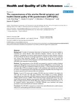
During the trials using the BWS system, participants were
mechanically supported in a harness with adjustable belts
and padded straps for the thighs, similar to the one used
by Norman et al. [17], which was attached to a horizontal
bar. A steel cable from an electric motor pulled the hori-
zontal bar upward and slid it through an upper rail as the
participants walked. A load cell connected the horizontal
bar to the cable and measured the amount of weight
borne by the BWS system, which was shown on a digital
display. In order to support the weight, participants stayed
still until the motor was activated by the experimenter,
who lengthened or shortened the cable to bear the desired
amount of body weight. Figure 1 illustrates the BWS sys-
tem used in the present study.
Passive reflective markers were placed on the nonparetic
and paretic sides of the body at the following anatomical
locations: head of the fifth metatarsal, lateral malleolus,
lateral epicondyle of the femur, greater trochanter, and
acromion, in order to define the foot, shank, thigh, and
trunk segments, respectively. The digitalization and the
reconstruction of all markers were performed using Ariel
Performance Analysis System - APAS (Ariel Dynamics,
Inc.) software, and filtering and posterior analyses were
performed using Matlab software (MathWorks, Inc. - Ver-
sion 6.5). Reconstruction of the real coordinates was per-
formed using the direct linear transformation (DLT)
procedure.
Data analysis
One intermediate stride per trial by each participant, for a
total of three selected trials for each condition, was ana-
lyzed. The trial selection was determined by the best visu-
alization of the markers and walking performance in an
uninterrupted trial. Through visual inspection, a stride
(walking cycle) was defined by two consecutive initial
contacts of the same limb to the ground along the progres-
sion line. In addition, walking events during a stride were
identified for subsequent calculation of walking temporal
organization (initial and terminal double stance, single
limb support, and swing period [27]). This procedure was
carried out for both nonparetic and paretic sides of the
body
All the data were digitally filtered using a 4
th
order and
zero-lag Butterworth filter and all markers were low-pass
filtered at 8 Hz. For joint and segmental angles, strides
were normalized in time from 0 to 100%, with a 1% step.
These cycles were referenced to the participants' neutral
angles measured during the calibration trial in each con-
dition and were then averaged to obtain the mean cycle
Partial view of the body weight support system used in the studyFigure 1
Partial view of the body weight support system used
in the study. The rail that the electric motor slides along,
the load cell, and one of the experimenters wearing the har-
ness are shown.
Journal of NeuroEngineering and Rehabilitation 2009, 6:43 />Page 4 of 10
(page number not for citation purposes)
for each participant. The same procedure was repeated to
obtain the mean cycle among participants.
The following variables were examined: mean walking
speed, calculated as the ratio between the distance
traveled and its duration (determined by the position of
the greater trochanter marker, which is closer to the center
of body mass); stride length, the distance between two
successive initial contacts of each foot to the ground
(determined by the position of the lateral malleolus
marker); stride speed, calculated as the ratio between
stride length and duration; durations of total double
stance and single limb support; ankle, knee, and hip joint
range of motion, calculated from the difference between
the maximum and minimum angles of these joints during
each stride cycle; and foot, shank, thigh, and trunk seg-
ment range of motion, calculated from the difference
between the maximum and minimum angles of these seg-
ments during each stride cycle. The movements of the seg-
ments were counter-clockwise (backward) and clockwise
(forward) rotations around the medial-lateral axis on the
sagittal plane, which denoted positive and negative val-
ues, respectively [28]. For example, a counter-clockwise
rotation of the trunk means trunk extension from neutral
position and a clockwise rotation means trunk flexion
from neutral position.
Statistical analysis
For all variables, data from three trials under each condi-
tion were averaged for each participant. A one-way analy-
sis of variance (ANOVA) was conducted, using the three
experimental conditions (no harness, 0% BWS, 30%
BWS) as factors. Four multivariate analyses of variance
(MANOVAs) were employed, using body side (nonparetic
and paretic) and the three experimental conditions as fac-
tors. The dependent variables were mean walking speed
for the ANOVA, cadence, stride length, and stride speed
for the first MANOVA; durations of initial double stance,
single limb support, terminal double stance, and swing
period for the second MANOVA; ankle, knee, and hip
joint range of motion for the third MANOVA; and foot,
shank, thigh, and trunk segmental range of motion for the
fourth MANOVA. When applicable, univariate analyses
and Tukey post hoc tests were employed. An alpha level of
0.05 was adopted for all statistical tests, which were per-
formed using SPSS software (Version 10.0).
Results
All participants performed the requested tasks. None used
assistive devices during walking performance; however,
three participants needed assistance from a physical ther-
apist that hold one of their hands, in order to support bal-
ance when walking with no harness. The results for
walking spatial-temporal parameters and for joint and
segmental pattern and range of motion follow.
Temporal-spatial gait parameters
Table 1 depicts mean and standard deviation (± SD) of the
walking cycle temporal-spatial parameters. Walking speed
was different among conditions, F(2,24) = 5.56, p = 0.02,
in which it was lower in the 30% BWS than in the no har-
ness condition. MANOVA revealed condition signifi-
cance, Wilks' Lambda = 0.54, F(6,44) = 2.65, p = 0.003,
and condition and body side interaction, Wilks' Lambda
= 0.37, F(6,44) = 4.68, p = 0.001. Univariate analyses indi-
cated condition effect and condition and body side inter-
action for stride length, F(2,24) = 8.39, p = 0.007, F(2,24)
= 12.41, p < 0.001, and stride speed, F(2,24) = 4.96, p =
0.029, F(2,24) = 16.31, p < 0.001, respectively. Stride
length was shorter and stride speed was lower in the 30%
BWS than in the no harness and 0% BWS conditions, and
in the 0% BWS than in the no harness condition. The
Table 1: Temporal-spatial parameters of walking during the stride cycle.
Outcome measures Conditions
No Harness 0% BWS 30% BWS
Nonparetic Paretic Nonparetic Paretic Nonparetic Paretic
Walking speed (m/s) 0.41 ± 0.24
a
0.38 ± 0.23 0.30 ± 0.14
a
Cadence (steps/min) 70.82 ± 20.63 70.73 ± 22.32 70.53 ± 21.02 71.48 ± 22.19 69.50 ± 14.53 72.13 ± 14.52
Stride Length (m) 0.63 ± 0.20 0.63 ± 0.20
a
0.58 ± 0.19 0.58 ± 0.18
b
0.48 ± 0.16 0.52 ± 0.16
a, b
Stride Speed (m/s) 0.40 ± 0.22 0.40 ± 0.23
a
0.36 ± 0.22 0.37 ± 0.22
b
0.28 ± 0.12 0.32 ± 0.13
a, b
Initial double stance (%) 26.59 ± 11.71 21.93 ± 12.52 27.11 ± 11.33 22.42 ± 11.53 27.68 ± 9.66 19.14 ± 7.92
Single limb support (%) 32.19 ± 9.34
1
18.49 ± 7.02
1
30.56 ± 9.19
2
18.09 ± 7.05
2
31.56 ± 8.30
3
18.42 ± 5.13
3
Terminal double stance (%) 21.82 ± 9.58 26.46 ± 12.07 23.54 ± 10.84 27.53 ± 11.73 22.23 ± 8.27 28.69 ± 9.16
Swing period (%) 19.40 ± 6.76
1
33.12 ± 10.18
1
18.79 ± 7.13
2
31.96 ± 9.81
2
18.54 ± 4.87
3
33.75 ± 8.74
3
Mean (± SD) values of mean walking speed, cadence, stride length, stride speed, duration of initial double stance, single limb support, terminal
double stance, and swing period, during stride cycle in the three conditions (no harness, 0% of BWS, and 30% of BWS) on nonparetic and paretic
body sides of individuals with chronic stroke (n = 13). Note: same letter indicates difference between conditions; same number indicates difference
between body sides.
Journal of NeuroEngineering and Rehabilitation 2009, 6:43 />Page 5 of 10
(page number not for citation purposes)
paretic side displayed longer stride length and faster stride
speed than the nonparetic side only in the 30% BWS con-
dition (Table 1).
Regarding temporal measures, MANOVA only revealed
significant body side effect, Wilks' Lambda = 0.14, F(4,9)
= 13.71, p = 0.001. Univariate analyses indicated that the
nonparetic side displayed longer single limb support,
F(1,12) = 53.36, p < 0.001, and shorter swing period dura-
tion, F(1,12) = 65.88, p < 0.001, than the paretic side of
the body (Table 1).
Joint and segmental angles
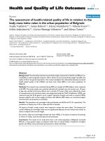
Figure 2 shows the mean (± SD) stride cycle of ankle,
knee, and hip angle patterns in the three conditions (no
harness, 0% BWS, and 30% BWS) for paretic and non-
paretic sides of the body. Qualitatively, the joints of either
side have a similar pattern amongst conditions. However,
joint angles between sides presented a remarkably differ-
ent pattern.
The ankle joint of the paretic side showed plantar flexion
during most of the gait cycle, and little dorsiflexion during
middle stance (approximately 40% of the cycle) in the
three conditions (Figure 2, upper panel). On the other
hand, the ankle of nonparetic side showed marked dorsi-
flexion later in the cycle. The knee joint (Figure 2, middle
panel) showed little flexion on the paretic side consider-
ing that this joint on the nonparetic side presented a much
larger flexion at swing period (approximately 85% of gait
Ankle, knee, and hip joint angles during the stride cycleFigure 2
Ankle, knee, and hip joint angles during the stride cycle. Mean (± SD) stride cycle of ankle, knee, and hip joint angles
for the individuals with chronic stroke walking with no harness (A), with 0% BWS (B), and 30% BWS (C) on nonparetic (gray
area) and paretic (line) body sides. Positive values denote ankle dorsiflexion, knee and hip flexion, and negative values denote
ankle plantar flexion, knee and hip extension (n = 13).
Journal of NeuroEngineering and Rehabilitation 2009, 6:43 />Page 6 of 10
(page number not for citation purposes)
cycle) in the three conditions. Finally, the hip joint (Figure
2, bottom panel) showed a flexor pattern with little exten-
sion during the entire cycle for both sides. However, the
hip on the nonparetic side showed greater flexion than the
hip on the paretic side in the three conditions.
Table 2 depicts mean (± SD) joint range of motion during
the walking cycle. MANOVA revealed joint range of
motion had significant difference for conditions, Wilks'
Lambda = 0.52, F(6,44) = 2.87, p = 0.02, body side, Wilks'
Lambda = 0.09, F(3,10) = 33.73, p < 0.001, and condition
and body side interaction tendency, Wilks' Lambda =
0.58, F(6,44) = 2.28, p = 0.053. The hip joint was influ-
enced by condition, F(2,24) = 10.49, p = 0.004, with a
greater range of motion in the no harness condition than
in the 30% BWS condition and a greater range of motion
in the 0% BWS than in the 30% BWS condition. Range of
motion was greater on the nonparetic side for the ankle,
F(1,12) = 21.98, p = 0.001, knee, F(1,12) = 41.91, p <
0.001, and hip, F(1,12) = 102.97, p < 0.001, than in the
paretic side (Table 2).
Figure 3 shows the mean (± SD) stride cycle of foot, thigh,
shank, and trunk angle patterns in the three conditions for
paretic and nonparetic sides of the body. Most of segmen-
tal angles displayed similar pattern in the three condi-
tions, however, there were some different patterns
between sides.
The foot remained close to neutral position during most
of the stance period on both sides. The foot on the non-
paretic side presented greater clockwise rotation and later
than the foot on the paretic side in all conditions (Figure
3, upper panel). The same pattern was observed for shank.
The thigh was the only segment that presented a similar
pattern between nonparetic and paretic sides during most
of the gait cycle. The thigh on the nonparetic side showed
a more counter-clockwise rotation than the thigh on the
paretic side (Figure 3, middle panel), except at the end of
the swing period. Finally, the trunk presented an opposite
orientation between nonparetic and paretic sides and was
close to neutral position with 30% BWS (Figure 3, bottom
panel).
Table 2 also displays mean (± SD) segmental range of
motion during the walking cycle. MANOVA revealed seg-
mental range of motion significant difference for condi-
tion, Wilks' Lambda = 0.35, F(8,42) = 3.67, p = 0.003,
body side, Wilks' Lambda = 0.13, F(4,9) = 14.85, p =
0.001, and condition and body side interaction, Wilks'
Lambda = 0.24, F(8,42) = 5.54, p < 0.001. Condition
influenced thigh range of motion, F(2,24) = 17.08, p =
0.001, with greater range of motion in the no harness than
in the 0% and 30% BWS conditions and greater range of
motion in the 0% BWS than in the 30% BWS condition.
Body side influenced foot, F(1,12) = 35.77, p < 0.001, and
thigh, F(1,12) = 22.34, p < 0.001, range of motion with
both segments showing a greater range of motion on the
nonparetic than on the paretic side. Finally, condition and
body side interaction was observed for the shank, F(2,24)
= 20.40, p < 0.001, and trunk, F(2,24) = 8.08, p = 0.007,
range of motion. Shank range of motion was decreased
throughout the no harness, 0% BWS, and 30% BWS con-
ditions on both sides, but with a greater decrease on the
nonparetic than on the paretic side. Trunk range of
motion was decreased throughout the no harness, 0%
BWS, and 30% BWS conditions only on the paretic side
and presented a smaller range of motion on the non-
paretic side in the no harness and 0% BWS conditions
than on the paretic side (Table 2).
Table 2: Joint and segmental range of motion during the stride cycle.
Outcome
measurements
Conditions
No Harness 0% BWS 30% BWS
Nonparetic Paretic Nonparetic Paretic Nonparetic Paretic
Joint (degrees)
Ankle 26.22 ± 4.53
1
18.23 ± 9.62
1
25.73 ± 6.03
2
17.90 ± 8.95
2
25.83 ± 7.36
3
17.40 ± 9.43
3
Knee 51.76 ± 6.23
1
27.25 ± 13.81
1
49.32 ± 5.43
2
26.91 ± 14.32
2
47.38 ± 6.46
3
24.74 ± 14.05
3
Hip 34.27 ± 6.49
1
19.72 ± 5.87
1, a
32.73 ± 5.58
2
18.21 ± 5.79
2, b
29.43 ± 5.75
3
17.72 ± 6.18
3, a, b
Segment (degrees)
Foot 58.67 ± 12.77
1
37.90 ± 14.88
1
54.03 ± 11.76
2
36.45 ± 14.40
2
51.74 ± 13.40
3
34.20 ± 15.61
3
Shank* 50.25 ± 9.80
1,4,5
34.21 ± 11.30
1,6,7, a
45.74 ± 8.77
2,4
32.10 ± 11.23
2,6,8, a, b
39.62 ± 7.92
3,5
29.65 ± 11.69
3,7,8, a, b
Thigh 31.43 ± 4.87
1
26.52 ± 5.79
1, a
29.75 ± 3.98
2
24.65 ± 5.31
2, a, b
26.51 ± 3.98
3
21.10 ± 5.97
3, a, b
Trunk* 8.23 ± 2.48 12.77 ± 3.11
1
7.88 ± 2.90 11.82 ± 3.53
2
7.91 ± 2.77 9.35 ± 3.45
1,2
Mean (± SD) values for range of motion of ankle, knee, and hip joints, and foot, thigh, shank, and trunk segments during stride cycle in the three
conditions (no harness, 0% of BWS, and 30% of BWS) on nonparetic and paretic body sides of individuals with chronic stroke (n = 13). Note: same
letter indicates difference between conditions; same number indicates difference between body sides; * indicates interaction
Journal of NeuroEngineering and Rehabilitation 2009, 6:43 />Page 7 of 10
(page number not for citation purposes)
Discussion
This study investigated spatial-temporal gait parameters,
and joint and segmental angles of individuals with
chronic stroke walking at self-selected comfortable speed
on ground level with and without BWS. The results
revealed that the use of BWS system leads to changes in
stride length and stride speed of individuals with chronic
stroke, but not on stance and swing period duration.
Regarding the joint range of motion, the hip was the only
joint that was influenced by the BWS system with the
paretic side presenting less hip joint range of motion dur-
ing walking in the 30% BWS condition than in the no har-
ness condition, and the nonparetic side presenting less
hip joint range of motion in the 30% BWS than in the no
harness and 0% BWS conditions. Finally, regarding the
segmental range of motion, shank and thigh segments
presented less range of motion in the 30% BWS condition
than in the other conditions and less range of motion in
the 0% BWS condition than in the no harness condition.
The trunk on the paretic side presented less range of
motion in the 30% condition than in the other conditions
and difference between paretic and nonparetic sides was
only observed in the 30% BWS condition. These results
did not support our initial suggestion that an individual
with stroke walking with BWS on ground level would
present a more stable and symmetrical gait pattern.
Foot, shank, thigh, and trunk segmental angles during the stride cycleFigure 3
Foot, shank, thigh, and trunk segmental angles during the stride cycle. Mean (± SD) stride cycle of foot, shank, thigh,
and trunk segmental angles for the individuals with chronic stroke walking with no harness (A), with 0% BWS (B), and 30%
BWS (C) on nonparetic (gray area) and paretic (line) body sides. Positive values denote counter-clockwise (backward) rotation
of segments and negative values denote clockwise (forward) rotation of segments (n = 13).
Journal of NeuroEngineering and Rehabilitation 2009, 6:43 />Page 8 of 10
(page number not for citation purposes)
At first glance, it seems that individuals with chronic
stroke had more difficulty walking with BWS on ground
level than without it. However, one of the most important
issues regarding this study is that the BWS system enabled
these individuals to perform the task on a surface that is
used in daily life activities and none required assistance to
keep their balance because the BWS system enabled them
to walk by themselves safely. In interventions, the BWS
provides physical support instead of the physical thera-
pist, who can then focus attention on the patient's walk-
ing performance. For example, the physical therapist can
focus on increased walking speed and its influence on spa-
tial-temporal parameters and joint patterns [9] in the
patient and correct gait pattern to favor a more symmetri-
cal gait [10,29]. The BWS system also provided steadiness
during the single limb support on the paretic side which
led to a greater joint range of motion during stepping.
These results are quite encouraging for gait training using
BWS on ground level on a long-term basis.
Another positive aspect of walking with BWS on ground
level is the better vertical alignment of the trunk through-
out gait cycle (Figure 3, bottom panel). We had investi-
gated the trunk segment from both sides of the body in
the sagital plane of motion because of the posture that
individuals with stroke usually adopt for walking. This
segment presented different ranges of motion between
nonparetic and paretic sides, which means that the indi-
viduals rotated the trunk (longitudinal axis of motion)
towards the opposite side, which presented the largest
range of motion. In the 30% BWS condition, the trunk
was close to neutral position (i.e. erect) and did not
present any difference between nonparetic and paretic
sides for range of motion. Trunk positioning is a critical
aspect of gait pattern, as its alignment is related to func-
tional performance [30], and it might contribute to a
decreased mechanical energy cost [31]. Therefore, BWS on
ground level contributes to aligning the trunk and pro-
vides advantages during gait performance.
Contrary to previous investigation of walking with BWS
on ground level [15], the participants in this study walked
slower in the 30% BWS than in the no harness condition.
This difference might be attributed to the different proce-
dures adopted in each case. While Lamontagne and Fung
[15] investigated individuals with acute stroke and classi-
fied them according to their walking speed as either low
or high functioning individuals, we evaluated individuals
with chronic stroke and did not classify them according to
their preferred walking speed. Also, we did not encourage
our patients to speed up along the pathway, as Lamon-
tagne and Fung [15] did and also had evaluated their par-
ticipants with stroke walking at preferred walking and
maximal walking speed.
Slow walking speed in the 30% BWS condition would be
due to decreased posterior muscle energy generation by
the lower limb at the end of terminal double stance. This
aspect has been described as fundamental to propel the
limb forward to control the walking speed [32]. We had
adopted 30% BWS for this study as it has been the most
common percentage of body weight support used during
gait training with BWS on treadmill and it was the per-
centage used in the previous study on ground level [15].
However, it seems that this percentage for walking with
BWS on ground level might not be as appropriate as it is
for walking with BWS on treadmill, because it may pre-
vent ground reaction force generation and, consequently,
the impulse to move the limb forward. In this way, future
studies using BWS on ground level in individuals with
chronic stroke should investigate a more appropriate per-
centage of body weight support for this type of surface.
Further, BWS systems that can be modulated dynamically
according to the gait phase have been proposed for tread-
mill [33] and should also be considered for ground level.
An unexpected finding was a longer stride length on the
paretic side than on the nonparetic side in the 30% BWS
condition. Any human walking on a straight line should
present the same stride length on both sides [34], but this
was not the case in the present study. One possible expla-
nation for this finding could be that individuals with
chronic stroke took advantage of the body weight support
on the single limb of the nonparetic side to generate a
longer and quicker step with the paretic limb.
Our results, as in the previous investigation [15], also
showed that BWS itself did not change gait asymmetry
between nonparetic and paretic sides among the experi-
mental conditions, which is a prominent characteristic of
hemiparetic gait [35,36]. However, it is possible that side
asymmetry might decrease only after a gait training period
with BWS on ground level, although this hypothesis still
needs to be further investigated.
Last but not least, the 0% BWS did not influence the mean
walking speed, temporal symmetry, ankle, knee, foot, and
trunk ranges of motion. Although the harness was
employed mainly to help with balance, it also contributed
to shortening the stride length, lowering stride speed, and
reducing hip, shank, and thigh range of motion when
compared to the no harness condition. These reductions
were lower in the 0% BWS condition than in the 30%
BWS condition. Thus, the use of harness itself was already
enough to change the gait pattern of individuals with
stroke. This result might be due to the BWS system
adopted in this study because it required the individuals
to move the motor along the rail and to a lack of sufficient
adaptation to this walking requirement before taking part
in the study. In future studies, use of a BWS system for
Journal of NeuroEngineering and Rehabilitation 2009, 6:43 />Page 9 of 10
(page number not for citation purposes)
ground level in which the motor is moved along the rail
by a specific controller rather than by the participant wear-
ing the harness, should be considered. Actually, we are
currently working on the system in order to implement
such a condition.
To our knowledge, this was the first study that considered
a more detailed description of walking with BWS on
ground level in individuals with stroke and it presented
some limitations. First, a full understanding of gait
requires more analyses than just the kinematic approach,
such as kinetic and electromyographyc analyses. Second,
the need to move the motor through the rail by the partic-
ipants creates a drag force as they walked and this can
influence walking performance and pattern. Third, only
the 0% and 30% of BWS were analyzed and participants
might take advantage of other percentages of body weight
unloading especially due to the difficulty in force produc-
tion to move forward in the 30% of BWS condition.
Finally, the adaptation period provided to the participants
might have not been long enough and this could have
masked some of the effects of BWS use. Despite all these
limitations, the use of BWS system overground seems to
be a useful and important strategy as a tool to provide an
alternative intervention and rehabilitation program for
individuals with stroke.
Conclusion
Individuals with stroke using BWS system on ground level
walked slower and with shorter stride length and slower
stride speed, respectively, than with no harness. BWS also
led to a reduction in hip, shank, and thigh range of
motion. However, this system did not change walking
temporal organization and the body side asymmetry of
individuals with stroke. The differences found in this
study might be attributed to the adjustments the individ-
uals had to make to walk with an unloading condition on
the lower limb, and to the brief period of adaptation to
the BWS system, as the use of the harness without support
of body weight (0% BWS condition) per se leads to some
alterations during the task performance.
Although the use of BWS system on ground level changed
some gait parameters, this system enabled individuals
with chronic stroke to walk safely and without physical
assistance. In interventions, the physical therapist can
focus on watching and correcting the individual's gait pat-
tern during performance instead of providing physical
assistance.
Competing interests
The authors declare that they have no competing interests.
Authors' contributions
COS and AMFB were responsible for conception and
design of the study, acquisition of data, analysis and inter-
pretation of data, and drafting the article. CLPM was
responsible to acquisition of data, analysis and interpreta-
tion of data, drafting the article. TFS and JAB were respon-
sible for interpretation of data and revising it critically for
scientific method and content. All authors read and
approved the final manuscript.
Acknowledgements
This work was supported by CNPq (Process #470421/2006-1). C.O. Sousa
and A.M.F. Barela are grateful to CNPq for their Masters scholarship
(830804/99-4) and Post-Doc fellowship (151893/2006-2), respectively, and
C.L. Prado-Medeiros is grateful to FAPESP for her doctoral scholarship
(200704503-6). All authors acknowledge P.H. Lobo da Costa for making
the use of the laboratory where this study took place possible, and thank
the individuals with stroke that participated in the study for their contribu-
tions.
References
1. Olney SJ, Richards C: Hemiparetic gait following stroke. Part I:
Characteristics. Gait Posture 1996, 5:136-148.
2. Hesse S, Uhlenbrock D, Werner C, Bardeleben A: A mechanized
gait trainer for restoring gait in nonambulatory subjects.
Arch Phys Med Rehabil 2000, 81:1158-1161.
3. Kerrigan DC, Xenopoulos-Oddsson A, Sullivan MJ, Lelas JJ, Riley PO:
Effect of a hip flexor-stretching program on gait in the eld-
erly. Arch Phys Med Rehabil 2003, 84:1-6.
4. Hesse S, Konrad M, Uhlenbrock D: Treadmill walking with par-
tial body weight support versus floor walking in hemiparetic
subjects. Arch Phys Med Rehabil 1999, 80:421-427.
5. Visintin M, Barbeau H, Korner-Bitensky N, Mayo NE: A new
approach to retrain gait in stroke patients through body
weight support and treadmill stimulation. Stroke 1998,
29:1122-1128.
6. Barbeau H, Rossignol S: Recovery of locomotion after chronic
spinalization in the adult cat. Brain Res 1987, 412:84-95.
7. Lovely RG, Gregor RG, Roy RR, Edgerton VR: Effects of training
on the recovery of full-weight-bearing stepping in the adult
spinal cats. Exp Neurol 1986, 92:421-435.
8. McCrea DA: Spinal circuitry of sensoriomotor control of loco-
motion. J Physiol 2001, 533:41-50.
9. van Hedel HJ, Tomatis L, Muller R: Modulation of leg muscle
activity and gait kinematics by walking speed and body-
weight unloading. Gait Posture 2006, 24:35-45.
10. Carr JH, Shepherd RB: Neurological rehabilitation: optimizing motor per-
formance Oxford: Butterworth-Heinemann; 1998.
11. Barbeau H, Wainberg M, Finch L: Description and application of
a system for locomotor rehabilitation. Med Biol Eng Comput
1987, 25:341-344.
12. Harris-Love ML, Macko RF, Whitall J, Forrester LW: Improved
hemiparetic muscle activation in treadmill versus over-
ground walking. Neurorehabil Neural Repair 2004, 18:154-160.
13. Lindquist ARR, Prado CL, Barros RML, Mattioli R, Lobo da Costa PH,
Salvini TF: Gait training combining partial body-weight sup-
port, a treadmill, and functional electrical stimulation:
effects on poststroke gait. Phys Ther 2007, 87:1144-1154.
14. Werner C, Bardeleben A, Mauritz K-H, Kirker S, Hesse S: Treadmill
training with partial body weight support and physiotherapy
in stroke patients: a preliminary comparison. Eur J Neurol
2002, 9:639-644.
15. Lamontagne A, Fung J: Faster is better: implications for speed-
intensive gait training after stroke. Stroke 2004, 35:2543-2548.
16. Richards CT, Malouin F, Wood-Dauphinee S, Williams JI, Bouchard JP,
Brunet D: Task-specific physical therapy for optimization of
gait recovery in acute stroke patients. Arch Phys Med Rehabil
1993, 74:612-620.
17. Norman KE, Pepin A, Ladouceur M, Barbeau H: A treadmill appa-
ratus and harness support for evaluation and rehabilitation
of gait. Arch Phys Med Rehabil 1995, 76:772-778.
18. Alton F, Baldey L, Caplan S, Morrisey MC: A kinematic compari-
son of overground and treadmill walking. Clin Biomech 1998,
13:434-440.
Publish with BioMed Central and every
scientist can read your work free of charge
"BioMed Central will be the most significant development for
disseminating the results of biomedical research in our lifetime."
Sir Paul Nurse, Cancer Research UK
Your research papers will be:
available free of charge to the entire biomedical community
peer reviewed and published immediately upon acceptance
cited in PubMed and archived on PubMed Central
yours — you keep the copyright
Submit your manuscript here:
/>BioMedcentral
Journal of NeuroEngineering and Rehabilitation 2009, 6:43 />Page 10 of 10
(page number not for citation purposes)
19. Bayat R, Barbeau H, Lamontagne A: Speed and temporal-distance
adaptations during treadmill and overground walking follow-
ing stroke. Neurorehabil Neural Repair 2005, 19:115-124.
20. Warabi T, Kato M, Kiriyama K, Yoshida T, Kobayashi M: Treadmill
walking and overground walking of human subjects com-
pared by recording sole-floor reaction force. Neurosci Res
2005, 53:343-348.
21. Lee SJ, Hidler J: Biomechanics of overground versus treadmill
walking in healthy individuals. J Appl Physiol 2008, 104:747-755.
22. Stolze H, Kuhtz-Buschbeck JP, Mondwurf C, Boczek-Funcke A, Johnk
K, Deuschl G, Illert M: Gait analysis during treadmill and over-
ground locomotion in children and adults. Electroencephalogr
Clin Neurophysiol 1997, 105:490-497.
23. Warabi T, Kato M, Kiriyama K, Yoshida T, Kobayashi N: Treadmill
walking and overground walking of human subjects com-
pared by recording sole-floor reaction force. Neurosci Res
2005, 53:343-348.
24. Wass E, Taylor N, Matsas A: Familiarisation to treadmill walk-
ing in unimpaired older people. Gait Posture 2005, 21:72-79.
25. Matsas A, Taylor N, McBurney H: Knee joint kinematics from
familiarised treadmill walking can be generalised to over-
ground walking in young unimpaired subjects. Gait Posture
2000, 11:46-53.
26. Barbeau H, Lamontagne A, Ladouceur M, Mercier I, Fung J: Optimiz-
ing locomotor function with body weight support training
and functional electrical stimulation. In Progress in motor control:
effects of age, disorders, and rehabilitation Volume 2. Edited by: Latash
ML, Levin MF. Champaign, IL: Human Kinetics; 2004:237-251.
27. Perry J: Gait analysis Throfare: Slack; 1992.
28. Barela AMF, Stolf SF, Duarte M: Biomechanics characteristics of
adults walking in shallow water and on land. J Electromyogr Kine-
siol 2006, 16:250-256.
29. Dobkin BH: An overview of treadmill locomotor training with
partial body weight support: a neurophysiologically sound
approach whose time has come for randomized clinical tri-
als. Neurorehabil Neural Repair 1999, 13:157-165.
30. Hirose D, Ishida K, Nagano Y, Takahashi T, Yamamoto H: Posture
of the trunk in the sagittal plane is associated with gait in
community-dwelling elderly population. Clin Biomech 2004,
19:57-63.
31. Chen G, Patten C, Kothari DH, Zajac FE: Gait differences
between individuals with post-stroke hemiparesis and non-
disabled controls at matched speeds. Gait Posture 2004,
22:51-56.
32. Riley PO, Della Croce U, Kerrigan DC: Propulsive adaptation to
changing gait speed. J Biomech 2001, 34:197-202.
33. Franz JR, Glauser M, Riley PO, Della Croce U, Newton F, Allaire PE,
Kerrigan DC: Physiological modulation of gait variables by an
active partial body weight support system. J Biomech 2007,
40:3244-3250.
34. Winter DA: The biomechanics and motor control of human gait: normal,
elderly, and pathological 2nd edition. Waterloo: University of Waterloo
Press; 1991.
35. Wall JC, Turnbull GI: Gait asymmetries in residual hemiplegia.
Arch Phys Med Rehabil 1986, 67:550-553.
36. Olney SJ, Richards C: Hemiparetic gait following stroke. Part I:
Characteristics. Gait Posture 1996, 5:136-148.