báo cáo hóa học: " The design and testing of a novel mechanomyogram-driven switch controlled by small eyebrow movements" docx
Bạn đang xem bản rút gọn của tài liệu. Xem và tải ngay bản đầy đủ của tài liệu tại đây (676.3 KB, 10 trang )
JNER
JOURNAL OF NEUROENGINEERING
AND REHABILITATION
Alves and Chau Journal of NeuroEngineering and Rehabilitation 2010, 7:22
/>Open Access
RESEARCH
© 2010 Alves and Chau; licensee BioMed Central Ltd. This is an Open Access article distributed under the terms of the Creative Com-
mons Attribution License ( which permits unrestricted use, distribution, and reproduc-
tion in any medium, provided the original work is properly cited.
Research
The design and testing of a novel
mechanomyogram-driven switch controlled by
small eyebrow movements
Natasha Alves
1,2
and Tom Chau*
1,2
Abstract
Background: Individuals with severe physical disabilities and minimal motor behaviour may be unable to use
conventional mechanical switches for access. These persons may benefit from access technologies that harness the
volitional activity of muscles. In this study, we describe the design and demonstrate the performance of a binary switch
controlled by mechanomyogram (MMG) signals recorded from the frontalis muscle during eyebrow movements.
Methods: Muscle contractions, detected in real-time with a continuous wavelet transform algorithm, were used to
control a binary switch for computer access. The automatic selection of scale-specific thresholds reduced the effect of
artefact, such as eye blinks and head movement, on the performance of the switch. Switch performance was estimated
by cued response-tests performed by eleven participants (one with severe physical disabilities).
Results: The average sensitivity and specificity of the switch was 99.7 ± 0.4% and 99.9 ± 0.1%, respectively. The
algorithm performance was robust against typical participant movement.
Conclusions: The results suggest that the frontalis muscle is a suitable site for controlling the MMG-driven switch. The
high accuracies combined with the minimal requisite effort and training show that MMG is a promising binary control
signal. Further investigation of the potential benefits of MMG-control for the target population is warranted.
Background
Individuals with severe physical disabilities often use
access technologies as an alternative means of communi-
cation, environmental control or computer access. By
providing a switching interface that the user is capable of
controlling, access technologies promote an individual's
independence and participation in daily living tasks [1].
Depending on the user's physical abilities, switching
interfaces may range from simple mechanical buttons to
brain-computer interfaces [2]. Often, individuals who are
severely disabled may retain the ability to contract certain
muscles. For example, individuals with high-level spinal
cord lesions may have sufficient muscle control to move
their head [3], and may therefore be able to use mechani-
cal head-switches, tilt switches [4], or head-operated joy-
sticks [5]. In cases where the individual lacks a high
degree of motor function, an alternative solution is to use
the remaining contractile ability of muscles.
Conventional muscle-based devices are controlled by
electromyogram (EMG) signals from viable muscle sites
of the hand, foot, cheek or forehead [6,7], and are com-
mercially available (eg. The Impulse™ Switch by
AbleNet
®
). The advantage of using muscle activity as the
switching control for access devices is that physical
movement is unnecessary, enabling the user to control
the device even when only weak volitional muscle activity
exists. Further, once the muscle site is located and the
sensor is attached to the skin, switch performance is not
compromised by misalignment of switch position due to
body movements. This is an advantage over non-contact
switches controlled by physical movement, such as infra-
red detectors (ex. IST switch by Words+
®
), optical detec-
tors [8], or vision-based movement detectors [9,10], that
are sensitive to the position of the sensor with respect to
the access site on the body.
* Correspondence:
1
Bloorview Research Institute, Bloorview Kids Rehab, Toronto, Ontario, Canada
Full list of author information is available at the end of the article
Alves and Chau Journal of NeuroEngineering and Rehabilitation 2010, 7:22
/>Page 2 of 10
In addition to exhibiting changes in electrical activity
detected by EMG, a contracting muscle also shows
changes in its mechanical activity. The mechanical index
of muscle contraction is known as the mechanomyogram
(MMG). MMG is generated from gross lateral movement
of the muscle at the initiation of a contraction, smaller
subsequent lateral oscillations at the resonant frequency
of the muscle, and dimensional changes of active muscle
fibers [11-13]. MMG may be measured by microphones
[14], piezoelectric contact sensors [15,16], accelerometers
[17] or laser distance sensors [18] on the surface of the
skin. Although MMG has found important applications
in the assessment of muscle pathologies such as pain [19],
fatigue [20,21] and disease [22], it has been under-studied
as a control signal for alternative access. MMG may offer
several advantages over conventional EMG muscle moni-
toring. It provides a better estimation of the inflection
points in motor-unit recruitment and firing rate [23].
Since it is a mechanical signal, it is not influenced by skin
impedance changes and does not require skin prepara-
tion. This makes it suitable for monitoring muscles when
the overlying skin is prone to perspiration. Because
MMG is typically measured by a single small sensor, it
occupies a smaller footprint on the skin than differential
EMG electrodes, making it suitable for non-invasive
monitoring of smaller muscles. The single-sensor mea-
surement is not dependent on the alignment along the
muscle fibre axis, and is therefore less prone to faulty sig-
nal recordings when the user or caregiver may be unfa-
miliar with muscle anatomy. In addition, since MMG
sensors are reusable, once purchased, they may be less
expensive than disposable EMG electrodes. Because of
these potential advantages, MMG has been investigated
as a control signal for upper-limb prostheses [24,25] and
powered orthotic devices [26]. Offline pattern recogni-
tion methods have shown that multi-site MMG signals
are discernable during different patterns of forearm mus-
cle contraction [27,28], indicating that MMG may find
applications in multifunction control of access devices.
In this study we demonstrate an MMG-based binary
switch and test its performance in detecting contractions
of the frontalis muscle during small eyebrow movements.
It has previously been reported that eyebrow movements
may be used as a switch for users with pervasive motor
impairments [8]. Although binary switches have limited
functionality, they are of profound importance in
enabling individuals with severe disabilities to achieve
interaction with, and control of, their environment. By
enabling the user to activate toys, speech output systems,
light displays, and computer access via scanning key-
boards, binary switches help the individual to overcome
barriers to access.
The challenge in the design of an MMG-driven switch
is to reliably convert the MMG signal into a switch-acti-
vation signal. To this end, we describe a real-time wave-
let-based contraction detection algorithm in sections A-
D. The switch is designed to harness small contractions of
the frontalis muscle in real-time, while being resilient to
artefact such as eye-blinks and head movements that
commonly compromise the MMG signal. In sections E
and F, we describe tests on able-bodied individuals to
demonstrate the real-time performance of the detection
algorithm, assessed in a single-switch paradigm, when
user-dependent errors are minimal. We further examine
the accessibility of the MMG switch by testing it on an
individual with severe physical disabilities. The paper
concludes with a presentation and discussion of the
empirical results.
Methods
A. Instrumentation
MMG was measured by a microphone-based sensor
manufactured according to the method of Silva et al. [29].
A program was written in LabView to perform real-time
data acquisition, contraction detection and switch activa-
tion. Microphone-detected MMG signals were continu-
ously sampled at 1 KHz (NI USB-6210, National
Instruments). The LabView program allowed online
modification of parameters such as switch debounce time
and activation thresholds, and provided the user with
visual and auditory feedback when a muscle contraction
was detected. On detecting a contraction, the DTR pin on
a serial port of the computer was asserted. The serial port
was interfaced with a conventional 1/8" mono-plug via an
opto-isolator (4N36, Motorola Inc) to provide a standard
switch output. A keyboard interface (KE-USB36, Hag-
strom Electronics) was used with the mono-plug for
computer access.
B. Contraction detection algorithm
Microphone signals were band-pass filtered with a 5
th
order Butterworth filter with a cut-off frequency range of
5-100 Hz. The low cut-off attenuates the effects of move-
ment [30], while the high cut-off attenuates any noise
beyond the accepted MMG signal range.
The contraction detection algorithm used in this study
is a modification of the off-line activity-detection algo-
rithm proposed by Alves and Chau [31]. In this study,
continuous-wavelet-transform (CWT) coefficients of the
MMG signal are compared to scale-specific thresholds to
identify voluntary muscle activity of the frontalis muscle
during small eyebrow raises. The CWT is defined as
CWT k a
a
xt
tk
a
dt
x mmg
mmg
(,; ) () ,
yy
=
−
⎛
⎝
⎜
⎞
⎠
⎟
−∞
∞
∫
1
(1)
Alves and Chau Journal of NeuroEngineering and Rehabilitation 2010, 7:22
/>Page 3 of 10
where x
mmg
is the filtered MMG signal, and ψ is a
mother wavelet shifted by k and scaled by a (k, a ᑬ).
In the contraction-detection scheme, CWT transform
coefficients at 14 scales, a, were compared to scale-spe-
cific thresholds, h(a), derived from baseline recordings. A
muscle contraction event, z, is detected at sample k when
the coefficients of at least j scales exceed their thresholds,
i.e.
and
where K
baseline
are the samples corresponding to the
baseline MMG signals and γ is the threshold-scaling fac-
tor.
The scaling-factor γ could be varied between 1.2 and
2.5 in increments of 0.2. The value of j was set to 1. CWT
analysis was performed on 100 ms long MMG signals,
using the sym7 mother wavelet at scales with pseudo-fre-
quencies that spanned the 5-100 Hz frequency range of
interest, i.e. a {7,9,10,12,14,15,17,20,23,28,35,46,69,115}.
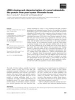
C. Post processing, noise detection and switch debouncing
Figure 1 shows the procedure for converting the continu-
ously acquired microphone signal, x, to a switch activa-
tion signal. CWT analysis was performed on the MMG
signal, x
mmg
, using non-overlapping sliding windows, 100
ms in length. The output of CWT analysis is a muscle
activity event, z [k], for each sample, k, of the windowed
MMG signal. To reduce the probability of spurious activ-
ity being detected as voluntary contractions, when fewer
than 10 ms of activity was detected in the 100 ms window,
the activity was not considered a valid muscle event, i.e.
where m is the current window, and K = 100 is the win-
dow size.
CWT coefficients of MMG signals during eyebrow
movement exceed those of artefact such as eyeblink and
head movement. However, high-amplitude artefacts are
observed in the MMG signal when the sensor is being
moved during activities such as donning, doffing or
adjusting the sensor position. While both contractions
and movement are detected in the microphone signal
associated with MMG (5-100 Hz), movement is more
prominent and differentiable in the high-frequency
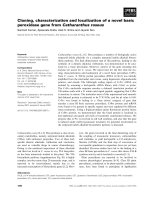
microphone signal (100-300 Hz). Figure 2 shows an
example of the low-frequency (MMG) and high-fre-
quency components of the microphone signal during
muscle contraction and sensor movement. The RMS of
the high-frequency signal, x
hf
, shows good separation
during contraction and sensor movement, and was there-
fore used to detect noise, n, at each window m of length K
= 100 samples, i.e.
where threshold
τ
is determined from the maximum
RMS of x
hf
during contraction. The noise event indicator
was asserted if noise was detected in any of the M preced-
ing windows, i.e.
zk
if CWT k a h a j
otherwise
x
a
mmg
[; ,]
,(,;)(;)
,
yg
yg y
=
>⋅
{}
≥
⎧
⎨
⎪
⎩
⎪
∑
1
0
,,
ha CWT ka
kK
x
baseline
mmg
(; ) max{ (,; )},
yy
=
∈
(3)
Muscle event m
if z k
otherwise
k
K
_[]
,[]
,
,=
≥
⎧
⎨
⎪
⎪
⎩
⎪
⎪
=
∑
110
0
1
(4)
nm
if
K
xk
otherwise
hf
k
K
[]
,[]
,
,=
≥
⎧
⎨
⎪
⎪
⎩
⎪
⎪
=
∑
1
1
0
2
1
t
(5)
Noise event m n m i
i
M
_[] [].=−>
=
−
∑
0
1
0
(6)
Figure 1 Switch activation scheme. Here x, x
mmg
and x
hf
are the microphone, MMG and high-frequency filtered signals, respectively; γ is the thresh-
old scaling factor; z is the muscle-contraction event signal; and
τ
is the threshold that separates contraction from sensor movement.
(2)
Alves and Chau Journal of NeuroEngineering and Rehabilitation 2010, 7:22
/>Page 4 of 10
In this implementation M was set to 5, thus disabling
the switch if noise was detected in the preceding 500 ms.
The switch was enabled when a muscle event was
detected and a noise event was absent. To avoid single
contractions that typically last longer than 100 ms from
being converted to multiple switch activations, the switch
output was debounced with an adjustable delay. The
delay was dependent on the speed at which the user could
comfortably raise their eyebrow, and could be adjusted
between 100-600 ms in 100 ms increments.
D. Events included in the baseline signal
The performance of the detection algorithm is pro-
foundly affected by the choice of thresholds, and hence,
the baseline signal that encompasses the artefact
expected during switch use. Even when the forehead is at
rest, the MMG signal recorded at the frontalis muscle is
affected by visually-observable periodic artefact due to
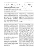
blood flow. As seen in Figure 3, the signal is further com-
promised by artefact due to eye-blinks and head move-
ment. The characteristic MMG signal when the eyebrow
is raised is an oscillatory wave whose amplitude initially
rises and then decays. While the high amplitude at the
initial burst of activity facilitates the detection of contrac-
tion onset, the eventual decay in activity encumbers
activity-detection during sustained contractions. This
limits the potential of a secondary switch activated by
sustained eyebrow raises.
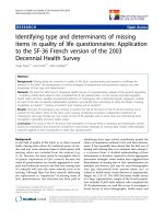
Figure 4 shows the maximum coefficients of the MMG
signal during events such as rest, eye-blink, head move-
ment, quick eyebrow raises and sustained frontalis con-
tractions. The scale-specific thresholds of the detection
algorithm are derived from the maximum coefficient of
baseline MMG signals at each scale. The baseline
includes MMG recorded during rest, blink and head
movement. A contraction is detected if the CWT coeffi-
Figure 2 Signal denoising. The microphone signal and RMS values of the low-frequency (MMG) and high-frequency filtered signals during contrac-
tion and movement.
Alves and Chau Journal of NeuroEngineering and Rehabilitation 2010, 7:22
/>Page 5 of 10
cient of at least one scale exceeds its baseline-derived
threshold. The coefficients of the steady-state MMG dur-
ing sustained contractions, while higher than the coeffi-
cients during rest, are confounded by those during
movement artefact; therefore, sustained muscle activity
cannot be detected. The signal transient at the initiation
of contraction, however, has sufficiently high CWT coef-
ficients to facilitate contraction-detection even during
low-effort eyebrow raises. A quick and small contraction
was therefore chosen as the preferred method for switch
activation.
The detection algorithm was evaluated in real-time to
monitor voluntary activity of the frontalis muscle and to
generate a switch output.
E. Protocol for performance testing
A convenience sample of ten able-bodied individuals (5
male), age 27 ± 2 years, provided written consent to par-
ticipate in the study. These participants, referred to as
A1-A10 in this study, had no previous history of muscu-
loskeletal illness. An adult with C1-C2 incomplete spinal
cord injury (SCI), referred to as B1, was also recruited.
B1's method of access included a sip-and-puff switch for
wheelchair control, a head tracker (TrackerPro
®
, Maden-
tec) for computer mouse emulation, and the dwell func-
tion (250 ms) of the head tracker for emulation of a
mouse click.
Participants were instrumented with an MMG sensor
[29] attached to the frontal belly of the occipitofrontalis
muscle of the forehead with an elastic strap, as shown in
Figure 5. The sensor was placed 1 cm above the eyebrow,
above the inside corner of the right eye. Once the sensor
was affixed, participants performed 30 s of 'baseline'
activities such as blinking, talking, smiling and moving
their head. Scale-specific thresholds were automatically
evaluated from the baseline MMG signals using the con-
traction-detection software written in LabView. The
threshold scaling factor was selectable in the 1.2-2.5
range, and was adjusted for each participant such that
false activations due to blinks and movement were
avoided and participants were able to activate the switch
by raising their eyebrows with minimal effort. Once par-
ticipants demonstrated that they could perform 10 con-
secutive cued switch activations correctly, the threshold
parameters were set and remained unchanged for the
remainder of the experiment.
Custom switch assessment software was written in
Visual Basic to present participants with audio-visual
stimuli and to record the times of switch activation and
stimulus presentation. Participants were presented with a
pseudo-random sequence of numbers at 2 s intervals, and
were asked to activate the switch by raising their eye-
brows slightly when the number "1" was presented. Par-
ticipants performed four trials of the experiment, with a
30 s break in between trials. One-hundred stimuli were
presented during each trial, with the actionable stimulus
(i.e. number 1) being presented 25% of the time.
Throughout the session, participants were encouraged
not to sit absolutely still, but rather to behave in a manner
that they normally would when seated at a desk: they
were free to blink, sway their chair slightly, move their
head and talk without moving their eyebrows or the strap.
The number of true positives (TP), true negatives (TN),
Figure 3 Typical MMG signal recorded from the frontalis muscle during quick and sustained eye-brow raises, eye blinks and head move-
ment.
Alves and Chau Journal of NeuroEngineering and Rehabilitation 2010, 7:22
/>Page 6 of 10
false positives (FP) and false negatives (FN) were
recorded during the cued stimulus tests.
In addition to responding to cued stimuli, participant
B1 typed a pangram for each of two selection modalities:
dwell and eyebrow-raise. For both typing tasks, B1 used
the head-tracker to point to a character on an on-screen
keyboard. For the first task, B1 dwelled at the character's
location for 250 ms to select it; this was the method B1
regularly used for typing for more than seven years. For
the second task, B1 raised his eyebrow to select the char-
acter. The time taken to complete each task was recorded.
After the data-collection trials were completed, all par-
ticipants practiced using the switch for 1 hour, perform-
ing activities such as typing using a scanning keyboard.
At the end of the hour, participants were asked to rate the
level of effort and fatigue associated with controlling the
eyebrow switch on a five-point linear scale: [1-Nothing at
all, not tired; 2- A little, not tired; 3- Moderate, a little
tired; 4- A lot, tired; 5-Too much, very tired]. In addition,
participants were asked to rate if they had to try multiple
times before activating the switch: [1-Never; 2- Very
infrequently; 3- Sometimes; 4- Very often; 5- Almost all
the time].
The experimental protocol was approved by the hospi-
tal and university research ethics boards, and was in com-
pliance with the Declaration of Helsinki.
Figure 4 Typical CWT coefficients of MMG recorded at the frontalis muscle. The maximum coefficients at 14 scales are shown for different con-
traction conditions. The dashed lines depict CWT coefficients of the artefact in the MMG signal during rest, eye blinks and head movements. The max-
imum coefficients across the artefacts are the scale-specific thresholds (x) for contraction-detection. The solid lines depict coefficients for the events
to be detected. Contractions are detected when the CWT coefficient of at least one scale is higher than the threshold. After the initial signal transient,
sustained contractions could not be detected.
Alves and Chau Journal of NeuroEngineering and Rehabilitation 2010, 7:22
/>Page 7 of 10
F. Performan ce M etrics
The sensitivity and specificity of the MMG switch were
evaluated from the cued stimulus test, and are given by
and
Sensitivity is a measure of correctly identified muscle
contractions, while specificity is a measure of correctly
rejected artefacts.
Trends in response delay were used to gauge if partici-
pants were fatigued from prolonged use of the eyebrow
switch. For each participant, a linear regression of
response delay against elapsed session time was evalu-
ated, and the 95% confidence-interval (CI) of the slope
was computed. Here it is assumed that response time
increases with increasing fatigue.
Results
The participant-chosen threshold scaling factor, γ, ranged
from 1.5 to 2.3, and was dependent on what the partici-
pant perceived to be baseline noise. The switch perfor-
mance metrics are shown in Table 1. The switch showed
almost perfect sensitivity and specificity for all partici-
pants. As reported by the participants, activities such as
batting eyelids or involuntary changing facial expressions
sometimes resulted in false detections. Participants
reported that multiple attempts to activate the switch
were infrequent. When required, the multiple attempts
usually included a very small contraction followed by a
stronger contraction. On average, participants rated that
switch activation required very little effort and was not
tiring to use. The response time of only one participant
(A10) had a small but significant (95% CI > 0) increase
over the course of the experiment.
For participant B1, the time required to complete the
typing task with the dwell switch was 63 s, while that for
the eyebrow switch was 54 s. No typing mistakes were
made for either switch modality. In addition, B1 reported
that he perceived the eyebrow switch to have a faster
response-time than the dwell switch.
Discussion
The CWT detection scheme showed very high sensitivity
and specificity in a switch paradigm where activation was
controlled by contractions of the frontalis muscle during
eyebrow raises. CWT detection has been shown to have
comparable sensitivity to RMS and absolute-value mus-
cle-activity detectors, while outperforming these detec-
tors in terms of specificity [31]. The MMG signal is non-
stationary during sustained contractions [27], warranting
the use of time-frequency analysis. The switch required
minimal training, and only the threshold scaling factor
needed adjustment before use. By using scale-specific
thresholds that are dependent on the baseline signal, the
detection scheme can estimate the noise level according
to measurement conditions, and does not require the
user to finely tune each threshold.
Sensitivity
TP
TP FN
=
+
×100,
(7)
Specificity
TN
TN FP
=
+
×100.
(8)
Figure 5 Schematic diagram of equipment set-up.
Alves and Chau Journal of NeuroEngineering and Rehabilitation 2010, 7:22
/>Page 8 of 10
The primary function of the frontalis is to raise the eye-
brow; hence, contraction of the frontalis often accompa-
nies movement of the skin proximal to the eyebrow.
Muscle-contraction detection has some notable advan-
tages over conventional movement-controlled switches.
First, commercially available non-contact movement-
triggered switches (ex. IST switch by Words+
®
) are sensi-
tive to the position of the transducer relative to the access
site, and may pose safety hazards when the transducer is
mounted by supports that are in close proximity to the
eye. Second, movement-based detectors often require
prominent movement, and hence, require more effortful
muscle contractions which may be fatiguing for the user.
This has been seen in the abandonment of an accelerom-
etry-based access solution, where movement of a head-
band during eyebrow raises was used for switch control
[32]. The muscle-based switch, in contrast, required little
effort for activation, as demonstrated by qualitative par-
ticipant feedback and the trends in response time. Fur-
ther, as a control site, the frontalis muscle is broad and
has a large surface area on the forehead, thus offering
flexibility with sensor placement.
The MMG signal is generated by the unfused mechani-
cal activities of motor units. The bulk movement of the
muscle and asynchronous activation of fibers at the initi-
ation and end of contraction creates a high-amplitude
transient that is easily detected. During a sustained con-
traction however, because of the fusion of motor unit
activity [33], the differentiation between muscle activa-
tion and the resting signal may not be as obvious. Thus,
fast muscle contractions may be more suitable for switch
control than sustained muscle contractions where a pro-
longed 'ON' time may be difficult to detect, especially
when the signal may be confounded by movement arte-
fact. Since the ON time is sometimes used to control a
secondary switch, this presents a limitation when com-
pared to EMG-based switches (ex. The Impulse™ Switch
by AbleNet
®
).
Microphones are less sensitive to motion artefact than
accelerometers [34], and may be the preferred method for
detecting MMG when the muscle site is prone to move-
ment. Nonetheless, signal artefact during eye blinks and
head movement, combined with the low-amplitude signal
during sustained contractions, constrained us to use the
signal transient for switch control. During eyebrow raises,
the transient is often accompanied by skin movement,
making it difficult to remove movement artefact using
source-separation methods suggested for the decoupled
microphone-accelerometer sensor employed in this study
[29,35]. While we were able to overcome the false detec-
tion of contractions during head-sway and sensor move-
ment by increasing the thresholds and analysing the high-
frequency signal, artefact due to vigorous head move-
ment, commonly seen in individuals with uncontrolled
Table 1: Performance metrics for the eyebrow switch.
Participant Contraction detection Attempt
rating
Effort rating Slope of response time
Sensitivity Specificity 95% CI of slope (ms/min)
B1 1.000 1.000 1 2 -10.25 -1.75
A1 1.000 1.000 1 2 -4.17 -0.31
A2 1.000 1.000 1 2 -9.71 -0.11
A3 1.000 1.000 1 2 -0.15 6.77
A4 1.000 0.997 2 2 -4.19 4.92
A5 0.990 1.000 2 2 -1.59 2.55
A6 1.000 1.000 2 3 -5.83 3.57
A7 1.000 1.000 2 1 -4.36 5.35
A8 0.990 1.000 1 2 -14.64 -5.84
A9 0.990 0.997 2 3 -2.33 9.54
A10 1.000 1.000 1 2 5.84 11.69
Average 0.997 ± 0.004 0.999 ± 0.001 1.45 ± 0.5 2.1 ± 0.5 -4.67 ± 5.5 3.30 ± 5.1
Multiple attempt rating: Did you have to try more than once before activating the switch? [1-Never; 2- Very infrequently; 3- Sometimes; 4-
Very often; 5- Almost all the time]
Effort rating: How much effort was required to activate the switch? [1-Nothing at all, not tired; 2- A little, not tired; 3- Moderate, a little tired;
4- A lot, tired; 5-Too much, very tired]
CI -confidence interval; Slope units: response time (ms)/elapsed experiment time (min)
Alves and Chau Journal of NeuroEngineering and Rehabilitation 2010, 7:22
/>Page 9 of 10
spasms or athetoid cerebral palsy, could not be removed
or automatically identified. These confounding move-
ments, however, affect a small portion of the population
that could stand to benefit from this access technology.
Movement artefacts could further be identified by
analysing temporal patterns typical of the user's uncon-
trolled movement; however, this may result in longer
switch response times, or may require additional instru-
mentation, such as tri-axis accelerometers.
As with other muscle-based control technologies [36],
accuracy could likely be gained by using additional infor-
mation available from larger windows of data. However,
the speed-accuracy trade-off should be considered in the
design of switching solutions. The delay introduced by
the control system, which includes the time for acquiring
data, processing data and actuating the device, should not
be perceivable by the user: for upper-limb prostheses the
acceptable delay is generally considered to be in the 200-
300 ms range [36,37]. In its current implementation, the
detection algorithm acquired and processed 100 ms of
MMG data before generating a switch response. For the
disabled participant, B1, although the time taken to com-
plete the typing task with the eyebrow was only slightly
less than that for the 250 ms dwell switch, the participant
qualitatively perceived a significant reduction in response
time. The appeal of active participation may have influ-
enced this perception.
The performance metrics indicate that the individual
with SCI could control the switch with accuracies compa-
rable to that of able-bodied individuals. While the high
sensitivity and specificity show the potential of the MMG
as a reliable switch control signal, it is important to note
that, for participant B1, the muscle site and its control
were largely unaffected by the SCI. A limitation of this
study is that it has not been trialed on individuals with
neuromuscular disability at the access site. Non-verbal
individuals with severe physical disabilities, due to condi-
tions such as quadriplegic cerebral palsy, are often left
without reliable access solutions and may therefore stand
to benefit most from emergent access technologies. Con-
trol challenges posed when detecting activity in atypical
muscles, and in discriminating between voluntary and
involuntary activity when muscle control is compromised
need to be further addressed and are deferred for future
studies.
Conclusion
An MMG-driven binary switch controlled by voluntary
activity of the frontalis muscle has been proposed. The
MMG-switch is designed to harness low-effort muscle
contractions in real-time, while being resilient to artefact
such as eye-blinks, head movements and sensor move-
ments. The switch showed high sensitivity and specificity
for cued response tests, was not fatiguing to use for pro-
longed periods, and required minimal effort to control.
These results suggest that MMG may be used as a non-
invasive access pathway for individuals who retain volun-
tary control of the frontalis muscle.
Competing interests
The authors declare that they have no competing interests.
Authors' contributions
NA designed and implemented the detection algorithm, designed the perfor-
mance tests, performed data collection, analyzed the data, and drafted the
manuscript. TC conceived the study, advised on the design and coordination
of the experiments, and edited the manuscript. All authors read and approved
the final version of the manuscript.
Acknowledgements
This work was supported in part by an Ontario Graduate Scholarship, Natural
Sciences and Engineering Research Council of Canada and the Canada
Research Chairs program. The authors acknowledge Mr. Ka Lun Tam for his
implementing the hardware interfaces, and Mr. Pierre Duez for programming
the stimulus presentation software.
Author Details
1
Bloorview Research Institute, Bloorview Kids Rehab, Toronto, Ontario, Canada
and
2
Institute of Biomaterials and Biomedical Engineering, University of
Toronto, Toronto, Ontario, Canada
References
1. Craig A, Tran Y, McIsaac P, Boord P: The efficacy and benefits of
environmental control systems for the severely disabled. Med Sci Monit
2005, 11(1):32.
2. Wolpaw JR, Birbaumer N, McFarland DJ, Pfurtscheller G, Vaughan TM:
Brain-computer interfaces for communication and control. Clin
Neurophysiol 2002, 113(6):767-791.
3. Dymond E, Potter R: Controlling assistive technology with head
movements-a review. Clin Rehabil 1996, 10(2):93.
4. Perring S, Summers A, Jones EL, Bowen FJ, Hart K: A novel accelerometer
tilt switch device for switch actuation in the patient with profound
disability. Arch Phys Med Rehabil 2003, 84(6):921-923.
5. Evans DG, Drew R, Blenkhorn P: Controlling mouse pointer position
using an infrared head-operatedjoystick. IEEE Trans Rehab Eng 2000,
8(1):107-117.
6. Gryfe P, Kurtz I, Gutmann M, Laiken G: Freedom through a single switch:
coping and communicating with artificial ventilation. J Neurol Sci 1996,
139(Suppl):132-133.
7. Huang CN, Chen CH, Chung HY: Application of facial electromyography
in computer mouse access for people with disabilities. Disabil Rehabil
2006, 28(4):231-237.
8. Lancioni GE, O'Reilly MF, Singh NN, Sigafoos J, Didden R, Oliva D,
Montironi G: Persons with multiple disabilities and minimal motor
behavior using small forehead movements and new microswitch
technology to control environmental stimuli. Percept Mot Skills 2007,
104(3 Pt 1):870-878.
9. Leung B, Chau T: A multiple camera tongue switch for a child with
severe spastic quadriplegic cerebral palsy. Disability & Rehabilitation:
Assistive Technology 2010, 5(1):58.
10. Memarian N, Venetsanopoulos AN, Chau T: Infrared thermography as an
access pathway for individuals with severe motor impairments. J
Neuroeng Rehabil 2009, 6:11.
11. Barry DT, Cole NM: Muscle sounds are emitted at the resonant
frequencies of skeletal muscle. IEEE Trans Biomed Eng 1990,
37(5):525-531.
12. Orizio C: Muscle sound: bases for the introduction of a
mechanomyographic signal in muscle studies. Crit Rev Biomed Eng
1993, 21(3):201-243.
Received: 11 January 2010 Accepted: 21 May 2010
Published: 21 May 2010
This article is available from: 2010 Alves and Chau; licensee BioMed Central Ltd. This is an Open Access article distributed under the terms of the Creative Commons Attribution License ( which permits unrestricted use, distribution, and reproduction in any medium, provided the original work is properly cited.Journa l of Neuro Engineeri ng and Reh abilitat ion 2010, 7:22
Alves and Chau Journal of NeuroEngineering and Rehabilitation 2010, 7:22
/>Page 10 of 10
13. Orizio C, Perini R, Diemont B, Maranzana Figini M, Veicsteinas A: Spectral
analysis of muscular sound during isometric contraction of biceps
brachii. J Appl Physiol 1990, 68(2):508-512.
14. Alves N, Chau T: Stationarity distributions of mechanomyogram signals
from isometric contractions of extrinsic hand muscles during
functional grasping. J Electromyogr Kinesiol 2008, 18(3):509-515.
15. Barry DT: Muscle sounds from evoked twitches in the hand. Arch Phys
Med Rehabil 1991, 72(8):573-575.
16. Watakabe M, Itoh Y, Mita K, Akataki K: Technical aspects of
mechanomyography recording with piezoelectric contact sensor. Med
Biol Eng Comput 1998, 36(5):557-561.
17. Barry DT: Vibrations and sounds from evoked muscle twitches.
Electromyogr Clin Neurophysiol 1992, 32(1-2):35-40.
18. Orizio C, Baratta RV, Zhou BH, Solomonow M, Veicsteinas A: Force and
surface mechanomyogram relationship in cat gastrocnemius. J
Electromyogr Kinesiol 1999, 9(2):131-140.
19. Madeleine P, Arendt-Nielsen L: Experimental muscle pain increases
mechanomyographic signal activity during sub-maximal isometric
contractions. J Electromyogr Kinesiol 2005, 15(1):27-36.
20. Shinohara M, Sogaard K: Mechanomyography for studying force
fluctuations and muscle fatigue. Exerc Sport Sci Rev 2006, 34(2):59-64.
21. Madeleine P, Jorgensen LV, Sogaard K, Arendt-Nielsen L, Sjogaard G:
Development of muscle fatigue as assessed by electromyography and
mechanomyography during continuous and intermittent low-force
contractions: effects of the feedback mode. Eur J Appl Physiol 2002,
87(1):28-37.
22. Barry DT, Gordon KE, Hinton GG: Acoustic and surface EMG diagnosis of
pediatric muscle disease. Muscle Nerve 1990, 13(4):286-290.
23. Akataki K, Mita K, Watakabe M: Electromyographic and
mechanomyographic estimation of motor unit activation strategy in
voluntary force production. Electromyogr Clin Neurophysiol 2004,
44(8):489-496.
24. Silva J, Heim W, Chau T: A self-contained, mechanomyography-driven
externally powered prosthesis. Arch Phys Med Rehabil 2005,
86(10):2066-2070.
25. Barry DT, Leonard JA Jr, Gitter AJ, Ball RD: Acoustic myography as a
control signal for an externally powered prosthesis. Arch Phys Med
Rehabil 1986, 67(4):267-269.
26. Antonelli MG, Zobel PB, Giacomin J: Use of MMG signals for the control
of powered orthotic devices: development of a rectus femoris
measurement protocol. Assist Technol 2009, 21(1):1-12.
27. Alves N, Chau T: Uncovering patterns of forearm muscle activity using
multi-channel mechanomyography. J Electromyogr Kinesiol in press.
(Eprint available online doi:10.1016/j.jelekin.2009.09.003).
28. Xie HB, Zheng YP, Guo JY: Classification of the mechanomyogram signal
using a wavelet packet transform and singular value decomposition
for multifunction prosthesis control. Physiol Meas 2009, 30(5):441-457.
29. Silva J, Chau T: Coupled microphone-accelerometer sensor pair for
dynamic noise reduction in MMG signal recording. Electronics Letters
2003, 39(21):1496-1498.
30. Madeleine P, Bajaj P, Sogaard K, Arendt-Nielsen L: Mechanomyography
and electromyography force relationships during concentric, isometric
and eccentric contractions. J Electromyogr Kinesiol 2001, 11(2):113-121.
31. Alves N, Chau T: Automatic detection of muscle activity from
mechanomyogram signals. Physiol Meas 2010, 31:461-476.
32. Blain S, McKeever P, Chau T: Bedside computer access for an individual
with severe and multiple disabilities: a case study. Disability &
Rehabilitation: Assistive Technology 2010:1-11.
33. Yoshitake Y, Shinohara M, Ue H, Moritani T: Characteristics of surface
mechanomyogram are dependent on development of fusion of motor
units in humans. J Appl Physiol 2002, 93(5):1744-1752.
34. Watakabe M, Mita K, Akataki K, Itoh Y: Mechanical behaviour of
condenser microphone in mechanomyography. Med Biol Eng Comput
2001, 39(2):195-201.
35. Silva J, Chau T: A Mathematical Model for Source Separation of MMG
Signals Recorded With a Coupled Microphone-Accelerometer Sensor
Pair. IEEE Trans Biomed Eng 2005, 52(9):1493-1501.
36. Englehart K, Hudgins B: A robust, real-time control scheme for
multifunction myoelectric control. IEEE Trans Biomed Eng 2003,
50(7):848-854.
37. Parker P, Englehart K, Hudgins B: Myoelectric signal processing for
control of powered limb prostheses. J Electromyogr Kinesiol 2006,
16(6):541-548.
doi: 10.1186/1743-0003-7-22
Cite this article as: Alves and Chau, The design and testing of a novel mech-
anomyogram-driven switch controlled by small eyebrow movements Jour-
nal of NeuroEngineering and Rehabilitation 2010, 7:22