báo cáo hóa học: " The effect of pharmacological treatment on gait biomechanics in peripheral arterial disease patients" pptx
Bạn đang xem bản rút gọn của tài liệu. Xem và tải ngay bản đầy đủ của tài liệu tại đây (672.29 KB, 9 trang )
JNER
JOURNAL OF NEUROENGINEERING
AND REHABILITATION
Huisinga et al. Journal of NeuroEngineering and Rehabilitation 2010, 7:25
/>Open Access
RESEARCH
© 2010 Huisinga et al; licensee BioMed Central Ltd. This is an Open Access article distributed under the terms of the Creative Commons
Attribution License ( which permits unrestricted use, distribution, and reproduction in
any medium, provided the original work is properly cited.
Research
The effect of pharmacological treatment on gait
biomechanics in peripheral arterial disease
patients
Jessie M Huisinga
1
, Iraklis I Pipinos
2
, Jason M Johanning
2
and Nicholas Stergiou*
1,3
Abstract
Background: Pharmacological treatment has been advocated as a first line therapy for Peripheral Arterial Disease
(PAD) patients suffering from intermittent claudication. Previous studies document the ability of pharmacological
treatment to increase walking distances. However, the effect of pharmacological treatment on gait biomechanics in
PAD patients has not been objectively evaluated as is common with other gait abnormalities.
Methods: Sixteen patients were prescribed an FDA approved drug (Pentoxifylline or Cilostazol) for the treatment of
symptomatic PAD. Patients underwent baseline gait testing prior to medication use which consisted of acquisition of
ground reaction forces and kinematics while walking in a pain free state. After three months of treatment, patients
underwent repeat gait testing.
Results: Patients with symptomatic PAD had significant gait abnormalities at baseline during pain free walking as
compared to healthy controls. However, pharmacological treatment did not produce any identifiable alterations on the
biomechanics of gait of the PAD patients as revealed by the statistical comparisons performed between pre and post-
treatment and between post-treatment and the healthy controls.
Conclusions: Pharmacological treatment did not result in statistically significant improvements in the gait
biomechanics of patients with symptomatic PAD. Future studies will need to further explore different cohorts of
patients that have shown to improve significantly their claudication distances and/or their muscle fiber morphology
with the use of pharmacological treatment and determine if this is associated with an improvement in gait
biomechanics. Using these methods we may distinguish the patients who benefit from pharmacotherapy and those
who do not.
Introduction
Peripheral Arterial Disease (PAD) is a condition of ath-
erosclerosis affecting the arteries of the lower extremities
which results in reduced blood flow at rest with further
reduction occurring with activity. Currently, PAD affects
12% of the general U.S. population and up to 20% of indi-
viduals 75 years and older [1]. During walking, PAD
patients typically experience intermittent claudication
symptoms. These symptoms include muscle aching and
cramping secondary to ischemia in the calf, thigh, or but-
tocks [1]. It has been shown that intermittent claudica-
tion is associated with alterations of basic temporal-
spatial (i.e. speed, step length) gait characteristics [2-4],
changes in functional status [5,6], increased risk of other
poor health outcomes [7], and increased risk for falls [7-
10]. The underlying cause of gait abnormalities in
patients with symptomatic PAD is not simply blood flow
restriction. Also associated is the chronic ischemia reper-
fusion cycle which results in mitochondriopathy and
reactive oxygen species damage leading to neuromuscu-
lar damage of the lower extremity [11,12]. Importantly,
these histological changes in the mitochondria are then
present in the absence of ischemia.
Despite our knowledge regarding the patho-physiology
of symptomatic PAD, pharmacological therapies are lim-
ited. Currently, only two medications are approved by the
FDA for treatment of intermittent claudication secondary
* Correspondence:
1
Nebraska Biomechanics Core Facility, University of Nebraska at Omaha, 6001
Dodge Street Omaha, NE, USA 68182, USA
Full list of author information is available at the end of the article
Huisinga et al. Journal of NeuroEngineering and Rehabilitation 2010, 7:25
/>Page 2 of 9
to PAD. The older of the two, pentoxifylline, acts by alter-
ing the hemorheological properties of blood leading to
reduced blood viscosity and hypercoagulability [13].
Research has found that pentoxifylline can improve the
respiration capacity of the mitochondria which may
result in changes in muscle physiology during physical
activity [14]. The other, cilostazol, increases the intracel-
lular concentration of the cyclic adenosine monophos-
phate in order to suppress platelet aggregation and
increase arterial dilation [15]. Although the purpose of
these medications is to eliminate symptoms and improve
the distance walked by patients with symptomatic PAD,
changes in the biomechanics of gait have not been docu-
mented as a result of pharmacological treatment in PAD
patients [16]. Especially, it is unknown if such treatment
can improve the biomechanics of gait of PAD patients
towards the level of normative healthy gait.
Recently, studies have been performed where biome-
chanical measures have been used to identify differences
between PAD patients and healthy controls [2,3,17-20].
Initial evaluations have clearly identified specific gait
deficiencies in PAD patients emphasizing the importance
of biomechanical measures to fully characterize the gait
handicap in these patients and delineate the underlying
mechanisms of this disease. In the present study, we
extended previous work utilizing the same kinematic and
kinetic biomechanical measures to assess the impact of
pharmacological treatment of PAD patients.
Therefore, the purpose of this study was to determine
the impact of pharmacological treatment on the biome-
chanics of gait of PAD patients as compared to healthy
controls. Biomechanical parameters have been success-
fully utilized to identify the effect of pharmacological
therapies in other gait related disorders such as osteoar-
thritis [21]. Thus, examining the effect of pharmacologi-
cal treatment is an important step in evaluating the
effectiveness of patient treatment. We hypothesized that
pharmacological treatment would produce changes in the
kinematics and kinetics of gait in PAD patients. It was
also hypothesized that differences would be present in
PAD patients as compared to healthy controls in the pre-
treatment collection and these differences would be
reduced in the post-treatment collection. By determining
whether gait function improves as a result of pharmaco-
logical treatment, it will be possible to design a more
effective treatment plan.
Methods
Subject inclusion and exclusion criteria
A total of 16 PAD patients and 14 healthy control subjects
matched in age, mass, height, BMI, and gender volun-
teered to participate in this study (Table 1). The partici-
pation of 16 PAD patients resulted in 30 total limbs
included for analysis, with two patients having unilateral
symptoms. From the 14 healthy controls, all 28 limbs
were used. The PAD patients received either pentoxifyl-
line or cilostazol as treatment for intermittent claudica-
tion. Cilostazol and pentoxifylline are both approved by
the FDA for treatment of claudication pain. PAD patients
were assigned to one of the pharmacological agents at the
discretion of the treating physician based on the patient's
medical history and participating medication formulary.
Therefore, the drug assignment within the study was not
random and the investigators were not blinded to the
treatment. All patients were treatment naïve with regards
to both cilostazol and pentoxifylline. All patients were
evaluated and treated in a standard fashion for non-inva-
sive treatment of symptomatic PAD. The study was not
specifically designed to compare pentoxifylline to cilosta-
zol, but instead to examine the overall effect of pharma-
cotherapy on the biomechanics of gait of PAD patients as
compared to healthy controls. Therefore, all patients
were grouped together for analysis without regard for the
pharmacological agent being taken.
PAD patients were recruited at the Nebraska Depart-
ment of Veterans Affairs (VA) hospital by two board cer-
tified vascular surgeons (coauthors I.P., J.J.). Patients were
specifically evaluated prior to study enrollment to ensure
that walking impairments were secondary to claudication
pain. Patients with ambulation limiting cardiac, pulmo-
nary, neuromuscular, or musculoskeletal disease or those
who experienced pain or discomfort during walking for
reasons other than claudication, such as arthritis, low
back pain, or other orthopedic problems, were excluded.
Additional exclusion criteria for the PAD patients were
severe congestive heart failure, severe hypertension
(>180/110), severe lung disease, severe ischemic heart
disease, severe arthritis, threatened limb loss (foot ulcers
or gangrene), uncontrolled hyperlipidemia or any other
process limiting the ability to walk.
The control subjects were recruited from the commu-
nity and were screened to exclude vascular disease. Ankle
brachial index (ABI), which is the ratio of systolic pres-
sure at the posterior tibial and dorsal pedis artery in the
ankle and the brachial artery in the arm, was measured to
confirm the level greater than 0.9 as part of inclusion cri-
teria where ABI < 0.9 indicates a PAD diagnosis. Control
subjects were screened in a similar manner as the PAD
patients and were excluded for the same ambulation lim-
iting problems. All participating patients gave informed
consent in the presence of one of the vascular surgeons
when they were seen and evaluated in the vascular clinic,
while control subjects gave informed consent in the pres-
ence of one of the secondary investigators at the time of
the data collection appointment. All procedures were
approved by the University of Nebraska Medical Center
and the local VA institutional review boards.
Huisinga et al. Journal of NeuroEngineering and Rehabilitation 2010, 7:25
/>Page 3 of 9
Experimental procedure and data collection
For all data collections, subjects arrived at the Biome-
chanics Laboratory and were prepared for data collection
by wearing a form fitting outfit and obtaining height,
body weight, and anthropometric data. Reflective mark-
ers were placed bilaterally according to anatomical posi-
tion and a modified Helen Hayes marker set [22]. Patients
walked through the 10 meter walk-way at a normal pace
without care of the position of the force platform. Then
they were asked to sit and rest for one minute before and
after each walking trial. The rest period was mandatory
to insure all trials were without ischemia and that
patients did not experience any claudication pain. We
collected only one limb at a time since only one force
platform is available in the laboratory, thus justifying the
usage of both limbs for our data analysis. The limb col-
lected first was randomly selected to insure fatigue was
not a factor in the results. Data were collected from heel
contact to toe off on the force platform, representing an
entire stance cycle. Five trials were collected from each
leg for a total of ten trials. On average patients completed
a total of 15 walkovers in order to obtain the ten success-
ful trials.
Absolute claudication distance was measured at the
end of the data collection after a period of five minutes of
rest to insure the beginning of test commenced while the
patients were pain-free. Patients walked on a treadmill at
a speed of 0.67 m/s and at a grade of 10% according to
published clinical guidelines [23]. Patients walked until
they were unable to continue due to claudication pain,
while the times of onset of claudication pain and absolute
claudication pain, which caused stoppage of ambulation,
were recorded.
The data collection procedure for the PAD patients was
identical for the pre-and post-pharmacological treatment
visit. The two collection times were separated by three
months to ensure medication effect to be fully present
and because three months is the same treatment period
used in previous pharmacological studies with PAD
patients [24,25]. Data collection procedure for the control
patients was identical to the procedure used for the PAD
patients. Control subjects, however, only had one data
collection performed with no repeat test. Ground reac-
tion forces were acquired using a floor mounted Kistler
force plate (Kistler 9281 B, Kistler Instrumentation Cor-
poration, Amhurst, NY) sampling at 600 Hz. The posi-
tions of the reflective markers were captured using a six
camera system (6 camera Eagle system, Motion Analysis
Corp., Santa Rosa, CA) sampling at 60 Hz. From the
ground reaction force data and the positions of the mark-
ers, joint kinetics and kinematics were calculated from
the sagittal plane of motion during the stance phase of
walking. A low-pass fourth-order Butterworth filter with
a 6 Hz cutoff was used to smooth the marker trajectories
during post data processing. Relative joint angles were
calculated by the methods described by Vaughn et al. [26]
and Nigg et al. [27]. A custom MatLab program was used
to calculate the joint kinetics and kinematics of each sub-
ject while inverse dynamics using linear and angular
Newtonian equations of motion were used to calculate
Table 1: Baseline characteristics of PAD patients and healthy control subjects.
Patient (N = 30 limbs) Control (N = 28 limbs)
Clinical Characteristics
Gender (Male/Female) 15/1 13/1
Age (years) 65.8 ± 9.51 64.7 ± 10.3
Body mass (kg) 79.97 ± 14.90 81.16 ± 21.45
Body height (m) 1.71 ± .46 1.73 ± .88
BMI 27.28 ± 4.99 26.9 ± 5.34
ABI
Right 0.55 ± 0.14 >.90
Left 0.60 ± 0.20 >.90
Hypertension (%) 75 0
Smoking (%) 31.3 0
Hyperlipidemia (%) 75 0
Diabetes mellitus (%) 0 0
Note: ABI is the ratio of systolic pressure at the posterior tibial and dorsal pedis artery in the ankle and the brachial artery in the arm where an
ABI < 0.9 is an indication of PAD.
Huisinga et al. Journal of NeuroEngineering and Rehabilitation 2010, 7:25
/>Page 4 of 9
the joint moments at each joint throughout the stance
phase of the gait cycle. The joint kinetics parameters were
scaled to body weight and body height [28]. The specific
dependent variables that were analyzed are listed in
Tables 2 and 3. These variables were selected based on
previous literature involving the biomechanics of gait of
PAD patients and the elderly [2,17,19,29-34].
All five overground walkovers were used to produce the
average for each leg corresponding to each measured
variable which was then used for statistical analysis. Inde-
pendent t-tests were performed to compare the depen-
dent variables of the PAD patients from both pre-
treatment and post-treatment to the healthy controls.
Dependent student t-tests were used to compare the PAD
patients pre-treatment to the post-treatment gait param-
eters. Statistical analysis was performed using SPSS 12.0.
Due to the large number of comparisons (joint angle and
moment variables), a Bonferroni correction was
employed and the α-level was adjusted to be at 0.005
(0.05/10). Effect size was calculated using the Cohen's d
method. Parametric statistics were utilized because gait
analysis data have showed to have good normality [35].
Additionally gait data from PAD patients has been shown
to exhibit strong tendency for normality among subjects
[16].
Results
Time-Distance Parameters
Walking speed was significantly less in the PAD patients
(p < 0.05) as compared to healthy controls (1.37 ± 0.15)
both pre- (1.17 ± 0.11) and post-treatment (1.18 ± 0.16).
An average improvement in absolute claudication dis-
tance of 20.14 ± 158.7 meters [pre-treatment (236.55
meters) and post-treatment (256.69 meters)] was noted
for the group as a whole with pharmacological treatment.
However due to the large standard deviations (78.13
meters for pre-treatment and 162.19 meters for post-
treatment), there was no significant difference (p = 0.619)
between pre- and post-treatment for the absolute claudi-
cation distance.
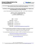
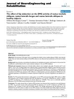
Joint Angles
Before the application of pharmacological treatment the
PAD patients had significantly increased range of motion
at the ankle (AROM) (Figure 1; Table 2) and significantly
decreased hip range of motion (HROM) as compared to
the controls (Figure 1; Table 2). There was no difference
in the knee parameters evaluated between the two
groups.
After pharmacological treatment, the same significant
differences were found between the controls and PAD
patients (Table 2). No significant differences were found
in the PAD patients comparing pre- and post-pharmaco-
logical treatment (Table 2).
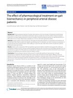
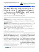
Joint Moments
Before the application of pharmacological treatment,
peak hip flexor moment (HFMM) was significantly
decreased in PAD patients compared to healthy controls
(Figure 2; Table 3). The adopted α-value being 0.005 after
the Bonferroni correction prohibits the identification of
some significant differences at the knee and hip. Specifi-
cally, the p-values for knee extension moment (KEMM; p
= 0.009), and hip extension moment (HEMM; p = 0.005)
are below p = 0.01 (Table 3). However, these results are
not statistically significant due to the Bonferroni correc-
tion used. These outcomes are observable in the graphi-
cal representation (Figure 2).
After pharmacological treatment, peak hip flexor
moment was still significantly decreased in PAD patients
compared to healthy controls (Figure 2; Table 3). Impor-
tantly, no significant differences were found in the PAD
patients between pre- and post- pharmacological treat-
ment (Table 3).
Discussion
The purpose of this study was to determine the impact of
pharmacological treatment on the biomechanics of gait
of PAD patients as compared to healthy controls. By
determining whether or not specific biomechanical
changes exist as a result of this treatment, it will be possi-
ble to determine if pharmacological treatment of patients
with PAD exerts an improvement in biomechanical
parameters. The pharmacological agents in the study
(pentoxifylline and cilostazol) are the two currently
approved FDA agents utilized to treat claudication. The
medications were considered together because the goal of
this study was not to compare the effects of a specific
Table 2: Group means and standard deviations for Joint Angle Ranges of Motion; Sig†, p < 0.005.
PAD pre PAD post Control p-values (effect size)
mean S.D. mean S.D. mean S.D. pre/post pre/con post/con
AROM 19.96 2.95 19.64 3.07 16.06 3.31 0.669 (0.11) < 0.001† (1.25) < 0.001† (1.12)
KROM 9.18 3.90 9.41 3.58 10.81 3.56 0.839 (0.06) 0.105 (0.43) 0.140 (0.29)
HROM 35.22 4.21 35.98 3.04 39.56 2.87 0.404 (0.21) < 0.001† (1.22) < 0.001† (1.21)
All mean values are in degrees of motion. AROM - Ankle range of motion; KROM - Knee range of motion; HROM - Hip range of motion.
Huisinga et al. Journal of NeuroEngineering and Rehabilitation 2010, 7:25
/>Page 5 of 9
pharmacological treatment but rather to determine if
pharmacological treatment resulted in any appreciable
change in biomechanical parameters. Comparisons
between the PAD patients pre- and post-treatment were
conducted with healthy controls. It was anticipated that
PAD patients would exhibit a large number of differences
as compared to healthy controls in the joint angles and
joint moments during walking in the pre-pharmacologi-
cal treatment phase. It was also anticipated that after the
conclusion of the treatment, significant improvements in
biomechanical parameters would be seen and the differ-
ences between the PAD patients and the healthy controls
would decrease and potentially disappear due to the posi-
tive effect of the pharmacological treatment.
Our results supported our previous findings that PAD
patients have significant gait abnormalities in the absence
of claudication pain as compared to control patients even
during the absence of ischemia [2,3,19,20,29,30]. How-
ever, significant improvements were not found in the gait
biomechanics of PAD patients due to pharmacotherapy.
Thus, our current evaluation showed that pharmacologi-
cal treatment did not affect the biomechanics of gait of
PAD patients.
Despite a lack of significant differences between the
pre- and post-pharmacological treatment conditions, a
closer look at the data can provide insight regarding the
movement patterns of the PAD patients. When evaluat-
ing patients with PAD as compared to control subjects,
little data exists to fully quantify the exact biomechanical
abnormalities that are present during walking. The evalu-
ation of the joint angles in our study showed that PAD
patients exhibited decreased hip flexion as the leg comes
in contact with the ground resulting in significantly
decreased hip range of motion during stance. The
decreased hip flexion indicates that PAD patients posi-
tion the leg closer to the body as they come in contact
with the ground. This is probably an adaptive strategy
employed by the PAD patients because the closer the leg
is to the body, the more securely and effectively they
move into single limb stance. Crowther et al. [30] also
reported decreased movement at the hip with reduced
extension during stance. Overall, the majority of abnor-
mal changes present in the PAD patients seem to be at the
Table 3: Group means and standard deviations for Joint Moments; Sig†, p < 0.005.
PAD pre PAD post Control p-values (effect size)
(%BWxBH) mean S.D. mean S.D. mean S.D. pre/post pre/con post/con
ADMM -3.41 0.97 -3.05 0.82 -3.74 1.11 0.030 (0.40) 0.241 (0.32) 0.011 (0.72)
APMM 14.84 1.62 15.54 1.69 15.05 1.57 0.036 (0.42) 0.632 (0.13) 0.266 (0.30)
KFMM 2.00 1.64 1.97 2.04 1.73 1.34 0.912 (0.02) 0.494 (0.19) 0.602 (0.14)
KEMM -6.59 2.50 -6.61 2.30 -8.42 2.48 0.950 (0.01) 0.009 (0.74) 0.007 (0.76)
HFMM 8.67 3.23 8.21 3.10 11.09 2.40 0.371 (0.14) 0.003† (0.86) < 0.001† (1.04)
HEMM -8.27 2.31 -8.69 2.45 -10.28 2.70 0.160 (0.17) 0.005 (0.80) 0.027 (0.62)
ADMM - Peak ankle dorsiflexion moment during early stance; APMM - Peak ankle plantarflexion moment during late stance; KFMM - Peak
knee flexion moment during stance; KEMM - Peak knee extension moment during stance; HFMM - Peak hip flexion moment during late
stance; HEMM - Peak hip extension moment during early stance.
Figure 1 Mean ensemble curves for Joint Angles.
Huisinga et al. Journal of NeuroEngineering and Rehabilitation 2010, 7:25
/>Page 6 of 9
hip and the ankle joints. These results are consistent with
findings of Celis et al. [17] who also found an increase in
the range of motion at the ankle during stance. However,
Celis et al. [17] did not find significant differences at the
hip. Chen et al. [2] found decreased hip flexion during
early stance but did not report decreased range of motion
at the hip during stance. Chen et al. [2] also reported
increased ankle plantarflexion during stance in PAD
patients as compared to controls. This agrees with the
increased range of motion at the ankle during stance in
PAD patients that was found in our study. The muscles
surrounding the ankle, the gastrocnemius and soleus in
particular, are the area of the leg most likely affected by
ischemia since the blood must travel farther to reach the
lower part of the leg. The abnormal biomechanical
parameters at the ankle, including the decreased force
production during late stance and the increased range of
motion during stance, are affected by changes that occur
further up in the kinetic chain at the hip joint. The
increase in the range of motion of the ankle during stance
was accompanied by a decrease in range of motion of the
hip during stance as it was found in our study. This is log-
ical when considering the entire kinetic chain. When
motion at one joint is decreased, another joint in the
kinetic chain may have to increase movement to maintain
forward progression. Thus, it is possible that the ankle
range of motion is increased before treatment as the hip
movement is decreased as a compensation mechanism.
The joint angle values found in this study are in general
agreement with those reported in previous studies
involving elderly populations [34,36].
Joint moment analysis showed no differences at the
ankle before or after treatment. At the hip, weakness is
observed in the hip flexors as evidenced by the decreased
hip flexor moment (HFMM) in PAD patients. The weak-
ness in the hip flexors would also account for the
decreased hip flexion at heel contact and overall smaller
hip range of motion, while decreased strength indicates
that control of the ankle range of motion was reduced
such that increased plantarflexion occurred at heel con-
tact and increased dorsiflexion occurred prior to toe-off.
Hip range of motion was decreased significantly in PAD
patients which contributed to decreases in maximum hip
flexor and extensor moments (Figure 2) that were noted
before and after pharmacological treatment. Chen et al.
[2] also found decreased peak hip extensor moment dur-
ing stance in PAD patients compared to controls. This is
likely due to decreased hip range of motion as was indi-
cated in the joint angle results. The decreased hip
moment values may also indicate an inability of PAD
patients to generate sufficient muscular contractions at
the hip during walking. Furthermore, the maximum knee
extension moment is related with the development of the
muscular contraction required to extend the knee and
move the entire body over the straight leg during single
support. Though this value was unchanged statistically,
PAD patients may exhibit a decreased ability to generate
this moment as indicated by the decreased knee extensor
moment which was approaching significance.
The PAD patients in the current study also had a slower
walking speed than healthy controls which is in agree-
ment with other studies of PAD patients [4,7]. This
altered gait speed suggests a baseline alteration in gait
function in patients with PAD. It should be noted that
despite pharmacological treatment in our current PAD
cohort, no walking speed changes were observed.
The lack of changes in the biomechanical gait parame-
ters with pharmacological treatment in the current study
may be multi-factorial in nature. Reasons for lack of gait
alterations may include treatment duration, medication
compliance, and lack of drug efficacy. Nonetheless, gait
adaptations may occur rapidly when changes are imposed
on the musculoskeletal system. Several researchers found
Figure 2 Mean ensemble curves for Joint Moments.
Huisinga et al. Journal of NeuroEngineering and Rehabilitation 2010, 7:25
/>Page 7 of 9
that supervised treadmill exercise over a 12-week period
improved absolute claudication distances [37-39],
improved peak oxygen uptake [38,39], and increased calf-
muscle strength and calf-muscle endurance [39]. These
results suggest that PAD patients may undergo significant
changes with respect to muscle strength and walking abil-
ity within a 12 week period if treated with a supervised
exercise program. Therefore, it is imperative that future
studies examine the underlying mechanisms for improve-
ment in gait parameters for both exercise and pharmaco-
logical treatment. If the mechanisms are found to be
different based upon biomechanical analysis, combina-
tion treatment consisting of treadmill exercise and phar-
macological treatment may be the most efficacious non-
operative treatment for symptoms of PAD patients.
Several limitations for this study may explain the
absence of significant changes found as a result of phar-
macological treatment. It should be noted that during the
biomechanical analysis of the kinematic and kinetic dif-
ferences between PAD patients and controls and within
PAD patients due to pharmacological treatment, walking
speed was not controlled or analyzed as a covariate.
Walking speed was not controlled during the data collec-
tions because it represents the natural self-selected walk-
ing speed of the populations. This decision is supported
by research with elderly populations where it is common
practice not to control for speed during biomechanical
comparisons [31-34]. Next, pentoxyfilline was used as
one of the treatment medications despite the fact that it
has a variable effect when compared to placebo at
improving walking distance in PAD patients [24]. This
was unavoidable since it is one of the approved medica-
tions for treatment of PAD and is frequently prescribed
for the treatment of intermittent claudication. Interest-
ingly, pentoxyfilline has been shown to variably improve
respiration capability in the mitochondria of skeletal
muscle of PAD patients [14]. Thus it was not unreason-
able to expect the drug to have an effect on the skeletal
muscle of patients in this study and for those physiologi-
cal changes to extend to gait alterations. Next, the medi-
cations were administered for a total of 12 weeks which is
half the time of the Dawson et al. [24] study which found
differences in walking distance due to pharmacological
treatment. However, 12 weeks is the same amount of time
that other studies involving PAD patients have used when
pharmacological treatment and exercise therapy was
employed and those studies did show significant
improvements in walking distance [25,38]. The method
of medication assignment was not ideal since the medica-
tions were not randomly assigned. It was not possible to
randomly assign treatment groups since the patients
recruited for this study had already been under the care
of a physician for treatment of their PAD symptoms and
the treating physicians had formulary restrictions regard-
ing which medication would most benefit an individual
patient. It should be noted that all patients in this study
were combined into one group for analysis regardless of
medication type. Because the focus of this study was not
to evaluate the effects of one medication versus another,
the groups were combined. However, based upon the lit-
erature and our current study, future studies should
include sufficient patients to have two separate treatment
groups for analysis. Importantly the results of our study
are valid for non-strenuous walking. It is possible that the
agents used here can elicit differences in the biomechan-
ics of gait of PAD patients after being administered for a
significant amount of time during strenuous walking such
as when they exhibit ischemia. This question is presently
being investigated in our laboratory. Furthermore, we
should mention that the chronic ischemia reperfusion
cycle which occurs in PAD results in damage to the mito-
chondria and overall neuromuscular damage of the lower
extremity that is present even when patients are not expe-
riencing claudication pain [11,12]. Finally, the control
group was not re-tested after an equivalent passage of
time experienced by the intervention group. The acquisi-
tion of a posttest observation for the control group would
have permitted analysis with a 2 × 2 pre-test/post-test
control group design and would have provided an ability
to control for other confounding variables as well as the
detection and evaluation of any interaction effect
between groups and time/intervention.
Conclusions
In conclusion, our study demonstrated that pharmaco-
logical treatment of PAD patients with intermittent clau-
dication did not result in detectable changes in gait
biomechanics during non-strenuous walking. Differences
as compared to healthy controls remained before and
after standard treatment with FDA approved drugs.
Therefore, such treatment for PAD does not translate to
biomechanical gait changes as hypothesized. The PAD
gait variables showed a large number of significant differ-
ences when compared to controls both pre- and post-
treatment. Future studies are needed to clarify whether
cilostazol or pentoxifylline is more effective, from a bio-
mechanical perspective, at improving gait during strenu-
ous walking and if a longer period of administering the
medication would have an effect on gait parameters. In
addition, new pharmacotherapy options may be neces-
sary in order to improve muscle function of the lower
extremities and the claudication pain which limits the
amounts of walking and other physical activity performed
by PAD patients.
Competing interests
We have read the submitted manuscript that includes our names as authors
and vouch for its accuracy. We certify that we have participated sufficiently in
the conception and design of this work and the analysis of the data (where
Huisinga et al. Journal of NeuroEngineering and Rehabilitation 2010, 7:25
/>Page 8 of 9
applicable), as well as the writing of the manuscript, to take public responsibil-
ity for its content. We believe the manuscript represents honest and valid work.
To the best of our knowledge, it contains no misrepresentations. We have
reviewed the final version of the submitted manuscript and approve it for pub-
lication. We warrant that the manuscript is original and its essential substance,
tables, or figures have not been previously published in part or in whole. The
manuscript or one with substantially similar content under our authorship or
the data within it has not been accepted for publication elsewhere and it is not
presently under review by any other publisher. The manuscript will not be sub-
mitted for publication elsewhere until a decision has been made on its accept-
ability for publication in Journal of NeuroEngineering and Rehabilitation. This
restriction does not apply to brief abstracts or press reports published in con-
nection with scientific meetings.
Authors' contributions
JH carried out gait analysis, performed data analysis/processing, performed
statistical analysis, drafted the manuscript, and revision of the manuscript to
the current version. IP participated in the design of the study, data interpreta-
tion, and approval of the manuscript version to be published. NS participated
in the design of the study, data interpretation, manuscript revisions, and
approval of the manuscript version to be published. JJ participated in data
interpretation, manuscript revisions, and approval of the manuscript version to
be published.
Acknowledgements
Support for this work was provided by the Nebraska Research Initiative, the NIH
(K25HD047194), the US Department of Education (H133G040118), the Univer-
sity of Nebraska at Omaha's University Committee on Research and Creative
Activity Grant, and the American Geriatrics Society's Hartford Foundation Den-
nis W. Jahnigen Award.
Author Details
1
Nebraska Biomechanics Core Facility, University of Nebraska at Omaha, 6001
Dodge Street Omaha, NE, USA 68182, USA,
2
Department of Surgery, University
of Nebraska Medical Center, 983280 Nebraska Medical Center, Omaha, NE, USA
68198-3280, USA and
3
Department of Environmental, Agricultural, and
Occupational Health, College of Public Health, University of Nebraska Medical
Center, 986075 Nebraska Medical Center, Omaha, NE, USA 68198-6075, USA
References
1. Scherer SA, Bainbridge JS, Hiatt WR, Regensteiner JG: Gait characteristics
of patients with claudication. Arch Phys Med Rehabil 1998, 79(5):529-31.
2. Chen SJ, Pipinos I, Johanning J, Radovic M, Huisinga JM, Myers SA, et al.:
Bilateral claudication results in alterations in the gait biomechanics at
the hip and ankle joints. J Biomech 2008, 41(11):2506-14.
3. Scott-Pandorf MM, Stergiou N, Johanning JM, Robinson L, Lynch TG,
Pipinos II: Peripheral arterial disease affects ground reaction forces
during walking. J Vasc Surg 2007, 46(3):491-9.
4. McDermott MM, Greenland P, Liu K, Guralnik JM, Criqui MH, Dolan NC, et
al.: Leg symptoms in peripheral arterial disease: Associated clinical
characteristics and functional impairment. JAMA 2001,
286(13):1599-606.
5. Myers SA, Johanning JM, Stergiou N, Lynch TG, Longo GM, Pipinos II:
Claudication distances and the walking impairment questionnaire
best describe the ambulatory limitations in patients with symptomatic
peripheral arterial disease. J Vasc Surg 2008, 47(3):550-5.
6. Regensteiner JG, Ware JE Jr, McCarthy WJ, Zhang P, Forbes WP, Heckman
J, et al.: Effect of cilostazol on treadmill walking, community-based
walking ability, and health-related quality of life in patients with
intermittent claudication due to peripheral arterial disease: Meta-
analysis of six randomized controlled trials. J Am Geriatr Soc 2002,
50(12):1939-46.
7. Gardner AW, Forrester L, Smith GV: Altered gait profile in subjects with
peripheral arterial disease. Vasc Med 2001, 6(1):31-4.
8. Gardner AW, Montgomery PS: The relationship between history of
falling and physical function in subjects with peripheral arterial
disease. Vasc Med 2001, 6(4):223-7.
9. Gardner AW, Killewich LA: Lack of functional benefits following
infrainguinal bypass in peripheral arterial occlusive disease patients.
Vasc Med 2001, 6(1):9-14.
10. Gardner AW, Montgomery PS: Impaired balance and higher prevalence
of falls in subjects with intermittent claudication. J Gerontol A Biol Sci
Med Sci 2001, 56(7):M454-8.
11. Pipinos II, Judge AR, Zhu Z, Selsby JT, Swanson SA, Johanning JM, et al.:
Mitochondrial defects and oxidative damage in patients with
peripheral arterial disease. Free Radic Biol Med 2006, 41(2):262-9.
12. Pipinos II, Sharov VG, Shepard AD, Anagnostopoulos PV, Katsamouris A,
Todor A, et al.: Abnormal mitochondrial respiration in skeletal muscle in
patients with peripheral arterial disease. J Vasc Surg 2003, 38(4):827-32.
13. Muller R: Pentoxifylline a biomedical profile. J Med 1979, 10(5):307-29.
14. Pipinos II, Boska MD, Shepard AD, Anagnostopoulos PV, Katsamouris A:
Pentoxifylline reverses oxidative mitochondrial defect in claudicating
skeletal muscle. J Surg Res 2002, 102(2):126-32.
15. Aronow WS: Management of peripheral arterial disease of the lower
extremities in elderly patients. J Gerontol A Biol Sci Med Sci 2004,
59(2):172-7.
16. Huisinga J, Pipinos I, Stergiou N, Johanning J: Treatment with
pharmacological agents in peripheral arterial disease patients does
not result in biomechanical gait changes. Journal of Applied
Biomechanics 2010 in press.
17. Celis R, Pipinos II, Scott-Pandorf MM, Myers SA, Stergiou N, Johanning JM:
Peripheral arterial disease affects kinematics during walking. J Vasc
Surg 2009, 49(1):127-32.
18. Ayzin Rosoky RM, Wolosker N, Muraco-Netto B, Puech-Leao P: Ground
reaction force pattern in limbs with intermittent claudication. Eur J
Vasc Endovasc Surg 2000, 20(3):254-9.
19. Crowther RG, Spinks WL, Leicht AS, Quigley F, Golledge J: Lower limb
movement variability in patients with peripheral arterial disease. Clin
Biomech 2008.
20. Crowther RG, Spinks WL, Leicht AS, Quigley F, Golledge J: Intralimb
coordination variability in peripheral arterial disease. Clin Biomech
2008, 23(3):357-64.
21. Shrader MW, Draganich LF, Pottenger LA, Piotrowski GA: Effects of knee
pain relief in osteoarthritis on gait and stair-stepping. Clin Orthop Relat
Res 2004:188-93.
22. Houck J, Yack HJ, Cuddeford T: Validity and comparisons of tibiofemoral
orientations and displacement using a femoral tracking device during
early to mid stance of walking. Gait Posture 2004, 19(1):76-84.
23. DiBianco R, Morganroth J, Freitag JA, Ronan JA Jr, Lindgren KM, Donohue
DJ, et al.: Effects of nadolol on the spontaneous and exercise-provoked
heart rate of patients with chronic atrial fibrillation receiving stable
dosages of digoxin. Am Heart J 1984, 108(4 Pt 2):1121-7.
24. Dawson DL, Cutler BS, Hiatt WR, Hobson RW, Martin JD, Bortey EB, et al.: A
comparison of cilostazol and pentoxifylline for treating intermittent
claudication. Am J Med 2000, 109(7):523-30.
25. Dawson DL, Cutler BS, Meissner MH, Strandness DE Jr: Cilostazol has
beneficial effects in treatment of intermittent claudication: Results
from a multicenter, randomized, prospective, double-blind trial.
Circulation 1998, 98(7):678-86.
26. Vaughan C, Davis B, O'Connor J: Dynamics of human gait Cape Town,
South Africa: Kiboho Publishers; 1999.
27. Nigg BM, Cole GK, Nachbauer W: Effects of arch height of the foot on
angular motion of the lower extremities in running. J Biomech 1993,
26(8):909-16.
28. Winter DA: Biomechanics and motor control of human movement. 3rd
edition. New York: John Wiley & Sons; 2005.
29. Crowther RG, Spinks WL, Leicht AS, Sangla K, Quigley F, Golledge J: Effects
of a long-term exercise program on lower limb mobility, physiological
responses, walking performance, and physical activity levels in
patients with peripheral arterial disease. J Vasc Surg 2008, 47(2):303-9.
30. Crowther RG, Spinks WL, Leicht AS, Quigley F, Golledge J: Relationship
between temporal-spatial gait parameters, gait kinematics, walking
performance, exercise capacity, and physical activity level in peripheral
arterial disease. J Vasc Surg 2007, 45(6):1172-8.
31. DeVita P, Hortobagyi T: Age causes a redistribution of joint torques and
powers during gait. J Appl Physiol 2000, 88(5):1804-11.
32. DeVita P: The selection of a standard convention for analyzing gait data
based on the analysis of relevant biomechanical factors. J Biomech
1994, 27(4):501-8.
Received: 5 February 2009 Accepted: 7 June 2010
Published: 7 June 2010
This article is available from: 2010 Huisinga et al; licensee BioMed Central Ltd. This is an Open Access article distributed under the terms of the Creative Commons Attribution License ( ), which permits unrestricted use, distribution, and reproduction in any medium, provided the original work is properly cited.Journal of NeuroEn gineerin g and Reha bilitatio n 2010, 7:25
Huisinga et al. Journal of NeuroEngineering and Rehabilitation 2010, 7:25
/>Page 9 of 9
33. Kerrigan DC, Lee LW, Nieto TJ, Markman JD, Collins JJ, Riley PO: Kinetic
alterations independent of walking speed in elderly fallers. Arch Phys
Med Rehabil 2000, 81(6):730-5.
34. Kerrigan DC, Todd MK, Della Croce U, Lipsitz LA, Collins JJ: Biomechanical
gait alterations independent of speed in the healthy elderly: Evidence
for specific limiting impairments. Arch Phys Med Rehabil 1998,
79(3):317-22.
35. Benedetti MG, Catani F, Leardini A, Pignotti E, Giannini S: Data
management in gait analysis for clinical applications. Clin Biomech
1998, 13(3):204-15.
36. Graf A, Judge JO, Ounpuu S, Thelen DG: The effect of walking speed on
lower-extremity joint powers among elderly adults who exhibit low
physical performance. Arch Phys Med Rehabil 2005, 86(11):2177-83.
37. Gardner AW, Killewich LA, Montgomery PS, Katzel LI: Response to
exercise rehabilitation in smoking and nonsmoking patients with
intermittent claudication. J Vasc Surg 2004, 39(3):531-8.
38. Wang J, Zhou S, Bronks R, Graham J, Myers S: Effects of supervised
treadmill-walking training on strength and endurance of the calf
muscles of individuals with peripheral arterial disease. Clin J Sport Med
2006, 16(5):397-400.
39. Hiatt WR, Regensteiner JG, Hargarten ME, Wolfel EE, Brass EP: Benefit of
exercise conditioning for patients with peripheral arterial disease.
Circulation 1990, 81(2):602-9.
doi: 10.1186/1743-0003-7-25
Cite this article as: Huisinga et al., The effect of pharmacological treatment
on gait biomechanics in peripheral arterial disease patients Journal of Neu-
roEngineering and Rehabilitation 2010, 7:25