báo cáo hóa học: " Hypoxia silences the neural activities in the early phase of the phrenic neurogram of eupnea in the piglet" docx
Bạn đang xem bản rút gọn của tài liệu. Xem và tải ngay bản đầy đủ của tài liệu tại đây (1.49 MB, 9 trang )
BioMed Central
Page 1 of 9
(page number not for citation purposes)
Journal of NeuroEngineering and
Rehabilitation
Open Access
Research
Hypoxia silences the neural activities in the early phase of the
phrenic neurogram of eupnea in the piglet
Metin Akay*
Address: Neural Engineering & Informatics Laboratory, Harrington Department of Bioengineering, Ira A. Fulton School of Engineering, Arizona
State University, Tempe, AZ 85287-9709, USA
Email: Metin Akay* -
* Corresponding author
Abstract
Objective: We investigated phrenic neurogram patterns during eupnea (normal breathing) and
severe hypoxia (gasping) during early maturation in the piglet.
Methods: We used continuous wavelet transform and short time Fourier transform methods to
examine the similarity of breathing patterns in both time and frequency domains during early
maturation. The phrenic neurogram was recorded during eupnea, severe hypoxia, and recovery
from severe hypoxia in piglets in three different age groups: 3–6 days, 10–15 days and 29–35 days.
Results: During the first week of postnatal age, respiratory patterns of phrenic activity were
marked by frequency components between 30 and 300 Hz during both the early (first half) and late
(second half) phases of the neurogram signals during eupnea. The results suggest that there is little
difference between the respiratory patterns in both time and frequency domains during eupnea
compared to gasping for the first week of postnatal age in piglets. After the first week of postnatal
age, the duration of the phrenic neurogram burst significantly increases and the patterns during the
early phase of the phrenic neurogram are different from those observed for gasping. However, the
patterns that mark the late phase of the phrenic neurograms are still the same as those of gasping.
Conclusion: Our most significant finding is that hypoxia silences the neural activity in the early
phase of phrenic neurogram regardless of maturation.
Introduction
Production of progressive brain hypoxia in an anesthe-
tized, vagotomized, peripherally-chemodenervated cat
results in depression of respiratory output and a stereotyp-
ical progression of respiratory pattern changes, as hypoxia
progresses [1-4]. Initially, the amplitude of the phrenic
neurogram is depressed, with a fall in phrenic firing fre-
quency only occurring as the hypoxia becomes more
severe. As arterial O
2
content falls, progressive respiratory
depression continues until the phrenic output is com-
pletely silenced. If hypoxia is allowed to progress beyond
this point, gasping will eventually ensue [6-8]. This form
of respiration is characterized by brief, intense inspiratory
efforts of the diaphragm and other respiratory muscles,
and has been interpreted as an attempt at "autoresuscita-
tion" [9-12]. This interpretation is based on the observa-
tion that animals asphyxiated to the point of apnea by
airway occlusion, will restore arterial oxygenation quickly
if the occlusion is removed and gasping ensues. If the ani-
mal fails to gasp, arterial oxygenation does not improve
Published: 30 November 2005
Journal of NeuroEngineering and Rehabilitation 2005, 2:32 doi:10.1186/1743-
0003-2-32
Received: 17 May 2005
Accepted: 30 November 2005
This article is available from: />© 2005 Akay; licensee BioMed Central Ltd.
This is an Open Access article distributed under the terms of the Creative Commons Attribution License ( />),
which permits unrestricted use, distribution, and reproduction in any medium, provided the original work is properly cited.
Journal of NeuroEngineering and Rehabilitation 2005, 2:32 />Page 2 of 9
(page number not for citation purposes)
and death occurs due to cardiovascular collapse inevitably
occurs.
The relationship of the medullary gasp to eupneic breath-
ing has been a point of contention for a number of years.
Lumsden originally conceived gasping as being the prod-
uct of a primitive medullary pattern generator which does
not contribute to eupneic breathing [6,13]. More recently,
St. John and associates have, over the course of several
studies, closely examined this question and have con-
cluded that gasping is the result of a unique medullary
pattern generator [14-16] in agreement with Lumsden's
finding. This conclusion was based on studies of gasping
produced by reversibly cooling the pontomedullary junc-
tion of decerebrate cats. Although the gasping produced
by this procedure has timing characteristics which differ
slightly from those seen during hypoxic gasping (e.g.,
shorter inspiratory time) [15], the qualitative changes
seen in the phrenic neurogram and other respiratory out-
puts during gasping following cooling, were the same as
those seen during hypoxic or asphyxic gasping [15,16].
With this model, it was first shown that gasping differs
fundamentally from eupnea, both in the pattern of assem-
bly of single phrenic motoneurons to produce a phrenic
burst, and in timing characteristics of the phrenic neuro-
gram. The central respiratory controller was also shown to
be unresponsive to peripheral chemoreceptor stimulation
during gasping. When gasping is produced in the decere-
brate cat under conditions of carbon monoxide hypoxia,
the discharge frequency of expiratory neurons falls sharply
with some units becoming totally silent. The discharge fre-
quency of inspiratory neurons is unchanged during gasp-
ing but, unlike during eupnea, all inspiratory neurons fire
simultaneously at the beginning of the inspiratory period
during gasping [17].
Respiratory control has been studied largely on the basis
of phenomenology. There have also been attempts to
apply empirical, analytical techniques to the study of cen-
tral respiratory patterning. Cohen [18] was the first to use
autospectral analysis of the phrenic neurogram to gain
insight into the central respiratory pattern generation.
Subsequently, numerous frequency domain analyses of
the phrenic neurogram during eupnea, and during manip-
ulations of various respiratory afferents, have been per-
formed. Virtually all respiratory outputs studied during
eupnea (e.g., phrenic and laryngeal neurograms; dia-
phragmatic electromyograms) have been shown to dis-
play two prominent peaks in their spectra: a medium-
frequency oscillation (MFO) in the frequency range of
20–50 Hz, and a high-frequency oscillation (HFO)
between 50–100 Hz [19-21]. A HFO spectral peak, which
is correlated to the phrenic neurogram HFO, has also been
noted in medullary inspiratory neuronal activity. Based
on these observations, the HFO has been considered to be
a characteristic of the central, respiratory pattern genera-
tor. The source of the MFO is more problematic [22].
Richardson and Mitchell [23] have proposed that the
MFO arises from the interaction of two pattern generators,
while Christakos et al. [24], interpret the MFO as a reflec-
tion of the rhythmic augmenting discharge of individual
phrenic motorneurons resulting from an augmenting
drive of supraspinal origin.
Richardson and Mitchell [24] compared the frequency
spectra of the phrenic neurogram during eupnea and
gasping in decerebrate cats. Hypoxic gasping in decere-
brate cats was associated with a high-frequency peak in
the phrenic neurogram at 120 Hz, as opposed to the 80
Hz peak seen during eupnea. Spectral analysis of occa-
sional eupneic, phrenic bursts which showed gasp-like
augmentation at the end of inspiration, revealed the pres-
ence of both eupneic and gasping high-frequency peaks.
The presence of a unique spectral peak during gasping was
presented as support for the idea that respiratory pattern
generation differs during eupnea and gasping.
Preliminary studies of Akay et al. [25] used the modified
Yule-Walker autoregression (AR) technique of spectral
analysis to analyze 19 eupneic and 13 gasping, phrenic
neurograms in anesthetized cats before and during CO-
hypoxia and hypoxic-hypoxia, in two preliminary experi-
ments. Our results suggested that eupnea is characterized
by three peaks in the AR spectrum, with the lowest peak
frequency between 30 and 60 Hz. During gasping a dis-
tinctive low-frequency peak was evident in the spectrum
below 30 Hz. During eupnea the power spectra of the
phrenic neurogram of both cats exhibited two prominent
peaks, the first at 40–55 Hz and the second at approxi-
mately 100 Hz. The frequencies of these peaks correspond
to those described in previous spectral analyses of the
phrenic neurogram during eupnea where the lower-fre-
quency peak has been described as medium-frequency
oscillation (MFO) and the higher-frequency peak as high-
frequency oscillation (HFO) [18,23]. In our results, the
transition from eupnea to gasping was characterized by
the loss of the MFO, and the appearance of a major peak
in the 10–30 Hz range. This shift to a lower frequency dur-
ing gasping contrasts with the finding of Richardson and
Mitchell [22] where gasping resulted in a new spectral
peak at a frequency higher than the eupneic HFO. The
shift of power to a lower frequency during gasping,
observed in our preliminary studies, suggests that there is
a synchronization of neuronal firing at a frequency of 20–
25 Hz during gasping. The maximal firing frequency of an
individual neuron is presumably determined by the kinet-
ics of the ion conductance changes associated with the
action potential propagation, which require a finite time
for activation and inactivation before a second action
potential can be propagated. A frequency of 20–25 Hz is
Journal of NeuroEngineering and Rehabilitation 2005, 2:32 />Page 3 of 9
(page number not for citation purposes)
slower than the maximal frequency observed in individ-
ual phrenic motoneurons during eupnea (50 Hz), but
may represent the maximum firing frequency of a respira-
tory neuron under the severe hypoxic conditions associ-
ated with gasping where channel conductance kinetics
may be compromised [25].
When viewed in the time domain, the phrenic neurogram
displays a characteristic "ramp" pattern during inspiration
and decrementing activity during a short post-inspiratory
period [6,12,13,15,26]. This pattern results from an
orderly recruitment of phrenic premotor and motor units
throughout the period of inspiration. Cohen et al. [27],
observed both low- and high-frequency neurogram pat-
terns in piglets at birth, but the high-frequency compo-
nent was shown to increase with age [27]. They also
claimed that high frequency oscillations arise from brain
stem respiratory neurons in the medulla and the low-fre-
quency component was not increased with age and was
believed to originate from respiratory efferent systems.
Later, Webber [28] showed in adult cats that both the
early and late phases of the phrenic neurogram have a
high frequency component, which is around 82 Hz. Only
the late phase has a low frequency component, which is
around 29 Hz.
We recently showed that the breathing activities for the
young group are not periodic signals, and that the charac-
teristics of phrenic neurograms rapidly change with
respect to time [29]. Furthermore our results showed that
the phrenic neurogram consists of several dominant burst
type activities (circular structured components) corre-
sponding to the early and late phases of the inspiratory
activity. However, dominant burst type activities (circular
structured components) were only present during the late
phase of the phrenic neurogram when maturation pro-
ceeds. These results suggest that the phrenic neurogram is
not a periodic signal and that its characteristics change
rapidly during maturation. The dominant burst type activ-
ities disappeared during the early phase of the phrenic
neurogram although the burst activity and the continuous
activity remained, but both them appear at the late phase
of the phrenic neurogram as maturation proceeds [29].
The objective of the study herein presented was to investi-
gate the similarity on the time-frequency respiratory pat-
ters during eupnea and severe hypoxia (gasping) and to
determine whether hypoxia results in changes in the time-
frequency patterns of the respiratory motor output. We
have examined the phrenic neurogram in both time and
frequency domains during the first few weeks of postnatal
life using time-frequency analysis methods to gain insight
into the behavior of the respiratory neural network during
eupnea and severe hypoxia.
Methodology
Experiment
Experiments were performed in decerebrate piglets of
both sexes. Piglets were divided into three age groups: 3–
6 days (n = 4), 10–15 days (n = 3) and 29–35 days (n =
3). The animals were anesthetized with 4% isoflurane in
O
2
. The trachea was then cannulated for subsequent deliv-
ery of anesthesia (2–3% isoflurane in O
2
). Cannulation of
the femoral artery and vein, peripheral chemodenerva-
tion, vagotomy, paralysis, and ventilation were per-
formed. The scalp and underlying muscles were cut and
the cerebral hemisphere and the diencephalon were
removed. After exposing the mesencephalon, a mid-collic-
ular cut was made and the remaining brain structures ros-
tral to the incision were removed. After completion of the
decerebration, anesthesia was removed. Piglets were
chemically paralyzed for the rest of the experiment. A
minimum of one hour was allowed to elapse between
removal of anesthesia and data collection. Piglets were
ventilated with 40% O
2
in N
2
during eupnea. Then, severe
hypoxia was produced by inhalation of 3–5% O
2
in N
2
until gasping was observed in the phrenic neurogram.
Phrenic neurogram activity was also recorded during 30
min of reoxygenation (40% O
2
in N
2
).
Data was digitized on line by using a commercial data
acquisition and analysis software program (ADI, Power-
lab). The phrenic nerve was isolated in the neck at the
level of C5 rootlet. The nerve was cut and placed on a
bipolar electrode for neuronal recording. The raw phrenic
neurogram was bandpass filtered (10 – 300 kHz) and
sampled at 1 kHz [29].
Continuous Wavelet Transform (CWT)
The continuous wavelet transform was utilized to analyze
the phrenic neurogram signals. This transformation can
be viewed as an inner product operation that allows one
to measure the similarity or cross-correlation between the
signal, s(t), and the wavelet function. The continuous
wavelet transform of s(t) is defined as:
where b is a translation (shift) in time and a is the scale
factor which represents a translation (shift) in frequency.
In the study, we used the Morlet based CWT transform
since it shows better time-frequency resolution compared
to other orthogonal wavelet transform methods. The
details of the Morlet based CWT are described elsewhere
[30].
Results
For each piglet, the time-frequency representations during
eupnea and severe hypoxia were estimated and compared.
cwt a b s t
a
tb
a
dt(,) () ( )=
−
∫
1
ψ
Journal of NeuroEngineering and Rehabilitation 2005, 2:32 />Page 4 of 9
(page number not for citation purposes)
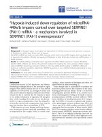
Figures 1 and 2 show the raw and the corresponding time-
frequency representation of the typical raw phrenic neuro-
grams of a 3-day old piglet during eupnea and severe
hypoxia, respectively. Although severe hypoxia (gasping)
reduced the time duration of phrenic neurograms during
inspiration and increased the expiratory duration, the
time-frequency representations during early and late
phases of phrenic neurogram during eupnea and gasping
showed components between 30 and 300 Hz and demon-
strated similarities. In addition, all 4 piglets in the young
group exhibited gasping patterns when they were exposed
to severe hypoxia.
For the mid-age group, only one of 3 animals had gasping
patterns and recovered when animals were reoxygenated.
Figures 3 and 4 shows the similar features for a 10 days
old piglet. The time frequency patterns were dominant
between 30 and 300 Hz at the late phase of the phrenic
neurogram during eupnea and about the same as those of
gasping.
Figure 4 show the time-frequency patters for a 30-day old
piglet. For the 29–35 days old age groups, the time fre-
quency patterns between 30 and 300 Hz are only present
for the late phase of the phrenic neurogram during eup-
nea. The time frequency patterns during gasping and the
late phase of phrenic neurogram during eupnea showed
considerable similarities. However, the patterns during
the early phase of phrenic neurogram was not dominant
and the signal components below 150 Hz were different
from those marking phrenic neurograms during eupnea.
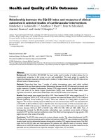
To investigate the similarity between the patterns in the
early and late phases of the phrenic neurogram during
eupnea and the patterns during gasping, time-frequency
patterns for each piglet over 10 consecutive phrenic bursts
during eupnea and 2–3 phrenic bursts during gasping
were estimated for each group. Then, we calculated the
mean total energies for four time-frequency regions,
divided first in time (first and second half of the phrenic
neurogram) and then frequency (above and below 150
Hz) during eupnea and the mean total energies below and
above 150 Hz during gasping. The mean ratio of the total
energies above and below 150 Hz for the early and late
phases of the phrenic neurogram during eupnea as well as
the phrenic burst during gasping were estimated. The
mean ratios for the early, late phase during eupnea and
gasping were 0.73 ± 0.1, 0.11 ± 0.1, 0.83 ± 0.19, respec-
tively for the young group. They were 0.52 ± 0.17, 0.67 ±
0.1, 0.73 ± 0.25, for the mid-group and finally they were
0.22 ± 0.09, 0.67 ± 0.12, 0.73 ± 0.18, for the old age
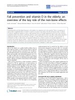
groups. Figure 7 summarizes the results. The mean ratios
for the early and late phases of the phrenic neurograms
during eupnea when compared to those of gasping were
not statistically significant for the young age group. As
maturation proceeds, the mean ratios for the early phase
of phrenic neurograms during eupnea and phrenic bursts
during gasping were statistically different although those
for the late phases of phrenic bursts during eupnea and
phrenic bursts during gasping remained statistically not
different. Statistical analysis was performed via an analysis
of variance (ANOVA) test.
Discussion and conclusion
Our previous study based on time-frequency analysis
methods showed that the time-frequency patterns at the
early and late phases of the phrenic neurogram were the
same for the 3–6 days old age group. As maturation pro-
ceeds, the early phase of the phrenic neurograms demon-
strated patterns below 150 Hz that were not dominant,
but the patterns for the last phase of phrenic neurograms
remained the same and were not influenced by matura-
tion. In this study, we estimated the time-frequency pat-
terns during early and late phases of phrenic neurograms
during eupnea and compared them with those of gasping
in order to investigate the similarities between these
patterns.
Our preliminary data indicated that the patterns during
early and late phases of the phrenic neurogram during
eupnea are similar to those during gasping for the 3–6
days old group.
The piglets in the young group were very resistive and
showed strong responses during gasping in all 4 piglets in
this study. However, the mid-group (10–15 days) failed to
gasp in 2 of 3 animals. But, all three animals in the old
group exhibited the gasping patterns like those in the 3–6
days old group. Therefore, we suggest that the animals in
the mid-group could be more vulnerable compared to
those in the young and old age groups. In addition, the
patterns during early and late phases of phrenic neuro-
gram were almost the same as those of gasping. As matu-
ration proceeds, the similarity between the late phase of
phrenic neurogram and gasping remained. Nevertheless,
hypoxia significantly reduced the phrenic activities in the
early phase of phrenic neurograms and caused a shift in
the associated frequency components toward the lower
frequency range (i.e., below 150 Hz). Hypoxia signifi-
cantly increased the expiratory duration and reduced the
inspiratory duration (especially, as maturation proceeds).
Our most significant finding is that hypoxia silences the
neural activity in the early phase of phrenic neurogram
regardless of maturation.
Although we do not know the exact mechanism underly-
ing these changes in the patterns of the phrenic neuro-
grams from eupnea to gasping, we speculate that gasping
silences phrenic neurons responsible for the neural activ-
ities in the early phase of the phrenic neurogram and does
Journal of NeuroEngineering and Rehabilitation 2005, 2:32 />Page 5 of 9
(page number not for citation purposes)
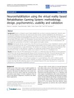
The raw phrenic neurogram and the corresponding time-frequency representation of the phrenic neurogram of a 3-day old piglet during eupnea (a) and gasping (b)Figure 1
The raw phrenic neurogram and the corresponding time-frequency representation of the phrenic neurogram of a 3-day old
piglet during eupnea (a) and gasping (b).
A
B
Journal of NeuroEngineering and Rehabilitation 2005, 2:32 />Page 6 of 9
(page number not for citation purposes)
The raw phrenic neurogram and the corresponding time-frequency representation of the phrenic neurogram of a 10-day old piglet during eupnea (a) and gasping (b)Figure 2
The raw phrenic neurogram and the corresponding time-frequency representation of the phrenic neurogram of a 10-day old
piglet during eupnea (a) and gasping (b).
A
B
Journal of NeuroEngineering and Rehabilitation 2005, 2:32 />Page 7 of 9
(page number not for citation purposes)
The raw phrenic neurogram and the corresponding time-frequency representation of the phrenic neurogram of a 30-day old piglet during eupnea (a) and gasping (b)Figure 3
The raw phrenic neurogram and the corresponding time-frequency representation of the phrenic neurogram of a 30-day old
piglet during eupnea (a) and gasping (b).
A
B
Journal of NeuroEngineering and Rehabilitation 2005, 2:32 />Page 8 of 9
(page number not for citation purposes)
not influence phrenic neurons responsible for the neural
activities in the late phase of the phrenic neurogram dur-
ing inspiration. In addition, it also significantly increases
the duration of the phrenic neurogram during expiration.
We also noted that patterns observed during gasping did
not change significantly as maturation proceeds. We spec-
ulate that severe hypoxia silences respiratory neurons
responsible for both early and late phases of phrenic neu-
rograms in 2 of 3 piglets in the mid-group. We suspect that
a reduction in the number of dendrites per cell after 2
weeks of maturation could be responsible for the failure
of gasping patterns in these piglets [31].
Acknowledgements
This work was supported by NIH grant (HL 65732). The authors thank K.
Johnson and Drs. N. Sekine, J. Bardonova, A. Curran and K. Moodie for
their technical support.
References
1. Melton JE, Neubauer JA, Edelman NH: CO
2
sensitivity of cat
phrenic neurogram during hypoxic respiratory depression. J
Appl Physiol 1988, 65(2):7536-7543.
2. Melton JE, Chae LO, Neubauer JA, Edelman NH: Extracellular
potassium homeostasis in the cat medulla during progres-
sive brain hypoxia. J Appl Physiol 1991, 70(4):1477-1482.
3. Melton JE, Wasicko MJ, Neubauer JA, Edelman NH: Patterns of
phrenic depression during progressive brain hypoxia. FASEB J
1988, 2(4):A510.
4. Neubauer JA, Melton JE, Edelman NH: Modulation of respiration
during brain hypoxia (review). J Appl Physiol 1990,
68(2):441-451.
5. Neubauer JA, Simone A, Edelman NH: Role of brain lactic acidosis
in hypoxic depression of respiration. J Appl Physiol 1988,
65(3):1324-1331.
6. Guntheroth WG, Kawabori I: Hypoxic apnea and gasping. J Clin
Invest 1975, 56(6):1371-1377.
7. Lawson EE, Thatch BJ: Respiratory patterns during progressive
asphyxia in newborn rabbits. J Appl Physiol 1977, 43:468-474.
8. Lumsden T: Observations on the respiratory centres. J Physiol
1923, 57:354-367.
9. Macefield G, Nail B: Phrenic and external intercostal motone-
uron activity during progressive asphyxia. J Appl Physiol 1987,
63(4):1413-1420.
The mean ratio of the total energies above and below 150 Hz for the early and late phases of the phrenic neurogram during eupnea as well as the phrenic burst during gasping for 3 different age groupsFigure 4
The mean ratio of the total energies above and below 150 Hz for the early and late phases of the phrenic neurogram during
eupnea as well as the phrenic burst during gasping for 3 different age groups.
Publish with BioMed Central and every
scientist can read your work free of charge
"BioMed Central will be the most significant development for
disseminating the results of biomedical research in our lifetime."
Sir Paul Nurse, Cancer Research UK
Your research papers will be:
available free of charge to the entire biomedical community
peer reviewed and published immediately upon acceptance
cited in PubMed and archived on PubMed Central
yours — you keep the copyright
Submit your manuscript here:
/>BioMedcentral
Journal of NeuroEngineering and Rehabilitation 2005, 2:32 />Page 9 of 9
(page number not for citation purposes)
10. Davis PJ, Macefield G, Nail BS: Respiratory muscle activity during
asphyxic apnoea and opisthotonus in the rabbit. Respir Physiol
1986, 65(3):285-294.
11. Macefield G, Nail B: Inspiratory augmentation during asphyxic
hyperpnoea and gasping: proprioceptive influences. Respir
Physiol 1986, 64(1):57-68.
12. Breckenridge CG, Hoff HE: Pontine and medullary regulation of
respiration in the cat. Am J Physiol 1950, 160:385-394.
13. Lumsden T: Observations on the respiratory centres in the
cat. J Physiol 1923, 57:153-160.
14. St John WM, Knuth KV: A characterization of the respiratory
pattern of gasping. J Appl Physiol: Resp, Environ & Exercise Physiol
1981, 50(1):984-993.
15. St John WM, Zhou D, Fregosi RF: Expiratory neural activities in
gasping. J Appl Physiol 1989, 66(1):223-231.
16. Zhou D, Wasicko MJ, Hu J, St John WM: Differing activities of
medullary respiratory neurons in eupnea and gasping. J Appl
Physiol 1991, 70(3):1265-1270.
17. St John WM, Bledsoe TA, Sokol HW: Identification of medullary
loci critical for neurogenesis of gasping. J Appl Physiol: Resp, Envi-
ron & Exercise Physiol 1984, 56(4):1008-1019.
18. Cohen MI: Synchronization of discharge, spontaneous and
evoked, between inspiratory neurons. Acta Neurobiol Exp 1973,
33(1):189-218.
19. Jacobi MS, Thach BT: Effect of maturation on spontaneous
recovery from hypoxic apnea by gasping. J Appl Physiol 1989,
66(5):2384-2390.
20. Mitchell RA, Herbert DA: Synchronized high frequency synaptic
potentials in medullary respiratory neurons. Brain Res 1974,
75(2):350-355.
21. Cohen MI, Feldman JL: Discharge properties of dorsal medul-
lary inspiratory neurons: relation to pulmonary afferent and
phrenic efferent discharge. J Neurophysiol 1984, 51(4):753-776.
22. Cohen MI, See WR, Christakos CN, Sica AL: High-frequency and
medium-frequency components of different inspiratory
nerve and their modification by various inputs. Brain Res 1987,
417(1):148-152.
23. Christakos CN, Cohen MI, See WR, Barnhardt R: Fast rhythms in
the discharges of medullary inspiratory neurons. Brain Res
1988, 463(2):362-367.
24. Richardson CA, Mitchell RA: Power spectral analysis of inspira-
tory nerve activity in the decerebrate cat. Brain Res 1982,
233(2):317-336.
25. Akay M, Melton JE, Welkowitz W, Edelman NH, Neubauer JA:
Autoregressive spectral analysis of phrenic neurogram dur-
ing eupnea and gasping. J Appl Physiol 1996, 81(2):530-540.
26. Suthers GK, Henderson-Smart DJ, Read DJC: Postnatal changes in
the rate of high frequency bursts of inspiratory activity in
cats and dogs. Brain Res 1977, 132(3):537-540.
27. Cohen HL, Gootman PM, Steele AM, Eberle LP, Rao PP: Age-
related changes in power spectra of efferent phrenic activity
in the piglet. Brain Res 1987, 426(1):179-182.
28. Webber CL: High-frequency oscillations within early and late
phases of the phrenic neurogram. J Appl Physiol 1989,
66(2):886-893.
29. Akay M, Sekine M: The Effects of Maturation on Early and Late
Phases of Phrenic Neurogram in Piglets during Maturation.
IEEE Trans on Biomedical Engineering, IEEE Trans on Biomedical
Engineering 2004, 51:1954-1959.
30. Akay M: Time Frequency and Wavelets in Biomedical Signal Processing
New York: Wiley-IEEE Press; 1997.
31. Jacobi MS, Gershan WM, Thach BT: Mechanism of failure of
recovery from hypoxic apnea by gaping in 17- to 23 day-old
mice. J Appl Physiol 1991, 71(3):1098-1105.