báo cáo hóa học: " Comparison of knee motion on Earth and in space: an observational stud" potx
Bạn đang xem bản rút gọn của tài liệu. Xem và tải ngay bản đầy đủ của tài liệu tại đây (480.2 KB, 8 trang )
BioMed Central
Page 1 of 8
(page number not for citation purposes)
Journal of NeuroEngineering and
Rehabilitation
Open Access
Research
Comparison of knee motion on Earth and in space: an observational
study
Mark C Pierre
1,2
, Kerim O Genc
1,2,5
, Micah Litow
1,2,5
, Brad Humphreys
6
,
Andrea J Rice
1,2
, Christian C Maender
7
and Peter R Cavanagh*
1,2,3,4
Address:
1
Department of Biomedical Engineering, Lerner Research Institute, Cleveland Clinic, Cleveland, OH, USA,
2
Center for Space Medicine,
Cleveland Clinic, Cleveland, OH, USA,
3
Department of Orthopaedic Surgery, Cleveland Clinic, Cleveland, OH, USA,
4
Orthopaedic Research
Center, Cleveland Clinic, Cleveland, OH, USA,
5
Case Western Reserve University, Cleveland, OH, USA,
6
ZIN Technologies, Inc., Brook Park, OH,
USA and
7
NASA-Johnson Space Center, Houston, TX, USA
Email: Mark C Pierre - ; Kerim O Genc - ; Micah Litow - ;
Brad Humphreys - ; Andrea J Rice - ; Christian C Maender - ;
Peter R Cavanagh* -
* Corresponding author
Abstract
Background: Spaceflight has been shown to cause atrophy, reduced functional capacity, and
increased fatigue in lower-limb skeletal muscles. The mechanisms of these losses are not fully
understood but are thought to result, in part, from alteration in muscle usage.
Methods: Knee-joint angles and lower-extremity muscle activity were measured continually, via
elecrogoniometry and surface electromyography respectively, from two subjects during entire
working days of activity on Earth and onboard the International Space Station (ISS).
Results: On Earth the distribution of angular positions of the knee was typically bimodal, with
peaks of >75 degrees of flexion and in almost full extension (<15 degrees of flexion). However, on
the ISS, a single peak in the mid-range of the available range of motion was seen. The knee joint was
also moved through fewer excursions and the excursions were smaller in amplitude, resulting in a
reduced span of angles traversed. The velocities of the excursions in space were lower than those
used on Earth.
Conclusion: These results demonstrate that, in space, overall knee-joint motion is reduced, and
there is a transformation in the type of muscle action compared to that seen on Earth, with more
isometric action at the expense of concentric and particularly eccentric action.
Background
Spaceflight has been shown to cause atrophy, reduced
functional capacity, and increased fatigue in skeletal mus-
cles of the lower limbs, with the greatest change observed
in "anti-gravity" muscles, primarily the leg extensors [1-5].
The mechanisms of these losses are not fully understood
but can be attributed in part to altered gene expression of
myofibril proteins [6,7] which is closely related to muscle
usage [8]. One of the primary functions of skeletal muscle,
as demonstrated by the leg extensors, is to routinely
develop forces against gravity. Active and passive tensions
have been shown to be essential for myofibril hypertro-
phy [9,10] and the reductions of either tension during
Published: 13 April 2006
Journal of NeuroEngineering and Rehabilitation 2006, 3:8 doi:10.1186/1743-0003-3-8
Received: 06 October 2005
Accepted: 13 April 2006
This article is available from: />© 2006 Pierre et al; licensee BioMed Central Ltd.
This is an Open Access article distributed under the terms of the Creative Commons Attribution License ( />),
which permits unrestricted use, distribution, and reproduction in any medium, provided the original work is properly cited.
Journal of NeuroEngineering and Rehabilitation 2006, 3:8 />Page 2 of 8
(page number not for citation purposes)
spaceflight most likely contribute to the muscle atrophy
and functional losses observed [11].
Joint angles can be good indicators of muscle length if
combined with an appropriate mathematical model of
the joint [12]. Even without such a model, inferences
about the relative lengths of joint muscles can be made. A
tendency for the knee to remain in a somewhat flexed
position during activities in space has been previously
reported [13,14] and this implies that the muscles cross-
ing the knee joint experience altered patterns of usage.
Therefore, the objective of this report is to document
knee-joint motion in the same subjects both onboard the
International Space Station (ISS) and on Earth.
Methods
Angles of the knee joint and muscle activity of the vastus
medialis (VM) and biceps femoris (BF) were measured
continually during entire working days of activity
(approximately 8 hours) in the same subjects on Earth
and onboard the ISS. The Institutional Review Boards of
the Cleveland Clinic Foundation (Cleveland, OH), and
NASA's Johnson Space Center (Houston, TX) approved
the protocol in advance and subjects provided written
informed consent before participating in the experiment.
A custom-built Lower Extremity Monitoring Suit (LEMS),
with incorporated electrogoniometers (Biometrics, Ltd.,
Cwmfelinfach, UK) and surface electrodes, recorded the
knee-joint angles and muscle activity on a wearable com-
puter, allowing crewmembers to move freely and unteth-
ered. The electrogoniometers were attached by secure
Velcro anchors to the lateral side of the right knee and
were calibrated to a 1 g-like standing position (full exten-
sion) at the start of each collection. Two crewmembers
participated in this study (Subject 1: 45 yrs, 80 kg, 1.7 m
pre-flight; Subject 2: 46 yrs, 75 kg, 1.8 m pre-flight). Sub-
ject 1 collected data for 4 typical working days (8.4 ± 0.6
hrs) on Earth and 6 days (9.7 ± 0.4 hrs) onboard the ISS.
Subject 2 collected data for 3 days (7.3 ± 0.1 hrs) on Earth
and 4 days (7.2 ± 0.8 hrs) onboard the ISS.
The angle of the knee joint was sampled continuously at
128 Hz throughout each working day. A knee angle of 0°,
as sampled during standing, was defined as full extension.
From these data, three parameters were calculated for the
entire dataset: 1) the angular position of the knee,
rounded to the nearest degree, at each sampling point; 2)
the amplitude and direction (flexion or extension) of all
excursions of >3° (Figure 1B); and 3) the average velocity
of each excursion. Typical data for an entire working day
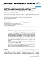
Data from a typical experimental trialFigure 1
Data from a typical experimental trial. (A) Knee angle recorded for an entire day of activity on Earth for Subject 1. Specific sec-
tions depicting walking, running, and sitting are indicated. Zero degrees indicates full knee extension. (B) Detailed view of a 4s
section of the above data demonstrating an excursion event. A transition from an extension excursion to flexion was detected
when the change in angular direction exceeded a 3° fluctuation.
Journal of NeuroEngineering and Rehabilitation 2006, 3:8 />Page 3 of 8
(page number not for citation purposes)
on Earth are presented in Figure 1A. To account for differ-
ent total sampling times, the number of occurrences of
each joint angle was divided by the duration of the data
collection in hours. The excursion was determined by
measuring the amplitude of continuous motion in one
direction, thus representing a monotonically increasing
(extension) or decreasing (flexion) knee angle, as demon-
strated in Figure 1B. A transition between flexion and
extension excursions was indicated by a change in the
direction of motion of >3°, thus discounting small fluctu-
ations in knee angle. The excursions were grouped into 1-
degree bins, and the number of occurrences at each excur-
sion was normalized by the duration of the data in hours.
The velocity of the excursions was linearly approximated
by dividing the magnitude of each excursion by the total
time for that excursion.
Muscle activity, collected by surface electromyography
(EMG), was sampled at 1024 Hz. EMG data were cleaned
using a 2000
th
-order band-pass finite impulse response
zero-phase distortion filter (a 1000
th
-order finite impulse
response filter was used with the data being first passed
forward and then in reverse; see MATLAB's filtfilt function
(Mathworks, Natick, MA, USA). The band-pass spectrum
of the filter was from 20 to 400 Hz. The EMG data were
enveloped by calculating an interval root mean square
(RMS) over a period of 1/64 of a second. Using the inter-
val RMS data from the resting calibration period, the
mean of the RMS and the standard deviation of the RMS
were calculated. The threshold value was then calculated
to be the 95% confidence interval of the interval RMS.
This methodology essentially creates a maximum envel-
oped RMS value during the threshold period. The muscle
is then considered to be active when the interval RMS is
greater than the threshold. Any muscle activation shorter
than 0.150 seconds was not considered.
These data were then correlated with concurrent joint
activity to determine whether the muscles of interest, VM
and BF, were acting concentrically (during knee extension
[VM] or flexion [BF]), eccentrically (during knee flexion
[VM] or extension [BF]), or isometrically (during periods
when the knee-joint angle did not change by more than
3° and the muscle was considered active). This character-
ization does not account for any differential length
changes of the passive and active elements in muscle;
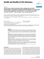
Typical histograms of the instantaneous angular position of the knee jointFigure 2
Typical histograms of the instantaneous angular position of
the knee joint. Data for both subjects are shown (A) on
Earth and (B) onboard the International Space Station (ISS).
On Earth 73.0 ± 11.5% of the instantaneous knee angles
occurred <15° and >75° ; onboard the ISS 74.6 ± 8.9%
occurred within 15–75°).
Table 1: Summary of results for angular position, excursion, and
velocity.
Onboard the ISS Earth
Mean Angular Position (deg)
Subj 1 36.9 ± 9.2 53.4 ± 13.1
Subj 2 48.2 ± 4.8 65.0 ± 6.2
Modal Angular Position (deg)
Subj 1 23.7 ± 9.0 86.0 ± 17.7
Subj 2 40.8 ± 13.4 89.0 ± 10.5
Total Number of Excursions per Hour
Subj 1 1.35 × 10
3
± 0.18 × 10
3
2.66 × 10
3
± 0.26 × 10
3
Subj 2 1.28 × 10
3
± 0.24 × 10
3
1.94 × 10
3
± 0.44 × 10
3
Total Excursion Magnitude per Hour (deg)
Subj 1 3.7 × 10
4
± 0.6 × 10
4
10.0 × 10
4
± 0.6 × 10
4
Subj 2 3.1 × 10
4
± 0.5 × 10
4
8.0 × 10
4
± 3.0 × 10
4
Mean Excursion Velocity (deg/sec)
Subj 1 65.7 ± 8.0 156 ± 13.9
Subj 2 63.0 ± 12.2 129 ± 55.2
Note: Standard deviations are for multiple collections under the same
conditions.
Journal of NeuroEngineering and Rehabilitation 2006, 3:8 />Page 4 of 8
(page number not for citation purposes)
rather it describes the length change of the muscle-tendon
unit.
Results
Angular position of knee joint
Figure 2 shows typical histograms of the instantaneous
angular position of the knee joint for both subjects during
typical days on Earth (Figure 2A) and onboard the ISS
(Figure 2B). A summary of data from all trials is presented
in Table 1.
The data collected on Earth shows a characteristic bimo-
dal distribution, with peaks at full extension and at
approximately 90° of flexion, whereas the data from
onboard the ISS show a predominantly unimodal distri-
bution, with a peak at approximately 30°-50° of flexion.
In space, 74.6 ± 8.9% of the knee-joint angles were
between 15° and 75°; on Earth, 73.0 ± 11.5% of the knee-
joint angles were either <15° or >75°, when averaged
across all days for both subjects. The mean knee-joint
angle onboard the ISS, averaged across all trials and both
subjects, was 41.4 ± 9.4°, with no statistical difference
between the individual subjects (p = 0.05). The average
modal knee-joint angle across all trials onboard the ISS
was 30.5 ± 13.5°.
Excursions of knee joint
Figure 3 shows typical histograms of the excursions of the
knee for different days of continuous data collection on
Earth and in space. Onboard the ISS, 80.1 ± 7.4 % of all
the excursions were less than 45° in magnitude compared
with 55.5 ± 5.1% on Earth. There were 44% fewer excur-
sions per hour onboard the ISS than on Earth (1320 ± 190
vs. 2360 ± 500, respectively). The sum of angles that the
knee swept through per hour was 63% smaller in space
(3.45 × 10
4
± 0.63 × 10
4
[in degrees] onboard the ISS vs.
9.43 × 10
4
± 2.2 × 10
4
[in degrees] on Earth).
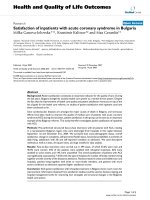
A histogram of the velocity of the excursions is shown in
Figure 4. Onboard the ISS, 47.7 ± 7.6% of all the excur-
sions occurred at velocities of <20°/s in magnitude,
whereas, on Earth, only 11.8 ± 3.2% of the excursions
were of <20°/s in magnitude.
Typical histograms of the excursion velocitiesFigure 4
Typical histograms of the excursion velocities. The instances
of different excursion velocities are shown for typical days of
activity (A) on Earth and (B) onboard the ISS.
Typical histograms of the knee excursion angles (>3 °)Figure 3
Typical histograms of the knee excursion angles (>3 °). Both
subjects are shown for typical days of activity (A) on Earth
and (B) onboard the ISS.
Journal of NeuroEngineering and Rehabilitation 2006, 3:8 />Page 5 of 8
(page number not for citation purposes)
Muscle activity
The relative amounts of concentric, eccentric, and isomet-
ric muscle action during days on Earth and on the ISS are
shown in Figure 5, and the changes in the type of muscle
action in space compared with Earth are shown in Figure
6. On average, there was 5.5% less concentric muscle
action, 9.4% less eccentric action, and 13.9% more iso-
metric muscle action in space than on Earth. Overall mus-
cle activity of the VM decreased onboard the ISS (14.2%
onboard the ISS vs. 22.1% on Earth) for Subject 1 but
increased for Subject 2 (25.6% onboard the ISS vs. 20.9%
on Earth). Overall muscle activity of the BF increased
onboard the ISS for both subjects (33.4% vs. 23.0% and
43.3% vs. 36.3% onboard the ISS vs. on Earth for Subjects
1 and 2, respectively).
Discussion
On Earth, the angular position of the knee joint is pre-
dominantly at the two ends of the used range of motion,
with the knee either flexed or extended (Figure 2A),
stretching either the knee extensors or the flexors.
Onboard the ISS, the knee typically is maintained at an
intermediate angular position around the average modal
value of 30.5 ± 13.5° (Figure 2B). The mean knee-joint
angle of 41.4 ± 9.4° observed onboard the ISS was not sig-
nificantly different (p < 0.05) from the "natural" micro-
gravity position of 47 ± 8° reported in the NASA
Standards 3000 [13]. The implications of these differences
are that single knee-joint flexor and extensor muscles are
not stretched to the same lengths onboard the ISS as they
are on Earth. Statements regarding the length of the two-
joint knee flexors and extensors would require the incor-
poration of both knee and hip angles into an anatomical
model [11], which is beyond the scope of this report.
However, given the maintained knee flexion, the flexed
hip posture (which has been commonly observed in space
[13]) would tend to equalize the lengths of the two-joint
The proportion of total muscle activity in a working day that was isometric, eccentric, or concentricFigure 5
The proportion of total muscle activity in a working day that was isometric, eccentric, or concentric. Both subjects and mus-
cles are shown for activity on Earth and onboard the ISS. Error bars indicate +/- 1 standard deviation (SD). Each set of error
bars is associated with the box containing the -1 SD bar.
Journal of NeuroEngineering and Rehabilitation 2006, 3:8 />Page 6 of 8
(page number not for citation purposes)
muscles toward their lengths during upright posture on
Earth.
The "natural" position of the knee experienced in space
most likely arises from the passive elastic properties of the
lower-extremity joints. Extensive research has examined
the passive properties of the knee joint by measuring the
moment produced when the knee joint is at different
angular positions throughout its range of motion [15-20].
It has been shown that the passive knee moment as a func-
tion of knee-joint angle is sigmoid in shape and that the
magnitude of the moment increases exponentially as the
angular position of the knee is further from a "neutral"
central position. When the knee-joint angle deviates from
the neutral position, passive restorative moments are pro-
duced from the imbalance in the elastic stiffness of the
knee flexors and extensors. The passive properties of the
hip are also likely to be critical to the "natural" posture
observed.
Reduction in knee excursions
Our data show that the total motion of the knee joint in
space is greatly reduced from what is typically experienced
on Earth. The data indicate both that a fewer number of
excursions occurred while subjects were onboard the ISS,
44% fewer per hour, and that excursions were of smaller
magnitude than on Earth. Overall, the knee was moved
through a reduced span of angles; the range of motion was
63% smaller onboard the ISS than on Earth.
Peaks in the histogram of excursion angles indicate a large
number of repeated motions, for instance, during walking
Change in the percentage duration of total muscle action onboard the ISS relative to the total muscle action duration on EarthFigure 6
Change in the percentage duration of total muscle action onboard the ISS relative to the total muscle action duration on Earth.
Mean data are presented for both subjects and both muscles. The action is either concentric, eccentric, or isometric. A posi-
tive value indicates a greater amount of that quantity onboard the ISS. Note the increase in the relative amount of isometric
action for all subjects and both muscles, primarily at the expense of eccentric action.
Journal of NeuroEngineering and Rehabilitation 2006, 3:8 />Page 7 of 8
(page number not for citation purposes)
or running (Figure 3). On Earth, both subjects exhibited
such peaks at varying magnitudes covering nearly the
entire range of angles used. Onboard the ISS, the ampli-
tudes of knee movements were limited and were predom-
inantly small, with 80.1 ± 7.4% of the excursions <45°.
During activities onboard the ISS, the crewmembers made
fewer and smaller-amplitude movements, resulting in less
change in the angular position of the knee joint. Onboard
the ISS, the knee-joint velocities indicated predominantly
slower movements than those on Earth. Velocities close to
zero result in quasi-isometric movements of relatively low
power [21]. The maintenance of the knee joint in a flexed
position during spaceflight may result in a similar flexor
bias in the estimation of joint position, one that has been
observed in the elbow joint [22].
Muscle activity and action
The increased duration of muscle activity observed on the
ISS for all but one of the four subject/muscle conditions
studied has a precedent in the work of Edgerton and col-
leagues [23], who found that, compared with pre- and
post-flight values, there was a marked increase in the daily
integrated EMG activity of the tibialis anterior and soleus
during spaceflight. However, the analysis presented here
refers only to the duration of above-threshold activity,
and thus the magnitude of activity (which was incorpo-
rated into the Edgerton group's data) is not considered.
This information is available and will be the topic of
future communications. The change to a more dominant
pattern of isometric action onboard the ISS is reasonable
based on the anti-gravity role of the muscles studied on
Earth. The marked reduction in knee-joint velocities
observed in space suggests a change in the pattern of mus-
cle use, which is likely to be associated with the change in
expression of the myosin phenotypes that has been
observed from human biopsy studies [7,24].
Among the limitations of the current experiment are the
potential for migration and/or misalignment of the goni-
ometers and the simplicity of the muscle-length models.
There is also a possibility that the electrode-skin interface
changed during the approximately 8-hour data collection
sessions, although we have previously shown that EMG in
response to a standard load measured on multiple occa-
sions over the course of a day in which the electrodes are
not removed is highly reliable [25]. However, all of the
above factors could have exerted an influence during
experiments on Earth or in space, and thus no bias in the
results is likely.
Conclusion
Onboard the ISS, the knee is operated in different ranges
of angles, excursions, total daily excursion, and velocities
than those observed during typical daily activity on Earth.
These differences imply that the muscles spanning the
knee joint are operating at altered lengths, velocities, and
power ranges, all of which may contribute to the muscle
atrophy and functional losses that have been observed in
microgravity.
Abbreviations
BF – Biceps Femoris
EMG – Electromyography
ISS – International Space Station
LEMS – Lower Extremity Monitoring Suit
RMS – Root Mean Square
SD – Standard Deviation
VM – Vastus Medialis
Competing interests
The author(s) declare that they have no competing inter-
ests.
Authors' contributions
MCP contributed to data analysis, interpretation of data,
and drafting the manuscript. KOG contributed to data
analysis and drafting the manuscript. ML and BH were
responsible for developing the data analysis algorithms
and assisted with manuscript revisions. AJR was responsi-
ble for project organization and contributed to data acqui-
sition and manuscript revision. CCM managed the in-
flight aspects of data collection. PRC conceived and
designed the experiment, contributed text, and critically
reviewed the manuscript. All authors have read and
approved the final manuscript.
Acknowledgements
This work was supported by NASA cooperative agreement NCC 9 153.
The authors would like to acknowledge the cooperation and dedicated
work of the subjects.
References
1. Desplanches D: Structural and functional adaptations of skele-
tal muscle to weightlessness. Int J Sports Med 1997, 18(Suppl
4):S259-64.
2. Edgerton R: Critical discussion of integrated physiology of
microG. Med Sci Sports Exerc 1996, 28:S107-8.
3. di Prampero PE, Narici MV: Muscles in microgravity: from fibres
to human motion. J Biomech 2003, 36:403-12.
4. Fitts RH, Riley DR, Widrick JJ: Physiology of a microgravity envi-
ronment invited review: microgravity and skeletal muscle. J
Appl Physiol 2000, 89:823-39.
5. Convertino VA: Physiological adaptations to weightlessness:
effects on exercise and work performance. Exerc Sport Sci Rev
1990, 18:119-66.
6. Jackman RW, Kandarian SC: The molecular basis of skeletal
muscle atrophy. Am J Physiol Cell Physiol 2004, 287:C834-43.
7. Caiozzo VJ, Haddad F, Baker MJ, Herrick RE, Prietto N, Baldwin KM:
Microgravity-induced transformations of myosin isoforms
Publish with BioMed Central and every
scientist can read your work free of charge
"BioMed Central will be the most significant development for
disseminating the results of biomedical research in our lifetime."
Sir Paul Nurse, Cancer Research UK
Your research papers will be:
available free of charge to the entire biomedical community
peer reviewed and published immediately upon acceptance
cited in PubMed and archived on PubMed Central
yours — you keep the copyright
Submit your manuscript here:
/>BioMedcentral
Journal of NeuroEngineering and Rehabilitation 2006, 3:8 />Page 8 of 8
(page number not for citation purposes)
and contractile properties of skeletal muscle. J Appl Physiol
1996, 81:123-32.
8. Roy RR, Baldwin KM, Edgerton VR: Response of the neuromus-
cular unit to spaceflight: what has been learned from the rat
model. Exerc Sport Sci Rev 1996, 24:399-425.
9. Sasa T, Sairyo K, Yoshida N, Fukunaga M, Koga K, Ishikawa M, Yasui
N: Continuous muscle stretch prevents disuse muscle atro-
phy and deterioration of its oxidative capacity in rat tail-sus-
pension models. Am J Phys Med Rehabil 2004, 83:851-6.
10. Vandenburgh HH, Hatfaludy S, Karlisch P, Shansky J: Skeletal mus-
cle growth is stimulated by intermittent stretch-relaxation
in tissue culture. Am J Physiol 1989, 256:C674-82.
11. Vandenburgh H, Chromiak J, Shansky J, Del Tatto M, Lemaire J:
Space travel directly induces skeletal muscle atrophy. FASEB
J 1999, 13:1031-8.
12. Delp SL, Loan JP: A graphics-based software system to develop
and analyze models of musculoskeletal structures. Comput
Biol Med 1995, 25:21-34.
13. NASA: Anthropometry and Biomechanics. (Section 3.0). .
NASA-STD-3000 1996, 3-1 to 3–76.
14. Andreoni G, Rigotti C, Baroni G, Ferrigno G, Colford NA, Pedotti A:
Quantitative analysis of neutral body posture in prolonged
microgravity. Gait Posture 2000, 12:235-42.
15. Pope MH, Crowninshield R, Miller R, Johnson R: The static and
dynamic behavior of the human knee in vivo. J Biomech 1976,
9:449-52.
16. Goddard R, Dowson D, Longfield MD, Wright V: Stiffness of the
knee in normal and osteoarthrosic subjects. Ann Rheum Dis
1970, 29:194.
17. Mansour JM, Audu ML: The passive elastic moment at the knee
and its influence on human gait. J Biomech 1986, 19:369-73.
18. Such CH, Unsworth A, Wright V, Dowson D: Quantitative study
of stiffness in the knee joint. Ann Rheum Dis 1975, 34:286-91.
19. Crowninshield R, Pope MH, Johnson R, Miller R: The impedance of
the human knee. J Biomech 1976, 9:529-35.
20. Lebiedowska MK, Fisk JR: Passive dynamics of the knee joint in
healthy children and children affected by spastic paresis. Clin
Biomech (Bristol, Avon) 1999, 14:653-60.
21. Adams GR, Caiozzo VJ, Baldwin KM: Skeletal muscle unweight-
ing: spaceflight and ground-based models. J Appl Physiol 2003,
95:2185-201.
22. McCall GE, Goulet C, Boorman GI, Roy RR, Edgerton VR: Flexor
bias of joint position in humans during spaceflight. Exp Brain
Res 2003, 152:87-94.
23. Edgerton VR, McCall GE, Hodgson JA, Gotto J, Goulet C, Fleis-
chmann K, Roy RR: Sensorimotor adaptations to microgravity
in humans. J Exp Biol 2001, 204:3217-24.
24. Zhou MY, Klitgaard H, Saltin B, Roy RR, Edgerton VR, Gollnick PD:
Myosin heavy chain isoforms of human muscle after short-
term spaceflight. J Appl Physiol 1995, 78:1740-4.
25. Ochia RS, Cavanagh PR: Reliability of surface EMG measure-
ments over 12 hours. J Electromyogr Kinesiol 2006 in press.