báo cáo hóa học: " Biofeedback for robotic gait rehabilitation" docx
Bạn đang xem bản rút gọn của tài liệu. Xem và tải ngay bản đầy đủ của tài liệu tại đây (1.66 MB, 11 trang )
BioMed Central
Page 1 of 11
(page number not for citation purposes)
Journal of NeuroEngineering and
Rehabilitation
Open Access
Review
Biofeedback for robotic gait rehabilitation
Lars Lünenburger*
†1
, Gery Colombo
1,2
and Robert Riener
1,3
Address:
1
Spinal Cord Injury Center, Balgrist University Hospital, Zurich, Switzerland,
2
Hocoma AG, Volketswil, Switzerland and
3
Rehabilitation
Engineering Group, Swiss Federal Institute of Technology (ETH), Zurich, Switzerland
Email: Lars Lünenburger* - ; Gery Colombo - ;
Robert Riener -
* Corresponding author †Equal contributors
Abstract
Background: Development and increasing acceptance of rehabilitation robots as well as advances
in technology allow new forms of therapy for patients with neurological disorders. Robot-assisted
gait therapy can increase the training duration and the intensity for the patients while reducing the
physical strain for the therapist.
Optimal training effects during gait therapy generally depend on appropriate feedback about
performance. Compared to manual treadmill therapy, there is a loss of physical interaction
between therapist and patient with robotic gait retraining. Thus, it is difficult for the therapist to
assess the necessary feedback and instructions. The aim of this study was to define a biofeedback
system for a gait training robot and test its usability in subjects without neurological disorders.
Methods: To provide an overview of biofeedback and motivation methods applied in gait
rehabilitation, previous publications and results from our own research are reviewed. A
biofeedback method is presented showing how a rehabilitation robot can assess the patients'
performance and deliver augmented feedback. For validation, three subjects without neurological
disorders walked in a rehabilitation robot for treadmill training. Several training parameters, such
as body weight support and treadmill speed, were varied to assess the robustness of the
biofeedback calculation to confounding factors.
Results: The biofeedback values correlated well with the different activity levels of the subjects.
Changes in body weight support and treadmill velocity had a minor effect on the biofeedback
values. The synchronization of the robot and the treadmill affected the biofeedback values
describing the stance phase.
Conclusion: Robot-aided assessment and feedback can extend and improve robot-aided training
devices. The presented method estimates the patients' gait performance with the use of the robot's
existing sensors, and displays the resulting biofeedback values to the patients and therapists. The
therapists can adapt the therapy and give further instructions to the patients. The feedback might
help the patients to adapt their movement patterns and to improve their motivation. While it is
assumed that these novel methods also improve training efficacy, the proof will only be possible
with future in-depth clinical studies.
Published: 23 January 2007
Journal of NeuroEngineering and Rehabilitation 2007, 4:1 doi:10.1186/1743-0003-4-1
Received: 28 April 2006
Accepted: 23 January 2007
This article is available from: />© 2007 Lünenburger et al; licensee BioMed Central Ltd.
This is an Open Access article distributed under the terms of the Creative Commons Attribution License ( />),
which permits unrestricted use, distribution, and reproduction in any medium, provided the original work is properly cited.
Journal of NeuroEngineering and Rehabilitation 2007, 4:1 />Page 2 of 11
(page number not for citation purposes)
Background
Robotic gait rehabilitation
Walking ability, though important for quality of life and
participation in social and economic life, can be adversely
affected by neurological disorders such as spinal cord
injury, stroke or traumatic brain injury. Rehabilitation of
patients with such disorders should include gait training
because there is evidence that the desired function or
movement has to be trained in a task-specific program
[1,2]. One contemporary approach is body-weight sup-
ported treadmill training in which the patient is sus-
pended over a treadmill and the patient's legs are guided
by therapists [3-9]. Several studies have shown beneficial
effects of this approach [10-12]. Because other studies
[13,14] did not find an advantage compared to conven-
tional therapy and systematic reviews [8,9] regard the evi-
dence as controversial, further studies are required. There
are some indications that an increased training intensity
might lead to clearer results [15-18]. However, the man-
ual form of this therapy in which the patient's legs are
guided by two therapists holding and moving them along
a gait-like trajectory is strenuous for the therapists and
labor- and cost-intensive. Depending on the patient's con-
dition, the therapists have to assist the stance leg by
extending the knee against the weight of the patient or
they have to flex the knee joint, possibly against spasticity,
and lift the leg through swing phase. The high physical
effort for the therapists often limits the training duration,
whereas the patient might benefit from a longer duration.
Recently developed rehabilitation robots [19,20] allow
delivering continuous support for the legs in a physiolog-
ical gait pattern, high repetition accuracy, and prolonged
training duration compared to manual treadmill training.
The loss of the physical contact between the therapist and
the patient is a disadvantage, yet can partly be overcome
by technology. The physical contact was often used by the
therapist to "feel" the patient's ability and activity. With
this information, the therapist can provide feedback to the
patient, give training instructions and help to improve the
patient's motivation. Because feedback on the current per-
formance may improve the training effect [21], a corre-
sponding, computerized feedback is desired for robotic
rehabilitation. As biological quantities are transferred to a
biological system (human) via artificial feedback, the term
"biofeedback" has been introduced and became widely
accepted.
The aim of this study was to develop a biofeedback system
for a gait training robot and test its usability in subjects
without neurological disorders.
Feedback and motivation
General considerations on feedback and motivation
To improve a certain motor function, it is helpful to know
the level of your success and your performance. For
human movements, this performance assessment is often
derived from afferents and reafference such as propriocep-
tive, force or visual sensory inputs. They can also be
described as intrinsic feedback [22]. This intrinsic feed-
back is generated by the movement itself (proprioception
or vision of the moving limb, but also sound of the foot-
steps). In contrast, extrinsic or augmented feedback may
be provided additionally by an outside source, such as a
therapist or coach. This extrinsic feedback is important for
learning some motor tasks [22]. For robotic rehabilita-
tion, the robot itself can be used to generate and display
the feedback.
Apart from its instructional aspect, feedback is also impor-
tant for motivation. Keeping patients informed about
their progress usually translates into greater effort during
task practice [chapter 10 of ref. [22]]. This higher effort,
e.g. in terms of enhanced endurance or higher compli-
ance, might help to improve training outcomes. Pursuing
and achieving goals usually motivates the subjects. This
requires measurements to compare the current status with
the desired goal. It is important to know the quantity and
quality of the movements performed by the patient.
In neuro-rehabilitation, the neurological disorder can
increase the need for artificial feedback. For people with
neurological disorders, interpretation of intrinsic feed-
back could be difficult or incorrect due to impaired som-
atosensory pathways.
Biofeedback principles in non-robotic gait rehabilitation
Biofeedback principles have been applied in gait rehabili-
tation of patients with stroke [23-31], cerebral palsy [32],
spinal cord injury [33], Spina Bifida [34] or arthritis [35].
Electromyographic (EMG) recordings [23-26,32,33], kin-
ematic quantities [25-30,34-38], and kinetic measures
[37,38] have been processed and displayed visually
[29,32], acoustically [27,28,30,37] or in combination
[23,26,33,35,38], as well as via vibrotactile stimuli
[34,36,37]. The application of biofeedback in stroke reha-
bilitation improved the patients' gait function according
to a recent systematic review [8].
During manual training therapists can estimate the
patients' performance in several ways. Apart from visual
observation therapists can base this estimation on the
amount of external assistance needed to perform the
movement correctly. However, because the therapist will
usually increase the assistance to maintain a physiological
gait pattern when the patient's performance reduces, the
patient does not have to walk with maximum effort (see
also comments on motivation above). Conversely, many
individuals with neurological disorders ambulate inde-
pendently and might still benefit from training. For these
individuals, assistance might be beneficial to achieve
Journal of NeuroEngineering and Rehabilitation 2007, 4:1 />Page 3 of 11
(page number not for citation purposes)
higher gait quality and delivers a basis for feedback. In
conclusion, the estimation of (maximum) walking capa-
bility of the patient might be difficult with this assistance-
based method. However, the estimation will reflect the
current performance correctly. The feedback of this per-
formance estimation might already be sufficient to
enhance the training.
This approach based on required assistance can be trans-
lated to rehabilitation robots that are equipped with force
sensors. However, the problems described above for the
estimation by the therapist basically also apply to robotic
implementation. With the most commonly used posi-
tion-controlled strategies, these force sensors register the
amount of robot-generated force assisting the patient to
follow the predefined gait pattern. The use of these force
or torque signals has an advantage over electromyo-
graphic muscle recording or standard videographic gait
analysis, because no additional time or equipment is
needed. Furthermore, electromyographic recordings regis-
ter muscle activity. The movement resulting from this
activity is usually difficult to identify especially when
many muscles act onto the same joint and in dynamic sit-
uations like walking. Videographic gait analysis is limited
by visual obstruction of the one leg by the other, or the
rehabilitation device. Additionally, when position control
strategies are applied, the visual gait analysis will mainly
identify the underlying predefined trajectory. Therefore,
we chose a force-based strategy described below for imple-
menting a biofeedback for a gait rehabilitation robot.
Force-based biofeedback in a rehabilitation
robot
One specific strategy presented in this paper is based on a
driven gait-orthosis DGO [20] (Lokomat
®
Pro Version 4,
by Hocoma AG, Volketswil, Switzerland). The DGO is a
bilateral robotic orthosis that is used in conjunction with
a body-weight support system to control the patient's leg
movements in the sagittal plane (Fig. 1). The DGO's hip
and knee joints are actuated by linear drives, which are
integrated in an exoskeletal structure. A passive foot lifter
induces an ankle dorsiflexion during the swing phase. The
legs of the patient are moved with highly repeatable pre-
defined hip and knee joint trajectories on the basis of an
impedance control strategy [39]. Knee and hip joint tor-
ques of the patient are determined from force sensors inte-
grated in the drives of the DGO.
Implementation of the biofeedback
The technical implementation of a force-biofeedback
strategy for the DGO has been described by the authors of
this paper [39,40]. For this strategy, the subject's legs are
guided by the DGO with high impedance (equivalent to
position control). With this high stiffness, changes in the
subject's behavior are best detectable because already
small deviations lead to large counteracting torques by the
robot. The torque outputs of the drives (with compensa-
tion for passive properties of the DGO) give direct infor-
mation about the patient's activity and performance. If the
patient actively moves according to the reference trajec-
tory, no interaction torques from the subject would act on
the robot. If the patient is passive and does not contribute
to the walking movement due to paresis or lack of moti-
vation, the robot has to exert torque in order to maintain
the desired reference trajectory. Thus, the robot has to
push the subject. Conversely, if the patient tries to move
faster than the reference trajectory, the robot requires less
torque or even has to decelerate the subject.
Biofeedback values are calculated for stance and swing
phase of the gait cycle as weighted averages of the torques
measured in the corresponding joint drives [39,40]. The
appropriate selection of the weight functions leads to pos-
itive biofeedback values when the patient performs thera-
peutically desirable activities. Specifically, active hip
flexion is required to bring the leg forward during the
swing phase, active knee flexion during early swing phase
and knee extension during late swing phase. During the
stance phase, the most important activity is weight bear-
ing by continuous, almost isometric knee extension,
whereas hip extension results from a combination of mus-
cle activity and passive motion of the treadmill. This
means that for each joint, except the knee joint during
stance phase, a torque pointing against the direction of
movement should produce a negative feedback, one
pointing parallel to the direction of motion a positive
feedback. Mathematically this can be implemented by
multiplication of the measured force and a weighting
function for each time during the gait cycle. Integration of
joint torques weighed according to this principle during
phases of the gait cycle delivers values that are compre-
hensive in summarizing the performance in the specific
gait phase and that are more robust against noise than the
continuous signal. Similar scaling for all values is
obtained by normalization (For the mathematical for-
mula see [39]). Because weighting functions that are pro-
portional to the angular velocity follow the described
principle, the present implementation employs these
functions for hip joint during stance phase and knee joint
during swing phase, as well as hip joint during swing
phase with a slight modification. This modification was
implemented because there is some indication for a pas-
sive pendulum-like motion of the leg in mid swing [41].
It reduces the importance of this phase by multiplication
of the weighting function with an additional smooth
function (quenching). In contrast to these three biofeed-
back calculations, the weighting function for the knee dur-
ing stance phase was chosen to be constant because it
takes the requirement of constant weight bearing better
into account. In summary, this biofeedback approach pro-
Journal of NeuroEngineering and Rehabilitation 2007, 4:1 />Page 4 of 11
(page number not for citation purposes)
vides four biofeedback values per stride and per leg that
become available immediately after each step.
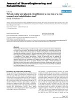
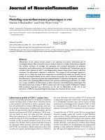
The most complete display shows all 8 values per stride in
an array of line graphs (Fig. 2A), each including the his-
tory for a modifiable number of recent strides. This allows
monitoring every aspect of gait performance that is evalu-
ated by the biofeedback. For supervision, a similar visual-
ization can be displayed on the therapist's monitor. Many
patients understand quickly which movement leads to
higher biofeedback values after verbal instruction by their
therapists. However, recurrently reminding the patients
usually improves their performance. Simultaneously, the
visualization for the patient can be adapted to emphasize
specific gait performance aspects and to avoid informa-
tion overload for the patient. Specifically, the display
should be accessible in the way that the patients are able
to perceive the information displayed to them, i.e. large
fonts readable while walking. The display should also be
intuitive. Otherwise, additional time would be required
for learning to understand and use the display and there-
fore shorten the available training time. Intuitive displays
are even more important in neuro-rehabilitation because
some patients with neurological disorders who require
gait retraining also sustain cognitive deficits (e.g. after
traumatic brain injury). Thus, such patients could benefit
from a reduction to one value per gait phase and a visually
more appealing display, such as a smiley face (Fig. 2B).
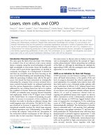
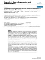
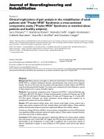
The driven gait orthosis LokomatFigure 1
The driven gait orthosis Lokomat. The driven gait orthosis Lokomat Pro (Hocoma AG, Volketswil, Switzerland) is a bilat-
eral robotic orthosis with actuated hip and knee joints that is used for body-weight supported treadmill training. (Photo cour-
tesy of Hocoma AG, Volketswil, CH)
Journal of NeuroEngineering and Rehabilitation 2007, 4:1 />Page 5 of 11
(page number not for citation purposes)
The biofeedback values are summarized by averaging the
values of a subset selected by the therapist. Averaging
results in an overall factor that is relatively unbiased. In
this way, the therapist can have the patient focus on spe-
cific aspects of walking. The possible performance loss in
the remaining aspects of walking that are not selected for
the feedback should be monitored by the therapists with
the help of the complete display on their monitor. When
selected, the smiley is continuously displayed on the
monitor in front of the patient and updated every step.
The shape of the smiley's mouth (an arc of a circle) is
determined from the obtained average biofeedback value
for the last step as well as threshold and scaling factors set
by the therapist. For averages larger than the therapist's
setting, the ends of the mouth point upward (smile), for
averages below the threshold, the ends of the arc point
downwards (frown). The arc lengthens with larger abso-
lute values resulting in a more prominent smile or frown
for high and low values respectively. The scaling factor
allows the therapist to adjust the sensitivity of the feed-
back to the functional abilities of the patient. In conclu-
sion, the smiley display allows for a goal-oriented training
with feedback, i.e. the patient should focus on specific
movements to reach the "goal" of a full smile.
Validation in subjects without neurological disorders
Three subjects without neurological disorder (2 female, 1
male), aged 24–30 years, without neurological disorders
were included in the study after giving informed consent
and approval by the regional ethics committee of the Can-
ton Zurich. The subjects walked in the DGO at two differ-
ent speeds (1.8 and 2.4 km/h). A dynamic body-weight
support system was used to support 25%, 50%, and 70%
of subject's body weight. Apart from the optimal setting of
the synchronization of the DGO and the treadmill, two
other settings were used that caused the DGO either to
walk about 10% slower or faster.
All subjects had previous experience in walking within the
DGO. During recording times of 30 seconds, the subjects
were instructed to walk in three different ways: (1) Pas-
sive: They should not contribute to the movement. (2)
Active: They should walk with the same pattern as the
DGO. (3) Exaggerated: They should exaggerate their
movements in order to increase the biofeedback values
that were displayed as line graphs. With the given time
and endurance limitations, not all of the 54 possible com-
binations could be tested in the single session performed.
Subject P1 completed 41, subject P2 45 and subject P3 42
trials. The actual joint angles and the joint moments were
digitally recorded with a sampling rate of 1 kHz.
For analysis, biofeedback values were re-calculated offline
(using Matlab, Mathworks Inc.) from the recorded tor-
ques according to the method described above, i.e. as
weighted averages of the force values using the described
weighting functions. (The analysis would have been pos-
sible by selecting strides from the automatically generated
biofeedback file. The recalculation was done for conven-
ience and easier automatic analysis). For illustration, the
torques and angles were cut into strides and normalized in
time to 100 samples per gait cycle. For purposes of corre-
lation with recorded joint torques and biofeedback values
using Spearman correlation in Matlab (Mathworks Inc.),
the walking instructions were coded as "passive" = 0,
"active" = 1, "exaggerated" = 2.
Torques acting during walking in the robot
Torques in the DGO joints were recorded during walking
with different instructed walking activity – passive, active,
exaggerated – and different settings of body weight sup-
port, treadmill speed and synchronization coefficient of
DGO and treadmill. The effect of different instructed
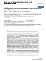
walking activities on the recorded torques are shown for
one example subject in Fig. 3. The traces show a large var-
iability within the 11–12 steps in each condition. The
largest variability was present in the "exaggerated" condi-
tion. The traces of the active condition are between the
traces of the passive and those of the active conditions for
most of the times.
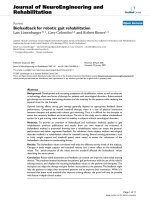
The correlation of the recorded torques at each time of the
gait cycle and the four external parameters, instructed
activity, patient coefficient, body weight support and
treadmill speed were calculated and are shown in Fig. 4
for the right hip and knee of the three subjects. In all three
subjects, the correlation of hip joint torque and instructed
activity was high (>0.5) during swing phase ranging from
about 55% to 100% of the gait cycle. The correlation of
hip torque and activity was inconsistent during stance
phase, being close to zero for 2 subjects and smaller than
-0.5 for one subject. For the knee joint, the correlation of
torque and activity was also small during stance phase.
During swing phase, the correlation of knee torque and
activity was positive during early swing, when the knee is
flexing, and negative (<-0.5) during late swing when the
knee is extending.
Changing the synchronization of DGO and treadmill
influenced the hip and knee joint torques during the
stance phase, especially at its end when the correlation
coefficients were >0.5 for the hip and <-0.5 for the knee
joint. The correlation coefficients of hip and knee torques
and treadmill speed were generally close to zero during
stance phase and had a consistent biphasic pattern during
swing phase. The correlation coefficients of hip and knee
torques and the amount of body weight support were gen-
erally closer to zero during the whole gait phase with larg-
est values in the hip during the stance phase.
Journal of NeuroEngineering and Rehabilitation 2007, 4:1 />Page 6 of 11
(page number not for citation purposes)
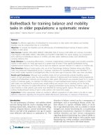
Visual displays of the biofeedbackFigure 2
Visual displays of the biofeedback. Screen shots of two standard displays of the biofeedback implemented for gait training.
Four biofeedback values become available after each step (e.g. left leg stance phase and right leg swing phase). These data can
be displayed in a line diagram (A), which is updated twice per stride. Each point represents the biofeedback value of one stride.
The values are displayed in independent subplots for each of the four joints. Swing and stance phase are color-coded. Both
axes can be adjusted by the therapist in order to adapt the feedback to the current training situation. It is possible to display a
selection of biofeedback values (e.g. only one leg, only swing phase, only knee joints) to help the patient focusing on specific
aspects. The selected subset of biofeedback values can also be averaged into one value that can be displayed by a smiley (B)
which smiles broader for higher and frowns for lower values of the biofeedback during the most recent step.
Journal of NeuroEngineering and Rehabilitation 2007, 4:1 />Page 7 of 11
(page number not for citation purposes)
Correlation of biofeedback and subject's activity
Biofeedback values were calculated as weighted averages
using the weight functions described above and illustrated
in Fig. 3. The resulting values for all four joints in two gait
phases during about 580 strides for each subject were cor-
related to the level of activity the subject was instructed to
perform (0 = passive, 1 = active, 2 = exaggerated). The rea-
son to use the instructed level of activity was that no other
quantification for gait performance was available that
would allow a concurrent validation. The implied propo-
sition that the subjects complied to the instruction is not
a strong assumption. Spearman correlation coefficients
were calculated because non-linear relations could be
expected. The results are shown in Fig. 5 and Table 1. Bio-
feedback values of the swing-phase correlated highly with
the instructed activity (range ρ = 0.63 to 0.82, mean ρ =
0.75; p < 0.01). The correlation of instructed activity and
the biofeedback values of the stance-phase was lower
(range ρ = -0.75 to 0.68, mean ρ = -0.01), especially in two
subjects, and sometimes even negative. The negative cor-
relation to the activity was not desired. However, it cannot
be completely avoided with the present calculation
method because the mechanical contact of the foot and
the treadmill during the stance phase results in the passive
torques acting onto the hip joint.
Other factors influencing the biofeedback
The correlation of biofeedback values and the synchroni-
zation settings of DGO and treadmill had large absolute
values (max 0.68, mean 0.39), and were higher for the
stance phase than for the swing phase. Because the syn-
chronization of the leg movements and the treadmill
influenced the forces between the treadmill and the stance
leg, it also affected the joint torques. These torques are
integrated into the biofeedback values, which indeed
show a correlation to the synchronization setting.
The correlations of the biofeedback values to the amount
of body weight support and to the treadmill speed are rel-
atively small. For the body weight support, the absolute
values of the correlation coefficients were on average 0.19
with a maximum of 0.38. For treadmill speed, the abso-
lute values were on average 0.14 with a maximum 0.33.
The influence of gait parameters other than the subject's
activity on the biofeedback values is therefore minor for
values addressing the swing phase. The stance-phase val-
ues are strongly influenced by the synchronization of
walking cadence and treadmill speed. The calculation of
these values will be updated to improve the robustness
against disturbances that is important for quantitative
analysis. For the use as a biofeedback, however, this effect
is less important because for adapting his or her motor
activity the patient will concentrate on the last several
steps and will take into account changes in the other
parameters. Furthermore, the currently used weighting
functions originate from basic biomechanical reasoning
(as described above) and can be understood as a first-
order approximation to robot-assisted walking.
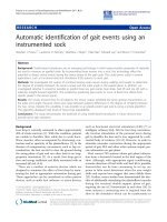
Example traces of joint torques during walking in the robot with different instructionsFigure 3
Example traces of joint torques during walking in the
robot with different instructions. Joint moment in the
hip and knee joint of the DGO were recorded while a sub-
ject without neurological disorders walked according to
three different instructions The other parameters, treadmill
speed, body weight support, synchronization between DGO
and treadmill were held constant. The instructions were:
Passive (black): Do not contribute to the movement. Active
(blue): Walk with the same pattern as the DGO. Exaggerated
(red): Exaggerate the movement pattern to increase the bio-
feedback values displayed to them as line graphs (red). The
weight functions used for calculation of the biofeedback val-
ues are illustrated as shaded areas.
Journal of NeuroEngineering and Rehabilitation 2007, 4:1 />Page 8 of 11
(page number not for citation purposes)
Clinical importance
Before trying to address the efficacy of the biofeedback for
rehabilitation, it is useful to check the usability and the
effect on compliance in patients. Preliminary results
obtained from patients with SCI gave positive responses
both from patients and therapists [39]. Six subjects with
incomplete spinal cord injury walked with different
instructions during five trials of 30 s each. They were
instructed to walk as powerfully as possible in two trials.
They were verbally instructed and motivated by a coach in
one trial (no visual display), whereas they used the bio-
feedback display in the other trial (no verbal instruction
and motivation). The biofeedback values during both
active trials were significantly higher than during the pas-
sive control trials for 5 out of 6 subjects with only a little
or no significant difference between the two active trials.
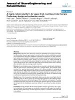
Correlation of the joint torques with the walking parameters during the gait cycleFigure 4
Correlation of the joint torques with the walking parameters during the gait cycle. The torques in the hip and knee
joints of the DGO were recorded during the walking sessions of three subjects and correlated to the different walking instruc-
tions ("passive" = 0, "active" = 1, "exaggerated" = 2; blue) and different walking parameters: synchronization of robot and tread-
mill ("patient coefficient" optimal and +/- 5 units; green), body weight support (25%, 50%, 70% of body weight; red) and
treadmill speed (1.8 and 2.4 km/h; cyan).
Journal of NeuroEngineering and Rehabilitation 2007, 4:1 />Page 9 of 11
(page number not for citation purposes)
Table 1: Correlation of biofeedback and subject's activity
Joint Hip right Knee right Hip left Knee left
Gait phase Stance Swing Stance Swing Stance Swing Stance Swing
Subject P1 -0.17 0.71 0.18 0.80 -0.17 0.71 0.19 0.80
Subject P2 0.31 0.82 -0.14 0.63 0.22 0.79 -0.29 0.72
Subject P3 0.68 0.79 -0.75 0.74 0.50 0.77 -0.69 0.72
Spearman correlation coefficients of the obtained biofeedback values and the level of activity with which they were instructed to walk in the DGO
(0 = passive, 1 = active, 2 = exaggerated). In three individuals, correlations are show independently for the four actuated joints and for stance and
swing phase.
Correlation of the biofeedback values with the instructed performance of subjects without neurological disordersFigure 5
Correlation of the biofeedback values with the instructed performance of subjects without neurological disor-
ders. Three subjects without neurological disorders were instructed to walk in the DGO with three different levels of activity
(passive, active, exaggerated) and with different treadmill speed, body weight support and synchronization of DGO and tread-
mill. Spearman correlation coefficients of the biofeedback values obtained during this walking and the instructed activity are
shown ("passive" = 0, "active" = 1, "exaggerated" = 2).
Journal of NeuroEngineering and Rehabilitation 2007, 4:1 />Page 10 of 11
(page number not for citation purposes)
One patient (the only one with ASIA impairment scale C
[42]) was not able to substantially modulate the biofeed-
back and did not regain independent walking function
during this therapy period. It was interpreted that the vis-
ual biofeedback is as effective as the continuous verbal
instruction for the observed short time periods. Subjects
reported in questionnaires that they felt positive about the
biofeedback and wanted to use it again. However, it will
be important to demonstrate clinical efficacy of the whole
rehabilitation period and potentially faster rehabilitation
with these new tools in future clinical studies.
Extension to other technologies
Virtual reality techniques developing from visualization
and simulation start to enter the rehabilitation domain
[for review see [43]]. The techniques, including large
screen 3D projections and head mounted display technol-
ogy that allow depth perception, permit the immersion of
the subject into an environment that is artificially gener-
ated in a computer. With an appropriate choice of the
environment, it should be possible to instruct and moti-
vate the subjects for training and rehabilitation. This
enhanced motivation and feedback has the potential to
improve the training efficacy and rehabilitation outcome.
Conclusion
Biofeedback is a necessary addition to robotic gait train-
ing. It can provide an online feedback about the patients'
performance to the training and allow the patient and the
therapist to assess the walking performance. This can help
to adapt and improve the training. The subjects might
draw additional motivation from the online feedback on
their performance.
Furthermore, the assessment of the patients' performance
might be used not solely as online feedback, but also for
evaluation of the rehabilitation progress. The integration
of robot-aided training with robot-aided assessment and
feedback has the potential to improve robotic rehabilita-
tion.
Abbreviations
DGO Driven gait orthosis
EMG Electromyography
Competing interests
LL is employed by the University of Zurich via a CTI
(Commission for Technology and Innovation) project
funded by the Swiss Bureau of Education and Technology
and Hocoma AG, Volketswil, Switzerland, which pro-
duces the Lokomat.
GC is founder, shareholder and CEO of Hocoma AG,
Volketswil, Switzerland, which produces the Lokomat.
GC is one of the inventors of the Lokomat.
Authors' contributions
LL conceived and designed the study, recruited subjects,
performed the acquisition and analysis of data, and
drafted the manuscript. GC and RR provided expert guid-
ance on experimental design, assisted with data interpre-
tation, helped drafting the manuscript and edited the
manuscript.
Acknowledgements
This work is partially supported by Commission for Technology and Inno-
vation (CTI) projects 6199.1-MTS and 7497.1 LSPP-LS and Swiss National
Science Foundation NCCR Neuro (project 7), Switzerland.
References
1. de Leon RD, Hodgson JA, Roy RR, Edgerton VR: Locomotor
capacity attributable to step training versus spontaneous
recovery after spinalization in adult cats. J Neurophysiol 1998,
79(3):1329-1340.
2. de Leon RD, Hodgson JA, Roy RR, Edgerton VR: Full weight-bear-
ing hindlimb standing following stand training in the adult
spinal cat. J Neurophysiol 1998, 80(1):83-91.
3. Wernig A, Müller S: Laufband Locomotion with Body-Weight
Support Improved Walking in Persons with Severe Spinal-
Cord Injuries. Paraplegia 1992, 30(4):229-238.
4. Barbeau H, Ladouceur M, Norman KE, Pepin A, Leroux A: Walking
after spinal cord injury: evaluation, treatment, and func-
tional recovery. Arch Phys Med Rehabil 1999, 80(2):225-235.
5. Hesse S, Werner C, Bardeleben A, Barbeau H: Body Weight-Sup-
ported Treadmill Training after Stroke. Current Atherosclerosis
Reports 2001, 3(4):287 -2294.
6. Colombo G, Wirz M, Dietz V: Effect of locomotor training
related to clinical and electrophysiological examinations in
spinal cord injured humans. Ann N Y Acad Sci 1998, 860:536-538.
7. Dietz V, Colombo G, Jensen L: Locomotor-Activity in Spinal
Man. Lancet 1994, 344(8932):1260-1263.
8. Teasell RW, Bhogal SK, Foley NC, Speechley MR: Gait retraining
post stroke. Top Stroke Rehabil 2003, 10(2):34-65.
9. Moseley AM, Stark A, Cameron ID, Pollock A: Treadmill training
and body weight support for walking after stroke. Cochrane
Database Syst Rev 2005:CD002840.
10. Pohl M, Mehrholz J, Ritschel C, Ruckriem S: Speed-dependent
treadmill training in ambulatory hemiparetic stroke
patients: a randomized controlled trial. Stroke 2002,
33(2):553-558.
11. Visintin M, Barbeau H, Korner-Bitensky N, Mayo NE: A new
approach to retrain gait in stroke patients through body
weight support and treadmill stimulation.
Stroke 1998,
29(6):1122-1128.
12. Wirz M, Zemon D, Rupp R, Scheel A, Colombo G, Dietz V, Hornby
G: Effectiveness of Automated Locomotor Training in
Patients with Chronic Incomplete Spinal Cord Injury: A Mul-
ticenter Trial. Arch Phys Med Rehabil 2005, 86:672-680.
13. Dobkin B, Apple D, Barbeau H, Basso M, Behrman A, Deforge D,
Ditunno J, Dudley G, Elashoff R, Fugate L, Harkema S, Saulino M, Scott
M: Weight-supported treadmill vs over-ground training for
walking after acute incomplete SCI. Neurology 2006,
66(4):484-493.
14. Liston R, Mickelborough J, Harris B, Hann AW, Tallis RC: Conven-
tional physiotherapy and treadmill re-training for higher-
level gait disorders in cerebrovascular disease. Age Ageing
2000, 29(4):311-318.
15. Dobkin BH: Rehabilitation and functional neuroimaging dose-
response trajectories for clinical trials. Neurorehabil Neural
Repair 2005, 19(4):276-282.
16. Kwakkel G, van Peppen R, Wagenaar RC, Dauphinee SW, Richards
C, Ashburn A, Miller K, Lincoln N, Partridge C, Wellwood I, Lang-
Publish with BioMed Central and every
scientist can read your work free of charge
"BioMed Central will be the most significant development for
disseminating the results of biomedical research in our lifetime."
Sir Paul Nurse, Cancer Research UK
Your research papers will be:
available free of charge to the entire biomedical community
peer reviewed and published immediately upon acceptance
cited in PubMed and archived on PubMed Central
yours — you keep the copyright
Submit your manuscript here:
/>BioMedcentral
Journal of NeuroEngineering and Rehabilitation 2007, 4:1 />Page 11 of 11
(page number not for citation purposes)
horne P: Effects of augmented exercise therapy time after
stroke - A meta-analysis. Stroke 2004, 35(11):2529-2536.
17. Kwakkel G, Wagenaar RC: Effect of duration of upper- and
lower-extremity rehabilitation sessions and walking speed
on recovery of interlimb coordination in hemiplegic gait.
Phys Ther 2002, 82(5):432-448.
18. Kwakkel G, Wagenaar RC, Koelman TW, Lankhorst GJ, Koetsier JC:
Effects of intensity of rehabilitation after stroke - A research
synthesis. Stroke 1997, 28(8):1550-1556.
19. Hesse S, Uhlenbrock D: A mechanized gait trainer for restora-
tion of gait. J Rehabil Res Dev 2000, 37(6701-708 [http://
www.rehab.research.va.gov/jour/00/37/6/pdf/hesse.pdf].
20. Colombo G, Joerg M, Schreier R, Dietz V: Treadmill training of
paraplegic patients using a robotic orthosis. J Rehabil Res Dev
2000, 37(6693-700 [ />pdf/colombo.pdf].
21. Basmajian JV: Muscles alive : their functions revealed by elec-
tromyography. 4th edition. Baltimore, Md. , Williams and Wilkins;
1978:495.
22. Schmidt RA, Wrisberg CA: Motor learning and performance.
2nd. edition. Campaign, Windsor, Leeds , Human Kinetics; 2000.
23. Cozean CD, Pease WS, Hubbell SL: Biofeedback and functional
electric stimulation in stroke rehabilitation. Arch Phys Med
Rehabil 1988, 69(6):401 -4015.
24. Moreland JD, Thomson MA, Fuoco AR: Electromyographic bio-
feedback to improve lower extremity function after stroke:
A meta-analysis. Arch Phys Med Rehabil 1998, 79(2):134-140.
25. Mandel AR, Nymark JR, Balmer SJ, Grinnell DM, Oriain MD: Electro-
myographic Versus Rhythmic Positional Biofeedback in
Computerized Gait Retraining with Stroke Patients. Arch
Phys Med Rehabil 1990, 71(9):649-654.
26. Colborne GR, Olney SJ, Griffin MP: Feedback of Ankle Joint
Angle and Soleus Electromyography in the Rehabilitation of
Hemiplegic Gait. Arch Phys Med Rehabil 1993, 74(10):1100-1106.
27. Morris ME, Matyas TA, Bach TM, Goldie PA: Electrogoniometric
Feedback - Its Effect on Genu Recurvatum in Stroke. Arch
Phys Med Rehabil 1992, 73(12):1147-1154.
28. Aruin AS, Sharma A, Larkins R, Chaudhuri G: Knee position feed-
back: its effect on management of pelvic instability in a
stroke patient. Disabil Rehabil 2000, 22(15):690-692.
29. Montoya R, Dupui P, Pages B, Bessou P: Step-Length Biofeedback
Device for Walk Rehabilitation. Med Biol Eng Comput 1994,
32(4):416-420.
30. Batavia M, Gianutsos JG, Kambouris M: An augmented auditory
feedback device. Arch Phys Med Rehabil 1997, 78(12):1389-1392.
31. Van Peppen RPS, Kwakkel G, Wood-Dauphinee S, Hendriks HJM, Van
der Wees PJ, Dekker J: The impact of physical therapy on func-
tional outcomes after stroke: what's the evidence? Clin Rehabil
2004, 18(8):833-862.
32. Bolek JE: A preliminary study of modification of gait in real-
time using surface electromyography. Appl Psychophysiol Bio-
feedback 2003, 28(2):129-138.
33. Petrofsky JS: The use of electromyogram biofeedback to
reduce Trendelenburg gait. Eur J Appl Physiol 2001,
85(5):491-495.
34. Phillips CA, Koubek RJ, Hendershot DM: Walking While Using a
Sensory Tactile Feedback-System - Potential Use with a
Functional Electrical-Stimulation Orthosis. Journal of Biomedi-
cal Engineering 1991, 13(2):91-96.
35. Hirokawa S, Matsumura K: Biofeedback Gait Training System
for Temporal and Distance Factors. Med Biol Eng Comput 1989,
27(1):8-13.
36. de Castro MCF, Cliquet A: Artificial sensorimotor integration in
spinal cord injured subjects through neuromuscular and
electrotactile stimulation. Artificial Organs 2000, 24(9):710-717.
37. Batavia M, Gianutsos JG, Vaccaro A, Gold JT: A do-it-yourself
membrane-activated auditory feedback device for weight
bearing and gait training: A case report. Arch Phys Med Rehabil
2001, 82(4):541-545.
38. Femery VG, Moretto PG, Hespel JMG, Thevenon A, Lensel G: A
real-time plantar pressure feedback device for foot unload-
ing. Arch Phys Med Rehabil 2004, 85(10):1724-1728.
39. Riener R, Lünenburger L, Jezernik S, Anderschitz M, Colombo G,
Dietz V: Patient-Cooperative Strategies for Robot-Aided
Treadmill Training: First Experimental Results. IEEE Trans
Neural Syst Rehabil Eng 2005, 13(3):380-394.
40. Lünenburger L, Colombo G, Riener R, Dietz V: Biofeedback in gait
training with the robotic orthosis Lokomat. 26th Annual Inter-
national Conference of the Engineering in Medicine and Biology Society
2004, 7:4888-91.
41. Selles RW, Bussmann JB, Wagenaar RC, Stam HJ: Comparing pre-
dictive validity of four ballistic swing phase models of human
walking. J Biomech 2001, 34(9):1171-1177.
42. Maynard FM, Bracken MB, Creasey G, Ditunno JF, Donovan WH,
Ducker TB, Garber SL, Marino RJ, Stover SL, Tator CH, Waters RL,
Wilberger JE, Young W: International standards for neurologi-
cal and functional classification of spinal cord injury. Spinal
Cord 1997,
35(5):266-274.
43. Holden MK: Virtual environments for motor rehabilitation:
review. Cyberpsychol Behav 2005, 8(3):187-211.