báo cáo hóa học: " Muscle and reflex changes with varying joint angle in hemiparetic stroke" pptx
Bạn đang xem bản rút gọn của tài liệu. Xem và tải ngay bản đầy đủ của tài liệu tại đây (391.28 KB, 15 trang )
BioMed Central
Page 1 of 15
(page number not for citation purposes)
Journal of NeuroEngineering and
Rehabilitation
Open Access
Research
Muscle and reflex changes with varying joint angle in hemiparetic
stroke
Mehdi M Mirbagheri*
1,2
, Laila Alibiglou
1,3
, Montakan Thajchayapong
1,4
and
William Z Rymer
1,2
Address:
1
Sensory Motor Performance Program, Rehabilitation Institute of Chicago, Chicago, USA,
2
Department of Physical Medicine and
Rehabilitation, Northwestern University, Chicago, USA,
3
Interdepartmental Neuroscience Program, Northwestern University, Chicago, USA and
4
Department of Mechanical Engineering, Northwestern University, Chicago, USA
Email: Mehdi M Mirbagheri* - ; Laila Alibiglou - ; Montakan Thajchayapong - m-
; William Z Rymer -
* Corresponding author
Abstract
Background: Despite intensive investigation, the origins of the neuromuscular abnormalities associated
with spasticity are not well understood. In particular, the mechanical properties induced by stretch reflex
activity have been especially difficult to study because of a lack of accurate tools separating reflex torque
from torque generated by musculo-tendinous structures. The present study addresses this deficit by
characterizing the contribution of neural and muscular components to the abnormally high stiffness of the
spastic joint.
Methods: Using system identification techniques, we characterized the neuromuscular abnormalities
associated with spasticity of ankle muscles in chronic hemiparetic stroke survivors. In particular, we
systematically tracked changes in muscle mechanical properties and in stretch reflex activity during changes
in ankle joint angle. Modulation of mechanical properties was assessed by applying perturbations at
different initial angles, over the entire range of motion (ROM). Experiments were performed on both
paretic and non-paretic sides of stroke survivors, and in healthy controls.
Results: Both reflex and intrinsic muscle stiffnesses were significantly greater in the spastic/paretic ankle
than on the non-paretic side, and these changes were strongly position dependent. The major reflex
contributions were observed over the central portion of the angular range, while the intrinsic
contributions were most pronounced with the ankle in the dorsiflexed position.
Conclusion: In spastic ankle muscles, the abnormalities in intrinsic and reflex components of joint torque
varied systematically with changing position over the full angular range of motion, indicating that clinical
perceptions of increased tone may have quite different origins depending upon the angle where the tests
are initiated.
Furthermore, reflex stiffness was considerably larger in the non-paretic limb of stroke patients than in
healthy control subjects, suggesting that the non-paretic limb may not be a suitable control for studying
neuromuscular properties of the ankle joint.
Our findings will help elucidate the origins of the neuromuscular abnormalities associated with stroke-
induced spasticity.
Published: 27 February 2008
Journal of NeuroEngineering and Rehabilitation 2008, 5:6 doi:10.1186/1743-0003-5-6
Received: 10 May 2007
Accepted: 27 February 2008
This article is available from: />© 2008 Mirbagheri et al; licensee BioMed Central Ltd.
This is an Open Access article distributed under the terms of the Creative Commons Attribution License ( />),
which permits unrestricted use, distribution, and reproduction in any medium, provided the original work is properly cited.
Journal of NeuroEngineering and Rehabilitation 2008, 5:6 />Page 2 of 15
(page number not for citation purposes)
Introduction
Injury to the central nervous system, as occurs in stroke,
results in several forms of motor and/or sensory impair-
ment including spasticity, a hallmark of the upper
motoneuron syndrome [1-7]. A widely accepted defini-
tion of spasticity, offered by Lance, describes spasticity as
a velocity-dependent joint resistance to stretch [8]. Most
scientific studies have focused on neural mechanisms
because the primary lesion causing spasticity is located in
the central nervous system. In recent years, there have
been reports that attribute the increased joint resistance to
structural and mechanical changes in skeletal muscles [9-
12]. Thus, despite decades of extensive research, the rela-
tive contributions of reflex mechanisms and of changes in
muscular and connective tissues remain unclear.
Changes in neuromuscular properties can be well charac-
terized by measuring joint dynamic stiffness, which is the
dynamic relationship between joint angular perturbation
as input and the resulting torque as output [13,14]. Joint
dynamic stiffness is determined by both intrinsic and
reflex mechanisms. Intrinsic stiffness arises from muscle
fibers, and from surrounding connective tissues, whereas
reflex stiffness arises from the neural response to muscle
stretch. These mechanisms coexist, are interdependent,
and can change dramatically over time. Since the mechan-
ical contributions of these various sources of stiffness vary
under different functional conditions such as joint posi-
tion and voluntary contraction levels [11,14], it is often
difficult to separate them, and consequently to fully char-
acterize the mechanical joint behavior [15]. This explains
why several attempts have been undertaken to separate
intrinsic and reflex torque and/or stiffness using electrical
stimulation [16-18] and nerve block [19] to suppress the
reflex response.
These experimental approaches have met with limited
success as described in detail in our previous publications
[11,14].
To explore the limitations of previous analytical
approaches briefly, in some cases sinusoidal inputs were
applied and Fourier analysis used to extract the compo-
nent of the output at the input frequency and all other
components discarded [20-23]. This analysis procedure
explicitly excludes nonlinear contributions to joint
dynamic stiffness, and would ignore almost all of the
reflex torque. Other studies have used indirect analyses to
relate the "path-length" of the Nyquist diagram to reflex
stiffness [20-23]. This method also assumes a linear
model, whereas reflex stiffness is strongly non-linear even
for small perturbations about an operating point
[13,14,24]. Consequently, the path-length approach is
likely to provide inaccurate estimates of reflex contribu-
tions to overall stiffness.
To address some of these limitations, we have developed
a parallel cascade system identification technique [13,14]
to characterize joint dynamic stiffness and to separate its
intrinsic and reflex components. In our published studies
of spinal cord injured persons using this technique, we
reported that overall ankle dynamic stiffness was abnor-
mally high. Both intrinsic and reflex mechanical
responses were significantly increased, but the major
mechanical abnormality arose from increased reflex stiff-
ness [11,25]. In contrast, Galiana et al. reported no signif-
icant difference in intrinsic stiffness of the ankle joint in
stroke subjects [26]. They also found that reflex stiffness
increased only in a minority of their subjects and was in a
normal range overall, as has also been reported by Sink-
jaer et al. [12].
The results of the Galiana et al. study showed that the
ankle range of motion (ROM) of their subjects was lim-
ited, and extended only to the neutral position (90°),
whereas our previous results indicated that the abnormal-
ities were manifested mostly at mid-range and beyond,
especially at full-dorsiflexion (DF) [11]. Thus, it is not sur-
prising that they did not observe significant changes in the
mechanical properties of the spastic ankle in stroke survi-
vors. Sinkjaer et al. also measured reflex torque in
response to a 4° stretch at a single position, however this
test did not detect abnormalities in reflex mechanical
properties.
On the other hand, it is also possible that the nature and
origin of spasticity are different in various neurological
disorders, such as between stroke and spinal cord injury.
Thus, the contributions of different neuromuscular com-
ponents to the spastic joint in the stroke population have
not been sufficiently investigated. This study addressed
these issues by examining the modulation of the abnor-
malities in intrinsic and reflex stiffness with changing
ankle joint angle over the complete range of available
angular motion in chronic, spastic stroke patients and in
normal subjects.
Our findings are that both intrinsic and reflex stiffness
increase abnormally in the spastic limb and that both
series of changes are strongly, but differently, position
dependent.
These findings are quite consistent with earlier published
findings obtained in subject with spinal cord injury (SCI)
[11,25], suggesting that although the cause and location
of injury are different in spastic stroke and SCI subjects,
the mechanical abnormalities were similar in most sub-
jects in the two groups.
Journal of NeuroEngineering and Rehabilitation 2008, 5:6 />Page 3 of 15
(page number not for citation purposes)
Methods
Subjects
Twenty individuals with a single hemispheric stroke (59.2
± 9.9 years), and eleven age-matched healthy subjects
(52.8 ± 10.9 years) participated in this study. All stroke
survivors had chronic stroke of between 2 and 18 years
(7.7 ± 4.4 years) duration, with different degrees of clini-
cally assessed spasticity. Both paretic and non-paretic
sides of the stroke subjects were tested. The healthy sub-
jects were used as an additional control.
Patients met the following inclusion criteria: stable medi-
cal condition, absence of aphasia or significant cognitive
impairment, absence of muscle tone abnormalities and
motor or sensory deficits in the non-paretic leg, absence of
severe muscle wasting or overt sensory deficits in the
paretic lower limb, and spasticity in the involved ankle
muscles for a duration of at least 1 year.
All subjects gave informed consent to the experimental
procedures, which had been reviewed and approved by
Northwestern University Institutional Review Board (IRB)
Board.
Clinical assessment
All stroke subjects were evaluated clinically using the
Modified 6-point Ashworth Scale (MAS) to assess spastic-
ity [27,28]. The MAS is a conventional clinical measure of
spasticity.
The experiment was carried out on both paretic and non-
paretic ankle joints. Although the non-paretic limb may
sometimes have minor detectable impairments [29], it
was designated as a control for the impaired limb because
it is not spastic and has similar musculo-tendon architec-
ture and limb mass. However, to control for possible
changes in the non-paretic side, we used healthy age-
matched subjects as additional controls.
Apparatus
The joint stretching motor device operated as a position
control servo driving ankle position to follow a command
input (Figure 1). Subjects were seated and secured in an
adjustable, chair with the ankle strapped to the footrest
and the thigh and trunk strapped to the chair.
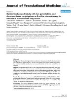
The apparatus including the joint stretching motor device, the height adjustable chair, and force and position sensorsFigure 1
The apparatus including the joint stretching motor device, the height adjustable chair, and force and position sensors.
Journal of NeuroEngineering and Rehabilitation 2008, 5:6 />Page 4 of 15
(page number not for citation purposes)
The seat and footrest were adjusted to align the ankle axis
of rotation with the axis of the force sensor and the motor
shaft. An oscilloscope mounted in front of the subject dis-
played a target signal and provided feedback of low-pass
filtered joint torque.
Recording
Ankle position was measured with a precision potentiom-
eter. Torque was recorded using a 6-axis torque trans-
ducer, mounted between the beam of the footrest and the
motor shaft. Displacements in the plantarflexion (PF)
direction were taken as negative and those in the dorsiflex-
ion (DF) direction as positive. An ankle angle of 90
degrees was considered to be the Neutral Position (NP)
and defined as zero. Torque was assigned a polarity con-
sistent with the direction of the movement that it would
generate (e.g. DF torque was taken as positive). Electromy-
ograms (EMGs) from tibialis anterior (TA) and lateral gas-
trocnemius (GS) were recorded using bipolar surface
electrodes (Delsys, Inc. Boston, MA). Position, torque,
and EMGs were filtered at 230 Hz to prevent aliasing, and
sampled at 1 kHz by a 16 bit A/D.
Procedures
Range of motion (ROM)
ROM was determined with the subject's ankle attached to
the motor and manually moved to maximum PF and DF.
Mean displacement amplitude was assessed 3 times by
slowly moving the joint until the examiner perceived rap-
idly increasing resistance or the subject reported discom-
fort. The typical angular range was from about 50° PF
(mean 49° ± 6°SD) to 20° DF (mean 21° ± 5° SD).
Paradigm
To identify overall stiffness properties and to separate the
reflex and intrinsic components, we used Pseudorandom
Binary Sequence (PRBS) position inputs with amplitude
of 0.03 rad and a switching interval of 150 ms. Our previ-
ously published results demonstrated that these perturba-
tions have a mean velocity low enough to avoid
attenuating reflex responses, contain power over a wide
enough bandwidth to identify the dynamics, and are well
tolerated by the spastic subjects [30].
Trials were conducted at different ankle positions from
full-PF to full-DF, at 5 degree intervals. Each position was
examined under Passive conditions, where subjects were
instructed to remain relaxed.
Following each trial, the torque and EMG signals were
examined for evidence of non-stationarities or co-activa-
tion of TA. If there was evidence of either, the data were
discarded and the trial was repeated.
Analysis procedures
Parallel cascade identification technique
Dynamic stiffness of the ankle is defined as the dynamic
relation between joint position (as input), and resulting
torque (as output). Reflex and intrinsic contributions to
ankle dynamic stiffness were identified using a parallel
cascade technique, described in detail in earlier publica-
tions [13,14]. Briefly, the method proceeded as shown in
Figure 2.
Intrinsic stiffness dynamics (top pathway) were estimated
in terms of a linear Impulse Response Function (IRF)
relating position and torque. The reflex pathway (bottom
pathway) was modeled as a differentiator in series with a
delay, a static non-linear element (closely resembling a
half-wave rectifier), and a dynamic linear element. Reflex
stiffness dynamics were estimated by determining the IRF
between half-waved rectified velocity as the input and
reflex torque as the output. The intrinsic and reflex stiff-
ness IRFs were convolved with the experimental input to
predict the intrinsic and reflex torque respectively.
Linear models were fitted to the estimated intrinsic and
reflex IRF curves using the Levenberg Marquardt nonlin-
ear least-square fit algorithm [31]. To make fitting easier,
the intrinsic stiffness IRF was inverted to give a compli-
ance IRF, which was described by a second-order model
having inertia, viscous and elastic parameters [14]. The
intrinsic elastic parameter also corresponds to the steady-
state, intrinsic stiffness gain.
The reflex stiffness was described by reflex delay and a
third-order model having gain, damping, and frequency
parameters. This model is more complex than the second-
order model used in our previous work [13]. This is
because we found that an additional pole was required to
accurately fit the reflex IRFs of the spastic joint [32].
Statistical analysis
We used a two-way ANOVA test, and standard t-tests to
analyze our results. Two-way ANOVA analyses were used
to test for significant main effects due to subject groups,
joint positions, or their interactions. The results could tell
us if there were significant differences due to main effects
and/or their interactions. Tukey post-hoc comparisons
were performed to find at which positions the differences
between groups were significant.
Standard t-tests procedures were used to test for signifi-
cant changes in intercepts and slopes of reflex stiffness as
a function of joint angle.
Results with p values less than 0.05 were considered signif-
icant.
Journal of NeuroEngineering and Rehabilitation 2008, 5:6 />Page 5 of 15
(page number not for citation purposes)
Results
Experimental data
To illustrate the form of data that are collected in our
experiments, we present a sequence of typical experimen-
tal records, together with results of model predictions.
Figure 3 shows a segment of a typical PRBS trial with the
amplitude of 0.03 rad and the switching-rate of 150 ms.
This record was acquired while the subject was relaxed.
Angular displacements in the positive (dorsiflexing) direc-
tion (Fig. 3A) evoked a short latency burst of activity in
gastrocnemius (GS) (Fig. 3B) while displacements in the
negative (plantarflexing) direction evoked no response.
The torque record (Fig. 3E) is similarly asymmetric, in that
dorsiflexing displacements evoked torque responses hav-
ing intrinsic and reflex components, while responses to
plantarflexing displacements have only the intrinsic com-
ponent. The intrinsic and reflex torque predicted by the
parallel-cascade identification model are shown in Fig. 3C
and Fig. 3D, respectively. The model's estimate of the
overall torque, given by the sum of the intrinsic and reflex
torques, is shown in thick curve superimposed on the
experimentally observed torque shown in thin curve (Fig.
3E). It is evident that the overall prediction was very good;
in this case, it accounted for 92.2% of the observed torque
variance. This was typical of all our data; the parallel-cas-
cade model routinely accounted for more than 90% of the
overall torque variance.
Figure 4 summarizes the intrinsic and reflex stiffness anal-
ysis for both paretic and non-paretic sides of a typical
stroke subject at the NP. The dashed curves in the first row
are the intrinsic compliance impulse response functions
(IRFs) estimated for the paretic (Fig. 4A) and non-paretic
(Fig. 4B) ankle. These were similar in shape although
compliance magnitude was slightly smaller in the paretic
that the non-paretic side indicating that stiffness (the
inverse of compliance) was slightly larger in the paretic
ankles. Second-order fits to these compliance IRFs, shown
by the superimposed solid curves, were very good. In both
cases, the Variance Accounted For (VAF
FIT
) was greater
than 98%, as was typical of all our data; VAF
FIT
for the
compliance IRF was always greater than 90%. The intrin-
sic torques predicted by these IRFs, shown in the Fig. 4C
The parallel cascade structure used to identify intrinsic and reflex stiffnessFigure 2
The parallel cascade structure used to identify intrinsic and reflex stiffness. Intrinsic dynamic stiffness is represented in the
upper pathway by the intrinsic stiffness impulse response function. Reflex dynamic stiffness is represented by the lower path-
way as a differentiator, followed by a static nonlinear element and then a linear impulse response function. The nonlinear ele-
ment is a half wave rectifier which shows the direction of stretch.
Journal of NeuroEngineering and Rehabilitation 2008, 5:6 />Page 6 of 15
(page number not for citation purposes)
A segment from a typical sequence trial for a spastic under relaxed conditionsFigure 3
A segment from a typical sequence trial for a spastic under relaxed conditions. A Position, B Half-wave rectified gastrocnemius
electromyogram (GS), C Predicted intrinsic torque, D Predicted reflex torque and E Predicted overall torque (thick curve)
superimposed on the actual torque (thin curve). Displacements in the PF direction were taken as negative and those in the DF
direction as positive. Torque was assigned a polarity consistent with the direction of the movement that it would generate (e.g.
PF torque was taken as negative).
−0.02
0
0.02
rad
POSITION
−0.2
−0.1
0
mV
GS EMG
−6
−3
0
Nm
PREDICTED INTRINSIC TORQUE
−6
−3
0
Nm
PREDICTED REFLEX TORQUE
0 1 2 3 4 5 6
−6
−3
0
Time (s)
Nm
ACTUAL & PREDICTED OVERALL TORQUE
Actual
Predicted
A
B
C
D
E
Journal of NeuroEngineering and Rehabilitation 2008, 5:6 />Page 7 of 15
(page number not for citation purposes)
Typical intrinsic and reflex dynamics and their predicted torques estimated for the Paretic (left column) and Non-paretic (right column)Figure 4
Typical intrinsic and reflex dynamics and their predicted torques estimated for the Paretic (left column) and Non-paretic (right
column). A, B Intrinsic compliances; C, D Predicted intrinsic torques; E, F Reflex stifnness; and G, H Predicted reflex tor-
ques. The dashed curves are the nonparametric IRF, the solid curve are the parametric fits.
0 0.6
−0.3
0
0.3
INTRINSIC COMPLIANCE IRF
rad/Nm
0 0.6
−0.3
0
0.3
IRF
FIT
0 7
−2
0
2
PREDICTED INTRINSIC TORQUE
Nm
0 7
−2
0
2
0 0.5
−50
−25
0
REFLEX STIFFNESS IRF
Nm.s/rad
0 0.5
−50
−25
0
IRF
FIT
0 7
−2
−1
0
PREDICTED REFLEX TORQUE
Nm
Time (s)
0 7
−2
−1
0
Time (s)
PARETIC
NON−PARETIC
A
B
C
D
E
F
G
H
Journal of NeuroEngineering and Rehabilitation 2008, 5:6 />Page 8 of 15
(page number not for citation purposes)
and 4D, were comparable in waveform although the mag-
nitude was slightly larger in the paretic than in the non-
paretic ankle, consistent with the differences in the com-
pliance IRFs. The small differences were expected since
these data were collected in the NP, at which typically
there was no significant difference in the intrinsic stiffness
between both sides.
The reflex stiffness IRFs, estimated from the paretic (Fig.
4E) and non-paretic (Fig. 4F) sides, are shown as dashed
lines. Third-order model fits to these reflex stiffness IRFs
were also very good as indicated by the superimposed
solid curves. These fits were always accurate; in this case,
VAF
FIT
was greater than 88% of the variance. The ampli-
tude of reflex stiffness IRF for the paretic side (Fig. 4E) was
approximately three times that of the non-paretic side
(Fig. 4F). The reflex torques predicted by these IRFs shows
that the peak-to-peak torque of the paretic limb in Fig. 4G
(~1.5 Nm) was approximately three times that of the non-
paretic limb in Fig. 4H (~0.5 Nm).
Group data: intrinsic and reflex stiffness
Figure 5 shows the intrinsic and reflex stiffness parameters
from the paretic limb plotted against the corresponding
control values from the non-paretic side for all stroke sub-
jects, and for all positions. The dotted line at 45 degrees
(the unity line) in each panel indicates what would be
expected if there were no change due to stroke. Points
above the line indicate abnormal increases following
stroke, while points below the line indicate decreases.
The reflex stiffness gain values (G
R
, panel A) for all sub-
jects were located well above the diagonal line, indicating
that G
R
was larger in the paretic than in non-paretic limbs
of the subjects. G
R
was the only reflex parameter that
changed consistently; it increased significantly for most
stroke subjects (p < 0.0001). The other three reflex param-
eters did not change significantly.
Similarly, the intrinsic stiffness gain (K, panel B) was sub-
stantially larger for the majority of stroke subjects (p <
0.0023). In contrast, the points for the intrinsic viscous
parameter (B, panel C) were mostly clustered around the
unity line, and did not show significant differences
between paretic and non-paretic limbs.
Position-dependency
Figure 6 shows group average results for reflex stiffness
gain as a function of ankle position for paretic, non-
paretic and normal groups. There was a significant differ-
ence between the paretic group, as compared with both
non-paretic and normal groups (p < 0.0001). Tukey post-
hoc comparisons showed that G
R
was significantly larger
in the paretic ankle than in the normal ankle at all posi-
tions (p < 0.005) and it was larger than the non-paretic
Paretic stiffness parameters plotted against non-paretic val-ues for all stroke subjectsFigure 5
Paretic stiffness parameters plotted against non-paretic val-
ues for all stroke subjects. A Reflex stiffness gain (G
R
), B
Intrinsic stiffness elasticity or gain (K), and C Intrinsic stiff-
ness viscosity (B).
0 2 4 6
0
2
4
6
REFLEX STIFFNESS GAIN (G
R
)
Non−paretic G
R
(Nm.s/rad)
Paretic G
R
(Nm.s/rad)
0 100 200
0
100
200
INTRINSIC STIFFNESS GAIN (K)
Non−paretic K (Nm/rad)
Paretic K (Nm/rad)
0 1 2 3
0
1
2
3
INTRINSIC STIFFNESS VISCOSITY (B)
Non−paretic B (Nm.s/rad)
Paretic B (Nm.s/rad)
A
B
C
Journal of NeuroEngineering and Rehabilitation 2008, 5:6 />Page 9 of 15
(page number not for citation purposes)
ankle at all positions except for the position -50° PF (p <
0.02). Differences in G
R
increased as the ankle was dorsi-
flexed. Statistical analyses confirmed that there was a sig-
nificant effect due to position for all groups (p < 0.0001).
Position dependence was similar in all groups; the reflex
stiffness gain first increased from mid-PF to mid-DF and
then declined. The slope of changes was larger in the
paretic than in the non-paretic and normal (P < 0.0001)
groups. Similarly, the intercept of the plots relating reflex
stiffness to jojnt angle increased significantly in the paretic
ankle as compared to other groups (p < 0.0001).
The peak value of G
R
was around NP in the stroke ankle
whereas it was around full-DF in the non-paretic and nor-
mal ankle. The group behavior was consistent but the
inter-subject variability was high at mid-ROM in the
stroke group as demonstrated by the large standard error
bars associated with the means.
As expected from the literature [29], the non-paretic side
of stroke survivors was not similar to healthy subjects; G
R
,
was significantly larger in the non-paretic than the normal
ankle (p < 0.001) and the differences were significant at
most positions; i.e. positions between -25° PF and 20°
DF, (p < 0.036).
Figure 7 summarizes the behavior of intrinsic stiffness
parameters with changes in ankle joint angle for all groups
(paretic, non-paretic and normal). Overall, the group
behavior was very consistent, as demonstrated by the nar-
row standard error bars.
For the intrinsic stiffness gain (K, top panel), there was a
significant difference between the paretic group and both
non-paretic and normal groups. K was significantly larger
in the paretic than the non-paretic (p < 0.038) and normal
(p < 0.03) ankle at dorsiflexed positions; i.e. at positions
between -10° PF and 15° DF. However, the intrinsic vis-
cous parameter (B, bottom panel) was significantly larger
in the paretic than in the normal subjects just for positions
between NP and 20° DF (p < 0.05).
Both K and B were strongly position dependent as con-
firmed by the statistical analysis (p < 0.0001); they first
decreased sharply from full PF to mid-PF, then increased
slowly from mid-PF to mid-DF, and finally it increased
sharply from mid-DF to full-DF. This position depend-
ency was consistent in all groups and was similar to our
previous finding for SCI subjects [11,25].
Position dependence of intrinsic stiffness for paretic, non-paretic and normal groups as functions of position (Group averages)Figure 7
Position dependence of intrinsic stiffness for paretic, non-
paretic and normal groups as functions of position (Group
averages). A Intrinsic stiffness gain (K); asterisks represent
points where differences between paretic group and both
non-paretic and normal control groups are statistically signif-
icant. B Intrinsic stiffness viscous parameter (B); asterisks
represent points where differences between paretic group
and normal control group was significant. Error bars indicate
± 1 standard error. NP: Neutral Position (90°).
−50 −40 −30 −20 −10 0 10 20
0
50
100
150
K (Nm/rad)
INTRINSIC STIFFNESS GAIN (K)
Paretic
Non−paretic
Normal
−50 −40 −30 −20 −10 0 10 20
0
0.5
1
1.5
2
INTRINSIC STIFFNESS VISCOSITY (B)
B (Nm.s/rad)
Plantarflexion Ankle Angle (deg) NP Dorsiflexion
A
B
Position dependence of Reflex stiffness gain (G
R
) for paretic, non-paretic and normal groups as functions of position (Group averages)Figure 6
Position dependence of Reflex stiffness gain (G
R
) for paretic,
non-paretic and normal groups as functions of position
(Group averages). Error bars indicate ± 1 standard error.
NP: Neutral Position (90°).
−50 −40 −30 −20 −10 0 10 20
0
1
2
3
Plantarflexion Ankle Angle (deg) NP Dorsiflexion
G
R
(Nm.s/rad)
REFLEX DYNAMIC STIFFNESS GAIN (G
R
)
Paretic
Non−paretic
Normal
Journal of NeuroEngineering and Rehabilitation 2008, 5:6 />Page 10 of 15
(page number not for citation purposes)
Intrinsic stiffness gain was similar in both non-paretic and
normal group (Fig. 7A) whereas the intrinsic viscous
parameter increased in the non-paretic group and was sig-
nificantly larger for a few positions, particularly in full DF
(i.e., at 15° and 20° DF) (p < 0.05) (Fig. 7B).
Group results: stroke effects
We investigated the position-dependency of stroke effects;
i.e. the differences between paretic and non-paretic sides
as ankle angle were changed systematically.
To characterize the amplitude of these changes, we com-
puted the percentage change caused by stroke (stroke
effects) at each joint position in stroke patients. Figure 8
shows the changes in G
R
, K, and B and as a function of
position. Panel A shows that G
R
, increased in stroke sub-
jects between ~100% at full-PF and ~350% around NP, by
an average of 211 ± 92%. The highest percentages of
changes obtained from mid-ROM. Panel B and C show
that K and B also increased by an average of 30 ± 19% and
10 ± 8%, respectively, which are much smaller than the
percentage of increase in reflex stiffness gain. However, an
increase of ~50% was observed for K only at dorsiflexed
positions which was considerable. These changes clearly
indicate that the abnormalities in intrinsic and reflex stiff-
ness are strongly position dependent.
Discussion
Our results revealed that both neural and muscular sys-
tems are altered in spastic limbs, but the changes are com-
plex and may depend on several factors. In this study, we
probed changes in intrinsic stiffness and changes in reflex
stiffness as a function of joint angle over the entire angular
range of motion, and found strong position dependency
in these neuromuscular abnormalities.
Summary of results
We used the parallel cascade system identification tech-
nique to characterize the mechanical changes associated
with spasticity in the ankle joint of chronic hemiplegic
stroke subjects. To our knowledge, this is the first study
that quantified the changes in neuromuscular properties
over the entire ROM, and used two different control
groups; i.e. the non-paretic limb in the stroke patients and
the normal limb in the healthy subjects.
Our major findings were that,
(i) overall dynamic joint stiffness was increased in paretic
side,
(ii) both reflex and intrinsic stiffness gain was larger in
paretic than in the non-paretic and normal limb and con-
tributed substantially to the increased stiffness,
(iii) these abnormalities were strongly dependent on joint
position; reflex stiffness was most pronounced at mid-
ROM whereas intrinsic stiffness were dominant during
DF,
(iv) the non-paretic side of people with stroke was not
similar to that of healthy ankle muscles in control sub-
jects. Reflex stiffness gain was significantly larger in them
than in healthy ankle muscles. Intrinsic viscosity was also
larger in the non-paretic than in the normal side but the
differences were not significant.
Increased intrinsic stiffness
We found that the intrinsic stiffness and viscous parame-
ter were larger in the stroke than in the normal subjects
(Figure 7), and the differences were significant for DF.
Increased intrinsic stiffness is consistent with enhance-
ment in passive stiffness of the ankle joint reported by
Sinkjaer et al. [12]. Surprisingly, Galiana et al. found no
significant differences between these groups [26]. This dis-
crepancy can be explained by two major differences
between the two studies.
First, Galiana et al. [26] studied a limited range of posi-
tions; e.g. from mid-PF to NP position, where the differ-
ences between intrinsic stiffness of stroke and normal
subjects were small, according to our findings. This
emphasizes the importance of considering the position
dependency of joint dynamic stiffness and its intrinsic
and reflex components. Second, the time post-injury
which can play a critical role in developing intrinsic struc-
tural remodeling was different between two studies; the
average time post-lesion used in their study (approxi-
mately 10.5 months) was much shorter than that in our
studies (approximately 92 months). Thus, lack of changes
in intrinsic stiffness observed by Galiana et al. [26] could
be due to shorter post-lesion times in their patients which
were potentially not long enough for the development of
substantial muscle fiber remodeling.
Recent cellular studies may explain the enhanced intrinsic
stiffness we observed in our stroke subjects with chronic
spasticity. Published studies of the tensile modulus of
muscle fibers demonstrated that intrinsic stiffness of spas-
tic muscle fibers is increased [33,34]. Furthermore, the
resting sarcomere length of cells is shorter in spastic mus-
cle cells [35,36]. Finally, although it has been proposed
that the isoform of titin, a large intracellular cytoskeletal
protein, may also be altered in spastic muscles and con-
tribute to these changes [33,37], recent findings reveal no
change in titin isoforms in spastic muscle [38].
In addition to altered muscle cell properties, changes in
proliferation of extracellar matrix material and in the
mechanical properties of this extracellular material in
Journal of NeuroEngineering and Rehabilitation 2008, 5:6 />Page 11 of 15
(page number not for citation purposes)
spastic muscle are described, based on biochemical meas-
urement of collagen concentration [39]. It has been sug-
gested that although spastic muscle contains a larger
amount of extracellular matrix material, the quality of that
material is much different from that in normal muscles
[33,35]. Taken together, the intra- and extracellular alter-
ations in muscle and its tissue that usually occur second-
ary to spasticity will likely change the intrinsic mechanical
properties of spastic limbs.
Intrinsic stiffness may also change for other structural rea-
sons such as alteration in fiber size and fiber type distribu-
tions and potentially also fiber length. Micrographs of
spastic muscles showed increased fiber size variability
[9,40,41] which may result from muscle fiber thinning
that usually occur near the end of some fibers [42,43]. The
occurrence of changes in fiber type distribution secondary
to spasticity is also accepted [35], although the nature of
these changes is controversial [44-46]. It has been also
widely reported that muscle fiber length and the number
of sarcomeres within the muscle decrease secondary to
Percentage change of stroke effects as functions of position (Group averages)Figure 8
Percentage change of stroke effects as functions of position (Group averages). A Reflex stiffness gain (G
R
), B Intrinsic stiffness
gain (K), C Intrinsic viscous parameter (B). Error bars indicate ± 1 standard error. NP: Neutral Position (90°). The dotted lines
reflect the mean percentage change over the range of motion.
−50 −40 −30 −20 −10 0 10 20
0
100
200
300
400
500
REFLEX STIFFNESS GAIN (G
R
)
Percentage Change
−50 −40 −30 −20 −10 0 10 20
0
20
40
60
80
100
INTRINSIC STIFFNESS GAIN (K)
Percentage Change
−50 −40 −30 −20 −10 0 10 20
0
20
40
60
80
100
INTRINSIC STIFFNESS VISCOSITY (B)
Plantarflexion Ankle Angle (deg) NP Dorsiflexion
Percentage Change
STROKE EFFECTS − POSITION
A
B
C
Journal of NeuroEngineering and Rehabilitation 2008, 5:6 />Page 12 of 15
(page number not for citation purposes)
spasticity, contributing to contracture [47,48]. Con-
versely, there is evidence indicating that serial sarcomere
number can be increased by lengthening the position at
which muscle immobilization occurs [49]. While patients
with stroke are not fully immobilized, particularly at
longer resting lengths, the lack of mobility of patients
post-stroke may provide some of the immobilization
stimulus. Although this adaptability to chronic length
changes cannot be generalized to all muscles [35] changes
in serial sarcomere number have also been reported for
the ankle extensor muscles [49]. It is also known that the
resting sarcomere length of spastic fibers is reduced in the
spastic muscle [33,34]. These changes associated with sar-
comere length reduction, and with the accumulation of
connective tissues in atrophic muscles secondary to the
lesions, may cause a left shift in passive length-force
curves that can explain the position dependency of altered
intrinsic stiffness found in the current study.
Abnormalities in reflex stiffness
Our results demonstrate that reflex stiffness gain was sig-
nificantly larger in stroke than normal subjects at most
positions; in some cases by as much as a factor of seven.
The differences were greatest at mid-ROM where reflex
stiffness was largest for the stroke group. In contrast, oth-
ers reported reflex torque [12] and stiffness [26] of spastic
subjects within the normal range. This discrepancy is at
least partially related to methodological differences as
described below.
In the Sinkjaer study [12] the amplitude of the torque
response was divided by that of the perturbation to pro-
vide units of stiffness, assuming that joint stiffness is a lin-
ear property that did not vary with displacement
amplitude or joint angle. Since both intrinsic and reflex
mechanics are known to be highly non-linear, and to be
dependent on joint angle, these measurements are
unlikely to provide an accurate measure of joint proper-
ties. The Sinkjaer study also attempted to separate intrin-
sic muscular contributions from reflex contributions
using electrical stimulation of the muscle nerve. There is
some uncertainty in interpreting experiments using elec-
trical stimulation to assess muscle mechanical properties
[12,16,18], since the order of motor axon recruitment
during electrical stimulation is very different from that
arising during normal voluntary contractions [50,51].
Thus, reflex contributions cannot be accurately estimated
by these methods.
Our results are at variance with those of Galiana et al. [26].
This difference could be due to their smaller sample size,
particularly since they report substantial increases in the
reflex response in four out of eleven patients.
Our findings demonstrate that reflex stiffness gain was
strongly dependent on position, similar to our previous
findings in subjects with SCI [11,25]. Indeed, reflex stiff-
ness gain was modulated greatly with ankle position; it
increased from PF to mid-DF in both stroke and normal
groups but it declined in the stroke subjects as the ankle
was moved toward full-DF. This abnormal modulation at
DF was not reported in the Galiana study, because their
subjects were examined over a more limited ROM, which
did not reach beyond the neutral joint angle.
The increases in reflex stiffness could be attributed to
greater excitation of the motoneuron pool by augmented
afferent or interneuronal input. Under these conditions,
recruitment of motoneurons would likely follow the nor-
mal sequence, from small to large. Alternatively, aug-
mented reflex stiffness gain could be due to an
inappropriate recruitment sequence, in the paretic limb,
in which recruitment rank order is disrupted, or even
reversed. This idea has been supported by others, who
suggest that CNS lesions may alter motor output by induc-
ing new connections including sprouting and strengthen-
ing of existing connections to optimize their performance
[52].
Abnormal modulation of reflex stiffness gain with ankle
position in the paretic limb was characterized by the three
major changes: (1) the slope of the gain was greater from
-30° PF to 5° DF, (2) the ordinal intercept was larger, and
(3) the reflex gain decreased sharply at the end of DF.
The first two changes can be explained by enhancement of
reflex gain as explained above. The decline in the reflex
gain at the end of DF, however, could be due potentially
to inhibitory effects of group III/IV muscle afferents which
are activated preferentially as muscles are stretched to near
maximum length [53]. The increased tension, which may
happen at full joint DF in normal subjects, was present at
the narrower joint angle in the spastic joints due to the
existence of muscle hypertonia. This abnormal modula-
tion can elicit strong reflex responses at undesirable pos-
ture and/or phase during movement, and consequently
contribute to movement impairments as observed in the
transition from stance to swing during walking in spastic
patients [54].
Non-paretic limb of stroke patients versus normal limb
Since the non-paretic limb of the stroke patients has ini-
tially similar mass, muscle architecture and neural con-
nectivity to the paretic limb, it seems that it could be the
best control because it reduces the inter-subject variabil-
ity. In contrast, it has been suggested that the non-paretic
limb in both the upper [55,56] and lower extremities [57-
59] is influenced to some extent by stroke. However, the
comparison between neuromechanical properties of the
Journal of NeuroEngineering and Rehabilitation 2008, 5:6 />Page 13 of 15
(page number not for citation purposes)
normal limb in healthy subjects and non-paretic limb in
persons with stroke has not been done yet. We postulated
that the non-paretic ankle would have different neu-
romuscular properties than the normal ankle. We there-
fore probed this hypothesis by comparing intrinsic
viscoelastic parameters and reflex stiffness gain between
stroke patients and aged-matched healthy subjects. Our
results revealed that all these parameters were larger in the
non-paretic than in the healthy subjects' ankle but the dif-
ferences were significant only for reflex stiffness. These
findings suggest that the non-paretic lower extremity of
people with stroke may not be used as an appropriate con-
trol for the study of neuromuscular properties.
Significance
The abnormalities in spastic, paretic joint stiffness, and its
intrinsic and reflex components, vary systematically with
position over the full angular range of motion. When the
ankle was fully plantarflexed, there was no systematic dif-
ference between the overall joint stiffness of spastic and
normal joints. In the mid range, overall joint stiffness was
greater in spastic limbs primarily due to abnormally high
reflex stiffness. At full DF, increased intrinsic stiffness pro-
vided the major contribution to the larger overall stiffness
in spastic joints. Thus, questions about the nature and ori-
gin of hypertonia may have quite different answers
depending upon where in the ROM tests are made. This
can explain some controversies in the literature regarding
the nature and origins of mechanical abnormalities asso-
ciated with spasticity.
Conclusion
Our findings revealed that in the paretic ankle of hemi-
paretic stroke survivors, both intrinsic and reflex stiff-
nesses were significantly increased, as compared to the
non-paretic ankle joints of stroke survivors, and to normal
controls. These abnormalities varied systematically and
differently with changing ankle position, over the full
angular range of motion; the differences reached their
maximum at NP for the reflex stiffness and at DF for the
intrinsic stiffness. These findings indicate that clinical per-
ceptions of increased tone may have quite different ori-
gins depending upon the angle where the tests are
initiated.
Furthermore, reflex stiffness was significantly larger in the
non-paretic limb of stroke patients than in healthy control
subjects, suggesting that the non-paretic limb should not
be used as control for studying neuromuscular properties
of the ankle joint.
Our findings will help elucidate the origins of the neu-
romuscular abnormalities associated with stroke-induced
spasticity and may facilitate the development of targeted
interventions for preventing these abnormal changes or
reversing them.
Abbreviations
ROM: Range of Motion; MAS: Modified Ashworth Scale;
DF: Dorsiflexion; PF: Platarflexion; EMGs: Electormy-
grams; GS: Gastrocnemius; TA: Tibiliais anterior; PRBS:
Pseudorandom Binary Sequence; IRF: Impulse Response
Function; VAF: Variance Accounted for; G
R:
Reflex stiffness
gain; K: Intrinsic stiffness gain; B: Intrinsic stiffness viscos-
ity; NP: Neutral Position.
Competing interests
The authors declare that they have no competing interests.
Authors' contributions
MMM designed the study, supervised data collection and
analysis, and participated in interpreting and writing the
manuscript. LA participated in performing the experi-
ments, interpreting data and writing the paper, MT partic-
ipated in analyzing data, and WZR participated in
interpreting data and writing the manuscript. All authors
read and approved the final manuscript.
Acknowledgements
We wish to acknowledge Richard Harvey, MD, Ross Bogey, DO, Krista Set-
tle, DPT, Thanan Lilaonitkul, BSc, and Elisa Pelosin, PT, for their collabora-
tions. This research was financed by the National Science Foundation (NSF
0302313).
References
1. Burne JA, Carleton VL, O'Dwyer NJ: The spasticity paradox:
movement disorder or disorder of resting limbs? J Neurol Neu-
rosurg Psychiatry 2005, 76:47-54.
2. Drolet M, Noreau L, Vachon J, Moffet H: Muscle strength changes
as measured by dynamometry following functional rehabili-
tation in individuals with spinal cord injury. Arc Phys Med Reha-
bil 1999, 80:791-800.
3. Duncan P, Wallace D, Studenski S, Lai S, Johnson D: Conceptuali-
zation of a new stroke-specific outcome measure: the stroke
impact scale. Top Stroke Rehabil 2001, 8:19-33.
4. Katz RT, Rymer WZ: Spastic hypertonia: Mechanisms and
measurement. Arch Phys Med Rehabil 1989, 70:144-155.
5. Sehgal N, McGuire JR: Beyond ashworth. Electrophysiologic
quantification of spasticity. Phys Med Rehabil Clin N Am 1998,
9(4):949-979.
6. Sommerfeld DK, Eek EU, Svensson AK, Holmqvist LW, von Arbin
MH: Spasticity after stroke: its occurrence and association
with motor impairments and acitivity limitations. Stroke
2004, 35:134-140.
7. Pandyan AD, Gregoric M, Barnes MP, Wood DE, Van Wijck FM, Burr-
idge JH, Hermens H, Johnson GR: Spasticity: Clinical percep-
tions, neruological realities and meaningful measurement.
Disabil Rehabil 2005, 27(1/2):2-6.
8. Lance JW: Symposium synopsis. In Spasticity: Disordered Motor Con-
trol Edited by: Feldman RG, Young RR, Koella WP. Chicago , Year
book publishers; 1980:485-494.
9. Dietz V, Ketelsen UP, Berger W, Quintern J: Motor unit involve-
ment in spastic paresis. Relationship between leg muscle
activation and histochemistry. J Neurol Sci 1986, 75(1):89-103.
10. Lieber RL, Runesson E, Einarsson F, Friden J: Inferior mechanical
properties of spastic muscle bundles due to hypertrophic but
compromised extracellular matrix material. Muscle Nerve
2003, 28(4):464-471.
Journal of NeuroEngineering and Rehabilitation 2008, 5:6 />Page 14 of 15
(page number not for citation purposes)
11. Mirbagheri MM, Ladouceur M, Barbeau H, Kearney RE: Intrinsic and
reflex stiffness in normal and spastic spinal cord injured sub-
jects. Exp Brain Res 2001, 141:446-459.
12. Sinkjaer T, Magnussen I: Passive, intrinsic and reflex-mediated
stiffness in the ankle extensors of hemiparetic patients. Brain
1994, 117:355-363.
13. Kearney RE, Stein RB, Parameswaran L: Identification of intrinsic
and reflex contributions to human ankle stiffness dynamics.
IEEE Trans Biomed Eng 1997, 44:493-504.
14. Mirbagheri MM, Barbeau H, Kearney RE: Intrinsic and reflex con-
tributions to human ankle stiffness: Variation with activation
level and position. Exp Brain Res 2000, 135:423-436.
15. Wood DE, Burridge JH, Van Wijck FM, Mcfadden C, Hitchcock RA,
Pandyan AD, Haugh A, Salazar-Torres JJ, Swain ID: Biomechanical
approaches applied to the lower and upper limb for the
measurement of spasticity: A systematic review of the liter-
ature. Disabil Rehabil 2005, 27(1/2):19-32.
16. Carter RR, Crago PE, Keith MW: Stiffness regulation by reflex
action in the normal human hand. J Neurophysiol 1990,
64:105-118.
17. Sinkjaer T, Toft E, Andreassen S, Hornemann BC: Muscle stiffness
in human ankle dorsiflexors: intrinsic and reflex compo-
nents. J Neurophysiol 1988, 60:1110-1121.
18. Toft E, Sinkjaer T, Andreassen S, Larsen K: Mechanical and elec-
tromyographic responses to stretch of the human ankle
extensors. J Neurophysiol 1991, 65:1402-1410.
19. Noth J, Matthew HR, Friedemann HH: Long latency reflex force
of human finger muscles in response to imposed sinusoidal
movements. Exp Brain Res 1984, 55:317-324.
20. Lehmann JF, Price R, deLateur BJ, Hinderer S, Traynor C: Spasticity:
Quantitative measurements as a basis for assessing effec-
tiveness of therapeutic intervention. Arch Phys Med Rehabil
1989, 70:6-15.
21. Meinders M, Price R, Lehmann JF, Questad KA: The stretch reflex
response in the normal and spastic ankle: Effect of ankle
position. Arch Phys Med Rehabil 1996, 77:487-492.
22. Price R, Bjornson KF, Lehmann JF, McLaughlin JF, Hays RM: Quanti-
tative measurement of spasticity in children with cerebral
palsy. Dev Med Child Neurol 1991, 33:585-595.
23. Rack PMH, Ross RF, Thilman AF: The ankle stretch reflexes in
normal and spastic subjects. Brain 1984, 107:637-654.
24. Mirbagheri MM, Settle K, Harvey R, Rymer WZ: Neuromuscular
abnormalities associated with spasticity of upper extremity
muscles in hemiparetic stroke. J Neurophysiol 2007, 98:629-637.
25. Mirbagheri MM, Ladouceur M, Barbearu H, E. KR: The effects of
long-term FES-assisted walking on intrinsic and reflex
dynamic stiffness in spastic SCI subjects. IEEE Trans Neural Sys-
tem Rehabil Eng 2002, 10(4):280-289.
26. Galiana L, Fung J, Kearney R: Identification of intrinsic and reflex
ankle stiffness components in stroke patients. Exp Brain Res
2005, 165(4):422-434.
27. Ashworth B: Preliminary trial of carisoprodol in multiple scle-
rosis. Practitioner 1964, 192:540-542.
28. Bohannon RW, Smith MB: Interrater reliability on a modified
Ashworth scale of muscle spasticity. Phys Ther 1987,
67:206-207.
29. Thilmann AF, Fellows SJ, Garms E: Pathological stretch reflex on
the "good" side of hemiparetic patients. J Neurol Neurosurg Psy-
chiatry 1990, 53:208-214.
30. Mirbagheri MM, Kearney RE, Barbeau H, Ladouceur M: Abnormal
stretch reflex mechanics in spastic spinal cord injured spastic
subjects. IEEE Eng Med Biol Soc 1997, 19:1648-1649.
31. Press WH, Flannery BP, Teukolsky SA, Betterling WT: Numerical
Recipes: The art of scientific computing cambridge. UK ,
Camb Univ Press 523-528; 1985.
32. Mirbagheri MM, Kearney RE, Barbeau H: Parametric modeling of
the reflex contribution to dynamic ankle stiffness in normal
and spinal cord injured spastic subjects.
IEEE Eng Med Biol Soc
1995, 17:1241-1242.
33. Foran JRH, Steinman S, Barash I, Chambers H, Lieber RL: Structruc-
tural and mechanical alterations in spastic skeletal muscle.
Dev Med Child Neurol 2005, 47:713-717.
34. Friden J, Lieber RL: Spastic muscle cells are shorter and stiffer
than normal cells. Muscle Nerve 2003, 27(2):157-164.
35. Lieber RL, Steinman S, Barash IA, Chambers H: Structural and
functional changes in spastic skeletal muscle. Muscle Nerve
2004, 29:615-627.
36. Wang K, McCarter R, Wright J, Beverly J, Ramirez-Mitchell R: Vis-
coelasticity of the sarcomere matrix of skeletal muscles. The
titin-myosin composite filament is a dual-stage molecular
spring. Biophys J 1993, 64(4):1161-1177.
37. Labeit S, Kolmerer B: Titins: giant proteins in charge of muscle
ultrastructure and elasticity. Science 1995, 270(5234):293-296.
38. Olsson MC, Kruger M, Meyer LH, Ahnlund L, Gransberg L, Linke
WA, Larsson L: Fibre type-specific increase in passive muscle
tension in spinal cord-injured subjects with spasticity. J Physiol
2006, 15(577):339-352.
39. Booth CM, Cortina-Borja MJ, Theologis TN: Collagen accumula-
tion in muscles of children with cerebral palsy and correla-
tion with severity of spasticity. Dev Med Child Neurol 2001,
43(5):314-320.
40. Ito J, Araki A, Tanaka H, Tasaki T, Cho K, Yamazaki R: Muscle his-
topathology in spastic cerebral palsy. Brain Dev 1996,
18(4):299-303.
41. Romanini L, Villani C, Meloni C, Calvisi V: Histological and mor-
phological aspects of muscle in infantile cerebral palsy. Ital J
Orthop Traumatol 1989, 15(1):87-93.
42. Ounjian M, Roy RR, Eldred E, Garfinkel A, Payne JR, Armstrong A,
Toga AW, Edgerton VR: Physiological and developmental impli-
cations of motor unit anatomy. J Neurobiol 1991, 22(5):547-559.
43. Trotter JA, Purslow PP:
Functional morphology of the endomy-
sium in series fibered muscles. J Morphol 1992, 212(2):109-122.
44. Marbini A, Ferrari A, Cioni G, Bellanova MF, Fusco C, Gemignani F:
Immunohistochemical study of muscle biopsy in children
with cerebral palsy. Brain Dev 2002, 24(2):63-66.
45. Rose J, Haskell WL, Gamble JG, Hamilton RL, Brown DA, Rinsky L:
Muscle pathology and clinical measures of disability in chil-
dren with cerebral palsy. J Orthop Res 1994, 12(6):758-768.
46. Sjostrom M, Fugl-Meyer AR, Nordin G, Wahlby L: Post-stroke
hemiplegia: crural muscle strength and structure. Scand J
Rheumatol 1980, 7:53-67.
47. Tabary JC, Tardieu C, Tardieu G, Tabary C, Gagnard L: Functional
adaptation of sarcomere number of normal cat muscle. J
Physiol (Paris) 1976, 72(3):277-291.
48. Williams PE, Goldspink G: Changes in sarcomere length and
physiological properties in immobilized muscle. J Anat 1978,
127(Pt 3):459-468.
49. Spector SA, Simard CP, Fournier M, Sternlicht E, Edgerton VR:
Architectural alterations of rat hind-limb skeletal muscles
immobilized at different lengths. Exp Neurol 1982,
76(1):94-110.
50. Davis R, Gesink JW: Evaluation of electrical stimulation as a
treatment for the reduction of spasticity. Bull Prosthet Res
1974:302-309.
51. Heyters M, Carpentier A, Duchateau J, Hainaut K: Twitch analysis
as an approach to motor unit activation during electrical
stimulation. Can J Appl Phys 1994, 19(4):451-461.
52. Carr LJ, Harrison LM, Evans AL, Stephens JA: Patterns of central
motor reorganization in hemiplegic cerebral palsy. Brain
1993, 116:1223-1247.
53. Schmit BD, Benz EN, Rymer WZ: Afferent mechanisms for the
reflex response to imposed ankle movement in chronic spi-
nal cord injury. Exp Brain Res 2002, 145:40-49.
54. Sinkjaer T: Muscle, reflex and central components in the con-
trol of the ankle joint in healthy and spastic man. Acta Neurol
Scand Suppl 1997, 170:1-28.
55. Desrosiers J, Bourbonnais D, Bravo G, Roy PM, Guay M: Perform-
ance of the 'unaffected' upper extremity of elderly stroke
patients. Stroke 1996, 27(9):1564-1570.
56. Colebatch JG, Gandevia SC: The distribution of muscular weak-
ness in upper motor neuron lesions affecting the arm. Brain
1989, 112 ( Pt 3):749-763.
57. Lindmark B, Hamrin E: Relation between gait speed, knee mus-
cle torque and motor scores in post-stroke patients. Scand J
Caring Sci 1995, 9(4):195-202.
58. Adams RW, Gandevia SC, Skuse NF: The distribution of muscle
weakness in upper motoneuron lesions affecting the lower
limb. Brain 1990, 113 ( Pt 5):1459-1476.
Publish with BioMed Central and every
scientist can read your work free of charge
"BioMed Central will be the most significant development for
disseminating the results of biomedical research in our lifetime."
Sir Paul Nurse, Cancer Research UK
Your research papers will be:
available free of charge to the entire biomedical community
peer reviewed and published immediately upon acceptance
cited in PubMed and archived on PubMed Central
yours — you keep the copyright
Submit your manuscript here:
/>BioMedcentral
Journal of NeuroEngineering and Rehabilitation 2008, 5:6 />Page 15 of 15
(page number not for citation purposes)
59. Watkins MP, Harris BA, Kozlowski BA: Isokinetic testing in
patients with hemiparesis. A pilot study. Phys Ther 1984,
64(2):184-189.