PRINCIPLES OF INTERNAL MEDICINE - PART 6 potx
Bạn đang xem bản rút gọn của tài liệu. Xem và tải ngay bản đầy đủ của tài liệu tại đây (580.77 KB, 41 trang )
This page intentionally left blank.
197
IX. DISORDERS OF THE KIDNEY AND
URINARY TRACT
QUESTIONS
DIRECTIONS: Each question below contains five suggested responses. Choose the
one best response to each question.
IX-1. A patient with lymphoma who is known to excrete
1.5 g urinary protein per day has a negative dipstick eval-
uation for urinary protein. The reason for the seeming
inconsistency is
(A) the size of the excreted protein is too small to be
picked up by the test strip
(B) the urine is not concentrated enough
(C) only heavy chain sequences are recognized by the
test strip
(D) Tamm-Horsfall protein blocks the reaction be-
tween the secreted protein and the test strip
(E) dipsticks preferentially detect albumin compared
with immunoglobulin because albumin is nega-
tively charged
IX-2. A 75-year-old female nursing home resident is
brought to the emergency department because of increas-
ing obtundation. She is found to communicate poorly.
Brief physical examination reveals diminished skin tur-
gor. Blood pressure is 100/60, pulse 120, respiratory rate
20, and temperature 37ЊC (98.6ЊF). Blood tests reveal the
following serum electrolytes: sodium 160 mmol/L, po-
tassium 5.0 mmol/L, bicarbonate 30 mmol/L, chloride
110 mmol/L. The most appropriate management at this
time would include administration of 5% dextrose in
(A) normal saline, 100 mL/h
(B) normal saline solution, 250 mL/h
(C) half normal saline, 100 mL/h
(D) half normal saline, 200 mL/h
(E) water, 150 mL/h
IX-3. Laboratory evaluation of a 19-year-old man being
worked up for polyuria and polydipsia yields the follow-
ing results:
Serum electrolytes (mmol/L): Na 144; K 4.0;
ϩϩ
Cl 107; HCO 25
ϪϪ
3
BUN: 6.4 mmol/L (18 mg/dL)
Blood glucose: 5.7 mmol/L (102 mg/dL)
Urine electrolytes (mmol/L): Na 28; K 32
ϩϩ
Urine osmolality: 195 mosmol/kg water
IX-3. (Continued)
After 12 h of fluid deprivation, body weight has fallen
by 5%. Laboratory testing now reveals the following:
Serum electrolytes (mmol/L): Na 150; K 4.1;
ϩϩ
Cl 109; HCO 25
ϪϪ
3
BUN: 7.1 mmol/L (20 mg/dL)
Blood glucose: 5.4 mmol/L (98 mg/dL)
Urine electrolytes (mmol/L): Na 24; K 35
ϩϩ
Urine osmolality: 200 mosmol/kg water
One hour after the subcutaneous administration of 5
units of arginine vasopressin, urine values are as follows:
Urine electrolytes (mmol/L): Na 30; K 30
ϩϩ
Urine osmolality: 199 mosmol/kg water
The likely diagnosis in this case is
(A) nephrogenic diabetes insipidus
(B) osmotic diuresis
(C) salt-losing nephropathy
(D) psychogenic polydipsia
(E) none of the above
IX-4. A 70-year-old man with diabetes mellitus and hy-
pertension has the following serum chemistries:
Electrolytes (mmol/L): Na 138; K 5.0; Cl 106;
ϩϩϪ
HCO 20
Ϫ
3
Glucose: 11 mmol/L (200 mg/dL)
Creatinine: 176
mol/L (2.0 mg/dL)
Which of the following may contribute to worsening
hyperkalemia?
(A) Propranolol
(B) Verapamil
(C) Theophyllin
(D) Carbenicillin
(E) Hydrochlorothiazide
IX-5. A 40-year-old male alcoholic presents with a 6-day
history of binge drinking. Serum chemistry tests reveal
the following:
Copyright 2001 The McGraw-Hill Companies. Click Here for Terms of Use.
IX. D
ISORDERS OF THE
K
IDNEY AND
U
RINARY
T
RACT —
Q
UESTIONS
198
IX-5. (Continued) IX-8. (Continued)
Electrolytes (mmol/L): Na 145; K 5.0; Cl 105;
ϩϩϪ
HCO 15
Ϫ
3
BUN: 7.1 mmol/L (20 mg/dL)
Creatinine: 133
g/L (1.5 mg/dL)
Glucose: 9.6 mmol/L (172 mg/dL)
The nitroprusside (Acetest) agent gives a minimally
positive result. Optimal therapy to ameliorate the patient’s
acid-base disorder would include 5% dextrose in
(A) water
(B) normal saline
(C) normal saline, insulin, and sodium bicarbonate
(D) half normal saline and insulin
(E) half normal saline, insulin, and sodium bicarbonate
IX-6. A 45-year-old woman who has had slowly progres-
sive renal failure begins to complain of increasing numb-
ness and prickling sensations in her legs. Examination re-
veals loss of pinprick and vibration sensation below the
knees, absent ankle jerks, and impaired pinprick sensation
in the hands. Serum creatinine concentration, checked
during her most recent clinic visit, is 790
mol/L (8.9 mg/
dL). The woman’s physician should now recommend
(A) a therapeutic trial of phenytoin
(B) a therapeutic trial of pyridoxine (vitamin B )
6
(C) a therapeutic trial of cyanocobalamin (vitamin B )
12
(D) initiation of renal replacement therapy
(E) neurologic referral for nerve conduction studies
IX-7. In patients with chronic renal failure, which of the
following is the most important contributor to renal os-
teodystrophy?
(A) Impaired renal production of 1,25-dihydroxyvita-
min D [1,25 (OH) D ]
323
(B) Hypocalcemia
(C) Hypophosphatemia
(D) Loss of vitamin D and calcium via dialysis
(E) The use of calcitriol
IX-8. A 50-year-old man is hospitalized for treatment of
enterococcal endocarditis. He has been receiving ampi-
cillin and gentamicin for the past 2 weeks but is persist-
ently febrile. Laboratory results are as follows:
Serum electrolytes (mmol/L): Na 145; K 5.0;
ϩϩ
Cl 110; HCO 20
ϪϪ
3
BUN: 14.2 mmol/L (40 mg/dL)
Serum creatinine: 300
mol/L (3.5 mg/dL)
Urine sodium: 20 mmol/L
Urine creatinine: 3000 mmol/L (35 mg/dL)
Which of the following is the most likely cause of this
patient’s acute renal failure?
(A) Tubular necrosis
(B) Insensible skin losses
(C) Renal artery embolism
(D) Cardiac failure
(E) Nausea and vomiting
IX-9. A 23-year-old man has recurrent episodes of hema-
turia over the past year. Each of the episodes seems to be
associated with an upper respiratory infection. Physical
examination currently is normal. Urinalysis reveals a rel-
atively bland sediment; dipstick is positive for both pro-
tein and blood. Renal biopsy most likely will reveal
(A) extensive extracapillary proliferation on light
microscopy
(B) diffuse mesangial proliferation on light microscopy
(C) autosomal dominant polycystic kidney disease
(D) diffuse mesangial deposition of IgA on immuno-
fluorescence
(E) deposition of C3 in capillary walls on immunofluo-
rescence
IX-10. The condition of a 50-year-old obese woman with
a 5-year history of mild hypertension controlled by a thia-
zide diuretic is being evaluated because proteinuria was
noted during her routine yearly medical visit. Physical
examination disclosed a height of 167.6 cm (66 in.),
weight 91 kg (202 lb), blood pressure 130/80 mmHg, and
trace pedal edema. Laboratory values are as follows:
Serum creatinine: 106
mol/L (1.2 mg/dL)
BUN: 6.4 mmol/L (18 mg/dL)
Creatinine clearance: 87 mL/min
Urinalysis: pH 5.0; specific gravity 1.018; protein 3ϩ;
no glucose; occasional coarse granular cast
Urine protein excretion: 5.9 g/d
IX. D
ISORDERS OF THE
K
IDNEY AND
U
RINARY
T
RACT —
Q
UESTIONS
199
IX-10. (Continued)
The results of a renal biopsy are shown below. Sixty percent of the glomeruli appeared
as shown (by light microscopy); the remainder were unremarkable.
The most likely diagnosis is
(A) hypertensive nephrosclerosis
(B) focal and segmental sclerosis
(C) minimal-change (nil) disease
(D) membranous glomerulopathy
(E) crescentic glomerulonephritis
IX-11. In a person who has carcinoma of the lung and the
depicted urinalysis, renal biopsy most likely will show
(A) minimal-change disease
(B) diffuse proliferative glomerulonephritis
(C) membranoproliferative glomerulonephritis
(D) membranous glomerulopathy
(E) focal glomerulosclerosis
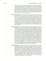
IX-12. Which of the following case histories would most
likely be associated with the urinary sediment depicted?
IX-12. (Continued)
(A) A 23-year-old man with newly diagnosed lympho
blastic lymphoma who is found to have a rising
creatinine level 2 days after the administration of
combination chemotherapy
(B) A 23-year-old woman 1 year after surgery per
formed because of morbid obesity
(C) A 45-year-old woman with a history of multiple
urinary tract infections with urea-splitting orga
nisms
IX. D
ISORDERS OF THE
K
IDNEY AND
U
RINARY
T
RACT —
Q
UESTIONS
200
IX-12. (Continued) IX-15. (Continued)
(D) A 40-year-old man with edema, hypoalbuminemia,
and proteinuria
(E) An 18-year-old man with flank pain, hematuria,
and a positive family history for renal stones in
youth
IX-13. A 72-year-old woman with rheumatic heart disease
is being treated with ampicillin and gentamicin for enter-
ococcal endocarditis. One week into the course she de-
velops a morbilliform skin rash and fever. Laboratory
evaluation is remarkable for a doubling of serum creati-
nine and blood urea nitrogen from their baseline values.
Urinalysis dipstick is positive for blood, protein, and
white cells. Ultrasonography reveals bilaterally enlarged
kidneys. Based on the available data, the most likely cause
of the patient’s azotemia is
(A) tubular necrosis caused by aminoglycoside
(B) membranous nephropathy resulting from endocar-
ditis
(C) enterococcal pyelonephritis
(D) cystitis
(E) hypersensitivity reaction to ampicillin
IX-14. A 40-year-old woman who has never had signifi-
cant respiratory disease is hospitalized for evaluation of
hemoptysis. Urinalysis reveals 2ϩ proteinuria and micro-
scopic hematuria. BUN concentration is 7.1 mmol/L
(20 mg/dL), and serum creatinine concentration is
177
mol/L (2.0 mg/dL). Serologic findings include nor-
mal complement levels and a negative assay for fluores-
cent antinuclear antibodies. Renal biopsy reveals granu-
lomatous necrotizing vasculitis with scattered
immunoglobulin and complement deposits. The most
likely diagnosis in this case is
(A) mesangial lupus glomerulonephritis
(B) Henoch-Scho¨nlein purpura
(C) microscopic polyarteritis
(D) Wegener’s granulomatosis
(E) Goodpasture’s syndrome
IX-15. Which of the following patients is most likely to
develop destruction of renal papillae with concomitant
tubulointerstitial damage?
(A) A middle-aged man who has consumed “moon-
shine” alcohol distilled in an automobile radiator
(B) An older man with early-stage prostate adenocarci-
noma
(C) A young adult woman with

-thalassemia
(D) An older woman who uses analgesics for chronic
headaches
(E) A middle-aged woman with her first episode of a
urinary tract infection which is associated with py-
uria, flank pain, and fever but responds well to a
short course of oral antibiotics
IX-16. A 45-year-old woman with long-standing systemic
lupus erythematosus (SLE) who has had intermittent
bouts of acute renal failure over the past 6 years presents
with anorexia. Physical examination is noncontributory.
Laboratory evaluation includes hematocrit 29%, white
count 5000 with a normal differential, and platelet count
27,500/
L. Renal biopsy shows sclerosis of 14/15 glo-
meruli, tubular atrophy, and interstitial fibrosis. The fol-
lowing values are also found:
Serum electrolytes (mmol/L): Na 136; K 6; Cl 90;
ϩϩϪ
HCO 20
Ϫ
3
BUN: 35.5 mmol/L (100 mg/dL)
Serum creatinine: 665
mol/L (7.5 mg/dL)
Anti-double-strand DNA and C3 levels have been sta-
ble. Renal biopsy shows obliterative sclerosing glomeru-
lar lesions. The most appropriate management strategy
would be
(A) high-dose intravenous methylprednisolone
(B) high-dose intravenous methylprednisolone and aza-
thioprine
(C) high-dose intravenous methylprednisolone and in-
travenous cyclophosphamide (500 mg/m )
2
(D) intravenous cyclophosphamide (500 mg/m ) plus
2
low-dose prednisone
(E) dialysis
IX-17. A 30-year-old woman with diabetic nephropathy
received a cadaveric renal allograft. On the third post-
operative day her serum creatinine concentration was
160
mol/L (1.8 mg/dL). She is being treated with
cyclosporine and prednisone. On the sixth postoperative
day she experiences a decrease in urine output from
1500 mL/d to 1000 mL/d; the serum creatinine concen-
tration increases to 194
mol/L (2.2 mg/dL). Her blood
pressure remains stable at 170/90 mmHg, and her tem-
perature is 37.2ЊC(99ЊF). The best initial step in manage-
ment would be to
(A) decrease the dose of cyclosporine
(B) obtain ultrasonography of the renal allograft
(C) obtain a biopsy of the renal allograft
(D) administer pulsed steroid therapy
(E) administer an intravenous bolus of furosemide
IX-18. A 55-year-old man undergoes intravenous pyelog-
raphy (IVP) as part of a workup for hypertension. A
3-cm solitary radiolucent mass is noted in the left kidney;
the study otherwise is normal. The man complains of no
symptoms referable to the urinary tract, and examination
of urinary sediment is within normal limits. Which of the
following studies should be performed next?
IX. D
ISORDERS OF THE
K
IDNEY AND
U
RINARY
T
RACT —
Q
UESTIONS
201
IX-18. (Continued) IX-20. (Continued)
(A) Repeat IVP in 6 months
(B) Early-morning urine collections for cytology (three
samples)
(C) Selective renal arteriography
(D) Renal ultrasonography
(E) CT scanning (with contrast enhancement) of the
left kidney
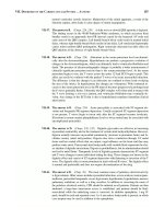
IX-19. A previously healthy 45-year-old man who devel-
oped weight gain, fatigue, and vomiting within the past
week presents to his physician. He had been seen 3
months earlier for a routine checkup, at which time a
physical examination, complete blood count, and serum
chemistries were all normal. Relevant physical findings
now include blood pressure of 155/110 mmHg and peri-
orbital edema. Serum studies reveal a BUN of 30 mmol/
L (85 mg/dL) and a creatinine of 796
mol/L (9 mg/dL).
Urinalysis reveals 2ϩ proteinuria and a microscopic ex-
amination of the sediment is depicted below. Which of
the following statements is correct?
(A) Renal biopsy is indicated
(B) The clinical scenario is typical of a patient who
presents with IgA nephropathy
(C) Extracapillary proliferation is probable
(D) Complete spontaneous resolution of the renal dis-
ease is likely
(E) A trial of high-dose glucocorticoids is contraindi-
cated
IX-20. Which of the following is a risk factor for carci-
noma of the bladder?
(A) Exposure to alcohol intake
(B) Use of cyclophosphamide
(C) History of renal carcinoma
(D) Positive family history
(E) Infestation with Schistosoma mansoni
IX-21. A 10-year-old girl complaining of profound weak-
ness, occasional difficulty walking, and polyuria is
brought to the pediatrician. Her mother is sure the girl has
not been vomiting frequently. The girl takes no medicines.
She is normotensive, and no focal neurologic abnormali-
ties are found. Serum chemistries include sodium
142 mmol/L, potassium 2.5 mmol/L, bicarbonate
32 mmol/L, and chloride 100 mmol/L. A 24-h urine col-
lection on a normal diet reveals sodium 200 mmol/d, po-
tassium 50 mmol/d, and chloride 30 mmol/d. Renal ultra-
sound demonstrates symmetrically enlarged kidneys
without hydronephrosis. A stool phenolphthalein test and
a urine screen for diuretics are negative. Plasma renin lev-
els are found to be elevated. Which of the following con-
ditions is most consistent with the above data?
(A) Conn’s syndrome
(B) Chronic ingestion of licorice
(C) Bartter’s syndrome
(D) Wilms’ tumor
(E) Proximal renal tubular acidosis
IX-22. Normal serum complement levels would be seen in
patients with hematuria, proteinuria, and hypertension re-
sulting from which of the following?
(A) Mixed essential cryoglobulinemia
(B) Hepatitis C– associated membranoproliferative glo-
merulonephritis
(C) Diffuse proliferative lupus nephritis
(D) Henoch-Scho¨nlein purpura
(E) Poststreptococcal glomerulonephritis
IX-23. In acute renal failure, dietary protein should be re-
stricted in which of the following?
(A) All patients
(B) All patients with BUN Ͼ100
(C) All patients with creatinine Ͼ10
(D) Only in patients who are well nourished on hospi-
tal admission
(E) If azotemia is advanced and dialysis is not an op-
tion
IX. D
ISORDERS OF THE
K
IDNEY AND
U
RINARY
T
RACT —
Q
UESTIONS
202
IX-24. A 43-year-old homeless man is brought into the emergency room. His past med-
ical history is significant for a long history of alcohol abuse. He is found obtunded with
evidence of clumsiness on neurologic examination. The emergency medical services
who brought the man to the emergency room believed that he had been drinking ethylene
glycol. Which set of laboratory values is most likely associated with the above clinical
scenario?
Na
ϩ
K
ϩ
Cl
Ϫ
HCO
3
Ϫ
pH
(Serum, mmol/L)
Serum Creatinine,
mol/L (mg/dL) Arterial Urine
(A) 140 2.5 114 14 265 (3.0) 7.30 6.2
(B) 139 6.3 108 19 265 (3.0) 7.35 5.0
(C) 139 5.1 104 21 265 (3.0) 7.37 5.0
(D) 143 4.8 100 10 265 (3.0) 7.25 5.0
(E) 135 4.5 107 21 265 (3.0) 7.37 5.0
IX-25. A 53-year-old woman with longstanding depression and a history of rheumatoid
arthritis is brought in by her daughter, who states that she found an empty bottle of
acetylsalicylic acid by her mother’s bedside. The patient is found confused and lethargic
and unable to provide a definitive history. What is the most likely set of laboratory
values?
Na
ϩ
K
ϩ
Cl
Ϫ
HC
O
3
Ϫ
Room Air ABG
(Serum, mmol/L)
Serum Creatinine
mol/L (mg/dL)
P
O
2
P
CO
2
pH
(A) 140 3.9 85 26 141 (1.6) 100 40 7.40
(B) 140 3.9 85 16 141 (1.6) 100 20 7.40
(C) 140 5.8 100 20 141 (1.6) 100 34 7.38
(D) 150 2.9 100 36 141 (1.6) 80 46 7.50
(E) 116 3.7 85 22 141 (1.6) 80 46 7.50
IX-26. A 37-year-old man is admitted with confusion.
Physical examination shows a blood pressure of 140/70
with no orthostasis, normal jugular venous pressure, and
no edema. Serum chemistries are notable for sodium
120 mmol/L, K 4.2 mmol/L, bicarbonate 24 mmol/L,
ϩ
and uric acid 0.177 mmol/L (2 mg/dL). The most likely
diagnosis is
(A) hepatic cirrhosis
(B) cerebral toxoplasmosis with SIADH
(C) Addison’s disease
(D) significant gastrointestinal fluid loss
(E) congestive heart failure
IX-27. A 56-year-old diabetic woman with end-stage renal
disease (ESRD) has been treated with peritoneal dialysis
(prescription of four 2-L exchanges per day) for 6 years.
She is 5 ft. 6 in. tall and weighs 70 kg (154 lb). The patient
complains of anorexia, abdominal discomfort, fatigue,
and insomnia. Medications include erythropoietin, cal-
cium carbonate, metoprolol, and a water-soluble vitamin
supplement. Laboratory studies are notable for hematocrit
38%, BUN 56 mg/dL, bicarbonate 14 meq/L, calcium
10.4 mg/dL, and phosphate 2.3 mg/dL.
IX-27. (Continued)
The most likely diagnosis is
(A) mycobacterial peritonitis
(B) dialysis disequilibrium
(C) uremia
(D) peritoneal carcinomatosis
(E) diabetic ketoacidosis
IX-28. Which of the following maneuvers may lead to the
development of hyperammonemia?
(A) Protein restriction
(B) A branched-chain amino acid –enriched protein
mixture
(C) The use of neomycin
(D) The use of lactulose
(E) The use of loop diuretics
IX-29. Nephrocalcinosis can be associated with
(A) the routine use of calcium-based phosphate binders
(B) the routine use of aluminum-based phosphate bind
ers
(C) calcitonin-related peptide
IX. D
ISORDERS OF THE
K
IDNEY AND
U
RINARY
T
RACT —
Q
UESTIONS
203
IX-29. (Continued) IX-33. (Continued)
(D) secondary hyperparathyroidism
(E) Crohn’s disease
IX-30. A 35-year-old man is in your clinic with the chief
complaint of progressive lower extremity edema. On lab
oratory analysis he is found to have a 24-h urine collection
that is significant for 5.3 g of protein. Which of the fol-
lowing statements is true?
(A) Lower serum lipid levels
(B) An elevated serum calcium value is likely to be
obtained
(C) The patient has an increased risk of a hemorrhage
(D) The most likely etiology is IgA glumerulonephritis
(E) The patient has an elevated thyroxin level
IX-31. A 60-year-old man with alcoholism presents to the
emergency department with severe confusion, vomiting,
and tachycardia. Blood pressure is 90/60, heart rate is 110,
and respiratory rate is 32. Laboratory studies are remark-
able for the following (mmol/L): Na 128, K 3.9, Cl
ϩϩϪ
90, bicarbonate 6. BUN was 12 mg/dL, and creatinine was
2.9 mg/dL. Acetest is negative. Urinalysis shows 4ϩ cal-
cium oxalate crystals. The most likely diagnosis is
(A) alcoholic rhabdomyolysis with acute tubular
necrosis
(B) alcoholic ketoacidosis
(C) renal tubular acidosis type 1
(D) ingestion of ethylene glycol
(E) alcoholic hepatitis with pancreatitis and multiple
organ dysfunction
IX-32. A 72-year-old man develops acute renal failure af-
ter cardiac catheterization. Physical examination is nota-
ble for diminished peripheral pulses, livedo reticularis, ep-
igastric tenderness, and confusion. Laboratory studies
include (mg/dL) BUN 131, creatinine 5.2, and phosphate
9.5. Urinalysis shows 10 to 15 WBC, 5 to 10 RBC, and
one hyaline cast per high-power field (HPF). The most
likely diagnosis is
(A) acute interstitial nephritis caused by drugs
(B) rhabdomyolysis with acute tubular necrosis
(C) acute tubular necrosis secondary to radiocontrast
exposure
(D) cholesterol embolization
(E) renal arterial dissection with prerenal azotemia
IX-33. The hyperlipidemia of nephrotic syndrome is char-
acterized by
(A) elevation of all plasma lipids but no increase in
atherogenesis
(B) elevation of total cholesterol but no increase in
atherogenesis
(C) selective elevation of low-density lipoprotein
(LDL) cholesterol with increased atherogenesis
(D) no response to HMG-CoA reductase inhibitors
(E) myositis in 20% of patients treated with lipid-low-
ering agents
IX-34. ACE inhibitors would be expected to slow the pro-
gression of renal insufficiency in which of the following
conditions?
(A) Analgesic nephropathy
(B) Contrast dye– associated nephropathy
(C) Chronic glomerulonephritis with Ͼ1 g/d protein-
uria
(D) Autosomal dominant polycystic kidney disease
(ADPKD)
(E) Amphotericin-induced nephropathy
IX-35. Which of the following statements about polycystic
kidney disease is true?
(A) Polycystic kidney disease is an autosomal reces-
sive disorder linked to a causative gene on the
short arm of chromosome 16.
(B) Erythropoietin levels are often low due to progres-
sive renal failure.
(C) Chronic diverticular disorder is a rare finding.
(D) Nephrotic-range proteinuria is an uncommon find-
ing.
(E) The development of an intracranial saccular aneu-
rysm (berry aneurysm) is associated with renal
failure and does not have a specific association
with polycystic kidney disease.
IX-36. Which of the following medications commonly
does not cause hypokalemia?
(A)

-Adrenergic agonists
(B) Theophylline
(C) Calcium channel blockers
(D) Diuretic therapy
(E) Amphotericin B
IX-37. In patients with urinary incontinence, which con-
dition puts them at highest risk for the development of
hydronephrosis?
(A) Alzheimer’s disease
(B) Guillain-Barre´ syndrome
(C) Normal-pressure hydrocephalus
(D) Low-grade astrocytoma
(E) Hypothyroidism
IX-38. Which of the following genetic abnormalities is as-
sociated with the development of hyperkalemia?
(A) 11

-hydroxylase deficiency
IX. D
ISORDERS OF THE
K
IDNEY AND
U
RINARY
T
RACT —
Q
UESTIONS
204
IX-38. (Continued)
(B) Liddle’s syndrome
(C) Bartter’s syndrome
(D) Gitelman’s syndrome
(E) Autosomal dominant polycystic kidney disease
IX-39. Which of the following statements is true concern-
ing acute poststreptococcal glomerulonephritis (PSGN)?
(A) The latent period appears to be longer when PSGN
is associated with cutaneous rather than pharyngeal
infections.
(B) Serologic evidence of a streptococcal infection can
usually be found regardless of antimicrobial ther-
apy.
(C) Antimicrobial therapy for streptococcal infection is
without value once the presence of renal disease is
established.
(D) Long-term antistreptococcal prophylaxis is indi-
cated after a prior documented case of PSGN.
(E) Progressive deterioration in renal function is more
common in children than in adults with PSGN.
IX-40. A 19-year-old man arrives in your office complain-
ing of generalized weakness, nausea, vomiting, and mal-
aise. He states the color of his urine over the past several
days has turned a “red,” or “smoky” color. He states that
he was well until approximately 10 days prior to his visit,
when he had severe pharyngitis with a high-grade fever,
although his pharyngitis and fever have now resolved.
Physical examination reveals a blood pressure of 180/96,
pulse of 98, and a temperature of 37.1ЊC (98.7ЊF). Lab-
oratory values reveal a serum creatinine of 177
mol/L
(2 mg/dL). Which of the following statements is true?
(A) The presence of dysmorphic red blood cells with
red blood cell casts and leukocyte casts on micro-
scopic examination is a universal finding.
(B) A 24-h urine collection reveals 4 g of protein.
(C) Mixed cryoglobulinemia is an unusual finding.
(D) The levels of serum C3 and CH50 are usually
within normal limits.
(E) Electron microscopy typically reveals large elec-
tron-dense immune deposits in the subendothelial,
subepithelial, and mesangial areas.
IX-41. Diseases involving the renal glomeruli are fre-
quently encountered. Both humoral and cellular mecha-
nisms play a part in the pathogenesis of glomerular injury.
Which of the following glomerular diseases is associated
with glomerulosclerosis as opposed to cellular prolifera-
tion?
(A) IgA nephropathy
(B) Diabetic nephropathy
(C) Poststreptococcal glomerulonephritis
(D) Henoch-Scho¨nlein purpura
(E) Glomerulonephritis related to hepatitis C infection
IX-42. An 87-year-old man presents to the emergency
room obtunded. On physical examination he has a pulse
of 120, blood pressure of 142/80, and weight of 72 kg
(158 lb). He has diminished skin turgor and dry mucous
membranes. He also has a fever of 38.9ЊC (102.0ЊF). Lab-
oratory analysis reveals the following results:
Serum electrolytes (mmol/L): Na 164; K 4.6;
ϩϩ
Cl 108; HCO 26
ϪϪ
3
BUN: 17.1 mmol/L (48 mg/dL)
Serum creatinine: 168
mol/L (1.9 mg/dL)
The most appropriate treatment plan would be which
of the following?
(A) D W at 300 mL/h
5
(B) D half-normal saline at 300 mL/h
5
(C) D W at 150 mL/h
5
(D) D W at 75 mL/h
5
(E) Ringer’s lactate at 160 mL/h
IX-43. A 48-year-old woman is hospitalized for elective
knee surgery. Routine preoperative laboratory evaluation
reveals the following:
Serum electrolytes (mmol/L): Na 138; K 3.5;
ϩϩ
Cl 110; HCO 20
ϪϪ
3
Blood glucose: 5.2 mmol/L (95 mg/dL)
Serum creatinine: 160
mol/L (1.8 mg/dL)
BUN: 7.1 mmol/L (20 mg/dL)
Urinalysis: pH 5.2; specific gravity 1.005; protein 1ϩ;
glucose 2ϩ; 3 to 5 white blood cells per high-power
field
The patient states that she voids several times during
the night but is unaware of any problem with her kidneys.
Which of the following disorders would be most likely
associated with the findings in this case?
(A) Multiple myeloma
(B) Diabetic nephropathy
(C) IgA nephropathy
(D) Penicillamine-induced nephropathy
(E) Lupus nephritis
IX-44. A 45-year-old woman presents with the third epi-
sode of nephrolithiasis. Laboratory studies disclose the
following:
Serum electrolytes (mmol/L): Na 134; K 2.5;
ϩϩ
Cl 106; HCO 18
ϪϪ
3
Serum chemistries: creatinine 97
mol/L (1.1 mg/dL);
calcium 2.4 mmol/L (9.5 mg/dL); albumin 40 g/L
(4.0 g/dL)
Arterial blood gas values: P 4 kPa (30 mmHg);
CO
2
P 14 kPa (108 mmHg); pH 7.30
O
2
Urine pH: 7.2
A plain film of the abdomen is shown below. Which of
the following statements about this clinical picture is cor-
rect?
IX. D
ISORDERS OF THE
K
IDNEY AND
U
RINARY
T
RACT —
Q
UESTIONS
205
IX-44. (Continued) IX-46. (Continued)
(A) The findings are consistent with the presence of
multiple myeloma.
(B) The findings are consistent with the presence of
medullary sponge kidney.
(C) There is evidence of type I distal renal tubular aci-
dosis (RTA).
(D) Family members are typically not affected.
(E) Intravenous pyelography is typically unremarkable.
IX-45. Which of the following statements is correct re-
garding renal transplantation?
(A) A potential living donor who does not share the
same blood type as the recipient cannot be consid-
ered even if the tissue types are HLA-identical.
(B) The degree of HLA mismatch with cadaveric do-
nor kidneys is a determinant of long-term graft
survival.
(C) Progressive renal failure in a transplant recipient,
termed chronic rejection, is not associated with re-
nal vascular damage.
(D) Allopurinol must be coadministered with azathio-
prine to prevent urate nephropathy associated with
drug-induced cell turnover.
(E) Cyclosporine inhibits interleukin (IL) 2 production
by cytotoxic (CD8ϩ) T cells.
IX-46. A 45-year-old man with a diagnosis of ESRD sec-
ondary to diabetes mellitus is being treated with peritoneal
dialysis. This is being carried out as a continuous ambu-
latory peritoneal dialysis (CAPD). He undergoes four
2-L exchanges per day and has been doing so for approx-
imately 4 years. Complications of peritoneal dialysis in-
clude which of the following?
(A) Hypotension after drainage of dialysate
(B) Hypoalbuminemia
(C) Hypercholesterolemia
(D) Hypoglycemia
(E) Pleural effusion
IX-47. A 45-year-old woman with a long history of asthma
now presents with progressive lower extremity skin rash
as well as renal insufficiency. On physical examination
she is short of breath, with audible wheezing. Her skin
examination reveals numerous raised papules, which are
erythematous in color, on both lower extremities. The le-
sions are nonblanching and raised, with areas of necrosis.
Which of the following statements is correct?
(A) The peripheral white blood count is within normal
limits.
(B) Antineutrophilic cytoplasmic autoantibodies
(ANCA) are found in a cytoplasmic distribution
consistent with antiproteinase-3 (PR3-ANCA).
(C) A history of a progressive lower extremity neurop-
athy is a rare finding.
(D) A normal chest x-ray is a typical finding.
(E) Patient has a history of coronary disease.
IX-48. You are called to see a 62-year-old man who has
recently undergone a transurethral resection of his pros-
trate. Postoperatively he is found to be confused and stu-
porous. The patient interoperatively received4Lof5%
dextrose as intravenous fluids. Clinically he is euvolemic
with a blood pressure of 142/82 mmHg. He weighs 68 kg.
Serum electrolytes (mmol/L) are obtained, which reveal
Na 114 and K 3.8, and serum osmolality is 230. The
ϩϩ
correct management decision would be which one of the
following?
(A) Free water restriction
(B) Normal saline at a rate of 180 mL/h for 3 h then
reevaluate with repeat serum chemistries
(C) 3% saline at 90 mL/h for 3 h and reevaluate with
repeat serum chemistries
(D) 3% saline at 180 mL/h for 3 h and reevaluate with
repeat serum chemistries
(E) Normal saline at 90 mL/h for 3 h and reevaluate
with repeat serum chemistries
IX-49. A 45-year-old patient with membranous glomeru-
lonephritis and renal insufficiency has nephrotic-range
proteinuria. On physical examination the patient has 3ϩ
lower extremity edema, and the patient’s serum albumin
is 21 g/L (2.1 g/dL) and serum creatinine is 106
mol/L
(1.2 mg/dL). An attempt to improve the lower extremity
edema is made with the oral loop diuretic furosemide.
Unfortunately, a poor response is obtained. Which of the
following mechanisms likely contributes to the subopti-
mal response to diuretics?
IX. D
ISORDERS OF THE
K
IDNEY AND
U
RINARY
T
RACT —
Q
UESTIONS
206
IX-49. (Continued)
(A) Decreased renal tubular secretion of furosemide
(B) Diminished bioavailability of furosemide
(C) Binding of furosemide to albumin in the tubular
fluid
(D) Decreased proximal reabsorption of sodium
(E) Decreased distal reabsorption of sodium
IX-50. A 46-year-old man with long-standing diabetes
mellitus and ESRD is undergoing hemodialysis. He has
completed his run of dialysis and you find him somewhat
confused, with a blood pressure of 86/42. Which of the
following factors most likely contributed to the post-
dialysis hypotension?
(A) Reduced temperature dialysate
(B) Concomitant use of antihypertensive therapy
(C) Impaired autonomic response
(D) Poor dietary intake during dialysis
(E) Hyperphosphatemia
IX-51. A 46-year-old man has long-standing hypertension,
and his current medications include hydrochlorothiazide.
He presents with right flank pain radiating to his groin.
Urinalysis reveals 3ϩ hematuria without proteinuria. He
has had a similar episode previously. Routine laboratory
analysis reveals a normal serum creatinine of 88
mol/L
(1.0 mg/dL), serum calcium of 2.6 mmol/L (10.3 mg/dL),
serum uric acid of 268
mol/L (4.5 mg/ dL), and a total
urine calcium of 200 mg per 24 h. The patient passes a
stone, and upon microscopic analysis of the stone it is
found to contain calcium. The most likely cause of the
patient’s recurrent nephrolithiasis is which one of the fol-
lowing?
(A) Idiopathic absorptive hypercalciuria
(B) A common complication of thiazide diuretic
(C) Diathesis
(D) Primary hyperparathyroidism
(E) Hypocitraturia
207
IX. DISORDERS OF THE KIDNEY AND
URINARY TRACT
ANSWERS
IX-1. The answer is E. (Chap. 47) Up to 150 mg/d of protein may be excreted by a normal
person. The bulk of normal daily excretion is made up of the Tamm-Horsfall mucoprotein.
Urine dipsticks may register a trace result in response to as little as 50 mg protein per liter
and are definitively positive once the urine protein exceeds 300 mg/L. A false negative
may occur if the proteinuria is due to immunoglobulins, which are positively charged. If
proteinuria is suspected or documented, a 24-h urine collection should be undertaken to
measure the absolute protein excretion. Urine immunoelectrophoresis also may identify
the particular immunoglobulin that is produced in excess.
IX-2. The answer is E. (Chap. 49) Because of the powerful effect of ADH secretion in the
setting of hypertonicity, severe persistent hypernatremia is possible only in patients who
cannot respond to thirst by ingesting water. A nursing home patient with a fever may lose
significant amounts of body fluid, which can result in dangerous levels of hypernatremia.
Manifestations of hypernatremia include central nervous system dysfunction such as neu-
romuscular irritability, seizures, obtundation, or coma. Calculation of water replacement
needs is based on total-body water, since water loss occurs from both intracellular and
extracellular sites. In this case, a 60-kg woman has a plasma sodium of 160 mmol/L, which
one would like to lower to 140 mmol/L. Total-body water is roughly 60% of weight
(36 L). To reduce the plasma sodium, this volume must be increased to 160/140 times
36 L, or about 41 L. Thus, a positive water balance of 5 L (41 Ϫ 36) is needed. This
deficit is best corrected fairly slowly, with the aim being to replace about half the water
deficit in the first day. If correction is done in this conservative fashion with close moni-
toring of electrolytes, progressive central nervous system dysfunction is not likely. If the
patient had signs of circulatory collapse indicating an associated sodium deficiency, treat-
ment would begin with normal saline to provide intracellular volume. In certain situations,
such as hyperosmolar diabetic coma, the plasma osmolarity is elevated because of hyper-
glycemia as well as hypernatremia. Therefore, initial treatment should consist of normal
saline to ensure circulatory integrity and insulin to lower plasma glucose and partially
reduce intracellular osmolarity. Finally, half normal saline could be used to slowly replace
the remaining water and salt deficits.
IX-3. The answer is A. (Chaps. 47, 329) Failure to concentrate urine despite substantial
hypertonic dehydration suggests a diagnosis of diabetes insipidus. A nephrogenic origin
will be postulated if there is no increase in urine concentration after exogenous vasopressin.
The only useful mode of therapy is a low-salt diet and use of a thiazide or amiloride, a
potassium-sparing distal diuretic agent. The resultant volume contraction presumably en-
hances proximal reabsorption and thereby reduces urine flow.
IX-4. The answer is A. (Chap. 47. Gennari, N Engl J Med 339:451– 458, 1998.) This man’s
electrolyte pattern is consistent with a hyporeninemic hypoaldosteronism state, or type IV
renal tubular acidosis. The defect is believed to be due to an insufficiency of both angio-
tensin- and adrenal mineralocorticoid-secreting capacity. Inhibition of the renin-angioten-
sin system by

-adrenergic blockade such as propranolol can cause hyperkalemia; in ad-
dition, nonsteroidal anti-inflammatory agents or angiotensin-converting enzyme (ACE)
inhibitors may also lead to hyperkalemia. The use of carbenicillin, theophyllin, and hy-
IX. D
ISORDERS OF THE
K
IDNEY AND
U
RINARY
T
RACT —
A
NSWERS
208
drochlorothiazide promote the loss of potassium through the distal renal tubule. Calcium
channel blockers such as verapamil have no significant effect on serum potassium con-
centrations at the usual doses.
IX-5. The answer is B. (Chap. 50. Wrenn, Am J Med 91:119, 1991.) A reasonable way to
approach the diagnosis of metabolic acidosis is to separate patients into those with an
increased anion gap and those with a normal anion gap (hyperchloremic acidosis). A
calculation of these unmeasured anions consists of the sum of plasma bicarbonate and
chloride minus the plasma sodium concentration (the normal value is 8 to 16 mmol/L).
Reasons for increased acid production include diabetic ketoacidosis, alcoholic ketoacidosis
(as in this patient), starvation, lactic acidosis caused by circulatory failure, certain drugs
and toxins, and poisoning resulting from salicylates, ethylene glycol, or methanol. Finally,
renal failure increases the anion gap because sulfate, phosphate, and organic acid ions are
not excreted normally. Normal anion gap acidosis is due to renal tubular dysfunction or
colonic losses. Since the ratio of

-hydroxybutyrate to acetoacetate is high in alcoholic
ketoacidosis, ketonemia can be missed by the routinely employed nitroprusside (Acetest)
reagent, which detects acetoacetate but not

-hydroxybutyrate. Patients suffering from
alcoholic ketoacidosis do well on infusions of glucose and saline. Neither insulin nor
alkali is required in these situations unless the acidosis is extreme (bicarbonate Ͻ6to
8 mmol/L).
IX-6. The answer is D. (Chap. 270) Development of advancing peripheral neuropathy is an
indication for dialysis. Delaying dialysis could allow the development of irreversible motor
deficits, such as foot drop. Prompt institution of dialysis, by contrast, usually prevents the
progression of uremic peripheral neuropathy and may ameliorate early sensory defects.
No pharmacologic agent would be of significant benefit in the clinical situation described.
IX-7. The answer is A. (Chap. 270. Ifudu, N Engl J Med 339:1054 – 1062, 1998.) Renal
osteodystrophy is a common complication of chronic renal disease, and the most common
complication secondary to impaired renal production of 1,25(OH) D . This leads to a
23
decreased calcium absorption within the gut as well as impaired renal phosphate excretion.
The resulting hyperphosphatemia causes a secondary hyperparathyroidism. Hyperparathy-
roidism is subsequently worsened by hypocalcemia, which is present because of the hy-
perphosphatemia and the decreased enzymatic conversion of 25-hydroxyvitamin D to
1,25(OH) D . Finally 1,25(OH) D deficiency worsens hyperparathyroidism as the former
23 23
is a direct inhibitor of parathyroid hormone secretion into the bone. The resultant decreased
serum calcium concentration leads to secondary hyperparathyroidism. In addition, other
causes of renal osteodystrophy include chronic metabolic acidosis, due to dissolution of
bone buffers and decalcification, and the long-term administration of aluminum-containing
antacids. There is no significant loss of vitamin D or calcium associated with currently
employed dialysis techniques, and the treatment of renal osteodystrophy often employs
calcitriol.
IX-8. The answer is A. (Chap. 50) To offer optimal management to patients with acute renal
failure, it is helpful to distinguish prerenal azotemia (generally managed with volume
replacement or amelioration of cardiac dysfunction) from intrinsic renal dysfunction. So-
dium reabsorption, which is quite avid in prerenal azotemia, is impaired in intrinsic renal
disease. However, creatinine is reabsorbed less efficiently than sodium in both conditions.
Therefore, the fractional excretion of sodium is very helpful in distinguishing between
these two etiologies of renal failure. The fractional excretion of sodium is calculated by
multiplying the urine sodium by the plasma creatinine, dividing this by the plasma sodium
times the urine creatinine, and multiplying by 100. In this case the result is approximately
1.4, which suggests that impaired reabsorption of sodium is ongoing and that intrinsic
renal failure is occurring. Only about 15% of patients receiving nephrotoxins such as
aminoglycosides or radiocontrast agents have renal failure associated with a fractional
excretion of sodium of Ͻ1% and so an elevated value in this case points in the direction
IX. D
ISORDERS OF THE
K
IDNEY AND
U
RINARY
T
RACT —
A
NSWERS
209
of nephrotoxic injury. The other causes of acute renal failure listed here are all associated
with prerenal azotemia and therefore with a more avid reabsorption of sodium than that
described.
IX-9. The answer is D. (Chap. 274) One of the more common forms of asymptomatic uri-
nary abnormalities is Berger’s disease, which may be a cause of recurrent hematuria of
glomerular origin. Such episodes of macroscopic hematuria and may be associated with
minor flulike illnesses or vigorous exercise. Skin rash, arthritis, and abdominal pain usually
are absent, which tends to distinguish this entity from Henoch-Scho¨nlein purpura. Occa-
sionally patients develop a nephrotic or nephritic syndrome. Serum IgA levels are increased
in about 50% of all cases, though serum complement is normal. Renal biopsy in these
situations may reveal a spectrum of changes, though diffuse mesangial proliferation or
focal and segmental proliferative glomerulonephritis is most common. The essential feature
of Berger’s disease is the finding of diffuse mesangial deposition of IgA on immunoflu-
orescence microscopy. IgG, C3, and properdin, but not C1q or C4, also may be found on
this study. Although the disease progresses slowly, about 50% of patients develop end-
stage renal failure within 25 years of the original presentation. Men with hypertension and
proteinemia (Ͼ1 g/d) are most likely to progress. Except for a recent report suggesting
that omega-3 fatty acids may play a role, specific therapy has not been useful. However,
glucocorticoids or antibiotics may reduce the frequency of episodic gross hematuria. IgA
deposition in the kidney and recurrent renal failure may occur in about 35% of those who
receive a renal allograft. Fortunately, such recurrent pathologic findings usually are not
associated with loss of renal function.
IX-10. The answer is B. (Chap. 274) The characteristic pattern of focal (not all glomeruli)
and segmental (not the entire glomerulus) glomerular scarring is shown. The history and
laboratory features are also consistent with this lesion: some associated hypertension, dim-
inution in creatinine clearance, and a relatively inactive urine sediment. The “nephropathy
of obesity” may be associated with this lesion secondary to hyperfiltration; this condition
may be more likely in obese patients with hypoxemia, obstructive sleep apnea, and right-
sided heart failure. Hypertensive nephrosclerosis exhibits more prominent vascular
changes and patchy, ischemic, totally sclerosed glomeruli. In addition, nephrosclerosis
seldom is associated with nephrotic-range proteinuria. Minimal-change disease usually is
associated with symptomatic edema and normal-appearing glomeruli as demonstrated by
light microscopy. This patient’s presentation is consistent with that of membranous ne-
phropathy, but the biopsy is not. With membranous glomerular nephritis all glomeruli are
uniformly involved with subepithelial dense deposits. There are no features of crescentic
glomerulonephritis present.
IX-11. The answer is D. (Chap. 274) Persons who have solid tumors and develop nephrotic
syndrome usually have membranous glomerulopathy. Diagnosis of the nephrotic syndrome
may precede recognition of the primary tumor. In several cases, tumor antigens have been
discovered in the glomerular deposits; the nephrotic syndrome may remit after effective
tumor therapy. Patients with Hodgkin’s disease may develop nephrotic syndrome on the
basis of minimal-change disease (diffuse epithelial foot process effacement on ultrastruc-
tural examination).
IX-12. The answer is A. (Chaps. 277, 279) Cystine crystals appear as flat hexagonal plates
and are found in association with cystine stones, which are caused by a hereditary defi-
ciency in tubular cystine transport. Struvite stones result from chronic urinary tract infec-
tion with Proteus spp. These bacteria degrade urea to carbon dioxide and ammonia, which
alkalinizes the urine, thereby favoring the formation of the insoluble triple salt
MgNH PO . Struvite crystals can appear in the urine as rectangular prisms. Patients with
44
proteinuria resulting from albuminuria exhibit a sediment characteristic of the nephrotic
syndrome with oval fat bodies. Patients with intestinal malabsorption with concomitant
steatorrhea, as in the case of a jejunoileal bypass done for obesity, may hyperabsorb oxalate
IX. D
ISORDERS OF THE
K
IDNEY AND
U
RINARY
T
RACT —
A
NSWERS
210
and form calcium oxalate renal stones. Calcium oxalate crystals appear bipyramidal or as
biconcave ovals. The sediment depicted here displays flat, square plates, which represent
one of the several forms uric acid crystals manifest. Hyperuricemia may accompany rapid
cell turnover (as occurs in the rapid lysis of lymphomas with large tumor burdens after
chemotherapy). In such settings aggressive hydration, the use of allopurinol, and urinary
alkalinization may provide effective prophylaxis against uric acid nephropathy.
IX-13. The answer is E. (Chap. 277) A number of drugs may elicit an acute interstitial ne-
phritis. The classic offender is methicillin, although ampicillin, penicillin, cephalothin,
thiazides, furosemide, and nonsteroidal anti-inflammatory drugs also have been associated
with this problem. Hematuria, fever, and skin rash may occur within 1 to 2 weeks of
exposure to the drug. Urinalysis reveals protein, pyuria, and eosinophiluria. Ultrasonog-
raphy discloses enlarged kidneys. A biopsy (usually not necessary, since withdrawal of
the offending drug leads to complete resolution) will reveal normal glomeruli but infiltra-
tion of the interstitium with polymorphonuclear leukocytes, lymphocytes, plasma cells,
and eosinophils.
IX-14. The answer is D. (Chap. 275) A variety of diseases involve both pulmonary and renal
(and often dermal) microvasculature and may present with either prominent pulmonary or
renal manifestations. When a firm diagnosis cannot be made serologically or by biopsy of
skin or lesions of the upper respiratory tract, renal biopsy may be necessary. In the case
described in this question, the serologic findings, though not specific, are typical of We-
gener’s granulomatosis, a diagnosis established by the renal biopsy report. Granulomas
are an uncommon microscopic finding in polyarteritis as well as in lupus nephritis and
Henoch-Scho¨nlein purpura, though a spectrum of pathologic abnormalities may be seen
in the latter two conditions. Antineutrophil antibodies in the serum are highly suggestive
of Wegener’s granulomatosis and other systemic vasculitides. The renal biopsy in Good-
pasture’s syndrome usually reveals linear immunoglobulin deposits.
IX-15. The answer is D. (Chap. 277. Bennett, DeBroe, N Engl J Med 320:1269 – 1271, 1989.
DeBroe, Elseviers, N Engl J Med 338:446 – 452, 1998.) Patients with damage to renal
papillae may be unable to excrete maximally concentrated urine owing to chronic tubular
damage. Moreover, the necrosed papillae can lead to the gradual development of renal
failure. Renal papillary necrosis has been classically associated with long-term analgesic
abuse. This is most commonly manifested by chronic use of acetaminophen or phenacetin.
In addition to analgesic abuse, renal papillary necrosis can also be caused by sickle cell
anemia, diabetic nephropathy, or acute obstructive uropathy. It is not associated with the
presence of early-stage prostate cancer in the absence of prostatic hypertrophy. In addition,
patients can present with renal papillary necrosis after multiple episodes of pyelonephritis,
but this is uncommon after a single uncomplicated episode. Aspirin can potentiate the
deleterious effects of chronic analgesic abuse by inhibiting the production of renal vaso-
dilatory prostaglandins. Ingestion of lead, such as that caused by lead leaching out from
an unusual distilling apparatus, can lead to a nephropathy manifested by tubular atrophy
and fibrosis of small renal arteries.
IX-16. The answer is E. (Chap. 275) The pathophysiology of nephrotoxic involvement by
SLE is thought to be immune complex deposition. Renal disease in SLE can range from
mild abnormalities of the urinalysis to a fulminant inflammatory process that leads to
progressive renal failure. Renal biopsy findings in patients with SLE who have worsening
renal function can range from minimal glomerular lesions to diffuse proliferative lupus
glomerulonephritis and membranous lupus glomerulonephritis. Patients with membranous
lupus glomerulonephritis may be managed conservatively with therapy directed toward
extrarenal manifestations. By contrast, those with more extensive or proliferative glomer-
ular lesions require a more aggressive approach using glucocorticoids (with or without
another immunosuppressive agent, such as azathioprine or cyclophosphamide). However,
little is gained by using immunosuppressant therapy in patients with advanced renal failure
IX. D
ISORDERS OF THE
K
IDNEY AND
U
RINARY
T
RACT —
A
NSWERS
211
characterized by obliterative sclerosing lesions of the glomeruli. If such patients have other
indications for dialysis, such as systemic symptoms and hyperkalemia, they are best man-
aged with dialysis followed by renal transplantation. Measurement of serologic evidence
of disease (e.g., double-stranded DNA autoantibodies or a decrease in serum complement
components) may be helpful. Patients with end-stage lupus nephritis can be managed
successfully with hemodialysis. Moreover, patients with SLE who have undergone renal
allografting rarely experience recurrence of disease in the new kidney.
IX-17. The answer is B. (Chap. 272) In the first week after renal transplantation the differ-
ential diagnosis of graft dysfunction includes early rejection, hypovolemia, cyclosporine
intoxication, acute tubular necrosis, urinary obstruction, and renal artery thrombosis. Cy-
closporine can mask many of the classic signs of rejection, such as fever and graft ten-
derness; renal biopsy often is needed to make the diagnosis. However, renal ultrasonog-
raphy should precede any manipulation to rule out mechanical outflow obstruction, as it
should in any patient with acute deterioration of renal function.
IX-18. The answer is D. (Chap. 94) The most important differential diagnosis in the case
presented is between a renal cell carcinoma and a benign cystic lesion. Urinalysis may be
normal in the presence of renal cell carcinoma, and urinary cytology is unfortunately of
little value in the diagnosis of this lesion. Ultrasonography will reveal whether the lesion
is cystic. If the lesion fulfills the criteria for a simple cyst (lack of internal echoes, smooth
borders, through transmission) and the patient does not have hematuria, the cyst can be
considered benign with a diagnostic accuracy of 97%. If greater assurance is required or
if there are changes on follow-up radiologic studies, needle aspiration should be carried
out. If the ultrasound appearance is not consistent with a simple cyst, contrast-enhanced
CT scanning, the optimal test for the diagnosis and staging of renal cell carcinoma, should
be performed.
IX-19. The answer is D. (Chap. 274. Hricik, Chung-Park, N Engl J Med 339:888 – 899, 1998.)
The syndrome described is typical of rapidly progressive glomerulonephritis (GN) with
rapid onset of acute renal failure in the setting of glomerular disease (manifested by red
blood cell casts and proteinuria). The patient’s vomiting is consistent with the development
of azotemia over a short time period. Renal biopsy is highly recommended early in the
course of such a disease to define the nature and severity of the glomerular lesion for both
prognostic and therapeutic purposes. The hallmark pathologic lesion associated with this
clinical scenario is crescentic glomerulonephritis, the manifestation of extracapillary en-
dothelial proliferation. Such a finding on renal biopsy carries an ominous prognosis, es-
pecially if crescents are present in 70% of glomeruli or if glomerular filtration rate (GFR)
is Ͻ5 mL/min. Spontaneous resolution rarely occurs except in cases associated with an
infectious cause, such as endocarditis and streptococcal disease. Though controlled trials
are lacking, it appears that high-dose methylprednisolone given parenterally (“pulse ster-
oids”) can stave off the need for hemodialysis in some patients. Plasmapheresis may benefit
some patients, especially those who have antiglomerular basement antibodies.
IX-20. The answer is B. (Chaps. 94, 222, 280) Carcinoma of the bladder typically affects
older men. Transitional cell carcinoma is the most common histologic subtype and is
associated with a more favorable prognosis than is adenocarcinoma or squamous carci-
noma. Squamous carcinomas occur more frequently in Egypt and are associated with S.
haematobium and not S. mansoni, which typically causes an infection of the intestines or
biliary tract. Risk factors for carcinoma of the bladder include exposure to the aromatic
amines, which result from cigarette smoke or products of the dye, rubber, and chemical
industries, but it is not associated with positive family history or a prior diagnosis of renal
carcinoma. Chronic bladder irritation, such as that produced by the metabolites of cyclo-
phosphamide or ifosfamide as well as by recurrent bladder stones or infections, also leads
to a higher incidence of carcinoma of the bladder.
IX. D
ISORDERS OF THE
K
IDNEY AND
U
RINARY
T
RACT —
A
NSWERS
212
IX-21. The answer is C. (Chaps. 49, 276. Narins, Am J Med 72:496, 1982.) The evaluation
of patients with hypokalemia should first include a consideration of redistribution of body
potassium into cells as occurs in alkalosis,

-agonist excess with refeeding syndrome and/
2
or insulin therapy, vitamin B therapy, patients with pernicious anemia, and periodic
12
paralysis. In periodic paralysis serum bicarbonate is normal. If the patient is hypertensive
and plasma renin is elevated, renovascular hypertension or a renin-secreting tumor (in-
cluding Wilms’) must be considered and appropriate imaging studies must be carried out.
If plasma renin levels are low, mineralocorticoid effect may be high, due either to endog-
enous hormone (glucocorticoid overproduction or aldosterone overproduction as in Conn’s
syndrome) or to exogenous agents (licorice or steroids). In a normotensive patient a high
serum bicarbonate excludes renal tubular acidosis. High urine chloride excretion makes
gastrointestinal losses less likely and implies primary renal potassium loss as might be
seen in diuretic abuse (ruled out by the urine screen) or Bartter’s syndrome. In Bartter’s
syndrome, hyperplasia of the granular cells of the juxtaglomerular apparatus leads to high
renin levels and secondary aldosterone elevations. Such hyperplasia appears to be second-
ary to chronic volume depletion caused by a hereditary (autosomal recessive) defect that
interferes with salt reabsorption in the thick ascending loop of Henle. Chronic potassium
depletion, which frequently initially presents in childhood, leads to polyuria and weakness.
IX-22. The answer is D. (Chap. 275. Hricik, Chung-Park, N Engl J Med 339:888 – 899, 1998.)
Cryoglobulinemia with renal involvement is associated with hypocomplementemia in the
majority of cases. In addition it has been well recognized that hepatitis C is often associated
with cryoglobulinemia. Diffuse proliferative lupus nephritis (WHO class IV) is the most
aggressive form of the disease and is also associated with hypocomplementemia. Early in
the course of postinfectious glomerulonephritis, immune complex deposition is in full force
and serum complements are low. Henoch-Scho¨nlein purpura, the systemic manifestation
of IgA nephropathy, is not associated with hypocomplementemia. Other causes of hypo-
complementemic glomerulonephritis are glomerulonephritis associated with bacterial en-
docarditis or other chronic infections as well as membranoproliferative glomerulonephritis.
IX-23. The answer is E. (Chap. 269) Years before dialysis was routinely available, it was
well established that protein restriction (prescribed or self-imposed) could alleviate some
of the symptoms of uremia; unfortunately, prolonged protein restriction led to the devel-
opment of malnutrition and its associated complications. In the setting of chronic renal
failure, a number of clinical studies have suggested that modest protein restriction may
slow the rate of progression of renal failure, particularly in patients with glomerular disease
and daily protein excretion rates Ͼ1 g/d. There are insufficient data in the setting of acute
renal failure to adequately assess the importance of protein intake. However, in view of
the hypercatabolism that accompanies many cases of acute renal failure, most practitioners
provide adequate protein to patients (e.g., Ն1.0 to 1.2 g protein per kg per day) and provide
dialysis if uremia ensues. There are no set laboratory “cutoffs” (BUN Ͼ100) that indicate
the need for dialysis.
IX-24. The answer is D. (Chap. 50. Adrogue, Madias, N Engl J Med 338:26 – 34, 1998.)
Ingestion of ethylene glycol can produce severe, high-anion-gap metabolic acidosis, caused
by the accumulation of toxic metabolites. Furthermore, the degree of acidosis is dispro-
portionate to the degree of renal insufficiency. Ethylene glycol intoxication often requires
a large amount of alkali to correct the severe acidemia. Additional measures include gastric
lavage, oral charcoal, and intravenous or oral ethanol. Ethanol has a higher affinity for
alcohol dehydrogenase and will therefore inhibit the generation of toxic metabolites from
the ethylene glycol. In severe cases hemodyalisis is required. In addition, forced diuresis
can prevent acute renal failure in some patients with ethylene glycol intoxication.
IX-25. The answer is B. (Chap. 50. Adrogue, Madias, N Engl J Med 338:26 – 34, 1998.)
This represents a respiratory alkalosis with a combined metabolic acidosis. This is typical
of salicylate toxicity. Salicylate intoxication can result in respiratory alkalosis, mixed res-
IX. D
ISORDERS OF THE
K
IDNEY AND
U
RINARY
T
RACT —
A
NSWERS
213
piratory alkalosis and metabolic acidosis, or, less commonly, a simple metabolic acidosis.
Respiratory alkalosis is caused by direct stimulation of the respiratory center by salicylate.
The accumulation of lactic acid and ketoacids leads to the concomitant metabolic acidosis.
The severity of the neurologic manifestations largely depends on the concentration of
salicylate in the central nervous system. Therapy is directed at limiting further drug ab-
sorption by administering activated charcoal and promoting the exit of salicylate from the
central nervous system. This can be accomplished by alkalinizing the serum, typically by
the addition of intravenous fluids with sodium bicarbonate with the goal of raising the
serum pH to between 7.45 and 7.50. Increasing the GFR will also enhance salicylate
excretion. Hemodialysis is reserved for severe cases, especially those involving fulminant
renal failure.
IX-26. The answer is B. (Chap. 49. Beck, N Engl J Med 301:528– 530, 1979.) Hyponatremia
can be broadly categorized as hypovolemic, euvolemic, or hypervolemic. Hepatic cirrhosis
in this case is unlikely because of the absence of edema. Gastrointestinal fluid loss is
unlikely because of normal blood pressure without orthostasis. Furthermore, depending on
whether the fluid loss is upper (vomiting with resultant alkalosis) or lower (diarrhea with
resultant acidosis), it often is accompanied by a disturbance in acid-base balance. Addi-
son’s disease is possible, although it often is associated with orthostasis, some degree of
hypotension, and hyperkalemia (due to aldosterone deficiency). The uric acid can be very
helpful in the differential diagnosis of hyponatremia. It is typically elevated in patients
with congestive heart failure and renal failure, two other important causes of hyponatremia,
and tends to be quite low in patients with SIADH.
IX-27. The answer is C. (Chap. 271) Mycobacterial peritonitis and fungal peritonitis are rel-
atively rare but important problems, particularly in patients who receive repeated courses
of antibacterial therapy for suspected or documented bacterial peritonitis. Dialysis dis-
equilibrium is a syndrome characterized by headache, confusion, and occasionally seizures;
it is seen in association with the excessively rapid correction of uremia with dialysis
(usually hemodialysis). It is thought to be related to cerebral edema caused by the rapid
removal of extracellular solute (urea) with resultant osmotic transfer of water into the cells.
Peritoneal carcinomatosis (from ovarian or widespread gastrointestinal carcinoma) is pos-
sible in this case, although there is no history of cancer.
IX-28. The answer is E. (Chap. 50) Hypokalemia impairs the renal excretion of ammonium,
which thereby results in hyperammonemia in cases of hepatic failure. The use of loop
diuretics promotes kaliuresis and therefore may lead to hyperammonemia. The alternative
choices typically improve hyperammonemia in selected cases.
IX-29. The answer is E. (Chap. 277) Nephrocalcinosis is an uncommon cause of interstitial
renal disease associated with a variety of metabolic disorders. The routine (with or after a
meal) use of calcium-based phosphate binders rarely results in hypercalcemia, although
injudicious use may lead to complications. Crohn’s disease and other abnormalities of ileal
fat absorption may cause nephrocalcinosis because of excessive absorption of dietary ox-
alate and calcium oxalate nephrolithiasis.
IX-30. The answer is E. (Chap. 274. Orth, Ritz, N Engl J Med 338:1202 – 1211, 1998.) The
patient has nephritic-range proteinuria as documented by a 24-h urine collection of Ͼ3.5g/
1.73 m . Such patients typically have hypoalbuminemia, peripheral edema, and hyperlip-
2
idemia as well as lipiduria. It is thought that the hypoalbuminemia is further compounded
by increased renal catabolism. The physiology of the edema formation in nephrotic syn-
drome is less well understood; it is thought that the presence of hypoalbuminemia results
in decreased intravascular oncotic pressure leading to leakage of extracellular fluid from
blood into the interstitial fluid. As a result, the intravascular volume falls, thereby stimu-
lating activation of the renin-angiotensin-aldosterone axis and consequently increasing the
release of vasopressin (antidiuretic hormone). In addition, there is suppression of the re-
IX. D
ISORDERS OF THE
K
IDNEY AND
U
RINARY
T
RACT —
A
NSWERS
214
lease of atrial natriuretic peptide, and this suppression contributes to the primary renal salt
and water retention that contributes to the formation of edema. Patients with nephrotic-
range proteinuria often have elevated serum lipid levels as a consequence of increased
hepatic lipoprotein synthesis. Both LDL-cholesterol and total cholesterol are increased in
the majority of patients, and, though not proven conclusively, this state of hyperlipidemia
may accelerate atherosclerosis as well as the progression of further renal disease.
Patients often present with a hypercoagulable state due to increased urinary loss of
antithrombin III as well as altered levels or activity of proteins C and S. There is also
impaired fibrinolysis. As a consequence of these alterations, patients can develop spon-
taneous peripheral arterial or venous thrombosis, renal vein thrombosis, and pulmonary
embolism.
Although IgA GN is the most common glomerulopathy, it only occasionally causes
nephrotic syndrome. The most common cause of nephrotic syndrome in adults is mem-
branous GN which represents approximately 40% of all cases. The most common cause
of nephritic syndrome in children is minimal-change glomerulopathy. Membranous GN
can be linked to neoplasms — either carcinoma, sarcoma, lymphoma, or more rarely leu-
kemia. This possibility should be specifically considered in older patients who present with
new-onset nephrotic syndrome. Other metabolic abnormalities include hypocalcemia and
secondary hyperparathyroidism as a consequence of vitamin D deficiency due to enhanced
urinary excretion of cholecalciferol-binding protein. In addition, loss of thyroxin-binding
globulin often results in depressed thyroxin levels. There is also an increased susceptibility
to infection, which may affect low levels of IgG urinary loss as well as increased catab-
olism.
IX-31. The answer is D. (Chap. 50) The key element is the anion gap, calculated as
(Na ϩ K) Ϫ Cl (normal 8 to 12). The anion gap is 32 in this case. Causes of high-anion-
gap acidosis include diabetic or alcoholic ketoacidosis, renal failure, and the excessive
ingestion of salicylates, methanol, ethanol, or ethylene glycol. The clues here are the
negative Acetest (making alcoholic and/or diabetic ketoacidosis less likely) and the pres-
ence of calcium oxylate crystals (oxylate is a by-product of ethylene glycol). Hemodialysis
should be provided in these cases of toxic ingestion, since the products are water-soluble
and can quickly cause damage to the CNS if not promptly removed.
IX-32. The answer is D. (Chap. 269) Cholesterol embolization (also known as atheroembolic
renal disease) is characterized by pyuria, progressive renal failure (usually nonoliguric),
and associated organ dysfunction (including bowel, pancreas, and CNS). Hypocomple-
mentemia and eosinophiluria also may be seen. The urinalysis is not compatible with acute
tubular necrosis because of the absence of granular casts.
IX-33. The answer is C. (Chap. 274. Joven et al., N Engl J Med, 323:579 – 584, 1990.)
Dyslipidemia is present in the vast majority of patients with nephrotic syndrome and
typically is characterized by a relatively selective increase in LDL cholesterol. There is
evidence of accelerated atherosclerosis in these patients. Although there appears to be an
increased risk of myositis in patients with renal failure treated with lipid-lowering agents,
the risk is relatively low (far below 20%). Combination therapy (e.g., an HMG-CoA re-
ductase inhibitor and a fibric acid derivative) should be used with caution.
IX-34. The answer is C. (Chaps. 270, 273. Maschio et al., N Engl J Med 334:939 – 945, 1996.)
Evidence continues to accumulate that ACE inhibition can slow the progression of chronic
renal insufficiency in a variety of disease states. In general, patients with protein excretion
rates 1 g/d tend to derive the greatest benefit. For reasons that are not entirely clear, patients
with ADPKD tend not to benefit from this therapy (and do not appear to benefit from a
low-protein diet).
IX-35. The answer is D. (Chap. 276. Gabow, N Engl J Med 329:332– 342, 1993.) Autosomal
dominant polycystic kidney disease is one of the most common hereditary disorders. Spon-
IX. D
ISORDERS OF THE
K
IDNEY AND
U
RINARY
T
RACT —
A
NSWERS
215
taneous mutations occur but account for only 10% or fewer of all cases. The majority of
cases are linked to a causative gene located on the short arm of chromosome 16, referred
to as ADPKD1. A second causative gene (ADPKD2) has not been identified but seems to
be at a locus distinct from ADPKD1, which appears to be the cause of the gene in Ն90%
of families in the white population.
A rare syndrome known as autosomal recessive polycystic kidney disease affects be-
tween 1 in 10,000 and 1 in 40,000 individuals in the United States, and the causative gene
has been localized to chromosome 6. Patients are typically diagnosed during the first year
of life, presenting with bilateral abdominal masses. Death in the neonatal period is common
due to pulmonary hypoplasia.
The most common complication of polycystic kidney disease is pain secondary to large
cystic kidneys; in addition, most patients are hypertensive. In addition to the chronic pain,
acute pain may represent cystic hemorrhage, infection, or, less commonly, a renal stone.
Proteinuria occurs in about 1/3 of patients but is typically mild, with Ͻ1 g per 24-h
collection. The rare patient with nephrotic-range proteinuria often has superimposed glo-
merular disease.
Certain endocrine functions of the kidney also appear to be altered. Renal secretion
seems to be elevated and likely causes a predilection for hypertension. Although polycy-
themia is rare, the hematocrit seems to be better maintained in patients with renal failure
due to ADPKD than in patients with other forms of renal disease.
Extrarenal complications are common and often include hepatic and pancreatic cysts;
and cardiac valvular abnormalities are also noted. The principal noncystic gastrointestinal
manifestation is the development of colonic diverticular disease, which has been reported
in as many as 82% of patients with ADPKD. The most devastating extrarenal manifesta-
tion, however, is the development of intracranial saccular aneurysms, often called berry
aneurysms. These are generally accepted to be a manifestation of polycystic kidney disease,
but occur with an unknown frequency.
IX-36. The answer is C. (Chap. 49. Gennari, N Engl J Med 339:451 – 458, 1998.) A wide
range of drugs have been implicated in causing hypokalemia as a significant side effect.
These drugs include the

sympathomimetic agonists of many drugs including decon-
2
gestants, bronchial dilators, and inhibitors of uterine contraction. The hypokalemia caused
by these drugs is sustained for up to 4 h. Intestinal ingestion of excess amounts of pseu-
doephedrine can cause severe hypokalemia.
Theophylline and caffeine are not sympathomimetic drugs; however, these agents stim-
ulate the release of sympathetic amines, which may also increase the Na , K -ATPase
ϩϩ
activity and thereby lead to hypokalemia. Severe hypokalemia is an almost invariable
feature of acute theophylline toxicity.
Although calcium channel blockers increase cellular uptake of potassium in experimen-
tal studies, these drugs typically have no effect on serum potassium concentration at the
usual doses. However, ingestion of large amounts of verapamil can cause hypokalemia.
The most common cause of drug-induced hypokalemia is diuretic therapy. Both thiazide
and loop diuretics block chloride-associated sodium reabsorption. As a result of the in-
creased delivery of sodium to the collecting tubules, there is a favorable electrochemical
gradient leading to increased potassium secretion.
Amphotericin B causes renal potassium wasting through the inhibition of the secretion
of hydrogen ions by the collecting duct. In addition, severe magnesium depletion is another
manifestation of amphotericin B usage.
IX-37. The answer is B. (Chap. 48. Resnick, N Engl J Med 320:1, 1989.) The force for
bladder emptying is provided by the detrusor muscle, which is innervated by parasym-
pathetic outflow from the sacral plexus. The involuntary control that prevents automatic
bladder emptying emanates from sympathetic innervation of the bladder outlet. A sacral
spinal reflex arc mediates automatic detrusor contraction when the intravesical pressure
exceeds 20 cmH O (a volume of 400 mL) unless it is inhibited by cortical centers via the
2
reticulospinal tracts. Diseases leading to damage of inhibitory neural pathways in the brain
IX. D
ISORDERS OF THE
K
IDNEY AND
U
RINARY
T
RACT —
A
NSWERS
216
or spinal cord, such as multiple strokes, Alzheimer’s disease, brain tumors, and normal-
pressure hydrocephalus, create detrusor instability. In this situation the bladder will empty
automatically before it is filled owing to unchecked operation of the spinal reflex arc. By
contrast, conditions leading to chronic overflow incontinence caused by obstruction at the
bladder neck or a hypotonic bladder caused by autonomic neuropathy can result in hydro-
nephrosis and impaired renal function. The most common example of outflow obstruction
is benign prostatic hypertrophy. Examples of conditions in which autonomic peripheral
neuropathy can lead to overflow incontinence include diabetes mellitus, hypothyroidism,
uremia, collagen vascular diseases, Guillain-Barre´ syndrome, and exposure to certain tox-
ins (including alcohol). Cholinergic agents such as bethanechol sometimes can aid bladder
emptying in those with overflow incontinence.
IX-38. The answer is E. (Chaps. 49, 276. Gennari, N Engl J Med 339:451 – 458, 1998.)
Mutation of the

-hydroxylase gene is a common cause of congenital adrenal hyperplasia.
This is a hereditary disease often leading to mineralo- or glucocorticoid excess and sub-
sequent hypokalemia with the development of metabolic alkolosis. In addition, Liddle’s
syndrome can lead to an apparent mineralocorticoid excess syndrome. Both Bartter’s and
Gitelman’s syndromes are genetic mutations that inactivate or impede the activity of the
chloride-associated Na transporter within the loop of Henle (Bartter’s) or the early distal
ϩ
tubule (Gitelman’s). This causes a metabolic alkalosis with hypokalemia. The presence of
hypertension is not a typical finding with the above two genetic disorders. ADPKD often
leads to chronic renal insufficiency with the development of hyperkalemia.
IX-39. The answer is A. (Chap. 274. Hricik et al., N Engl J Med 339:888 – 899, 1998.)
Studies during epidemics of streptococcal disease have shown that the latent period be-
tween symptomatic pharyngitis and the appearance of PSGN is 6 to 10 days. The latent
period after cutaneous infection is more difficult to establish but appears to be longer.
Persons who receive early antimicrobial therapy for streptococcal infection may develop
glomerulonephritis but not mount the immune response to the streptococcal enzymes (e.g.,
streptolysin O) on which laboratory testing for antecedent streptococcal infection is based.
Antimicrobial therapy is recommended for persons who have acute glomerulonephritis and
continuing streptococcal infection. Long-term prophylaxis, however, is unwarranted be-
cause affected persons are not markedly predisposed to recurrent episodes of PSGN. For
unknown reasons, PSGN leads to permanent, progressive renal insufficiency more often
in adults than in children.
IX-40. The answer is E. (Chap. 274. Hricik, Chung-Park, N Engl J Med 339:888 – 899, 1998.)
This patient presents with characteristic symptoms and findings of PSGN. PSGN is an
acute reversible disease and is characterized by spontaneous recovery in the vast majority
of patients. The typical urinalysis has a urinary sediment that is nephritic in nature, dis-
playing dysmorphic red blood cells, red cell casts, leukocytes, occasional leukocyte casts,
and proteinuria. However, fewer than 5% of patients will actually develop nephrotic-range
proteinuria. The serum creatinine is typically elevated in the range of 88 to 177
mol/L
(1 to 2 mg/dL), and compliment levels such as C3 and CH50 are typically depressed in
Ͼ90% of cases. The vast majority of patients will have transient hypergammaglobulinemia
as well as mixed cryoglobulinemia. Most patients will present with gross hematuria, head-
ache, and generalized symptoms of anorexia, nausea, vomiting, and malaise. Physical
examination may reveal hypervolemia, edema, and hypertension. The edema probably
results from increased sodium retention due to the decrease in the GFR. Swelling of the
renal capsule can cause occasional flank or back pain. Diagnosis is made in Ͼ90% of
patients with circulating antibodies against streptococcal enzymes such as antistreptolysin
O (ASO), anti-DNAse B, and anti-streptokinase (ASKase). Electron microscopy often
reveals the presence of electron-dense immune deposits in the subendothelial, subepithe-
lial, and mesangial areas. More extensive immunoglobulin deposition throughout the glo-
merular capillary wall is associated with a worse prognosis.
IX. D
ISORDERS OF THE
K
IDNEY AND
U
RINARY
T
RACT —
A
NSWERS
217
IX-41. The answer is B. (Chaps. 273, 274. Hricik, Chung-Park, N Engl J Med 339:888 – 899,
1998.) The glomerular diseases can be classified according to the presence or absence
of proliferative changes. In the primary renal disorders, proliferative changes are common
in IgA nephropathy, IgM nephropathy, and mesangioproliferative glomerulonephritis as
well as in membranoproliferative glomerulonephritis. Proliferative changes are not seen
in focal segmented glomerulosclerosis, membranous glomerulopathy, minimal-change dis-
ease, or thin basement membrane disease. Glomerular diseases can also be caused by
systemic disease such as lupus nephritis or PSGN, the glomerulonephritis associated with
either hepatitis B or C infection. All of these are associated with proliferative changes
within the glomerular capsule. In addition, the glomerular diseases associated with We-
gener’s granulomatosis, polyarteritis nodosa, or Henoch-Scho¨nlein purpura are also as-
sociated with proliferative changes.
Diabetes mellitus is a major cause of renal morbidity and mortality. Diabetic glomeru-
losclerosis is associated with morphologic changes that include capillary basement mem-
brane thickening, diffuse diabetic glomerulosclerosis, and nodular glomerulosclerosis. The
typical lesions in nodular glomerulosclerosis, or Kimmelstiel-Wilson syndrome, take the
form of ovoid spherical hyaline deposits within the periphery of the glomeruli. Cellular
proliferative changes are not typical of diabetic glomerular nephropathy.
IX-42. The answer is C. (Chap. 49. Adrogue, Madias, N Engl J Med 342:1493 – 1499, 2000.)
This 87-year-old man presents with a hypernatremia caused by pure water depletion, likely
due to insensible losses. The most appropriate infusion would be replenishment of free
water with 5% dextrose. The estimated total-body water volume is 50% of his weight,
which is 36 L (0.5 ϫ 72). The retention of 1 L of 5% dextrose will reduce the serum
sodium concentration by approximately 4.4 mmol/L [(0 Ϫ 164) Ϭ (36ϩ1) ϭϪ4.4 mmol/ L].
The goal of treatment is to reduce the serum sodium concentration slowly. This can be
achieved by lowering the serum sodium by 10 mmol/L over a 24-h period. Therefore,
2.3 L of 5% dextrose (10 Ϭ 4.4) is required. In addition, 1.5 L is added to compensate
for average obligatory water losses over the 24-h period; therefore, approximately 3.8 L
will be administered over a 24-h period, which corresponds to a rate of 160 mL/h. In
addition, the serum glucose concentration will need to be closely monitored and insulin
therapy initiated at the first indication of hyperglycemia. Hyperglycemia could significantly
aggravate the patient’s free water balance. Also, close monitoring of the patient’s clinical
status and laboratory values will be required during the first 6 to 24 h.
IX-43. The answer is A. (Chap 277) Glycosuria (with a normal blood glucose concentration),
proteinuria, and hyperchloremic acidosis constitute evidence of proximal renal tubular
dysfunction [type II renal tubular acidosis (RTA)]. Frequent nocturia, presumably resulting
from an impaired ability to concentrate urine, also suggests renal insufficiency caused by
tubulointerstitial disease. Multiple myeloma may present in this manner, and similar renal
abnormalities may be associated with analgesic abuse and Sjo¨gren’s syndrome. The find-
ings in this case are not characteristic of primary glomerular diseases, such as diabetic
nephropathy, or membranous glomerulonephritis.
IX-44. The answer is B. (Chaps. 276, 277, 279) This patient has recurrent renal calculi and
nephrocalcinosis and has a normal serum calcium level. The serum electrolyte pattern is
typical of distal (type I) RTA and not proximal (type II) RTA. There is evidence of renal
potassium wasting and hyperchloremic metabolic acidosis with the presence of alkaline
urine. Nephrocalcinosis and distal RTA are consistent with hypervitaminosis D, medullary
sponge kidney, hyperparathyroidism, sarcoidosis, or multiple myeloma. However, in all
these conditions except medullary sponge kidney, there is an increased serum calcium
concentration responsible for nephrocalcinosis. Intravenous pyelography is abnormal in
patients with medullary sponge kidney. Intravenous pyelography shows a typical “paint-
brush” pattern in the renal papillae, with tiny papillary cysts that contain calcium deposits.
Although medullary sponge kidney is usually sporadic, some cases have an apparent au-
tosomal dominant inheritance pattern.
IX. D
ISORDERS OF THE
K
IDNEY AND
U
RINARY
T
RACT —
A
NSWERS
218
IX-45. The answer is B. (Chap. 272) Living volunteer donors should be healthy, have normal
renal arteries, and have the same blood group as the recipient. The one exception to the
last rule occurs in the case of a type O donor, who can donate to a recipient with any
blood group, since no endothelial antigens in the ABO system are present to engender
rejection. The donor and recipient should be as closely HLA-matched as possible, and the
mixed lymphocyte response (MLR) should be absent. DNA typing techniques have ob-
viated the need for MLR testing in related donors. In the case of cadaveric donor kidneys,
there is a direct relationship between the degree of HLA incompatibility and graft loss.
For example, there is a projected 10-year graft survival rate of 27% if there are five HLA
mismatches but a 52% rate if there is only one mismatch. Chronic rejection frequently is
due to nephrosclerosis, which often is initially characterized by proliferation of the intima
(with eventual fibrosis) in the renal vasculature. Prophylaxis against rejection includes the
use of cyclosporine, which inhibits production of the immunostimulatory molecule IL-2
by helper-inducer T lymphocytes (CD4ϩ) and the mercaptopurine analogue azathioprine.
Azathioprine is metabolized by the purine degradative pathway to uric acid via the action
of xanthine oxidase. Thus, coadministration of the xanthine oxidase inhibitor allopurinol
could interfere with drug catabolism and lead to a dangerously toxic effect of a given dose
of azathioprine.
IX-46. The answer is B. (Chap. 271. Pastan, Bailey, N Engl J Med 338:1428 – 1437, 1998.)
Peritonitis is the most common serious complication of peritoneal dialysis. Patients typi-
cally present with abdominal pain, fever, and a cloudy peritoneal dialysate. Persistent or
recurrent peritonitis may require the removal of the catheter. Further complications include
losses of amino acids as well as albumin, which may be as much as 5 to 15 g/d. In addition,
patients can absorb glucose through the peritoneal dialysate, resulting in hyperglycemia
not hypoglycemia. The resulting hyperglycemia can cause a hypertriglyceridemia, espe-
cially in patients with diabetes mellitus. Leakage of the dialysate fluid into the pleural
space can also occur and can be diagnosed by analysis of the pleural fluid, which typically
has an elevated glucose concentration. Rapid fluid shifts are uncommon with peritoneal
dialysis, and this approach may be favored for patients with congestive heart failure or
unstable angina.
IX-47. The answer is E. (Chaps. 275, 317. Jennette, Falk, N Engl J Med 337:1512 – 1523,
1997.) The patient in question has Churg-Strauss syndrome. Churg-Strauss syndrome
has three phases: allergic rhinitis with asthma and eosinophilia; eosinophilic infiltrative
disease such as eosinophilic pneumonia; and systemic small-vessel vasculitis. The vas-
culitic phase usually develops within 3 years of onset of asthma. Approximately 70% of
patients with Churg-Strauss syndrome have ANCA. The antibodies are usually perinuclear
and not cytoplasmic, consistent with antimyeloperoxidase (MPO-ANCA) antibodies. The
cytoplasmic ANCA antiproteinase-3 is typically found in Wegner’s granulomatosis. Many
patients with Churg-Strauss syndrome develop an eosinophilic pneumonia or evidence of
eosinophilic infiltrative disease. In addition, patients can develop gastroenteritis, coronary
arteritis, and myocarditis, which are the principal causes of morbidity and mortality, re-
sulting in over 50% of deaths. In addition, patients often complain of frequent neuropathy.
IX-48. The answer is C. (Chap. 49. Adrogue, Madias, N Engl J Med 342:1581 – 1589, 2000.)
There is no definitive consensus on the optimal treatment of patients with symptomatic
hyponatremia. The correction of the hyponatremia should be sufficient to reverse the clin-
ical manifestations associated with it. In this situation, the patient is obtunded and confused
with significant neurologic compromise. Even seizures can be induced by hyponatremia
and can be stopped by rapid increases of the serum sodium. However, one must be cautious
to avoid the catastrophic development of osmotic demyelination. In this situation the pa-
tient’s total-body water is 34 L (0.5 ϫ 68). The planned treatment should increase the
serum sodium by 1 mmol/L per hour, and this is best accomplished with hypertonic saline.
A liter of 3% saline will increase the serum sodium by 11.4 mmol/L [(513 Ϫ 114) Ϭ
IX. D
ISORDERS OF THE
K
IDNEY AND
U
RINARY
T
RACT —
A
NSWERS
219
(34 ϩ 1) ϭ 11.4]. A rise of 3 mmol/L over 3 h corresponds to approximately 0.27 L
(3 Ϭ 11.4), or approximately 90 mL/h.
IX-49. The answer is C. (Chap. 270. Brater, N Engl J Med 339:387– 395, 1998.) Several
mechanisms result in decreased diuretic response in patients with nephrotic syndrome.
These include diminished nephron response to the diuretics as well as binding of the
diuretic to urinary protein. However, in most patients with nephrotic syndrome as well as
those with cirrhosis, renal tubular secretion of furosemide is normal, unless the patient has
significant renal insufficiency. In addition, patients with nephrotic syndrome have an in-
crease of both proximal and distal reabsorption of sodium. These factors can be overcome
by higher doses and more frequent administration of loop diuretics. In addition, the addition
of a thiazide diuretic may also enhance efficacy. If these strategies fail and the patient has
severe hypoalbuminemia [Ͻ20 g/L (2g/dL)], a trial of a loop diuretic with albumin can
be tried.
IX-50. The answer is C. (Chap. 271. Pastan, Bailey, N Engl J Med 338:1428– 1437, 1998.)
Hypotension is the most common acute complication after hemodialysis. Numerous factors
appear to increase the risk of hypotension, including excessive ultrafiltration with inade-
quate compensatory vascular filling. In addition, patients often have impaired vasoactive
or autonomic responses, especially those patients with long-standing diabetes mellitus. In
addition, patients may also have impaired cardiac reserve, and most patients are on sig-
nificant hypertensive therapy, which in the setting of increased ultrafiltration may lead to
a more rapid precipitation of hypotension.
Although less common, food ingestion during or shortly after hemodialysis can also
lead to hypotension secondary to vascular redistribution. The temperature of the dialysate
seems also to be important in the regulation of blood pressure. Temperatures of 37ЊC may
cause heat retention and vasodilation. Low temperature dialysates often cause an increase
in vascular resistance. Hyperkalemia, hypokalemia, or hypocalcemia, but not hyperphos-
phatemia, are associated with hypotension.
IX-51. The answer is A. (Chap. 279. Preminger, Semin Nephrol 12:200 – 216, 1992.) The
differential diagnosis of calcium renal calculi is broad and includes idiopathic hypercal-
ciuria. Calcareous calculi represent approximately 75% of all renal stones; the remaining
25% of the renal calculi are noncalcareous and are composed of either uric acid struvite
or cystine. Calcareous calculi are composed of either calcium oxalate or calcium phosphate.
The causes of calcareous stone formation include hypercalciuria, hyperoxaluria, hyperur-
icosuria, hypomagnesuria, and hypocitruria. The association of hypercalciuria with recur-
rent calcium nephrolithiasis has long been recognized. Of the variety of causes of hyper-
calciuria, the most common is absorptive hypercalciuria. This results in an increase in
intestinal calcium absorption and therefore an increased filtered load. The exact cause of
the hyperabsorption is unknown, and it is thought that this process develops through a
vitamin D– independent pathway. Most patients with this process have a normal serum
calcium level, a normal serum parathyroid level, and a normal fasting 24-h urinary calcium
level. Primary hyperparathyroidism can be diagnosed from an elevated serum calcium
level with an elevated serum parathyroid level. In addition, patients typically have elevated
fasting 24-h urinary calcium levels. Normal serum calcium and a normal 24-h urinary
calcium make the diagnosis of primary hyperparathyroidism unlikely. Hyperoxaluria is
typically secondary to inflammatory bowel disease or dietary overindulgence in oxalate-
rich foods, but it may also be caused by an excessive amount of vitamin C ingestion. This
is thought to cause stone formation because of reduced urinary output due to fluid losses
from the intestinal tract. The resultant hypocitruria caused by hypokalemia as well as
metabolic acidosis in addition to low urinary magnesium concentrations may also cause
stone formation. Hypocitruria is a common finding but remains uncommon as a sole eti-
ologic cause of recurrent nephrolithiasis. The use of thiazide diuretics decreases the urinary
calcium excretion and therefore decreases the frequency of calcium stone formation.
This page intentionally left blank.