báo cáo hóa học: " On the potential role of glutamate transport in mental fatigue" ppt
Bạn đang xem bản rút gọn của tài liệu. Xem và tải ngay bản đầy đủ của tài liệu tại đây (489.76 KB, 9 trang )
BioMed Central
Page 1 of 9
(page number not for citation purposes)
Journal of Neuroinflammation
Open Access
Hypothesis
On the potential role of glutamate transport in mental fatigue
Lars Rönnbäck* and Elisabeth Hansson
Address: Institute of Clinical Neuroscience, Göteborg University, Göteborg, Sweden
Email: Lars Rönnbäck* - ; Elisabeth Hansson -
* Corresponding author
AstrogliamicrogliaTNF-αIL-1βIL-6extracellular glutamate ([Glu]
ec
)glutamate transport
Abstract
Mental fatigue, with decreased concentration capacity, is common in neuroinflammatory and
neurodegenerative diseases, often appearing prior to other major mental or physical neurological
symptoms. Mental fatigue also makes rehabilitation more difficult after a stroke, brain trauma,
meningitis or encephalitis. As increased levels of proinflammatory cytokines are reported in these
disorders, we wanted to explore whether or not proinflammatory cytokines could induce mental
fatigue, and if so, by what mechanisms.
It is well known that proinflammatory cytokines are increased in major depression, "sickness
behavior" and sleep deprivation, which are all disorders associated with mental fatigue.
Furthermore, an influence by specific proinflammatory cytokines, such as interleukin (IL)-1, on
learning and memory capacities has been observed in several experimental systems. As glutamate
signaling is crucial for information intake and processing within the brain, and due to the pivotal
role for glutamate in brain metabolism, dynamic alterations in glutamate transmission could be of
pathophysiological importance in mental fatigue. Based on this literature and observations from our
own laboratory and others on the role of astroglial cells in the fine-tuning of glutamate
neurotransmission we present the hypothesis that the proinflammatory cytokines tumor necrosis
factor-α, IL-1β and IL-6 could be involved in the pathophysiology of mental fatigue through their
ability to attenuate the astroglial clearance of extracellular glutamate, their disintegration of the
blood brain barrier, and effects on astroglial metabolism and metabolic supply for the neurons,
thereby attenuating glutamate transmission. To test whether our hypothesis is valid or not, brain
imaging techniques should be applied with the ability to register, over time and with increasing
cognitive loading, the extracellular concentrations of glutamate and potassium (K
+
) in humans
suffering from mental fatigue. At present, this is not possible for technical reasons. Therefore, more
knowledge of neuronal-glial signaling in in vitro systems and animal experiments is important.
In summary, we provide a hypothetic explanation for a general neurobiological mechanism, at the
cellular level, behind one of our most common symptoms during neuroinflammation and other
long-term disorders of brain function. Understanding pathophysiological mechanisms of mental
fatigue could result in better treatment.
Published: 04 November 2004
Journal of Neuroinflammation 2004, 1:22 doi:10.1186/1742-2094-1-22
Received: 30 August 2004
Accepted: 04 November 2004
This article is available from: />© 2004 Rönnbäck and Hansson; licensee BioMed Central Ltd.
This is an Open Access article distributed under the terms of the Creative Commons Attribution License ( />),
which permits unrestricted use, distribution, and reproduction in any medium, provided the original work is properly cited.
Journal of Neuroinflammation 2004, 1:22 />Page 2 of 9
(page number not for citation purposes)
Background
Mental fatigue with reduced capacity for attention, con-
centration, and learning, as well as subsequent distur-
bance of short-term memory, is a common symptom in
diseases with general or patchy neuroinflammation, such
as multiple sclerosis (MS) and neurodegenerative dis-
eases, such as Ahlzheimer's and Parkinson's diseases [1-
6]. The mental fatigue often appears prior to other more
prominent mental, cognitive, or physical symptoms from
the nervous system in these diseases. Mental fatigue is also
common during the rehabilitation after meningitis or
encephalitis (postinfectious mental fatigue), stroke or
brain trauma (posttraumatic mental fatigue), being espe-
cially troublesome when major neurological symptoms
have disappeared and the patient is on his way back to
work. According to the International Classification of Dis-
eases, 10th revision (ICD-10), mental fatigue is covered
by the diagnoses "mild cognitive disorder" or "neurasthe-
nia" and according to the Diagnostic and Statistical Manual
of Mental Disorders, 4th edition [7], mental fatigue is
included in the group of "mild neurocognitive disorders".
According to the diagnostic classification by Lindqvist and
Malmgren [8], mental fatigue is one of the symptoms of
the "astheno-emotional syndrome".
Although mental fatigue is not exactly the same as depres-
sion, where the patient has a feeling of not being able to
do anything, there are overlaps and both disorders have
behavioral manifestations such as reduction in motiva-
tion that would appear similar in animal models, where
affective state is either irrelevant or difficult to assess. Even
the "sickness behavior" [9] contains a component of
fatigue. Mental fatigue is also prominent after sleep depri-
vation. In addition to the fatigue itself, the patient with
mental fatigue often suffers from loudness and light sen-
sitivity, irritability, affect lability, stress intolerance, and
headaches [8].
Mental fatigue appears as a decreased ability to intake and
process information over time. Mental exhaustion
becomes pronounced when cognitive tasks have to be per-
formed for longer time periods with no breaks (cognitive
loading). Often, the symptoms are absent or mild in a
relaxed and stress-free environment. To explore the possi-
ble cellular neurobiology of mental fatigue, we start by
looking at some components important for information
intake and processing within the central nervous system,
namely glutamate neurotransmission, and focus on the
clearance of extracellular glutamate ([Glu]
ec
).
Glutamate neurotransmission is indispensable
for information intake and processing within the
central nervous system
Glutamate neurotransmission is crucial in information
intake and information processing within the brain [see
[10]]. Glutamate transmission is also indispensable for
long-term potential (LTP) formation, the cellular correlate
to memory formation [see [11]].
In brain, the [Glu]
ec
has to be maintained at approxi-
mately 1–3 µM in order to assure a high precision (high
signal-to-noise ratio) at normal glutamate neurotransmis-
sion [12] and also, to avoid excitotoxic actions of gluta-
mate on neurons. The clearance of glutamate from the
extracellular space is achieved by high-affinity, sodium
(Na
+
)-dependent electrogenic uptake transporters. The
glutamate aspartate transporter (GLAST) and glutamate
transporter 1 (GLT-1) are most abundantly located on
astrocytes surrounding synapses of glutamate-bearing
neurons [13]. In fact GLAST and GLT-1 have different
expression patterns. GLAST is the major transporter for
glutamate uptake during development while expression
of GLT-1 increases with the maturation of the nervous sys-
tem. Glutamate transporter 1 expression seems to follow
the formation and maturation of synapses and especially
synaptic activity [14]. Even more convincing for the role
of astroglia in keeping the [Glu]
ec
low, it has been demon-
strated with knockout techniques in rats that loss of GLT-
1 or GLAST produces elevated [Glu]
ec
and neurodegenera-
tion characteristic of excitotoxicity, while the loss of neu-
ronal glutamate transporter does not elevate [Glu]
ec
[15].
Regulation of astroglial glutamate transporter
capacity – role of proinflammatory cytokines
A large number of factors have been shown to affect the
activity and expression of the glutamate transporters GLT-1
and GLAST. For example, GLT-1 is stimulated by phospho-
rylation by protein kinase C (PKC), while GLAST is inhib-
ited by PKC at a non-PKC consensus site [16]. The synthesis
of GLT-1 has been shown to be stimulated by factors acting
via receptor tyrosine kinases and pathways dependent on
phosphatidylinositol-3-kinase (PI3K) and the nuclear tran-
scription factor NFκB. One mechanism of regulation of
GLT-1 is related to formation of cysteine bridges. Gluta-
mate transporter 1 contains cysteines that are sensitive to
oxidative formation of cysteine bridges. Oxidative species
such as hydrogen peroxide can readily oxidize the func-
tional sulfhydryl groups of cysteines, to form disulfide
bridges which exert an inhibitory effect towards glutamate
transports [17]. Examples of factors or altered conditions
that impair astroglial glutamate transport are arachidonic
acid, lactic acid, cytokines, and leukotrines, nitric oxide
(NO), β-amyloid protein, peroxynitrate, and glucocorti-
coids. The altered conditions could be disturbed energy
metabolism with lowering of adenosine triphosphate
(ATP) levels or lowering of pH. Notable is the finding that
many of these substances or conditions also decrease astro-
glial gap junction communication and even disintegrate
the BBB, thus impairing the astroglial support of the gluta-
mate neurotransmission [for references, see [18]].
Journal of Neuroinflammation 2004, 1:22 />Page 3 of 9
(page number not for citation purposes)
Proinflammatory cytokines tumor necrosis factor-α (TNF-
α), interleukin (IL)-1β and IL-6 have since long been
known to impair astroglial glutamate uptake even if the
mechanisms are not fully understood. The inhibitory
function of TNF-α was established as early as the 1990s,
when TNF-α was shown to inhibit astroglial glutamate
uptake [19]. Hu and coworkers [20] reported a dose-
dependent inhibition of astrocyte glutamate uptake by a
mechanism involving nitric oxide (NO). In a study from
2001, Liao and Chen [21] demonstrated that TNF-α
potentiates glutamate-mediated oxidative stress, which
results in a decrease in glutamate transporter activity.
Recently, Wang and coworkers [22] showed a reduced
expression of GLT-1 and GLAST, and also, an impaired
glutamate transport in human primary astrocytes, by TNF-
α. The nuclear factor NFκB has been suggested to be
involved in this regulation [23]. Even IL-1β and IL-6 have
been shown to impair astroglial glutamate uptake capac-
ity by involvement of oxidative stress or NO [20,24,25].
Even dysregulation of the blood brain barrier (BBB) is
seen early in neuroinflammation, and parallels the release
of proinflammatory cytokines [26-28]. Mechanisms for
disruption of the BBB in neuroinflammation are incom-
pletely understood, but appear to involve direct effects of
cytokines on endothelial regulation of BBB components.
Exposure of endothelium to TNF-α interrupts the BBB by
disorganizing cell-cell junctions. Furthermore, TNF-α has
been shown to depress calcium (Ca
2+
) signaling between
BBB endothelial cells by reducing gap junction coupling
and inhibiting triggered ATP release [29].
Could glutamate neurotransmission be
dynamically regulated by extracellular
glutamate levels?
As stated above, already when the [Glu]
ec
exceeds some 3–
5 µM, the efficiency of the glutamate signaling is consid-
ered to be reduced [12]. There is prolonged postsynaptic
and adjacent glial receptor activation [30], with less preci-
sion (with a decreased signal-to-noise ratio) in the gluta-
matergic transmission. As a consequence, the information
taken into the brain will be less distinct. In addition, acti-
vation of astroglial networks, with induction of Ca
2+
oscil-
lations, both within and between the gap junction-
coupled astroglial syncytia [31-33], and with subsequent
astroglial glutamate release [34] could increase the excita-
bility level in neighboring neuronal circuits. The overall
result may be that more, and larger, neuronal circuits
would be activated over time [35,36]. This conclusion is
further supported by studies demonstrating that inhibi-
tion of GLT-1 could facilitate hippocampal neurotrans-
mission [37] and lead to increased neuronal excitability,
as seen in for example hepatic encephalopathy [38].
Increased [Glu]
ec
would also lead to astroglial cell swell-
ing, with a resulting decrease in the extracellular space vol-
ume, and locally further increased [Glu]
ec
[39-42]. The
astroglial swelling would give rise to relative depolariza-
tion of the astroglial cell membrane, with a further
decreased astroglial glutamate uptake capacity, and in
addition, a decreased capacity of the astrocytes to remove
[K
+
]
ec
[43,44]. Even moderately increased (up to 8–10
mM) [K
+
]
ec
levels have been shown in experimental sys-
tems to inhibit glutamate release [45].
Recent data indicate a dynamic and fine-tuning regulation
of the glutamatergic transmission. One mechanism by
which neurons regulate excitatory transmission is by alter-
ing the number and composition of glutamate receptors
at the postsynaptic plasma membrane. This has been
shown for the NMDA receptor in experimental systems
and could have prominent importance for dynamic proc-
esses as learning and memory [46]. Of great importance in
this context are also studies where stimulation of metabo-
tropic glutamate receptors (mGluR3 and mGluR5) have
been shown to critically and differentially modulate the
expression of glutamate transporters [47] thus creating a
substrate for a fine-tuning of the glutamate neurotrans-
mission. Even the proinflammatory cytokine IL-1β could
act as a regulator of glutamate transmission, as it was
shown recently that this cytokine inhibits glutamate
release and reduces LTP as a consequence of the formation
of reactive oxygen species [11].
Furthermore, in states of decreased astroglial glutamate
uptake capacity, even astroglial glucose uptake, and con-
sequently the supply of metabolic substrates to the neu-
rons, has been reported to decrease [48-50] and there may
be relative energy insufficiency at the cellular level in neu-
ronal circuits. In addition, glutamate release from the pre-
synaptic terminals could decrease due to factors such as a
decreased glutamine supply of the neurons.
Experimental investigations in the rat and monkey have
demonstrated a feedback loop from the left basal frontal
cortex, with an inhibitory influence on the locus coeruleus
in the brain stem [51]. If this loop also exists in humans,
a slight increase in the neuronal firing due to slightly ele-
vated [Glu]
ec
in the basal frontal cortex could lead to a
decrease in the noradrenaline and serotonin (5-HT)
release in the cerebral cortex, which would also decrease
glucogenolysis [52,53] and, furthermore, impair meta-
bolic substrates for cortical neurons.
Thus, it might be that glutamate neurotransmission could
be regulated by changing astroglial glutamate transporter
capacity, and thus, increases in [Glu]
ec
levels could be one
factor to impair glutamate transmission.
Journal of Neuroinflammation 2004, 1:22 />Page 4 of 9
(page number not for citation purposes)
Proinflammatory cytokines and
neuroinflammatory and degenerative diseases,
major depression, sickness behavior, and sleep
deprivation
There is an extensive literature on inflammatory response
with microglial activation and the production of proin-
flammatory cytokines (TNF-α, IL-1β and IL-6) in neuroin-
flammatory/infectious and neurodegenerative diseases as
well as in stroke and trauma [5,54]. The inflammatory
activation starts early in some neurodegenerative disease
such as Alzheimer's and Parkinson's diseases, being prom-
inent for long time in these diseases and also in neuroin-
flammatory diseases, in meningitis, encephalitis and in
trauma or stroke [see [54]].
Several groups have also described enhanced production
of proinflammatory cytokines in major depression [see
[55]] and sickness behavior [9,56,57]. This is interesting
as there are overlaps between mental fatigue and these dis-
orders. Furthermore, proinflammatory cytokines are acti-
vated in sleep deprivation [58], a state where mental
fatigue is often prominent.
In states of anxiety and stress, often experienced as sec-
ondary to mental fatigue, increased glucocorticoid levels
have been demonstrated. Interestingly, long-term
increases in glucocorticoids have been demonstrated to
result in the production of both TNF-α and IL-1β [59].
Could mental fatigue be the consequence of a
dysfunction in a specific brain region?
In the search for pathophysiological correlates to fatigue
in MS, Roelcke and co-workers [60] demonstrated
reduced glucose metabolism in the frontal cortex and
basal ganglia in MS patients with fatigue. A hypotheses by
Chaudhuri and Behan [6] also focused on basal ganglia as
one part of the brain crucial for mental fatigue to appear.
Using patients with chronic fatigue syndrome, which is
not however exactly the same as mental fatigue, studies
have revealed prefrontal and temporal cortices, anterior
cingulate and cerebellum as regions possibly involved in
fatigue [61]. Interestingly these later studies also pointed
at a possible connection between glutamate transmission
and fatigue. Even if the mental fatigue is not the central
problem in attention deficit hyperactivity disorder
(ADHD), some of the symptoms in this disorder is similar
to the symptom complex associated with mental fatigue,
and there is some support for glutamate being involved in
the disorder and its treatment [62] and also, at least hypo-
thetically, a deficient astroglial metabolism due to
decreased noradrenaline and serotonin levels [63]. Until
now there is no evidence for a specific brain region being
affected in mental fatigue. On the contrary, it seems that
mental fatigue could appear from disturbances of differ-
ent neuronal systems. We will therefore present a hypoth-
esis (figure 1) where the functional disturbance of mental
fatigue at the cellular level is coupled to the fine-tuning of
the glutamate neurotransmission.
Mental fatigue – a stereotypical reaction upon
brain function disturbance – a hypothesis
focusing on impaired glutamate
neurotransmission (figure 1)
It may be that mental fatigue is a stereotypical reaction to
disturbance of "higher" brain functions. The brain, with
its billions of specialized neurons and supporting glial
cells, works as a "whole" organ. Every disturbance of brain
homeostasis, no matter where the anatomical localization
is, would therefore attenuate brain capacity for informa-
tion processing and, as a consequence, information
intake. One way to diminish information intake and
processing at the cellular level would be to impair gluta-
mate neurotransmission by attenuating the glial support
and especially diminishing the astroglial capacity to clear
[Glu]
ec
. The initial consequence would be slightly
increased [Glu]
ec
, with less precision in glutamate trans-
mission. This would disintegrate the "filter", which nor-
mally selects information and prevents it from reaching
the cerebral cortex. We can take the sound from a low-fre-
quency fan as an example. This sound is normally sorted
out after hearing it for a while. If this sound is handled
with less precision by auditory recognition systems, it will
continually be recognized by brain centers as "new" infor-
mation and be processed in the cerebral cortex as long as
the sound is on. The "filter" that normally restrains
already recognized information from reaching higher
brain centers, has been "opened". From a physiological
point of view, it seems appropriate that the individual,
and not the brain at the synaptic level, should determine
which information should reach, and be processed by, the
cerebral cortex. The decreased attention, increased loud-
ness and light sensitivity, and irritability could be physio-
logical ways of avoiding overstimulation of higher cortical
centers. In case the individuals cannot protect themselves
from too much sensory stimulation, the filter's opening
leads to overstimulation of the cerebral cortex. Here, the
final shutdown of the glutamate transmission could be
one mechanism underlying mental exhaustion (figure 1).
In line with these theoretical proposals, increased [Glu]
ec
has in fact been demonstrated in MS, meningitis, and
encephalitis, Alzheimer's disease, ischemia and traumatic
brain injury [64-69]. Furthermore, it has been shown in
experimental studies that even extracellular K
+
is involved
in the post-traumatic hyperexcitability, and a recent study
has proposed that the larger extracellular K
+
increase
evoked by neuronal activity is a consequence rather than
the primary mechanism underlying post-traumatic hyper-
excitability [70].
Journal of Neuroinflammation 2004, 1:22 />Page 5 of 9
(page number not for citation purposes)
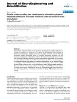
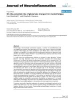
Figure 1
;'LU=
EC
ASTROGLIALSWELLING
ASTRODEPOL
+
UPTAKE
;+
=
EC
'LURELEASE
'LUTRANSMISSION
-ENTALFATIGUEEXHAUSTION
3IGNALTONOISE
IN'LUTRANSMISSION
0RECISIONIN
INFOINTAKE
INFOPROCESSING
ECSPACE
NEURONALEXCITABILITY
,#
.!(4
ASTROGLIAL
METABOLISM
METABOLSUPPORT
LOUND
LIGHT
DEPRESSION
ATTENTION
]
SENS
4.&n
A
),nB
),n
.ORMAL
Schematic drawing of cellular regulation of extracellular glutamate concentrations ([Glu]
ec
) in normal brain function (left), and in the presence of the proinflammatory cytokines tumor necrosis factor-α (TNF-α), interleukin (IL)-1β, and IL-6 (right)Figure 1
Schematic drawing of cellular regulation of extracellular
glutamate concentrations ([Glu]
ec
) in normal brain function
(left), and in the presence of the proinflammatory cytokines
tumor necrosis factor-α (TNF-α), interleukin (IL)-1β, and IL-
6 (right). Possible pathophysiology underlying mental fatigue
at the cellular level is outlined below. To the left:
Two neu-
ronal cell bodies with processes (white) make contact with
each other through a synapse (center). Astrocytic (pink)
processes encapsulate the synapse and cover also the ablumi-
nal side of the blood vessel wall (right). The endothelial cells
covering the luminal (blood) side of the vessel wall and the
astrocytic processes make up the blood brain barrier (BBB).
An oligodendroglial cell (bluish), with its myelin encapsulating
the axon, and a microglial cell (yellow) are seen. The astro-
cytes, with their high-affinity glutamate transporters, are the
main site for keeping [Glu]
ec
low. Even neurons express
glutamate transporters, as do oligodendroglial cells, and
endothelial cells at their abluminal side. To the right:
TNF-α,
IL-1β and IL-6 attenuate astroglial glutamate uptake transport
and disintegrate the BBB, allowing glutamate from the blood
to enter the brain. The overall result is slightly increased
[Glu]
ec
. Tumor necrosis factor-alfa also decreases oligoden-
droglial cell glutamate uptake [78], while microglial glutamate
uptake has been demonstrated to increase (Persson, M.,
Hansson, E., and Rönnbäck, L, to be published), though not
to levels to compensate for the decreased astroglial gluta-
mate uptake capacity. Due to increased [Glu]
ec
, astroglial
swelling is shown. Below:
Hypothetic cellular events underly-
ing mental fatigue. Slightly increased [Glu]
ec
could make the
glutamate neurotransmission less distinct (decrease the sig-
nal-to-noise ratio). At the cellular level, there would be
astroglial swelling, which in turn would decrease the local
extracellular (ec) volume and, as a consequence, lead to fur-
ther increased [Glu]
ec
. Astroglial swelling also depolarizes
the astroglial cell membrane, which further attenuates the
electrogenic glutamate uptake and, in addition, the astroglial
K
+
uptake capacity. As a consequence, even [K
+
]
ec
may rise.
The increased [K
+
]
ec
, together with decreased glutamine
production and reduced glucose uptake concomitant with
the decreased glutamate uptake, could lead to decreased
presynaptic glutamate release and thereby decreased gluta-
mate transmission, which, according to our hypothesis, is
one cellular correlate to mental fatigue/exhaustion. Increased
extracellular glutamate levels in the prefrontal region could
lead to inhibition of the brain stem nuclei locus coeruleus
(LC) and raphe nuclei and thereby inhibit noradrenaline (NA)
and serotonin (5-HT) release in the cerebral cortex resulting
in decreased astroglial metabolism and neuronal metabolic
supply. Increased neuronal excitability may be part of the
loudness and light sensitivity often accompanying the mental
fatigue. In addition, the decrease in noradrenaline and serot-
onin release might be part of decreased attention and the
appearance of depression often accompanying the mental
fatigue.
Journal of Neuroinflammation 2004, 1:22 />Page 6 of 9
(page number not for citation purposes)
The theory also involves the possibility of a disturbed
noradrenaline/serotonin turnover in the cerebral cortex
due to a slight hyperexcitability in the frontal cortex. Inter-
estingly, increased [Glu]
ec
in the prefrontal cortex has
been reported by Bossuet and coworkers [67] in asympto-
matic simian immunodeficiency virus (SIV)mac251-
infected macaques without major brain involvement,
being consistent with our theory at least in this set of ani-
mal experiments. If valid even in humans, a disturbed
noradrenaline/serotonin turnover in the cerebral cortex
could be coupled to the disturbed attention and depres-
sion often occurring in addition to the mental fatigue [see
[71-73]].
Testing of the hypothesis
It is not possible at present to ultimately prove whether or
not the altered neuronal-glial interactions in glutamater-
gic transmission induced by proinflammatory cytokines
could serve as a model to explain cellular mechanisms
underlying mental fatigue. Brain imaging techniques able
to determine and follow [Glu]
ec
and [K
+
]
ec
over time
would be important to use in humans suffering from
mental fatigue. Today, this is not possible for technical
reasons. Instead, we must use experimental systems to
learn about glial cell biology and neuron-glia-neuron sig-
naling and interactions, and thus test specific parts of the
hypothesis. Neuroactive substances produced by, or
altered conditions related to, the production of proin-
flammatory cytokines could be evaluated with regard to
their effects on astroglial support of glutamate transmis-
sion, and especially glutamate transport capacity. The role
of the intact astroglial network in higher brain functions
(cognition and behavior) could be studied in animal
models. Effects of astroglial dysfunction with regard to
glutamate transport capacity would be of special interest.
Even clinical studies with different treatment strategies
could be important in casting some light on the accuracy
of the hypothesis. Of utmost importance in all such stud-
ies would be test batteries making it possible to objectify
and even quantify the degree of mental fatigue.
Why do the symptoms persist in some patients?
Normally, mental fatigue and the associated symptoms
disappear when the brain dysfunction is over. In some
patients, the symptoms persist. We have at present no
explanation for this, but if our hypothesis is correct, there
could be a genetic failure preventing astroglial glutamate
transporters from upregulating. Another explanation for
why the symptoms persist could be that the pathological
stimulation by brain plasticity creates new neuronal net-
works [18,36].
Aspects of treatment
Providing information about mental fatigue, its cause and
the prognosis, is of utmost importance for breaking the
vicious circle, which comes with the risk for secondary
anxiety and depression. Furthermore, it is important for
the patient to imagine and learn how much sensory stim-
ulation they can tolerate prior to feeling too exhausted.
Due to recent results on changes in cell signaling and neu-
ronal plasticity [18,36], it may be important to identify
the symptoms and treat them as early as possible to avoid
formation of new and functionally disturbing neuronal
circuits due to overstimulation of neuronal-glial units. If
our hypothesis is correct, it may be possible to further
improve the symptoms by suppressing the production of
proinflammatory cytokines and, thereby, restoring the
normal astroglial glutamate uptake. In this context, xan-
thine derivatives may be of use [74]. Another substance,
worth considering, may be minocycline, a synthetic tetra-
cycline derivative that has been shown to attenuate micro-
glial activation and, consequently, the production of
proinflammatory cytokines [75]. During recent years sub-
stances, which enhances glutamate uptake have been
identified. Nicergoline [76], different growth factors
including pituitary adenylate cyclase-activating polypep-
tide (PACAP) [77], some low molecular weight factors
[23] as well as metabotropic glutamate agonists [47] have
all been able to stimulate glutamate transport in experi-
mental systems and could be of interest in the pharmaco-
therapy of mental fatigue. Interestingly, even AMPA
receptor modulators have been demonstrated as cognitive
enhancers [10].
List of abbreviations used
ADHD attention deficit hyperactivity disorder
AMPA alpha-amino-3-hydroxy-5-methyl-4-isoxazolepro-
pionate
ATP adenosine triphosphate
BBB blood brain barrier
Ca
2+
calcium
Ec extracellular
GLAST glutamate aspartate transporter
GLT-1 glutamate transporter-1
[Glu]
ec
extracellular glutamate concentration
5-HT 5-hydroxytryptamine
ICD-10 International Classification of Diseases, 10
th
revi-
sion
IL-1/-6 interleukin-1/-6
Journal of Neuroinflammation 2004, 1:22 />Page 7 of 9
(page number not for citation purposes)
K
+
potassium
[K
+
]
ec
extracellular potassium concentration
LC locus coeruleus
LTP long term potential
MS multiple sclerosis
Na
+
sodium
NA noradrenaline
NFκB nuclear transcription factor kappaB
NMDA N-methyl-D-aspartate
NO nitric oxide
PACAP pituitary adenylate cyclase-activating polypeptide
PI3K phosphatidylinositol-3-kinase
PKC protein kinase C
Siv mac simian immunodeficiency virus macaques
TNF-α tumor necrosis factor alpha
Competing interests
The author(s) declare that they have no competing
interests.
Authors' contributions
Equal contributions by both authors.
Acknowledgments
This work, performed in the authors' laboratories, was supported by the
Swedish Research Council (grant No. 21X-13015; 21BL-14586), Swedish
Council for Working Life and Social Research, Edith Jacobsson Foundation,
Rune and Ulla Amlöv Foundation for Neurological and Rheumatological
Research, and John and Brit Wennerström Foundation for Neurological
Research. The authors are grateful to Eva Kraft, Göteborg, Sweden, for
drawing Figure 1.
References
1. Colosimo C, Millefiorini E, Grasso MG, Vinci F, Fiorelli M, Koudriavt-
seva T, Pozzilli C: Fatigue in MS is associated with specific clin-
ical features. Acta Neurol Scand 1995, 92:353-355.
2. Krupp LB, Pollina DA: Mechanisms and management of fatigue
in progressive neurological disorders. Curr Opin Neurol 1996,
9:456-460.
3. Ford H, Trigwell P, Johnson M: The nature of fatigue in multiple
sclerosis. J Psychosom Res 1998, 45:33-38.
4. Schreurs KM, de Ridder DT, Bensing JM: Fatigue in multiple scle-
rosis: reciprocal relationships with physical disabilities and
depression. J Psychosom Res 2002, 53:775-781.
5. Flachenecker P, Bihler I, Weber F, Gottschalk M, Toyka KV, Rieck-
mann P: Cytokine mRNA expression in patients with multiple
sclerosis and fatigue. Mult Scler 2004, 10:165-169.
6. Chaudhuri A, Behan PO: Fatigue and basal ganglia. J Neurol Sci
2000, 179:34-42.
7. American Psychiatric Association: Diagnostic and statistical man-
ual of mental disorders. 4th edition. Washington DC: American
Psychiatric Association; 1994.
8. Lindqvist G, Malmgren H: Organic mental disorders as hypo-
thetical pathogenetic processes. Acta Psychiatr Scand Suppl 1993,
88(Suppl 373):5-17.
9. Kelley KW, Bluthe RM, Dantzer R, Zhou JH, Shen WH, Johnson RW,
Broussard SR: Cytokine-induced sickness behavior. Brain Behav
Immun 2003, 17(Suppl 1):S112-118.
10. Lynch G: AMPA receptor modulators as cognitive enhancers.
Curr Opin Pharmacol 2004, 4:4-11.
11. Vereker E, O'Donnell E, Lynch MA: The inhibitory effect of inter-
leukin-1beta on long-term potentiation is coupled with
increased activity of stress-activated protein kinases. J
Neurosci 2000, 20:6811-6819.
12. Yudkoff M, Nissim I, Daikhin Y, Lin Z-P, Nelson D, Pleasure D, Ere-
cinska M: Brain glutamate metabolism: neuronal-astroglial
relationships. Dev Neurosci 1993, 15:343-350.
13. Huang YH, Bergles DE: Glutamate transporters bring competi-
tion to the synapse. Curr Opin Neurobiol 2004, 14:346-352.
14. Perego C, Vanoni C, Bossi M, Massari S, Basudev H, Longhi R, Pietrini
G: The GLT-1 and GLAST glutamate transporter are
expressed morphologically distrinct astrocytes and regu-
lated by neuronal activity in primary hippocampal
cocultures. J Neurochem 2000, 75:1076-1084.
15. Rothstein JD, Dykes-Hoberg M, Pardo CA, Bristol LA, Jin L, Kuncl R,
Kanai Y, Hediger MA, Wang Y, Schielke JP, Welty DF: Knockout of
glutamate transporters reveals a major role of astroglial
transport in excitotoxicity and clearance of glutamate. Neu-
ron 1996, 16:675-686.
16. Danbolt NC: Glutamate uptake. Prog Neurobiol 2001, 65:1-105.
17. Anderson CM, Swanson RA: Astrocyte glutamate transport:
review of properties, regulation, and physiological functions.
Glia 2000, 32:1-14.
18. Hansson E, Rönnbäck L: Altered neuronal-glial signaling in
glutamatergic transmission as a unifying mechanism in
chronic pain and mental fatigue. Neurochem Res 2004,
29:989-996.
19. Fine SM, Angel RA, Perry SW, Epstein LG, Rothstein JD, Dewhurst S,
Gelbard HA: Tumor necrosis factor alpha inhibits glutamate
uptake by primary human astrocytes. Implications for
pathogenesis of HIV-1 dementia. J Biol Chem 1996,
271:15303-15306.
20. Hu S, Sheng WS, Ehrlich LC, Peterson PK, Chao CC: Cytokine
effects on glutamate uptake by human astrocytes. Neuroimmu-
nomodulation 2000, 7:153-159.
21. Liao SL, Chen CJ: Differential effects of cytokines and redox
potential on glutamate uptake in rat cortical glial cultures.
Neurosci Lett 2001, 299:113-116.
22. Wang Z, Pekarskaya O, Bencheikh M, Chao W, Gelbard HA, Ghor-
pade A, Rothstein JD, Volsky DJ: Reduced expression of gluta-
mate transporter EAAT2 and impaired glutamate transport
in human primary astrocytes exposed to HIV-1 gp120. Virology
2003, 312:60-73.
23. Su Z-z, Leszczyniecka M, Kang D-c, Sarkar D, Chao W, Volsky DJ,
Fisher PB: Insights into glutamate transport regulation in
human astrocytes: Cloning of the promoter for excitatory
amino acid transporter 2 (EAAT2). Proc Natl Acad Sci 2003,
100:1955-1960.
24. Ye ZC, Sontheimer H: Cytokine modulation of glial glutamate
uptake: a possible involvement of nitric oxide. Neuroreport
1996, 7:2181-2185.
25. Chao CC, Hu S, Ehrlich L, Peterson PK: Interleukin-1 and tumor
necrosis factor-alpha synergistically mediate neurotoxicity:
involvement of nitric oxide and of N-methyl-D-aspartate
receptors. Brain Behav Immun 1995, 9:355-365.
26. Minager A, Alexander JS: Blood-brain barrier disruption in mul-
tiple sclerosis. Mult Scler 2003, 9:540-549.
27. Lynch NJ, Willis CL, Nolan CC, Roscher S, Fowler MJ, Weihe E, Ray
DE, Schwaeble WJ: Microglial activation and increased synthe-
Journal of Neuroinflammation 2004, 1:22 />Page 8 of 9
(page number not for citation purposes)
sis of complement component C1q precedes blood-brain
barrier dysfunction in rats. Mol Immunol 2004, 40:709-716.
28. Sibson NR, Blamire AM, Bernades-Silva M, Laurent S, Boutry S, Muller
RN, Styles P, Anthony DC: MRI detection of early endothelial
activation in brain inflammation. Magn Reson Med 2004,
51:248-252.
29. Vandamme W, Braet K, Cabooter L, Leybaert L: Tumour necrosis
factor alpha inhibits purinergic calcium signaling in blood-
brain barrier endothelial cells. J Neurochem 2004, 88:411-421.
30. Hansson E, Rönnbäck L: Astrocytic receptors and second mes-
senger systems. Adv Molec Cell Biol 2004, 31:475-501.
31. Cotrina ML, Lin JH, Alves-Rodrigues A, Liu S, Li J, Azmi-Ghadimi H,
Kang J, Naus CC, Nedergaard M: Connexins regulate calcium
signaling by controlling ATP release. Proc Natl Acad Sci USA
1998, 95:15735-15740.
32. Blomstrand F, Khatibi S, Muyderman H, Hansson E, Olsson T, Rön-
nbäck L: 5-Hydroxytryptamine and glutamate modulate
velocity and extent of intercellular calcium signalling in hip-
pocampal astroglial cells in primary cultures. Neuroscience
1999, 88:1241-1253.
33. Carmignoto G: Reciprocal communication systems between
astrocytes and neurones. Progr Neurobiol 2000, 62:561-581.
34. Muyderman H, Ängehagen M, Sandberg M, Björklund U, Olsson T,
Hansson E, Nilsson M: α
1
-adrenergic modulation of metabo-
tropic glutamate receptor-induced calcium oscillations and
glutamate release. J Biol Chem 2001, 276:46504-46514.
35. Hansson E, Olsson T, Rönnbäck L, eds: On astrocytes and gluta-
mate neurotransmission. Landes Bioscience Company, Austin,
Texas, USA, Springer Verlag, Heidelberg, Germany; 1997.
36. Hansson E, Rönnbäck L: Glial neuronal signaling in the central
nervous system. FASEB J 2003, 17:341-348.
37. Tozaki H, Kanno T, Nomura T, Kondoh T, Kodama N, Saito N,
Aihara H, Nagata T, Matsumoto S, Ohta K, Nagai K, Yajima Y,
Nishizaki T: Role of glial glutamate transporters in facilitatory
action of FK960 on hippocampal neurotransmission. Brain Res
Mol Brain Res 2001, 97:7-12.
38. Butterworth RF: Neurotransmitter dysfunction in hepatic
encephalopathy: new approaches and new findings. Metab
Brain Dis 2001, 16:55-65.
39. Kempski O, Staub F, Jansen M, Baethmann A: Molecular mecha-
nisms of glial swelling in acidosis. Adv Neurol 1990, 52:39-45.
40. Kimelberg HK, Goderie SK, Higman S, Pang S, Waniewski RA: Swell-
ing-induced release of glutamate, aspartate, and taurine
from astrocyte cultures. J Neurosci 1990, 10:1583-1591.
41. Hansson E: Metabotropic glutamate receptor activation
induces astroglial swelling. J Biol Chem 1994, 269:21955-21961.
42. Hansson E, Rönnbäck L: Astrocytes in glutamate
neurotransmission. FASEB J 1995, 9:343-350.
43. Orkand RK: Glial-interstitial fluid exchange. Ann NY Acad Sci
1986, 481:269-272.
44. Walz W: Role of glial cells in regulation of the brain ion
microenvironment. Progr Neurobiol 1989, 33:309-333.
45. Meeks JP, Mennerick S: Selective effects of potassium elevations
on glutamate signaling and action potential conduction in
hippocampus. J Neurosci 2004, 24:197-206.
46. Perez-Otano I, Ehlers MD: Learning from NMDA receptor traf-
ficking: clues to the development and maturation of gluta-
matergic synapses. Neurosignals 2004, 13:175-189.
47. Aronica E, Gorter JA, Ijlst-Keizers H, Rozemuller AJ, Yankaya B, Leen-
stra S, Troost D: Expression and functional role of mGluR3 and
mGluR5 in the human astrocytes and glioma cells: opposite
regulation of glutamate transporter proteins. Eur J Neurosci
2003, 17:2106-2118.
48. Magistretti P, Pellerin L: Regulation by neurotransmitters of
glial energy metabolism. Adv Exp Med Biol 1997, 429:137-143.
49. Loaiza A, Porras OH, Barros LF: Glutamate triggers rapid glu-
cose transport stimulation in astrocytes as evidenced by
real-time confocal microscopy. J Neurosci 2003, 23:7337-7342.
50. Hertz L: Intercellular metabolic compartmentation in the
brain: past, present and future. Neurochem Int 2004, 45:285-296.
51. Sara SJ, Hervé-Minvielle A: Inhibitory influence of frontal cortex
on locus coeruleus neurons. Proc Natl Acad Sci USA 1995,
92:6032-6036.
52. Subbarao KV, Hertz L: Effect of adrenergic agonists on glycog-
enolysis in primary cultures of astrocytes. Brain Res 1990,
536:220-226.
53. Hsu CC, Hsu CS: Effect of isoproterenol on the uptake of
[14C]glucose into glial cells. Neurosci Res 1990, 9:54-58.
54. Kreutzberg GW: Microglia: a sensor for pathological events in
the CNS. Trends Neurosci 1996, 19:312-318.
55. Anisman H, Hayley S, Turrin N, Merali Z: Cytokines as a stressor:
implications for depressive illness. Int J Neuropsychopharmacol
2002, 5:357-373.
56. Hosoi T, Okuma Y, Nomura Y: The mechanisms of immune-to-
brain communication in inflammation as a drug target. Curr
Drug Targets Inflamm Allergy 2002, 1:257-262.
57. Banks WA, Farr SA, Morley JE: Entry of blood-borne cytokines
into the central nervous system: effects on cognitive
processes. Neuroimmunomodulation 2003, 10:319-327.
58. Krueger JM, Majde JA: Humoral links between sleep and the
immune system: research issues. Ann NY Acad Sci 2003,
992:9-20.
59. Dinkel K, MacPherson A, Sapolsky RM: Novel glucocorticoid
effects on acute inflammation in the CNS. J Neurochem 2003,
84:705-716.
60. Roelcke U, Kappos L, Lechner-Scott J, Brunnschweiler H, Huber S,
Ammann W, Plohmann A, Dellas S, Maguire RP, Missimer J, Radu EW,
Steck A, Leenders KL: Reduced glucose metabolism in the fron-
tal cortex and basal ganglia of multiple sclerosis patients
with fatigue: a 18F-fluorodeoxyglucose positron emission
tomography study. Neurology 1997, 48:1566-1571.
61. Kuratsune H, Yamaguti K, Lindh G, Evengård B, Hagberg G, Mat-
sumura K, Iwase M, Onoe H, Takahashi M, Machii T, Kanakura Y,
Kitani T, Langstrom B, Watanabe Y: Brain regions involved in
fatigue sensation: reduced acetylcarnitine uptake into the
brain. NeuroImage 2002, 17:1256-1265.
62. Carrey N, MacMaster FP, Fogel J, Sparkes S, Waschbusch D, Sullivan
S, Schmidt M: Metabolite changes resulting from treatment in
children with ADHD: a 1H-MRS study. Clin Neuropharmacol
2003, 26:218-221.
63. Todd RD, Botteron KN: Is attention-deficit/hyperactivity disor-
der an energy deficiency syndrome? Biol Psychiatry 2001,
50:151-158.
64. Guerra-Romero L, Tureen JH, Fournier MA, Makrides V, Tauber MG:
Amino acids in cerebrospinal and brain interstitial fluid in
experimental pneumococcal meningitis. Pediatr Res 1993,
33:510-513.
65. Perry VL, Young RS, Aquila WJ, During MJ: Effect of experimental
Escherichia coli meningitis on concentrations of excitatory
and inhibitory amino acids in the rabbit brain: in vivo micro-
dialysis study. Pediatr Res 1993, 34:187-191.
66. Stover JF, Pleines UE, Morganti-Kossmann MC, Kossmann T, Lowit-
zsch K, Kempski OS: Neurotransmitters in cerebrospinal fluid
reflect pathological activity. Eur J Clin Invest 1997, 27:1038-1043.
67. Bossuet C, Vaufrey F, Condé F, Chrétien F, Pichon J, Hantraye P, Le
Grand R, Dormont D, Gras G: Up-regulation of glutamate con-
centration in the putamen and in the prefrontal cortex of
asymptomatic SIVmac251-infected macaques without
major brain involvement. J Neurochem 2004, 88:928-938.
68. Liang Z, Valla J, Sefidvash-Hockley S, Rogers J, Li R: Effects of estro-
gen treatment on glutamate uptake in cultured human
astrocytes derived from cortex of Ahlzheimer's disease
patients. J Neurochem 2002, 80:807-814.
69. Arundine M, Tymianski M: Molecular mechanisms of glutamate-
dependent neurodegeneration in ischemia and traumatic
brain injury. Cell Mol Life Sci 2004, 61:657-668.
70. Santhakumar V, Voipio J, Kaila K, Soltesz I: Post-traumatic hyper-
excitability is not caused by impaired buffering of extracellu-
lar potassium. J Neurosci 2003, 23:5865-5876.
71. Kratochvil CJ, Vaughan BS, Harrington MJ, Burke WJ: Atomoxetine:
a selective noradrenaline reuptake inhibitor for the treat-
ment of attention-deficit/hyperactivity disorder. Expert Opin
Pharmacother 2003, 4:1165-1174.
72. Abrams JK, Johnson PL, Hollis JH, Lowry CA: Anatomic and func-
tional topography of the dorsal raphe nucleus. Ann N Y Acad Sci
2004, 1018:46-57.
73. Marien MR, Colpaert FC, Rosenquist AC: Noradrenergic mecha-
nisms in neurodegenerative diseases: a theory. Brain Res Rev
2004, 45:38-78.
74. Schubert P, Rudolphi K: Interfering with the pathologic activa-
tion of microglial cells and astrocytes in dementia. Ahlzheimer
Dis Assoc Disord 1998, 12(suppl 2):S21-S28.
Publish with Bio Med Central and every
scientist can read your work free of charge
"BioMed Central will be the most significant development for
disseminating the results of biomedical research in our lifetime."
Sir Paul Nurse, Cancer Research UK
Your research papers will be:
available free of charge to the entire biomedical community
peer reviewed and published immediately upon acceptance
cited in PubMed and archived on PubMed Central
yours — you keep the copyright
Submit your manuscript here:
/>BioMedcentral
Journal of Neuroinflammation 2004, 1:22 />Page 9 of 9
(page number not for citation purposes)
75. Tikka T, Fiebich BL, Goldsteins G, Keinanen R, Koistinaho J: Minoc-
ycline, a tertracycline derivative, is neuroprotective against
excitotoxicity by inhibiting activation and proliferation of
microglia. J Neurosci 2001, 21:2580-2588.
76. Nishida A, Iwata H, Kudo Y, Kobayashi T, Matsuoka Y, Kanai Y, Endou
H: Nicergoline enhances glutamate uptake via glutamate
transporters in rat cortical synaptosomes. Biol Pharm Bull 2004,
27:817-820.
77. Figiel M, Maucher T, Rozyczka J, Bayatti N, Engele J: Regulation of
glial glutamate transporter expression by growth factors.
Exp Neurol 2003, 183:124-135.
78. Pitt D, Nagelmeier IE, Wilson HC, Raine CS: Glutamate uptake by
oligodendrocytes: Implications for excitotoxicity in multiple
sclerosis. Neurology 2003, 61:1113-1120.