báo cáo hóa học: " Primary glia expressing the G93A-SOD1 mutation present a neuroinflammatory phenotype and provide a cellular system for studies of glial inflammation" potx
Bạn đang xem bản rút gọn của tài liệu. Xem và tải ngay bản đầy đủ của tài liệu tại đây (672.32 KB, 9 trang )
BioMed Central
Page 1 of 9
(page number not for citation purposes)
Journal of Neuroinflammation
Open Access
Research
Primary glia expressing the G93A-SOD1 mutation present a
neuroinflammatory phenotype and provide a cellular system for
studies of glial inflammation
Kenneth Hensley*
1,2
, Haitham Abdel-Moaty
1,3
, Jerrod Hunter
1
,
Molina Mhatre
1
, Shenyun Mou
1
, Kim Nguyen
1
, Tamara Potapova
1
,
Quentin N Pye
1
, Min Qi
1
, Heather Rice
1
, Charles Stewart
1
,
Katharine Stroukoff
1
and Melinda West
1
Address:
1
Free Radical Biology and Aging Research Program, Oklahoma Medical Research Foundation (OMRF), 825 NE 13th Street, Oklahoma
City, OK, 73104, USA ,
2
Department of Cell Biology, University of Oklahoma Health Science Center (OUHSC), Oklahoma City, OK, 73104, USA
and
3
University of Oklahoma College of Engineering, Bioengineering Program, Norman, OK, USA
Email: Kenneth Hensley* - ; Haitham Abdel-Moaty - ;
Jerrod Hunter - ; Molina Mhatre - ; Shenyun Mou - shenyun-
; Kim Nguyen - ; Tamara Potapova - ;
Quentin N Pye - ; Min Qi - ; Heather Rice - ;
Charles Stewart - ; Katharine Stroukoff - ; Melinda West - melinda-
* Corresponding author
Abstract
Detailed study of glial inflammation has been hindered by lack of cell culture systems that
spontaneously demonstrate the "neuroinflammatory phenotype". Mice expressing a glycine →
alanine substitution in cytosolic Cu, Zn-superoxide dismutase (G93A-SOD1) associated with
familial amyotrophic lateral sclerosis (ALS) demonstrate age-dependent neuroinflammation
associated with broad-spectrum cytokine, eicosanoid and oxidant production. In order to more
precisely study the cellular mechanisms underlying glial activation in the G93A-SOD1 mouse,
primary astrocytes were cultured from 7 day mouse neonates. At this age, G93A-SOD1 mice
demonstrated no in vivo hallmarks of neuroinflammation. Nonetheless astrocytes cultured from
G93A-SOD1 (but not wild-type human SOD1-expressing) transgenic mouse pups demonstrated a
significant elevation in either the basal or the tumor necrosis alpha (TNFα)-stimulated levels of
proinflammatory eicosanoids prostaglandin E
2
(PGE
2
) and leukotriene B
4
(LTB
4
); inducible nitric
oxide synthase (iNOS) and •NO (indexed by nitrite release into the culture medium); and protein
carbonyl products. Specific cytokine- and TNFα death-receptor-associated components were
similarly upregulated in cultured G93A-SOD1 cells as assessed by multiprobe ribonuclease
protection assays (RPAs) for their mRNA transcripts. Thus, endogenous glial expression of G93A-
SOD1 produces a metastable condition in which glia are more prone to enter an activated
neuroinflammatory state associated with broad-spectrum increased production of paracrine-acting
substances. These findings support a role for active glial involvement in ALS and may provide a
useful cell culture tool for the study of glial inflammation.
Published: 25 January 2006
Journal of Neuroinflammation 2006, 3:2 doi:10.1186/1742-2094-3-2
Received: 01 September 2005
Accepted: 25 January 2006
This article is available from: />© 2006 Hensley et al; licensee BioMed Central Ltd.
This is an Open Access article distributed under the terms of the Creative Commons Attribution License ( />),
which permits unrestricted use, distribution, and reproduction in any medium, provided the original work is properly cited.
Journal of Neuroinflammation 2006, 3:2 />Page 2 of 9
(page number not for citation purposes)
Introduction
Although the proximal cause of paralysis in amyotrophic
lateral sclerosis (ALS) is the death of motor neurons, it is
becoming widely accepted that motor neuron death in
ALS is not cell autonomous but depends upon active and
passive roles for ambient glial cells. The neuron-cell
autonomy of ALS pathogenesis has been strongly ques-
tioned by a number of studies over the past several years.
In work published during 2001, Rouleau et al. created a
strain of transgenic mice that express mutant SOD1 specif-
ically in neurons. These mice display no frank pathology
even at 1.5 years of age [1]. Caroni's group subsequently
reported similar findings [2]. Selective expression of
mutant SOD1 only in astroglia, causes a type of astroglio-
sis but fails to produce motor neuron disease [3] in the
absence of simultaneous mutant SOD1 expression in neu-
rons. Nonetheless, Cleveland and colleagues recently
showed that the rate of disease progression in mutant
SOD1 chimeric mice depends on the extraneuronal
expression of mutant SOD1 [4]. The survival of chimeric
mice was dependent upon mutant SOD1 expression in
neurons, but also highly dependent on the number of
ambient mutant SOD1-expressing non-neuronal cells.
These studies provide strong incentive to consider glial
involvement in ALS.
With the advent of transgenic mouse models for familial
amyotrophic lateral sclerosis (FALS), it has become more
possible to study inflammatory and autoimmune features
of the disease at distinct time points during the progres-
sion of the illness. Using the G93A-SOD1 mutant mouse
model for ALS, Gurney et al. reported dramatically
increased numbers of MHC-II
+
microglia and concomi-
tant astroglial activation beginning prior to onset of paral-
ysis and increasing during the paralytic phase [5]. Several
recent studies have built upon these early studies by doc-
umenting reproducible, age-dependent elaboration of
pro-inflammatory cytokines during the onset and progres-
sion phases of disease in the G93A-SOD1 mouse [6-11].
Tumor necrosis factor-α (TNFα) and its principle receptor
TNF-RI are particularly elevated at pre- and post-sympto-
matic stages of disease [6-9], suggesting a rationale for the
application of this cytokine in cell culture studies of ALS-
linked glial activation. The time-course of cytokine up-reg-
ulation closely mirrors the time-course of protein oxida-
tive damage, and begins approximately two weeks prior to
the point of actual motor neuron death [7,12]. In addition
to cytokines and reactive oxygen species, eicosanoids such
as PGE
2
are elevated and pharmacological antagonism of
PGE
2
-synthesizing inducible cyclooxygenase (COX-II)
improves prognosis in the murine model [13]. Likewise
arachidonic acid 5-lipoxygenase (5LOX) is elevated in
G93A-SOD1 spinal cords and the 5LOX antagonist nordi-
hydroguaiaretic acid (NDGA) slows disease progression
in the ALS mouse [14]. These findings suggest a robust,
multi-faceted neuroinflammatory response, antagonism
of which may slow the progression of ALS.
In order to better understand the contributions of astro-
glia to neuroinflammation in the ALS context, and to cre-
ate a tool for the study of neuroinflammatory signal
transduction, primary cortical astrocytes were cultured
from neonatal mice bearing G93A-SOD1 mutations. The
cells were characterized for their ability to synthesize sali-
ent biomolecules including cytokines, eicosanoids, and
reactive oxygen species. G93A-SOD1 transgenic astroglia
were found to synthesize higher-than-normal levels of
TNFα, COX-II, 5LOX, and PGE
2
even in the absence of
deliberate stimulation. When challenged with TNFα
alone or in combination with IFNγ, selective subsets of
cytokines were further induced. Leukotriene B
4
, nitric
oxide and protein oxidation increased more markedly in
G93A-SOD1 glia challenged with TNFα or interferon-γ
IFNγ than in similarly treated nontransgenic cells. Expres-
sion of high copy numbers of wild-type human SOD1 had
no effect or slightly diminished the inflammatory indices.
These findings suggest that SOD1 mutations fundamen-
tally alter the phenotype of astrocytes, placing the cells in
a metastable condition that is hypersensitive to certain
types of ligand-induced activation.
Materials and methods
Animals
Mice expressing high copy numbers of human mutant
G93A-SOD1 were obtained from Jackson Laboratories
(Bar Harbor ME; strain designation B6SJL-Tg(SOD1
G93A)1Gur/J; [15-17]. In some control experiments mice
were used that express equivalent protein levels of wild-
type human SOD1 (B6SJL-TgN-(SOD1 G93A)-2Gur; Jack-
son Laboratories). Transgenic mice were maintained in
the hemizygous state by mating G93A males with B6SJL-
TGN females. All animal procedures were approved by the
OMRF Institutional Animal Care and Use Committee
(IACUC).
Astrocyte culture
Primary mouse neocortical astrocytes were cultured by
slight modifications of previously described methods [18]
from G93A-SOD1 mice, matched nontransgenic litterma-
tes, or wildtype-human SOD1 expressing mice. In all cases
the cortex was used to maximize astroglial yield. Briefly,
the neocortex was removed from 7 day old pups under
aseptic conditions and large blood vessels carefully
removed. Tissue was rinsed and triturated in cold Ca
++
/
Mg
+
free HBSS buffer, then centrifuged at 300 × g for five
minutes. The resulting pellet was resuspended in 30 mL of
50% Dulbecco's Modified Essential Medium (DMEM)
and 50% F12 media containing 10% heat-inactivated fetal
bovine serum, 1% glutamine, and 1% streptomycin and
penicillin. The 30 mL suspension was placed into a 75
Journal of Neuroinflammation 2006, 3:2 />Page 3 of 9
(page number not for citation purposes)
cm
2
tissue culture flask. Cells obtained from individual
mouse pups were plated in separate flasks. Media was
replenished 7 days following the initial plating. Between
6–10 days after initial plating, glia became fully confluent
at which time they were subcultured at a 1:4 dilution into
6- or 24-well plates. In all cases, unless otherwise speci-
fied, astroglial cultures were not futher subcultured. Fur-
thermore each experiment compared genotype-specific
cell responses between parallel cultures of identical pas-
sage number, obtained from paired neonatal pups (litter-
mates in the case of G93A-SOD1 +/- and -/- mice). Paired
cultures were prepared on the same day and subject to
medium changes and manipulations in exactly parallel
fashion so as to avoid artifacts arising from differences in
medium self-conditioning, clonal selection, or other
uncontrolled variables. Specific experiments were con-
ducted deliberately on astroglia that had been subcultured
intentionally for up to 5 passages. Statistically significant
genotype-specific differences in cytokine-stimulated
nitrite production and other variables, as indicated, were
maintained at least to the fifth passage in these experi-
ments. Purity of cultures was routinely assessed by immu-
nocytochemistry using fluorescein-conjugated anti-OX-
42 antibody (Chemicon, Temecula CA USA) to identify
microglia, and rhodamine-conjugated rabbit anti-glial
fibrillary protein (GFAP) antibody (Chemicon) to iden-
tify astroctyes.
Cytokine treatments
In all experiments cell cultures were stimulated at full con-
fluence (110,000 cells/cm
2
). Cells were treated with
recombinant murine TNFα and/or interferon gamma
(IFNγ) (BD Pharmingen, San Diego CA USA) as indicated
in specific experiments. Cytokines were predissolved in
4% fatty acid-free bovine serum albumin (BSA) in 0.9%
saline at 100-fold working concentration. Vehicle control
treatments used 4% BSA: saline only. Because TNFα activ-
ity varied somewhat from lot to lot, each lot was pre-
tested to determine the concentration of applied cytokine
that would yield a measurable effect within the linear
range of cell response. For cytokine treatments, culture
medium was replaced with fresh medium. After 2 hours
equilibration, cytokines or vehicle were diluted 1:100 into
cell culture medium. Viability was routinely assessed by
means of tetrazolium reduction assays (Aqueous
OneStep
®
, Promega, Gaithersburg MD USA).
Ribonuclease protection assays
Multiprobe ribonuclease protection assays (RPAs) were
performed as described [14,7,8]. Cells or brain cortices
were lysed in TRIzol™ mRNA isolation reagent (Life Tech-
nologies, Gaithersburg MD). Total RNA was quantified
spectrophotometrically at 260 nm. Panels of mRNA were
detected using commercial RPA kits (Riboquant™,
Pharmingen, San Diego, CA). Radiolabeled probes were
synthesized from DNA templates containing a T7 RNA
polymerase promoter (Pharmingen). Templates were
transcribed in the presence of 100 µCi [γ-
32
P]UTP to yield
radioactive probes of defined size for each mRNA. Probes
were hybridized with 5–10 µg total RNA, then treated
with RNAse A and T1 to digest single-stranded RNA. Intact
double-stranded RNA hybrids were resolved on 5% poly-
acrylamide/8 M urea gels. Dried gels were visualized using
a phosphorimager (Molecular Dynamics, Sunnyvale CA)
and bands quantified using instrument-resident densit-
ometry software (ImageQuant™, Molecular Dynamics).
Within each sample, the density of each apoptosis-associ-
ated mRNA band was normalized to the sum of the L32 +
GAPDH bands.
Eicosanoid assays
PGE
2
and LTB
4
were measured in cell culture medium
using commercially available enzyme linked immuno-
sorbent assays (ELISAs; Cayman Chemical, San Diego CA
USA).
Nitrite assay
Cell culture medium was assayed for NO
2
-
by the Griess
assay as described [8]. Samples were mixed 1:1 with a mix-
ture of equal portions sulfanilamide and napthylethylen-
ediamine reagents (LabChem, Gaithersburg MD USA).
External standards were prepared in fresh cell culture
medium. The diazo product was measured spectrophoto-
metrically at 560 nm.
Western blots
Cells were lysed in 10 mM sodium acetate pH 6.5 contain-
ing 0.1% triton X-100, 100 µM sodium orthovanadate
and 1:1000 diluted mammalian protease inhibitor cock-
tail (Sigma Chemical, St. Louis MO USA). After centrifu-
gation, samples were assayed for total protein by Lowry
assay [19], adjusted to constant concentration, mixed 1:1
with loading dye (50% glycerol, 10% Tris, 0.01%
bromophenol blue) and electrophoresed across 4–20%
gradient polyacrylamide gels. Protein concentration per
well of confluent, matched cultures did not differ signifi-
cantly amongst the genotypes (data not illustrated). Sam-
ples were electroblotted onto polyvinylidene difluoride
(PVDF) membranes, blocked overnight in 4% BSA then
probed with one of the following antibodies at 1:2000
dilution: rabbit polyclonal anti-iNOS (Chemicon); rabbit
polyclonal anti-COX-II (Chemicon); mouse monoclonal
anti-5LOX (Transduction Laboratories, Lexington KY
USA); or mouse monoclonal anti-actin clone AC15
(Sigma Chemical) followed by the appropriate peroxi-
dase-conjugated secondary antibody. Blots were devel-
oped using enhanced chemiluminescence (Amersham
Biosciences, Buckinghamshire UK).
Journal of Neuroinflammation 2006, 3:2 />Page 4 of 9
(page number not for citation purposes)
Protein carbonylation
Protein carbonylation was measured using methods simi-
lar to those previously published [7]. Cells were lysed in
20 mM 2-(N-morpholino)-ethanesulfonate (MES) buffer
pH 5.5 containing 0.1% triton X-100, 5 mM biotin-LC-
hydrazide (Pierce Biotechnology, Rockford IL USA) and
100 µM butylated hydroxytoluene (BHT). Samples were
incubated 2 H at 37°C, centrifuged, and supernatant was
electrophoresed and blotted as describe above. Blots were
blocked overnight in 4% BSA, probed with 50 ng/mL
streptavidin-conjugated horseradish peroxidase (Pierce)
and visualized by chemiluminescence. Control experi-
ments omitted the biotin-LC-hydrazide reagent or substi-
tuted biotin for biotin-hydrazide; in neither case was any
labeling observed.
Statistics
Data were evaluated by analysis of variance (ANOVA) fol-
lowed by post-hoc comparisons to assess genotype-specific
differences in particular endpoints amongst nontrans-
genic, G93A-SOD1
+
and human wildtype-expressing glial
cell cultures. All analyses were conducted using GraphPad
Prism
®
software (GraphPad, San Diege CA USA).
Results
When fresh cortical tissue was excised from G93A-SOD1
and non-transgenic neonatal pups at 7 days of age, no
genotype-dependent differences were observed with
respect to PGE
2
concentration as measured by ELISA
(NonTg = 281 ± 200 pg/mg protein; G93A-SOD1 = 336 ±
134 pg/mg protein; N = 6/group); COX-II expression or
5LOX expression as measured by immunoblot (not
shown); or cytokine expression patterns assessed by RPAs
(data not shown). Nonetheless cultured astroglia demon-
strated clear genotype-dependent differences in these sev-
eral parameters, as described below.
Primary astrocyte cultures from G93A-SOD1 or nontrans-
genic mice were almost exclusively astrocytic based on
immunocytochemical staining with anti-glial fibrillary
acidic protein (GFAP) (not illustrated). In initial cultures,
microglia were occasionally evident; however, these cells
were not retained throughout multiple serial passages.
Both nontransgenic and transgenic cells displayed typical
morphological attributes of cultured astrocytes. G93A-
SOD1 cells tended to be slightly more elongated than
nontransgenic cells though no formal attempt was made
to quantify or statistically analyze this feature. There was
no discernible difference in rates of tetrazolium reduction
amongst the genotypes, under any of the conditions
tested. Viability of cells treated with maximum concentra-
tions of stimulatory cytokines (40 ng/mL TNFα plus 50 U/
mL IFNγ) did not differ significantly from that of
untreated cells, based on tetrazolium reduction assays, at
time points up to 48 hours post-stimulation.
Specific cytokine expressioin differences occur in G93A-
SOD1 astrocytes
Multiprobe RPA methods were used to assess genotype-
dependent differences in cytokine-stimulated cytokine
expression between nontransgenic and G93A-SOD1 glia.
Medium was replaced and cells were stimulated for 4
hours, which was found to represent the approximate
peak for TNFα-stimulated cytokine mRNA transcription.
A number of observations were evident in these experi-
ments. First, G93A-SOD1 cells demonstrated lower levels
of "housekeeping" messages L32 and GAPDH than did
non-transgenic, matched cell cultures (Fig. 1). This may
reflect a fundamental alteration in mRNA distribution
with an over-expression of "non-housekeeping" genes
such that the ratio of L32 and GAPDH to total mRNA, is
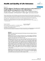
Representative multiprobe ribonuclease protection assay results indicating selective increase in certain cytokine mRNAs in G93A-SOD1 astrocyte cultures, either in the absence of deliberate stimulation (basal condition) or after 4 hours treatment with recombinant murine IFNγ (50 U/mL), TNFα (40 ng/mL) or bothFigure 1
Representative multiprobe ribonuclease protection assay
results indicating selective increase in certain cytokine
mRNAs in G93A-SOD1 astrocyte cultures, either in the
absence of deliberate stimulation (basal condition) or after 4
hours treatment with recombinant murine IFNγ (50 U/mL),
TNFα (40 ng/mL) or both. Each lane represents pooled
mRNA from at 6 wells of cells.
Journal of Neuroinflammation 2006, 3:2 />Page 5 of 9
(page number not for citation purposes)
fundamentally skewed in G93A-SOD1+ gial cultures.
Thus, when equivalent amounts of message (based on UV
absorption of RNA extracts) was loaded onto polyacryla-
mide gells, the cytokine: housekeeping message ratio
could be noticeably affected.
With this possible qualification, G93A-SOD1 cells were
found to (1) express more TNFα message in the basal state
than did non-transgenic cells and (2) hyper-express TNFα
message after either IFNγ or TNFα challenge (Fig. 1).
TNFα stimulation produced, on average, 4-fold greater
increase in TNFα message when G93A-SOD1 glia were
stimulated than when nontransgenic cells were stimu-
lated (% change in TNFα bands, without normalization to
L32 + GAPDH = 1132 ± 618% in G93A-SOD1 cells vs. 242
± 120% in nontransgenic cells, respectively, N = 5 experi-
ments). TRAIL (TNF-Related Apoptosis-Inducing Ligand)
was likewise very markedly upregulated in G93A-SOD1
cells following cytokine challenge, relative to nontrans-
genic cells (Fig. 1). Several other pro-inflammatory
cytokines or apoptosis-related transcripts were differen-
tially regulated in the G93A-SOD1 cells (Table 1). Numer-
ous other transcripts did not differ notably in their levels
as a function of genotype or stimulus (Fig. 1, Table 1). IL-
6, which has some neuroprotective functions [20], tended
to decrease in G93A-SOD1 cultures.
Eicosanoid synthesis is increased in G93A-SOD1-
expressing glia
Confluent, primary glia from G93A-SOD1 or nontrans-
genic neonatal mice, or from mice expressing high copy
numbers of wildtype human SOD1 (wt-hSOD1) were
stimulated with IFNγ, TNFα, or both for 24 hours and
medium was assayed by ELISA for LTB
4
and PGE
2
. As illus-
trated in Figure 2, PGE
2
production was elevated three-
fold in the G93A-SOD1 cells even in the absence of exog-
enous cytokines. The elevated production of PGE
2
per-
sisted through at least five serial passages of the astroglial
cultures. COX-II protein was likewise increased in G93A-
SOD1 glia. The G93A-SOD1 cells were resistant to addi-
tional cytokine-induced prostaglandin synthesis; how-
ever, the amount of PGE
2
produced by unstimulated
G93A-SOD1 glia was 2-fold greater than the amount that
could be stimulated from nontransgenic cells by com-
bined IFNγ plus TNFα (Fig. 2). A somewhat different pat-
tern was observed for LTB
4
. Synthesis of this 5LOX
metabolite was also elevated in G93A-SOD1 glia under
basal conditions. However, LTB
4
was synergistically
Table 1: Cytokine and apoptosis-associated message levels in nontransgenic and G93A-SOD1 astrocyte cultures in the basal
(unstimulated) condition and 4 hours following stimulation with 50 U/mL IFNγ, 20 ng/mL TNFα or a combination of both cytokines.
Data represent pooled samples from 6 wells in a typical experiment (see Fig. 1). Band intensities were normalized to the sum of
GAPDH + L32 message levels prior to comparison between genotypes.
mRNA Unstimulated G93A-SOD1 as %
of unstimulated nonTg
Stimulated message level as % of unstimulated level, within genotypes
NonTg stimulated with: G93A-SOD1 stimulated with:
IFNγ TNFα IFNγ + TNFα IFNγ TNFα IFNγ + TNFα
Caspase 8 237 108 98 108 181 177 233
FADD 168 90 114 126 106 114 100
FAF 94 94 87 70 101 100 92
FAP 129 136 103 151 149 128 220
FAS 145 186 622 757 868 777 1392
IL1α 329 136 252 103 100 203 110
IL1β 644 152 1740 1101 161 724 223
IL1-RA 147 157 114 159 180 163 373
IL6 34 148 768 991 1461 1228 2755
IL18 307 221 104 92 199 205 194
IL12p35 341 141 239 263 108 71 103
IL12p40 325 121 137 165 197 94 1402
IFNγ 158 121 480 798 534 605 1792
MIF 70 83 129 133 95 110 108
RIP 159 130 152 225 230 161 258
TNFα 428 700 512 1511 2302 1669 3352
TNF-RI 166 132 99 131 180 112 176
TGFβ1 353 99 122 145 94 116 115
TGFβ3 155 90 96 87 94 132 148
TRADD 182 111 101 125 125 105 115
TRAIL 284 300 458 749 1307 734 1644
Journal of Neuroinflammation 2006, 3:2 />Page 6 of 9
(page number not for citation purposes)
inducible by IFNγ + TNFα in both genotypes (Fig. 2). The
relative increase in LTB
4
during cytokine stimulation was
similar between the genotypes but LTB
4
remained at least
2-fold elevated in G93A-SOD1 cultures relative to non-
transgenic cultures, under all experimental conditions.
These data begin to suggest fundamental perturbations to
glial arachidonic acid metabolism as a function of the
mutant SOD1 transgene.
iNOS expression and nitric oxide synthesis is increased in
G93A-SOD1 glia
Primary glia cultured from 7 day old G93A-SOD1 or non-
transgenic pups were treated with increasing concentra-
tions of TNFα plus or minus IFNγ. As an indicator of nitric
oxide production, nitrite was measured in the cell culture
medium 48 hours later. Measurable NO
2
-
formation
required IFNγ in both non-transgenic and G93A-SOD1
astrocytes, and abundant nitrite production was only
observed 48 hours after cytokine stimulation (Fig. 3).
Under combined IFNγ and TNFα stimulation, TNFα-stim-
ulated G93A-SOD1 glia produced significantly more NO
2
-
than did nontransgenic cells (Fig. 3). The G93A-SOD1
enhanced NO
2
-
production was maintained through at
least 5 serial passages of cell cultures (not illustrated). Ele-
vated levels of iNOS protein could be detected in G93A-
SOD1 astrocytes relative to nontransgenic cells (Fig. 3).
G93A-SOD1 astrocytes experience exacerbated protein
carbonylation under cytokine challenge
Protein carbonyl accumulation is a well-accepted indica-
tor of oxidative damage [7,20]. Recently biotin hydrazide
and similar reagents have been adapted to monitor carbo-
nylation in cell and tissue lysates [7]. The use of biotin
hydrazide allows the sensitive detection of oxidized pro-
teins by means of streptavidin conjugates, without resort-
ing to antibody methods that are often hindered by low
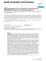
Comparison of basal and cytokine-stimulated PGE
2
and LTB
4
production by nontransgenic primary mouse astrocytes, G93A-SOD1 mouse astrocytes, or wild type human SOD1-expressing mouse astrocytesFigure 2
Comparison of basal and cytokine-stimulated PGE
2
and LTB
4
production by nontransgenic primary mouse astrocytes, G93A-
SOD1 mouse astrocytes, or wild type human SOD1-expressing mouse astrocytes. Insets show western blot analysis of basal
COX-II and 5-LOX protein expression. Data bars indicate mean ± SD of 6 wells of cells from a typical experiment. p < 0.05
overall by ANOVA; * indicates specific difference between nontransgenic and G93A-SOD1 cultures assessed by Bonferroni
post-hoc tests.
Journal of Neuroinflammation 2006, 3:2 />Page 7 of 9
(page number not for citation purposes)
signal: noise and nonspecific binding artifacts. For these
reasons, experiments were undertaken to assess genotype-
related differences in glial protein carbonylation through
means of the biotin labeling technique.
Cells were stimulated with 40 ng/mL TNFα plus 50 U/mL
IFNγ, or vehicle for 48 hours and lysed for carbonyl assess-
ment. The 48 hours timepoint was chosen as duration of
treatment sufficient to induce obvious increases in protein
carbonylation within nontransgenic astrocytes. The inclu-
sion of IFNγ was also necessary to insure this effect. As
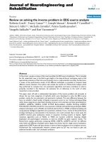
shown in Fig. 4, G93A-SOD1 glia contained approxi-
mately 2-fold greater levels of protein carbonyl than did
nontransgenic cells, in the absence of an applied cytokine
challenge. After exposure to TNFα + IFNγ, protein carbo-
nyl levels increased in both nontransgenic and G93A-
SOD1 cells. Whereas the cytokine-stimulated increase in
carbonylation was approximately 2-fold in nontransgenic
cells, it was approximately 150-fold in G93A-SOD1 astro-
glia (Fig. 4; estimates for relative levels of carbonylation
were made by repeated serial dilution of the labeled sam-
ples). Curiously, no major protein carbonylation band
assignable to SOD1 was found in any G93A-SOD1 astro-
cyte lysates whereas a major carbonylated protein identi-
fiable as SOD1 was previously demonstrated in spinal
cord extracts from symptomatic G93A-SOD1 mice [7,12].
Discussion
The role of astrocytes in paracrine inflammatory networks
has become increasingly appreciated in recent years. In
this capacity astrocytes likely respond to neural damage,
infection, or tumorigenisis in such a way as to modulate
necessary innate immune responses. Contrastingly,
chronic unremitting neuroinflammation has been widely
implicated in diverse neurodegenerative diseases. In
murine models of ALS, neuroinflammation is robust as
indicated by broad-spectrum cytokine upregulation plus
oxidative stress, astroglial morphological changes, and
microglial proliferation [6-8,14,9-12,5]. Aberrations in
eicosanoid production, largely mediated by inducible
cyclooxygenase-II (COX-II) [13] but perhaps also by
5LOX [14] represent another major component of the
neuroinflammatory phenotype that might be amenable
to therapeutic intervention. Thus far it has been difficult
to separate the cell type-dependent contributions to the
neuroinflammatory phenomenon. This limitation has
prevented detailed molecular dissection of relevant path-
ways that are perturbed by the insertion of mutant SOD1
transgenes, and has slowed the development of new ther-
apeutic modalities. The ability to recapture certain aspects
of neuroinflammation in primary astrocyte cultures will
likely facilitate detailed studies of signal transduction
pathways that are sensitive to mutant SOD1.
The findings from the present study corroborate recent
reports of cytokine hyper-expression in the CNS of mutant
Basal and cytokine-stimulated protein carbonylation is increased in G93A-SOD1 astrocyte culturesFigure 4
Basal and cytokine-stimulated protein carbonylation is
increased in G93A-SOD1 astrocyte cultures. Cells were
stimulated for 48 hours with 50 U/mL IFNγ plus 40 ng/mL
TNFα, lysed in the presence or absence of biotin-LC-
hydrazide (+ or - label as indicated), blotted onto a PVDF
membrane and probed with streptavidin-conjugated horse-
radish peroxidase.
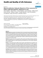
iNOS protein expression and NO
2
-
formation in cultured nontransgenic or G93A-SOD1+ astrocytes in the basal state and after 48 hours stimulation with recombinant murine TNFα (40 ng/mL) plus IFNγ (50 U/mL)Figure 3
iNOS protein expression and NO
2
-
formation in cultured
nontransgenic or G93A-SOD1+ astrocytes in the basal state
and after 48 hours stimulation with recombinant murine
TNFα (40 ng/mL) plus IFNγ (50 U/mL). Bars represent mean
± SD from 6 wells of cells in a typical experiment; * p < 0.05
for stimulated G93A-SOD1
+
cells relative to correspondingly
treated nontransgenic cells, by two-tailed t-test.
Journal of Neuroinflammation 2006, 3:2 />Page 8 of 9
(page number not for citation purposes)
SOD1 mice preceding motor neuron death [6-8]. In par-
ticular the new data suggest that astrocytes cultured from
7 day old neonates reside in a metastable state that is
exquisitely prone to activation, resulting in elevated
expression of specific cytokines, upregulation of eicosa-
noid biosynthetic pathways, and increased oxidant pro-
duction. The act of plating and culturing the cells seemed
sufficient to induce expression of TNFα, COX-II and to a
lesser extent 5LOX and iNOS. None of these inflamma-
tory correlates were detectably elevated in cortical tissue
extracted directly from the same transgenic neonates.
Nonetheless, cultured glia from the same animals showed
clear evidence for activation of the respective gene induc-
tive pathways. Thus, glial over-expression of mutant
SOD1 (but not wild-type SOD1) elicits a fundamental
influence upon multiple gene regulatory pathways.
One of the most important, unaccomplished necessities
in understanding ALS is to elucidate the toxic gain-of-
function(s) inherent to SOD1 mutants. In this study we
have demonstrated a cellular gain-of-function inasmuch
as G93A-SOD1 fundamentally alters astrocyte response to
relevant pro-inflammatory cytokines such as TNFα.
Efforts are currently underway to discern the molecular
mechanism(s) by which G93A-SOD1 alters glial sensitiv-
ity. One likely mode of action is through accumulation of
mutant SOD1 within the mitochondrial intermembrane
space [21,22] which may facilitate electron transport
chain deficits, either directly or indirectly [23-26]. We
have previously documented that mitochondrial inhibi-
tors such as antimycin-A that disrupt electron transport,
are sufficient to stimulate cytokine transcription in pri-
mary astrocyte cultures [27]. Thus factors including, but
not restricted to reactive oxygen species may be released
from glial mitochondria secondary to accumulation of
mutant SOD1. These mitochondria-derived oxidants, lip-
ids and proteins then can act through redox-sensitive
mitogen-activated protein kinases [27] or directly upon
transcription factors [28] to facilitate gene expression
events thereby plausibly accounting for some of the
hypersensitivity inherent to the G93A-SOD1 glial cul-
tures. These concepts deserve closer scrutiny in future
research and are under active investigation within our lab-
oratory.
A major question that remains to be answered is whether
or not increased cytokine and eicosanoid production in
G93A-SOD1 central nervous system tissue, actually
endangers ambient neurons. Most cytokines, including
TNFα and IL6, that we find upregulated in primary glial
cultures or in vivo [7,8], exert pleiotropic effects and can be
trophic to pure neurocultures. In the presence of microglia
however these cytokines trigger production of diffusible
oxidants and could dysregulate key metabolic pathways,
such as the kynurenine pathway, leading to production of
excitotoxins (eg. quinolinic acid) and other paracrine fac-
tors that might injure neaby neurons [29]. Research ongo-
ing in our laboratory is underway in attempts to address
this issue.
Competing interests
The authors declare they have no competing interests that
impinge upon data presented within this manuscript.
Authors' contributions
KH supervised the work presented in this manuscript. MW
managed the mouse breeding program. QP and SM pre-
pared and maintained cell cultures. H A-M, JH, MM, KN,
TP, MQ, HR, and KS performed or assisted with biochem-
ical assays for nitrite, eicosanoids, and immunoblots. CS
performed ribonuclease protection assays.
Acknowledgements
This work was supported in large part by the ALS Association; the Okla-
homa Center for the Advancement of Science and Technology (HR02-
149RS); and the National Institutes of Health (AG20783, NS044154). We
thank the Oklahoma Medical Research Imaging Core Facility for their assist-
ance and Mrs. Marilyn Bonham-Leyba for assistance with manuscript prep-
aration.
References
1. Pramatarova A, Laganiere J, Roussel J, Brisebois K, Rouleau GA: Neu-
ron-specific expression of mutant superoxide dismutase 1 in
transgenic mice does not lead to motor impairment. J Neuro-
sci 2001, 21:3369-3374.
2. Lino MM, Schneider C, Caroni P: Accumulatrion of SOD1
mutants in postnatal neurons does not cause motoneuron
pathology of motoneuron disease. J Neurosci 2002,
22:4825-4832.
3. Gong YH, Parsadanian AS, Andreeva A, Snider WD, Elliott JL:
Restricted expression of G86R Cu/Zn superoxide dismutase
in astrocytes results in astrocytosis but does not cause
motoneuron degeneration. J Neurosci 2000, 20:660-665.
4. Clement AM, Nguyen MD, Roberts EA, Garcia ML, Boillee S, Rule M,
McMahon AP, Doucette W, Siwek D, Ferrante RJ, Brown RH Jr, Julien
J-P, Goldstein LSB, Cleveland DW: Wild-type nonneuronal cells
extend survival of SOD1 mutant motor neurons in ALS
mice. Science 2003, 302:113-118.
5. Hall ED, Oostveen JA, Gurney ME: Relationship of microglia and
astrocytic activation to disease onset and progression in a
transgenic mouse model of familial ALS. Glia 1998,
23:249-256.
6. Chen L-C, Smith AP, Ben AP, Zukic B, Ignacio S, Moore D, Lee NM:
Temporal gene expression patterns in G93A/SOD1 mouse.
ALS Mot Neur Dis 2004, 5:164-171.
7. Hensley K, Floyd RA, Mou S, Pye QN, West M, Williamson K, Stewart
C: Temporal patterns of cytokine and apoptosis gene expres-
sion in spinal cords of the G93A-SOD1 mouse model of
amyotrophic lateral sclerosis. J Neurochem 2002, 82:365-374.
8. Hensley K, Fedynyshyn J, Ferrel S, Floyd RA, Gordon B, Grammas P,
Hamdheydari L, Mhatre M, Mou S, Pye QN, Stewart S, West M, West
S, Williamson KS: Message and Protein level elevations of
tumor necrosis factor alpha (TNFα) and TNF-modulating
cytokines in spinal cords of the G93A-SOD1 mouse model
for amyotrophic lateral sclerosis. Neurobio Dis 2003, 14:74-80.
9. Yoshihara T, Ishigaki S, Yamamoto M, Liang Y, Niwa J-I, Takeuchi H,
Doyu M, Sobue G: Differential expression of inflammation- and
apoptosis-related genes in spinal cords of a mutant SOD1
transgenic mouse model of familial amyotrophic lateral scle-
rosis. J Neurochem 2002, 80:158-167.
10. Elliott JL: Cytokine upregulation in a murine model of familial
amyotrophic lateral sclerosis. Brain Res Mol Brain Res 2001,
95:172-178.
Publish with BioMed Central and every
scientist can read your work free of charge
"BioMed Central will be the most significant development for
disseminating the results of biomedical research in our lifetime."
Sir Paul Nurse, Cancer Research UK
Your research papers will be:
available free of charge to the entire biomedical community
peer reviewed and published immediately upon acceptance
cited in PubMed and archived on PubMed Central
yours — you keep the copyright
Submit your manuscript here:
/>BioMedcentral
Journal of Neuroinflammation 2006, 3:2 />Page 9 of 9
(page number not for citation purposes)
11. Nguyen MD, Julien JP, Rivest S: Induction of proinflammatory
molecules in mice with amyotrophic lateral sclerosis: No
requirement for proapoptotic interleukin-1-beta in neurode-
generation. Ann Neurol 2001, 50:630-639.
12. Andrus PK, Fleck TJ, Gurney ME, Hall ED: Protein oxidative dam-
age in a transgenic mouse model of familial amyotrophic lat-
eral sclerosis. J Neurochem 1998, 71:2041-2048.
13. Drachman DB, Frank K, Dykes-Howber M, Teismann P, Almer G,
Przedborski S, Rothstein JD: Cyclooxygenase 2 inhibition pro-
tects motor neurons and prolongs survival in a transgenic
mouse model of ALS. Ann Neurol 2002, 52:771-772.
14. West M, Mhatre M, Ceballos A, Ferrell S, Floyd RA, Gordon B, Gram-
mas P, Hamdheydari L, Mai T, Mou S, Pye Q, Stewart C, West S, Wil-
liamson K, Hensley K: The arachidonic acid 5-lipoxygenase
inhibitor nordihydroguaiaretic acid inhibits TNFα activation
of microglia and extends survival of G93A-SOD1 transgenic
mice. J Neurochem 2004, 91:133-143.
15. Gurney ME, Pu H, Chiu AY, Dal Canto MC, Polchow CY, Alexander
DD, Caliendo J, Hentati A, Kwon YW, Deng H-X, Chen W, Zhai P,
Sufit RL, Siddique T: Motor neuron degeneration in mice that
express a human Cu Zn superoxide dismutase mutation. Sci-
ence 1994, 264:1772-1775.
16. Gurney ME, Cutting FB, Zhai P, Doble A, Taylor CP, Andrus PK, Hall
ED: Benefit of vitamin E, riluzole and gabapentin in a trans-
genic model of familial amyotrophic lateral sclerosis. Ann
Neurol 1996, 39:147-157.
17. Gurney ME, Fleck TJ, Himes CS, Hall ED: Riluzole preserves
motor function in a transgenic model of familial amyo-
trophic lateral sclerosis. Neurology 1998, 50:62-66.
18. Robinson K, Stewart CA, Pye QN, Nguyen X, Kenney L, Salsman S,
Floyd RA, Hensley K: Redox-sensitive protein phosphatase
activity regulates the phosphorylation state of p38 protein
kinase in primary astrocyte culture. J Neurosci Res 1999,
55:724-732.
19. Lowry OH, Rosebrough NJ, Farr AL, Randall RJ: Protein measure-
ment with the Folin phenol reagent. J Biol Chem 1951,
193:265-275.
20. Loddick SA, Turnbull AV, Rothwell NJ: Cerebral interleukin-6 is
neuroprotective during permanent focal cerebral ischemia
in the rat. J Cereb Blood Flow Metab 1998, 18:176-179.
21. Higgins CJM, Jung C, Ding HL, Xu ZS: Mutant Cu, Zn-superoxide
dismutase that causes motoneuron degeneration is present
in mitochondria in the central nervous system. J Neurosci
2002, 22(RC215):1-6.
22. Higgins CJM, Jung CW, Xu ZS: ALS-associated mutant SOD1-
G93A causes mitochondrial vacuolation by expansion of the
intermembrane space and involvement of peroxisomes.
BMC Neurosci 2003, 4:1-14.
23. Jung C, Higgins CMJ, Xu Z: Mitochondrial electron transport
chain complex dysfunction in a transgenic mouse model for
amyotrophic lateral sclerosis. J Neurochem 2002, 83:535-545.
24. Bendotti C, Calvaresi N, Chiveri L, Prelle A, Moggio M, Braga M, Silani
V, DeBiasi S: Early vacuolization and mitochondrial damage in
motor neurons of FALS mice are not associated with apop-
tosis or with changes in cytochrome oxidase histochemical
reactivity. J Neurol Sci 2001, 191:25-33.
25. Mattiazzi M, D'Aurelio M, Gajewski CD, Martushova K, Kiaei M, Beal
MF, Manfredi G: Mutated human SOD1 causes dysfunction of
oxidative phosphorylation in mitochondria of transgenic
mice. J Biol Chem 2002, 277:29626-29633.
26. Guegan C, Vila M, Rosoklija G, Hays AP, Przedborski S: Recruit-
ment of the mitochondrial-dependent apoptotic pathway in
amyotrophic lateral sclerosis. J Neurosci 2001, 21:6569-6576.
27. Gabbita SP, Robinson KA, Stewart CA, Floyd RA, Hensley K: Redox
regulatory mechanisms of cellular signal transduction. Arch
Biochem Biophys 2000, 376:1-13.
28. Grether-Beck S, Felsner I, Brenden H, Krutmann J: Mitochondrial
cytochrome c release mediates ceramide-induced AP-2 acti-
vation and gene expression in keratinocytes. J Biol Chem 2003,
278:498-507.
29. Chiarugi A, Cozzi A, Ballerini C, Massacesi L, Moroni F: Kynurenine
3-mono-oxygenase activity and neurotoxic kynurenine
metabolites increase in the spinal cord of rats with experi-
mental allergic encephalomyelitis. Neurosci 2001, 102:687-695.