báo cáo hóa học: " Cholesterol synthesis inhibitors protect against platelet-activating factor-induced neuronal damage" docx
Bạn đang xem bản rút gọn của tài liệu. Xem và tải ngay bản đầy đủ của tài liệu tại đây (360.22 KB, 8 trang )
BioMed Central
Page 1 of 8
(page number not for citation purposes)
Journal of Neuroinflammation
Open Access
Short report
Cholesterol synthesis inhibitors protect against platelet-activating
factor-induced neuronal damage
Clive Bate*, Louis Rumbold and Alun Williams
Address: Department of Pathology and Infectious Diseases, Royal Veterinary College, Hawkshead Lane, North Mymms, Herts, AL9 7TA, UK
Email: Clive Bate* - ; Louis Rumbold - ; Alun Williams -
* Corresponding author
Abstract
Background: Platelet-activating factor (PAF) is implicated in the neuronal damage that
accompanies ischemia, prion disease and Alzheimer's disease (AD). Since some epidemiological
studies demonstrate that statins, drugs that reduce cholesterol synthesis, have a beneficial effect
on mild AD, we examined the effects of two cholesterol synthesis inhibitors on neuronal responses
to PAF.
Methods: Primary cortical neurons were treated with cholesterol synthesis inhibitors (simvastatin
or squalestatin) prior to incubation with different neurotoxins. The effects of these drugs on
neuronal cholesterol levels and neuronal survival were measured. Immunoblots were used to
determine the effects of simvastatin or squalestatin on the distribution of the PAF receptor and an
enzyme linked immunoassay was used to quantify the amounts of PAF receptor.
Results: PAF killed primary neurons in a dose-dependent manner. Pre-treatment with simvastatin
or squalestatin reduced neuronal cholesterol and increased the survival of PAF-treated neurons.
Neuronal survival was increased 50% by 100 nM simvastatin, or 20 nM squalestatin. The addition
of mevalonate restored cholesterol levels, and reversed the protective effect of simvastatin.
Simvastatin or squalestatin did not affect the amounts of the PAF receptor but did cause it to
disperse from within lipid rafts.
Conclusion: Treatment of neurons with cholesterol synthesis inhibitors including simvastatin and
squalestatin protected neurons against PAF. Treatment caused a percentage of the PAF receptors
to disperse from cholesterol-sensitive domains. These results raise the possibility that the effects
of statins on neurodegenerative disease are, at least in part, due to desensitisation of neurons to
PAF.
Background
The hypothesis that brain cholesterol levels can affect the
progression of Alzheimer's disease (AD) is now widely
accepted [1]. One consequence of this hypothesis is the
increasing interest in the use of statins as treatments for
AD and other mild neurodegenerative disorders [2]. These
drugs inhibit 3-hydroxy-3-methylglutaryl-co-enzyme A
(HMG-CoA) reductase, the rate-limiting step in choles-
terol production, and it is commonly thought that the
main effects of statins are related to their cholesterol-low-
ering activity [3](Figure 1). While HMG-CoA reductase
inhibitors reduce cholesterol levels, they also inhibit the
Published: 18 January 2007
Journal of Neuroinflammation 2007, 4:5 doi:10.1186/1742-2094-4-5
Received: 27 September 2006
Accepted: 18 January 2007
This article is available from: />© 2007 Bate et al; licensee BioMed Central Ltd.
This is an Open Access article distributed under the terms of the Creative Commons Attribution License ( />),
which permits unrestricted use, distribution, and reproduction in any medium, provided the original work is properly cited.
Journal of Neuroinflammation 2007, 4:5 />Page 2 of 8
(page number not for citation purposes)
production of isoprenoids such as the geranyl and far-
nesyl pyrophosphates [4]. Recently the effect of statins on
non-sterol pathways such as isoprenoid production has
been elucidated, and the role of isoprenoids on AD patho-
genesis reviewed [5]. Such observations suggest that the
effects of statins are not through cholesterol reduction
alone. In the current study we compared the effects of sim-
vastatin, a HMG-CoA reductase inhibitor, and squalesta-
tin, an inhibitor of squalene synthase, which inhibits
cholesterol production without affecting the production
of non-sterol products [6] (Figure 1), on neurons.
Studies using platelet-activating factor (PAF) antagonists
have indicated that PAF is involved in neuronal loss fol-
lowing human immunodeficiency virus infection [7], in
kainic acid-induced epilepsy models [8] and in AD [9].
The effects of PAF are mediated via a specific receptor [10]
which is coupled to cell-specific signalling pathways by G
proteins [11]. In the current study we examined the effects
of simvastatin and squalestatin on the sensitivity of pri-
mary cortical neurons to PAF and a variety of neurotoxins.
We report that neurons treated with simvastatin or squal-
estatin demonstrate increased resistance to the otherwise
toxic effects of PAF, but remain sensitive to neuronal
injury induced by arachidonic acid or staurosporine. The
protective effects of these drugs were associated with a sig-
nificant reduction in neuronal cholesterol content, and
the dispersal of the majority of PAF receptors from within
detergent-resistant membranes.
Methods
Neuronal cultures
Primary cortical or cerebellar neurons were prepared from
the brains of mouse embryos (day 15.5) after mechanical
dissociation, cell sieving and isolation on histopaque
(Sigma, Poole, UK). Neuronal precursors were plated
(500,000 cells per well in 48 well plates coated with 5 μg/
ml poly-L-lysine) in Hams F12 containing 5% fetal calf
serum (FCS) for 2 hours. Cultures were shaken (600 r.p.m
for 5 minutes) and non-adherent cells removed by 2
washes in phosphate buffered saline (PBS). Neurons were
subsequently grown in neurobasal medium (NBM) con-
taining B27 components (Invitrogen, Paisley, UK) for 7
days. Neurons were subsequently incubated with drug
combinations for 24 hours before the addition of neuro-
toxins. The viability of neurons was determined 5 days
later by the addition of 25 μM thiazolyl blue tetrazolium
(MTT); neuronal survival was reported as a percentage of
control cultures (untreated neurons).
Neuronal membrane and lipid raft extracts
Treated neurons were scraped off plates and lysed at 1 ×
10
6
cells per ml in distilled water containing 2 mM phe-
nylmethylsulphonylflouride (PMSF). Membranes were
isolated following physical disruption and a post nuclear
supernatant was collected after centrifugation (300 × g for
5 mins). Neuronal membranes were collected by centrifu-
gation (14,000 × g for 1 hr at 4°C). To dissociate lipid raft
and non-raft membranes, total membranes were isolated
as above and solubilised in a buffer containing 1% Triton
X-100, 100 mM NaCl, 10 mM EDTA, 10 mM Tris-HCl, pH
7.8 and 5 mM PMSF. The mixture was incubated at 4°C
for 1 hr; soluble material (bulk membrane) was collected
after centrifugation (14,000 × g for 30 mins at 4°C). The
insoluble pellet (lipid raft) was suspended in 100 mM
NaCl, 10 mM Tris-HCl pH 7.8, 5 mM PMSF and 0.2%
SDS. For immunoblot studies, pellets were dissolved in an
extraction buffer containing 10 mM Tris-HCl, pH7.8, 100
mM NaCl, 10 mM EDTA, 0.5% Nonidet P-40, 0.5%
sodium deoxycholate and 5 mM PMSF at 1 × 10
6
cells per
ml. Sequential log 2 dilutions were made and 50 μl were
added to duplicate wells. For western blot analysis sam-
ples were diluted 1 in 1 with Laemmli buffer (Bio-Rad)
containing 2-mercaptoethanol and boiled for 5 minutes.
20 μl of each sample was subjected to electrophoresis on
a 15% polyacrylamide gel and proteins were transferred
onto a Hybond-P PVDF membrane (Amersham Biotech,
UK) by semi-dry blotting. Membranes were blocked using
10% milk powder in Triz-buffered saline containing 0.2%
Tween 20. PAF receptors were detected with polyclonal
antibodies raised against a synthetic peptide containing
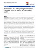
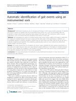
Cholesterol biosynthesis pathwayFigure 1
Cholesterol biosynthesis pathway. A biochemical path-
way showing the major steps by which cholesterol is synthe-
sised. Also shown are HMG-CoA reductase and squalene
synthase, the enzymes that are inhibited by simvastatin and
squalestatin respectively.
Journal of Neuroinflammation 2007, 4:5 />Page 3 of 8
(page number not for citation purposes)
amino acids 1–17 of the human PAF receptor (Cayman
Chem, Ann Arbor, USA), a secondary anti-rabbit IgG
extravidin conjugate followed by a biotin-alkaline phos-
phatase and visualised with 4-nitrophenol phosphate
(Sigma). Membranes were also probed for β-actin using a
mouse monoclonal antibody (Sigma, Dorset, UK). Pro-
tein concentration was determined using a micro-BCA
protein assay kit (Pierce, Cramlington, UK) and amounts
of cholesterol were measured using a fluorometric Amplex
Red cholesterol assay kit with excitation at 550 nM and an
emission detection at 590 nm (Invitrogen, Paisley, UK),
according to the manufacturer's instructions. The
amounts of PGE
2
in cell extracts were determined using a
competitive enzyme immunoassay kit (Amersham Bio-
tech, Amersham, UK) according to the manufacturer's
instructions.
Enzyme-liked immunoassay (ELISA) for the PAF receptor
Total cell membranes or lipid raft fractions were sus-
pended in carbonate buffer at 1 × 10
6
cells per ml, plated
into 96 well immunoplates (50 μl per well) and incubated
for 1 hour at room temperature to allow proteins to
adhere. Non-specific binding sites were blocked by 10%
milk powder and PAF receptors were detected by rabbit
polyclonal antibodies to the human PAF receptor. Bound
antibodies were detected with an anti-rabbit IgG alkaline-
phosphatase conjugate and developed with 4-nitrophenol
phosphate in diethanolamine buffer. Absorbance was
measured on a microplate reader at 450 nM and PAF
receptor content was calculated by reference to a standard
curve. Samples were expressed as "units PAF receptor"
where 100 units was arbitrarily defined as the amount of
PAF receptor in 1 × 10
6
untreated cells. A standard curve
was generated from this sample using sequential log 2
dilutions (range 100 to 1.56 units).
Drugs
PAF (1-O-Hexadecyl-2-acetyl-sn-glycerol-3-phospho-
choline) and simvastatin were obtained from Calbiochem
(Nottingham, UK). Lyso-PAF (1-O-Hexadecyl-2-sn-glyc-
erol-3-phosphocholine), arachidonic acid, mevalonate,
squalene and staurosporine were obtained from Sigma
(Poole, UK). Squalestatin was a gift from GlaxoSmithK-
line, Stevenage, UK.
Statistical analysis
Comparison of treatment effects were carried out using
one and two way analysis of variance techniques as appro-
priate.
Results
Treatment with simvastatin or squalestatin reduces the
neurotoxicity of PAF
The addition of PAF, but not lyso-PAF (an inactive metab-
olite of PAF), caused a dose-dependent reduction in the
survival of primary cortical neurons; with an LD
50
~30 nM
(Figure 2). To determine if cholesterol depletion affected
neuronal responses to PAF, neurons were pre-treated with
varying concentrations of squalestatin or simvastatin for
24 hours, prior to the addition of 100 nM PAF. Treatment
of neurons with either squalestatin or simvastatin resulted
in a dose-dependent increase in neuronal survival (Figure
3). While the concentration of simvastatin required to
reduce the toxicity of PAF by 50% was 100 nM, the con-
centration of squalestatin required to provide a similar
level of protection was 20 nM. Neurons treated with 500
nM squalestatin were not completely resistant to PAF,
however the concentration of PAF required to kill 50% of
squalestatin-treated neurons was 4 μM, approximately
100 times more than that required in untreated neurons
(Figure 4). The effect of simvastatin or squalestatin on the
amounts of cholesterol in primary cortical neurons was
also determined. After 24 hours, the cholesterol content of
neurons treated with 100 nM squalestatin was signifi-
cantly less than that of untreated neurons (297 ng choles-
terol/mg protein ± 32 v 496 ± 42, n = 9, P < 0.05).
Similarly, the cholesterol content of neurons treated with
500 nM simvastatin was also significantly less than that of
untreated neurons (331 ng cholesterol/mg protein ± 50 v
496 ± 42, n = 9, P < 0.05). To determine if cortical neurons
treated with simvastatin/squalestatin were resistant to
other neurotoxins, they were incubated with different
concentrations of staurosporine or arachidonic acid. The
survival of neurons treated with staurosporine, an activa-
tor of the ceramide pathway that induces apoptosis [12],
was not significantly different after pre-treatment with
either 100 nM squalestatin or 500 nM simvastatin (Figure
5). Similarly, the toxicity of arachidonic acid, a precursor
to the production of neurotoxic prostaglandins [13], was
not significantly different between untreated neurons and
neurons treated with 100 nM squalestatin or 500 nM sim-
vastatin (Figure 6).
Squalene reverses the effects of squalestatin on neurons
To confirm that the protective effect of simvastatin and
squalestatin were though inhibition of cholesterol synthe-
sis, we sought to reverse the effects of these drugs with two
precursors of cholesterol synthesis, mevalonate or
squalene. We found no significant differences in the cho-
lesterol content of untreated neurons and neurons treated
with 100 μM mevalonate or with 50 μM squalene. While
the addition of mevalonate or squalene reversed the effect
of simvastatin on neuronal cholesterol levels, only
squalene was able to reverse the effect of squalestatin on
neuronal cholesterol (Table 1). The addition of either 100
μM mevalonate or 50 μM squalene alone did not affect
the survival of neurons, nor did pre-treatment with meval-
onate or squalene alter the survival of neurons subse-
quently treated with 100 nM PAF. While the addition of
mevalonate or squalene reversed the protective effect of
Journal of Neuroinflammation 2007, 4:5 />Page 4 of 8
(page number not for citation purposes)
simvastatin, only squalene was able to reverse the protec-
tive effect of squalestatin (Table 2).
Effect of squalestatin on the cellular location of the PAF
receptor
To determine if the protective effect of squalestatin was
due to changes in the expression of PAF receptors, we
examined membranes isolated from primary cortical neu-
rons for the presence of PAF receptors. We were unable to
distinguish any differences in the amounts of the PAF
receptor in the total membrane fraction of untreated neu-
rons and neurons treated with 100 nM squalestatin by
dotblot (Figure 7). When an ELISA was used to quantify
the amounts of PAF receptor in cells, no significant differ-
ences were observed between untreated and simvastatin-
treated cells (100% ± 5 v 102% ± 7, n = 12, p > 0.05), or
squalestatin-treated cells (100% ± 5 v 106% ± 9, n = 12, p
> 0.05), showing that the resistance of these neurons to
PAF was not due to a reduction in the number of PAF
receptors. Since the PAF receptor is linked to various G-
proteins [11] and many of the G-proteins are found in
cholesterol sensitive lipid rafts [15] we questioned
whether the PAF receptor may also be found in these
domains. Fractionation of neuronal membranes revealed
that in untreated neurons most of the PAF receptors reside
within detergent-resistant membranes synonymous with
lipid rafts. Following treatment with 100 nM squalestatin,
a significant proportion of the PAF receptors were
detected in the non-raft fraction of neuronal membranes
(Figure 7). ELISA studies demonstrated significant differ-
ences between the amounts of PAF receptor detected in
detergent-resistant membranes from untreated neurons
and simvastatin-treated neurons (100% ± 9 v 38% ± 5, n
= 12, p < 0.05) or squalestatin-treated neurons (100% ±
12 v 29% ± 4, n = 12, p < 0.05). The detergent-soluble frac-
tion (non-raft membrane extract) from untreated neurons
or neurons treated with 500 nM simvastatin or 100 nM
squalestatin contained similar amounts of β-actin (Figure
7).
Squalestatin reduces PAF-induced prostaglandin E
2
production
Previous studies have shown that levels of prostaglandin
E
2
are elevated in Alzheimer's disease [14] and in this
study we demonstrated that the addition of PAF caused a
dose-dependent increase in prostaglandin E
2
production.
There was no significant difference in the production of
prostaglandin E
2
between untreated neurons and neurons
treated with lyso-PAF. In neurons pre-treated with 100 nM
squalestatin the effects of PAF on prostaglandin E
2
pro-
duction were significantly reduced (Figure 8).
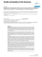
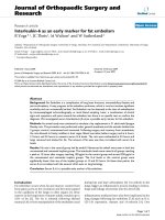
Simvastatin and squalestatin protect neurons against PAFFigure 3
Simvastatin and squalestatin protect neurons against
PAF. The survival of primary cortical neurons pre-treated
for 24 hours with different concentrations of simvastatin (❍)
or squalestatin (●) prior to the addition of 100 nM PAF. Val-
ues shown are the mean average neuronal survival ± SD from
9 observations.
PAF kills cortical neurons in a dose-dependent mannerFigure 2
PAF kills cortical neurons in a dose-dependent man-
ner. The survival of primary cortical neurons incubated with
different concentrations of PAF (❍) or lyso-PAF (●). Values
shown are the mean average neuronal survival ± SD from 9
observations.
Journal of Neuroinflammation 2007, 4:5 />Page 5 of 8
(page number not for citation purposes)
Discussion
In the present study, the effects of simvastatin or squales-
tatin on primary cortical neurons were examined. The
amounts of cholesterol in neurons were significantly
reduced by treatment with either simvastatin or squalesta-
tin. The difference in the effects of these drugs is explained
by their pharmacological targets (Figure 1). Cholesterol
regulation within cells is under tight feedback control and
is sensitive to the concentration of cholesterol in the
endoplasmic reticulumn (ER). Reduced cholesterol in the
ER results in production of HMG-CoA reductase [15]
which catalyses mevalonate production and overcomes
the effect of simvastatin (Figure 1) and in these studies the
addition of exogenous mevalonate increased cholesterol
production in simvastatin-treated cells. Since squalestatin
inhibits squalene synthase, an enzyme further down the
cholesterol synthetic pathway (Figure 1), the synthesis of
new HMG-CoA reductase or the addition of exogenous
mevalonate did not reverse the effects of squalestatin on
neuronal cholesterol content. Although squalestatin is the
more specific research tool, it does not cross the blood-
brain barrier, which limits its therapeutic value.
Although PAF plays roles in the normal functioning of
neurons, higher concentrations of PAF have been impli-
cated in the neurotoxicity of epilepsy, ischaemia, human
immunodeficiency virus infection, prion diseases and AD
[16] PAF is not stored in a preformed state; rather it is syn-
thesised in neurons in response to specific stimuli that
activate phospholipase A
2
[17]. In this study, cortical neu-
rons were killed by nanomolar concentrations of PAF, but
not by lyso-PAF, a non-acetylated structural analogue of
PAF that does not bind to the PAF receptor [18]. Pre-treat-
ment with simvastatin or squalestatin protected neurons
against the otherwise toxic effects of PAF; the IC
50
for sim-
vastatin was ~100 nM, and the IC
50
for squalestatin was
~20 nM. We first tested simvastatin, a HMG-CoA reduct-
ase inhibitor, since it is one of the statins that penetrates
the blood-brain barrier and is in clinical use [3]. However,
although simvastatin reduces neuronal cholesterol con-
tent it also reduces the production of non-sterol products
such as the isoprenoid precursors [4]. The modification of
proteins by isoprenoids is essential for the function of a
wide variety of proteins including the Ras-related G pro-
teins[19]. Since some have argued that the effects of stat-
ins are mediated through inhibition of non-sterol
products rather than cholesterol reduction [20]).) we also
Simvastatin and squalestatin do not protect neurons against staurosporineFigure 5
Simvastatin and squalestatin do not protect neurons
against staurosporine. The survival of untreated primary
cortical neurons (❍) or neurons pre-treated for 24 hours
with 500 nM simvastatin (ᮀ) or 100 nM squalestatin (●)
prior to the addition of different concentrations of stau-
rosporine. Values shown are the mean average neuronal sur-
vival ± SD from 9 observations.
Squalestatin-treated neurones are susceptible to high con-centrations of PAFFigure 4
Squalestatin-treated neurones are susceptible to
high concentrations of PAF. The survival of untreated
primary cortical neurons (❍) or neurons pre-treated for 24
hours with 100 nM squalestatin (●) prior to the addition of
varying concentrations of PAF. Values shown are the mean
average neuronal survival ± SD from 9 observations.
Journal of Neuroinflammation 2007, 4:5 />Page 6 of 8
(page number not for citation purposes)
examined the effects of squalestatin, which inhibits
squalene synthase, thus reducing cholesterol production
without affecting the production of non-sterol products
[6]. The observation that pre-treatment with squalestatin
protects neurons against PAF suggests that cholesterol
depletion is responsible for the observed neuroprotection.
Furthermore, the protective effects of these drugs were
reversed by the addition of squalene, a precursor of cho-
lesterol synthesis that does not affect the production of
non-sterol products (Figure 1). It is worth noting that neu-
rons pre-treated with squalestatin were not completely
resistant to PAF; approximately 50 times more PAF was
required to kill squalestatin-treated cells than to kill
untreated cells. Thus, it seems likely that the signalling
pathways responsible for PAF-induced neurotoxicity
remain intact in squalestatin-treated neurons but that the
reduced cholesterol content of membranes hinders their
activation.
Although pre-treatment with statins confers protection
against amyloid-β peptides [21], prions [22] and PAF, it
does not protect against all neurotoxic insults. Specifi-
cally, neurons treated with simvastatin or squalestatin
remain sensitive to other neurotoxins including stau-
rosporine and arachidonic acid. Staurosporine has been
reported to have a number of effects that include activa-
tion of the ceramide pathway which is implicated in neu-
ronal apoptosis [12]. We conclude that, in cortical
neurons, the downstream pathways that lead to neuronal
death activated by these neurotoxins are not sensitive to
treatment with statins.
The effects of PAF are mediated via a specific receptor with
seven transmembrane spanning segments [10]. We found
no evidence that the protective effects of squalestatin or
simvastatin were due to reduced amounts of the PAF
receptor. Instead we identified effects of squalestatin and
simvastatin on the location of PAF receptors within lipid
rafts. In untreated neurons greater than 90% of the PAF
receptors were found in lipid rafts. This observation is sig-
nificant since the activation of downstream signalling
pathways by PAF is dependent on interaction with pertus-
sis toxin-sensitive G proteins [11], which also reside
within lipid rafts [23]. The present results are consistent
with the concept that PAF activates the PAF receptor in a
lipid raft platform containing PAF receptors and G-pro-
teins. The formation of some lipid rafts is cholesterol-
dependent and therefore susceptible to treatment with
cholesterol synthesis inhibitors [22]. Following treatment
with squalestatin, significantly less PAF receptor was
found within lipid rafts and more was found in the nor-
mal cell membrane. We propose that the PAF receptors
Table 1: Squalene reverses the effects of squalestatin on the amounts of cholesterol in neurons
Treatment Substrate
None 50 μM Squalene 100 μM Mevalonate
Neuronal cholesterol content (ng/mg protein)
None 496 ± 42 472 ± 53 482 ± 53
100 nM Squalestatin 297 ± 32 475 ± 32 284 ± 29
500 Nm Simvastatin 331 ± 50 501 ± 46 485 ± 68
The amounts of cholesterol in primary cortical neurons treated for 24 hours with different combinations of either squalestatin or simvastatin
together with either squalene or mevalonate as shown. Values shown are the mean average ± SD from 6 observations.
Simvastatin and squalestatin do not protect neurons against arachidonic acidFigure 6
Simvastatin and squalestatin do not protect neurons
against arachidonic acid. The survival of untreated pri-
mary cortical neurons (❍) or neurons pre-treated for 24
hours with 500 nM simvastatin (ᮀ) or 100 nM squalestatin
(●) prior to the addition of different concentrations of ara-
chidonic acid. Values shown are the mean average neuronal
survival ± SD from 9 observations.
Journal of Neuroinflammation 2007, 4:5 />Page 7 of 8
(page number not for citation purposes)
outside lipid rafts fail to stimulate the G-proteins respon-
sible for activation of downstream signalling pathways.
This hypothesis is consistent with out observation that
pre-treatment with squalestatin reduced PAF-induced
prostaglandin E
2
production.
Conclusion
High concentrations of PAF are thought to contribute to
neuronal damage in some neurodegenerative diseases.
The current study demonstrates that inhibitors of choles-
terol synthesis reduce the cholesterol content of neurons
and greatly increases the resistance of these cells to PAF.
This protective effect was associated with the dispersal of
PAF receptors from within detergent-resistant mem-
branes, or lipid rafts, and into the bulk cell membrane.
The reduction of PAF receptors within detergent-resistant
Squalestatin reduces PAF-induced prostaglandin E
2
productionFigure 8
Squalestatin reduces PAF-induced prostaglandin
E
2
production. The amounts of prostaglandin E
2
(pg/ml) pro-
duced by untreated neurons incubated with different concen-
trations of PAF (●) or lyso-PAF (▲) or neurons pre-treated
with 100 nM squalestatin and incubated with varying concen-
trations of PAF (❍). Values shown are the mean prostaglan-
din E
2
± SD from 9 observations.
Table 2: Squalene reverses the protective effects of squalestatin
Treatment Substrate
None 50 μM Squalene 100 μM Mevalonate
Neuronal survival (% of control)
None 31 ± 6 32 ± 4 30 ± 5
100 nM Squalestatin 95 ± 7 40 ± 9 93 ± 5
500 nM Simvastatin 94 ± 4 42 ± 9 34 ± 8
The survival of primary cortical neurons treated for 24 hours with combinations of either squalestatin or simvastatin together with either squalene
or mevalonate as shown, and subsequently incubated with 100 nM PAF. Values shown are the mean average neuronal survival ± SD from 12
observations.
Squalestatin alters the cellular location of the PAF receptorFigure 7
Squalestatin alters the cellular location of the PAF
receptor. (A) Dotblots showing the amounts of PAF recep-
tor in sequential dilutions (log 2 dilutions from neat) of
extracts from untreated neurons and neurons treated for 24
hours with 100 nM squalestatin. (B) Western blot showing
the amounts of PAF receptors in lipid raft/non-raft fractions
from untreated neurons and neurons treated with 100 nM
squalestatin. (C) Western blot showing the amounts of β-
actin in cell extracts from untreated neurons (Con), or neu-
rons treated with 500 nm simvastatin (Sim) or 100 nM squal-
estatin (Sq).
Publish with Bio Med Central and every
scientist can read your work free of charge
"BioMed Central will be the most significant development for
disseminating the results of biomedical research in our lifetime."
Sir Paul Nurse, Cancer Research UK
Your research papers will be:
available free of charge to the entire biomedical community
peer reviewed and published immediately upon acceptance
cited in PubMed and archived on PubMed Central
yours — you keep the copyright
Submit your manuscript here:
/>BioMedcentral
Journal of Neuroinflammation 2007, 4:5 />Page 8 of 8
(page number not for citation purposes)
membranes was accompanied by a reduction in prostag-
landin E
2
production. We speculate that interactions
between PAF and PAF receptors residing within the bulk
cell membrane (outside lipid rafts) have a reduced capac-
ity to stimulate downstream signalling pathways that lead
to neuronal death. These results raise the possibility of
using statins as adjuvant therapy for neurodegenerative
diseases in which PAF has been implicated, such as ischae-
mia, stroke and AD. However, neuronal damage occurs
via a variety of mechanisms in vivo and squalestatin- or
simvastatin-treated neurons remain sensitive to other
neurotoxins.
Abbreviations
Alzheimer's disease (AD), platelet-activating factor (PAF),
Competing interests
The author's declare that they have no competing inter-
ests.
Authors' contributions
CB was responsible for the conception, planning and per-
formance of experiments, and for writing this manuscript.
LR prepared toxicity assays, western and dot blot analysis.
AW contributed to the planning of experiments, interpre-
tation of results and the writing of the manuscript.
Acknowledgements
This work was supported by a grant from the European Commission FP6 –
Network of Excellence "Neuroprion".
References
1. Puglielli L, Tanzi RE, Kovacs DM: Alzheimer's disease: the choles-
terol connection. NatNeurosci 2003, 6(4):345-351.
2. Wolozin B, Kellman W, Ruosseau P, Celesia GG, Siegel G:
Decreased prevalence of alzheimer disease associated with
3-hydroxy-3-methyglutaryl coenzyme A reductase inhibi-
tors. ArchNeurol 2000, 57(10):1439-1443.
3. Hess DC, Fagan SC: Pharmacology and clinical experience with
simvastatin. ExpertOpinPharmacother 2001, 2(1):153-163.
4. Maltese WA, Sheridan KM: Isoprenylated proteins in cultured
cells: subcellular distribution and changes related to altered
morphology and growth arrest induced by mevalonate dep-
rivation. JCell Physiol 1987, 133(3):471-481.
5. Cole SL, Vassar R: Isoprenoids and Alzheimer's disease: A
complex relationship. Neurobiology of Disease 2006,
22(2):209-222.
6. Baxter A, Fitzgerald BJ, Hutson JL, McCarthy AD, Motteram JM, Ross
BC, Sapra M, Snowden MA, Watson NS, Williams RJ: Squalestatin
1, a potent inhibitor of squalene synthase, which lowers
serum cholesterol in vivo. JBiolChem 1992,
267(17):11705-11708.
7. Perry SW, Hamilton JA, Tjoelker LW, Dbaibo G, Dzenko KA, Epstein
LG, Hannun Y, Whittaker JS, Dewhurst S, Gelbard HA: Platelet-
activating factor receptor activation. An initiator step in
HIV-1 neuropathogenesis. JBiolChem 1998,
273(28):17660-17664.
8. Bazan NG: The neuromessenger platelet-activating factor in
plasticity and neurodegeneration. ProgBrain Res 1998,
118:281-291.
9. Bate C, Salmona M, Williams A: The role of platelet activating
factor in prion and amyloid-b neurotoxicity. Neuroreport 2004,
15:509-513.
10. Shukla SD: Platelet-activating factor receptor and signal
transduction mechanisms. The FASEB Journal 1992,
6(6):2296-2301.
11. Clark GD, Zorumski CF, McNeil RS, Happel LT, Ovella T, McGuire
S, Bix GJ, Swann JW: Neuronal platelet-activating factor recep-
tor signal transduction involves a pertussis toxin-sensitive G-
protein.
NeurochemRes 2000, 25(5):603-611.
12. Wiesner DA, Dawson G: Staurosporine induces programmed
cell death in embryonic neurons and activation of the cera-
mide pathway. J Neurochem 1996, 66(4):1418-1425.
13. Hatae T, Wada M, Yokoyama C, Shimonishi M, Tanabe T: Prostacy-
clin-dependent apoptosis mediated by PPAR delta. JBiolChem
2001, 276(49):46260-46267.
14. Montine TJ, Sidell KR, Crews BC, Markesbery WR, Marnett LJ, Rob-
erts LJ, Morrow JD: Elevated CSF prostaglandin E2 levels in
patients with probable AD. Neurology 1999, 53(7):1495-1498.
15. Brown MS, Goldstein JL: A proteolytic pathway that controls
the cholesterol content of membranes, cells, and blood. Proc-
NatlAcadSciUSA 1999, 96(20):11041-11048.
16. Bazan NG, Rodriguez TEB, Allan G: Mediators of injury in neuro-
trauma: intracellular signal transduction and gene expres-
sion. JNeurotrauma 1995, 12(5):791-814.
17. Aihara M, Ishii S, Kume K, Shimizu T: Interaction between neu-
rone and microglia mediated by platelet-activating factor.
Genes Cells 2000, 5(5):397-406.
18. Prescott SM, Zimmerman GA, Stafforini DM, McIntyre TM: Platelet-
activating factor and related lipid mediators. AnnuRevBiochem
2000, 69:419-445.
19. Zhang FL, Casey PJ: Protein prenylation: molecular mecha-
nisms and functional consequences. AnnuRevBiochem 1996,
65::241-269.
20. Diomede L, Albani D, Sottocorno M, Donati MB, Bianchi M, Fruscella
P, Salmona M: In vivo anti-inflammatory effect of statins is
mediated by nonsterol mevalonate products. Arterioscler-
ThrombVascBiol 2001, 21(8):1327-1332.
21. Paris D, Townsend KP, Humphrey J, Obregon DF, Yokota K, Mullan
M: Statins inhibit A beta-neurotoxicity in vitro and A beta-
induced vasoconstriction and inflammation in rat aortae.
Atherosclerosis 2002, 161:293-299.
22. Bate C, Salmona M, Diomede L, Williams A:
Squalestatin cures
prion-infected neurones and protects against prion neuro-
toxicity. JBiolChem 2004, 279((15)):14983-14990.
23. Oh P, Schnitzer JE: Segregation of heterotrimeric G proteins in
cell surface microdomains. G(q) binds caveolin to concen-
trate in caveolae, whereas G(i) and G(s) target lipid rafts by
default. MolBiolCell 2001, 12(3):685-698.