báo cáo hóa học: " Toll-like receptor 2 signaling is a mediator of apoptosis in herpes simplex virus-infected microglia" pdf
Bạn đang xem bản rút gọn của tài liệu. Xem và tải ngay bản đầy đủ của tài liệu tại đây (256.91 KB, 7 trang )
BioMed Central
Page 1 of 7
(page number not for citation purposes)
Journal of Neuroinflammation
Open Access
Research
Toll-like receptor 2 signaling is a mediator of apoptosis in herpes
simplex virus-infected microglia
Rajagopal N Aravalli, Shuxian Hu and James R Lokensgard*
Address: Center for Infectious Diseases and Microbiology Translational Research, University of Minnesota Medical School, Minneapolis, MN
55455, USA
Email: Rajagopal N Aravalli - ; Shuxian Hu - ; James R Lokensgard* -
* Corresponding author
Abstract
Background: Information regarding the response of brain cells to infection with herpes simplex
virus (HSV)-1 is needed for a complete understanding of viral neuropathogenesis. We have recently
demonstrated that microglial cells respond to HSV infection by producing a number of
proinflammatory cytokines and chemokines through a mechanism involving Toll-like receptor 2
(TLR2). Following this cytokine burst, microglial cells rapidly undergo cell death by apoptosis. We
hypothesized that TLR2 signaling might mediate the cell death process as well.
Methods: To test this hypothesis, we investigated HSV-induced cell death of microglia obtained
from both wild-type and TLR2
-/-
mice. Cell death was studied by oligonucleosomal ELISA and
TUNEL staining, and the mechanisms of apoptosis were further analyzed using murine apoptosis-
specific microarrays. The data obtained from microarray analysis were then validated using
quantitative real-time PCR assays.
Results: HSV infection induced apoptotic cell death in microglial cells from wild-type as well as
TLR2 cells. However, the cell death at 24 h p.i. was markedly lower in knockout cells. Furthermore,
microarray analyses clearly showed that the expression of pro-apoptotic genes was down-
regulated at the time when wild-type cells were actively undergoing apoptosis, indicating a
differential response to HSV in cells with or without TLR2.
Conclusion: We demonstrate here that HSV induces an apoptotic response in microglial cells
which is mediated through TLR2 signaling.
Background
In the central nervous system (CNS), microglial cells gen-
erate the first line of defense against invading pathogens
[1]. They are key immune cells that survey the brain
parenchyma. During early onset of infection, microglia
become activated and produce proinflammatory
cytokines and chemokines. Production of these proin-
flammatory mediators may result in the infiltration of
lymphocytes across the blood-brain barrier to sites of viral
infection [2]. Microglia are functionally very similar to
macrophages in that they clear up dead neurons and other
cell debris by phagocytosis [1,2]. Therefore, efficient
immune functions by microglial cells may be critical in
controlling a number of CNS infections.
Published: 30 April 2007
Journal of Neuroinflammation 2007, 4:11 doi:10.1186/1742-2094-4-11
Received: 15 February 2007
Accepted: 30 April 2007
This article is available from: />© 2007 Aravalli et al; licensee BioMed Central Ltd.
This is an Open Access article distributed under the terms of the Creative Commons Attribution License ( />),
which permits unrestricted use, distribution, and reproduction in any medium, provided the original work is properly cited.
Journal of Neuroinflammation 2007, 4:11 />Page 2 of 7
(page number not for citation purposes)
Herpes simplex virus 1 (HSV-1) is a neurotropic virus that
infects a wide range of mammalian cells. Following pri-
mary infection of epithelial cells, HSV gains access to the
nervous system and establishes latency in ganglionic neu-
rons. Viral reactivation from this latent state may result in
herpes encephalitis. A number of studies have demon-
strated that Toll-like receptor (TLR) signaling in microglia
is critical in generating innate immune responses against
viral pathogens in the CNS [3-7]. In cell lines, HSV infec-
tion has been shown to activate signaling from TLR2 and
TLR9 [4,8,9]. While TLR2 is localized on the cell surface,
TLR9 is expressed intracellularly on lysosomal mem-
branes. In a recent report, TLR2-deficient neonatal mice
were found to be less susceptible to encephalitis caused by
HSV, suggesting that TLR2 plays an important role in dis-
ease pathogenesis [4].
We have previously shown that microglial cells respond to
HSV-1 by producing a large number of proinflammatory
immune mediators in a TLR2-dependent manner [3].
Interestingly, however, these cells undergo apoptotic cell
death following immune mediator production [10].
Although activation of TLR signaling has been shown to
induce apoptosis in cell lines [11,12], little is known
about TLR involvement in cell death of primary brain
cells. In this study, we hypothesized that TLR2 signaling
induces HSV-mediated microglial cell apoptosis.
Methods
Preparation of microglial cell cultures
Wild type and TLR2
-/-
C57BL/6 mice were purchased from
the Jackson Laboratories (Bar Harbor, ME). Purified
microglial cell cultures (>99% pure), as determined by
MAC-1 antibody staining (Roche Applied Science, Indian-
apolis, IN), were prepared from these mice using a previ-
ously described method with minor modifications [13].
Growth medium for microglial cell cultures was Dul-
becco's modified Eagle's medium (DMEM) with 10%
heat-inactivated fetal calf serum (HyClone Laboratories,
Logan, UT) and antibiotics. For microarray analysis and
real-time PCR assay, 1 × 10
6
cells/sample were used. For
oligonucleosomal ELISA and TUNEL staining, 2 × 10
5
cells were used.
Virus
A highly neurotropic HSV-1 17 syn
+
strain, propagated
and purified from rabbit skin fibroblasts, was used for
infection studies at a multiplicity of infection (MOI) of 2.
After adding virus, culture plates were incubated at 37°C
for the indicated time points.
Oligonucleosomal ELISA
A sandwich ELISA-based system (Roche Applied Science)
was used to detect nucleosomes generated due to DNA
fragmentation during apoptosis. The assay was performed
at the indicated time points as per the manufacturer's
instructions. Data are representative of three independent
experiments, and bars represent the mean + SD of tripli-
cate samples.
TUNEL staining
Wild type and TLR2
-/-
microglial cells were infected with
HSV (MOI = 2) and DNA fragmentation was determined
by terminal deoxynucleotidyl transferase (TdT)-mediated
dUTP-X nick end labeling (TUNEL) using the ApopTag
®
peroxidasein situ apoptosis detection kit (Millipore,
Temecula, CA). Microglial cells were cultured on Lab-Tek
chamber slides at a density of 2 × 10
5
cells per well. At the
end of the incubation period, cells were fixed in 4% para-
formaldehyde for 20 min followed by a staining proce-
dure according to the manufacturer's protocol.
Microarrays
Mouse-specific OligoGEArray
®
apoptosis microarrays
(OMM-12) (SuperArray, Frederick, MD) were used for our
studies, and hybridization procedures were performed per
manufacturer's instructions. Wild type and TLR2
-/-
micro-
glial cells were treated with HSV, and total RNA was
extracted after 8 h and 24 h post-infection (p.i.) using the
RNeasy mini kit (Qiagen, Valencia, CA). Following the
chemiluminescent detection steps, positive spots on
arrays were scanned using a Kodak Image Station 2000R
(Molecular Imaging Systems, Rochester, NY) and were
quantified using GEA analysis suite software (SuperAr-
ray). Data were analyzed as relative induction after each
gene was normalized to the house-keeping gene GAPDH.
Quantitative real-time PCR
cDNA was synthesized using 1 µg of total RNA from unin-
fected and infected wild-type and TLR2
-/-
microglia, at 8 h
and 16 h p.i,. using Superscript II reverse transcriptase
(Invitrogen, Carlsbad, CA) and oligo dT
6–12
primers
(Sigma-Genosys, The Woodlands, TX). PCR was per-
formed with the FullVelocity SYBR Green QPCR master
mix (Stratagene, La Jolla, CA). The PCR conditions for the
Mx3000P QPCR System (Stratagene) were: 40 denatura-
tion cycles of 95°C for 10 s, annealing at 60°C for 10 s
and elongation at 72°C for 10 s. The relative product lev-
els were quantified using the 2(-Delta Delta C(T)) method
[14] and were normalized to β-actin, and are representa-
tive of three independent experiments. Forward and
reverse primer sequences used in the study: caspase-3: 5'-
gggcctgaaataccaagtca-3' and 5'-aaatgaccccttcatcacca-3';
Dsip1: 5'-ggtggccctagacaacaaga-3' and 5'-tcaagcagctcac-
gaatctg-3'; CIDE-B: 5' ctggaactcagctcctccac-3' and 5'- cctc-
caggaccagtgttagc-3'; caspase-2: 5'- cagctccaagaggtttttcg-3'
and 5'- acatccaggggattgtgtgt-3'; Tnfrsf12a: 5'-gattcggcttggt-
gttgatg-3' and 5'-cagtccatgcacttgtcgag-3'; RipK2: 5' cagct-
gggatggtatcgttt-3' and 5'- tggttaaggcaggcttcagt-3'.
Journal of Neuroinflammation 2007, 4:11 />Page 3 of 7
(page number not for citation purposes)
Results and discussion
TLR2 signaling mediates HSV-induced apoptosis in murine
microglia
To test the hypothesis that TLR2 signaling is involved in
the induction of apoptosis in HSV-infected microglia, 2 ×
10
5
cells/sample were infected with the neurotropic HSV-
1 strain 17 syn
+
. We have previously demonstrated that
HSV infects both wild-type and TLR2
-/-
microglia with
similar efficiencies [3], and that apoptosis in virus-
infected wild-type microglia peaks at 24 h p.i. [10]. Fol-
lowing these observations, we harvested cells at 8 h and
24 h p.i., to reflect early and peak apoptotic time points,
and performed oligonucleosomal ELISA assay to detect
nucleosomes generated as a result of DNA fragmentation.
As shown in Fig. 1A, cell death was not observed at signif-
icant levels in either wild-type or TLR2
-/-
microglia at 8 h
p.i. However, apoptosis was induced in wild-type cells at
24 h p.i. The extent of HSV-induced cell death observed in
TLR2
-/-
microglia at 24 h p.i. was 40% of that seen in wild-
type microglia. To confirm apoptotic death in these cells,
TUNEL assay was performed using wild type and TLR2
-/-
microglial cells following a 24 h infection with HSV. As
shown in Fig. 1B, HSV-induced apoptosis was found to be
markedly lower in TLR2
-/-
microglia than in wild type
cells, further demonstrating that TLR2 signaling plays a
role in regulating microglial cell apoptosis in response to
HSV.
Differential expression of apoptotic genes in HSV-infected
wild-type and TLR2
-/-
microglial cells
To further investigate differences in cell death between
HSV-infected wild-type and TLR2
-/-
cells, and to study the
expression profiles of apoptotic genes, we performed
microarray analyses using mouse-specific apoptosis
microarrays. These arrays contained most murine apop-
totic genes. Since gene expression occurs several hours
ahead of DNA fragmentation, 8 h and 16 h p.i. time
points were selected for this study. Furthermore, an induc-
tion or down-regulation of at least two-fold or higher of a
given gene was considered significant in either inducing
or blocking apoptosis. As shown in Tables 1 and 2, the
expression profiles of the apoptotic genes were markedly
different between wild-type and in TLR2
-/-
microglial cells.
At 8 h p.i., the expression of most apoptotic genes
remained unchanged in wild-type cells, while TLR2
-/-
cells
showed induction of few genes. At 16 h p.i., however, pro-
apoptotic genes such as caspase-3 and caspase-8 were
highly expressed in wild-type cells demonstrating that
they were actively undergoing apoptosis. Interestingly,
these genes were not expressed in TLR2
-/-
cells at this time
point. Moreover, many pro-apoptotic genes were down-
regulated in TLR2
-/-
cells at 16 h p.i. when compared with
their expression at 8 h p.i.
Validation of apoptotic gene expression in HSV-infected
microglia
To further confirm these findings, we performed quantita-
tive real-time PCR for six different apoptotic genes
selected from the microarray data. These genes were found
to be either up-regulated or down-regulated in HSV-
infected wild-type microglia and were down-regulated in
TLR2
-/-
cells (Tables 1 &2). As shown in Fig. 2, the expres-
sion levels of caspase-2, caspase-3, Cide-B and Dsip1
increased in wild-type cells between 8 h to 16 h p.i.
whereas they were down-regulated from basal expression
in uninfected controls. On the other hand, Tnfrsf12a and
RipK2 were down-regulated both in wild-type and TLR2
-/
-
cells. These data further demonstrate that TLR2
-/-
cells
have significantly lower levels of pro-apoptotic gene
expression than wild-type cells, and that TLR2 signaling
mediates apoptotic cell death in HSV-infected microglial
cells. We have previously shown that the levels of TNF-α
expression in TLR2
-/-
microglia were approximately 50%
of those seen in wild-type cells [3]. In this study, apoptotic
death in TLR2
-/-
cells was found to be 40% of that in wild-
type microglia and, therefore, it is possible that TNF-α, as
well as other immune mediators, might eventually trigger
apoptosis in cells lacking TLR2.
Conclusion
In this study, we showed that TLR2 signaling induces
apoptosis in HSV-infected microglia. Although the virus
infects both wild-type and TLR2
-/-
microglial cells with
similar efficiencies [3], apoptotic cell death was signifi-
cantly lower in TLR2
-/-
cells. In addition, a large number of
pro-apoptotic genes were clearly down-regulated in TLR2
-
/-
cells at a time when wild-type cells were actively under-
going apoptosis. We have previously demonstrated that at
early time points the production of proinflammatory
immune mediators did not occur in TLR2
-/-
microglia but
they were produced robustly in wild-type cells [3]. How-
ever, TNF-α was still expressed in TLR2
-/-
cells at approxi-
mately 50% of the level seen in wild-type cells, and it is
possible that immune mediators such as TNF-α produced
early in infection, might induce apoptosis. In a recent
study, we deduced apoptotic pathways occurring in pri-
mary glial cells infected with HSV and found that TNF-α
pathway was active in HSV-infected microglial cells [10].
Taken together, these data indicate that HSV infection of
microglial cells activates TLR2 signaling which, in turn,
induces the production of immune mediators and eventu-
ally leads to cell death.
Competing interests
The author(s) declare that they have no competing inter-
ests.
Journal of Neuroinflammation 2007, 4:11 />Page 4 of 7
(page number not for citation purposes)
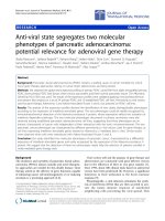
HSV infection induces apoptosis in murine microglial cellsFigure 1
HSV infection induces apoptosis in murine microglial cells. Wild-type and TLR2
-/-
C57BL/6 microglial cells were infected with
HSV at a MOI of 2. (A) The cells were examined for apoptotic DNA fragmentation using an oligonucleosomal ELISA at 8 and
24 h p.i. Data are presented as optical density (OD) per 10
4
cells and are representative of three independent experiments
using cells isolated from different brain specimens. (B) TUNEL staining of wild-type and TLR2
-/-
microglia at 24 h p.i. After fixing
and staining the wells, TUNEL positive cells from at least five fields were counted for each well. Data presented were repre-
sentative of three independent experiments.
A
B
0.0
0.1
0.2
0.3
0.4
TLR2 KO
C57BL/6
8h
24h
4
OD/10 cells
Wt
Wt+HSV
TLR2 KO
TLR2 KO+HSV
% TUNEL positive cells
0
10
20
30
40
Journal of Neuroinflammation 2007, 4:11 />Page 5 of 7
(page number not for citation purposes)
Table 1: Expression of apoptotic genes in HSV-infected microglial cells from C57BL/6 mice
Symbol Gene Fold change
8 h 16 h
Casp3 Caspase-3 2.39 3.79
Card15 Caspase recruitment domain family member 15 - 2.04
Casp11 Caspase-11 0.85 2.38
Casp8 Caspase-8 -0.67 2.41
Dsip1 TSC22 domain family 3 -0.26 3.04
Tnfrsf12a TNF receptor superfamily member 12a 2.32 0.84
Atf5 Activating transcription factor -2.24 -1.22
Bcl10 B-cell leukemia/lymphoma 10 -2.50 -0.94
Bid BH3 interacting domain death agonist -2.15 -1.03
Dad1 Defender against cell death -2.17 -1.40
Mapk8ip1 MAP kinase interacting protein 1 -4.02 -4.06
Rnf7 Ring finger protein 7 -4.44 -1.81
Polb Polymerase B -5.08 -1.82
Prdx2 Peroxiredoxin 2 -3.49 -2.14
Ltbr Lymphotoxin B receptor -3.00 -1.98
Tnfrsf21 TNF receptor superfamily member 21 -4.70 -5.88
Traf3 TNF receptor-associated factor 3 -3.52 -2.07
Table 2: Expression of apoptotic genes in HSV-infected TLR2KO microglial cells.
Symbol Gene Fold change
8 h 16 h
Tnfsf10 TNF ligand superfamily member 10 8.66 5.59
Casp12 Caspase-12 4.15 1.00
RipK2 Receptor (Tnfrsf)-interacting kinase2 3.38 2.87
Casp8ap2 Caspase-8-associated protein 2 3.36 0.66
Fasl Fas ligand 3.15 1.37
Casp3 Caspase-3 2.91 1.46
Tnf Tumor necrosis factor 2.71 1.35
Cflar Casp-8 and FADD-like apoptosis regulator 2.50 0.90
Tnfrsf5 TNF superfamily member 5 2.35 0.93
Bag4 BCL2-associated athanogene 4 2.16 0.58
Bad Bcl-associated death promoter -3.00 -5.07
Akt Thymoma viral proto-oncogene1 -3.76 -2.56
Als2cr2 Als chromosome region candidate 2 -3.97 -2.38
Bax Bcl2-associated X protein -2.35 -1.49
Bcl10 B-cell leukemia/lymphoma 10 -2.71 -3.44
Birc5 Baculoviral IAP repeat-containing 5 -2.19 -8.87
Bcl2l14 Bcl2-like 14 (apoptosis facilitator) -2.08 -4.08
Bid BH3 interacting domain death agonist -0.97 -2.21
Bnip3l BCL2/adenovirus E1B-interacting protein -4.94 -5.14
Birc6 Baculoviral IAP repeat-containing 6 -1.35 -3.19
Bnip2 BCL2/adenovirus E1B-interacting protein -1.04 -2.56
Api5 Apoptosis inhibitor 5 -4.01 -2.56
Dsip1 TSC22 domain family 3 -2.55 -1.50
Cideb Cell-death inducing DNA fragmentation factor 2 -1.78 -3.42
Cradd CASP2 and RIPK1 adaptor domain containing protein -1.54 -6.32
Fadd Fas-associated death domain -1.21 -2.92
Faim Fas apoptotic inhibitory molecule -1.81 -3.18
Hells Helicase lymphoid specific -0.84 -2.24
Il10 Interleukin 10 -1.27 -2.21
Mapk8ip1 MAP kinase interacting protein 1 -0.87 -5.75
Zc3hc1 C3HC type zinc finger protein -0.93 -2.25
Nfkb1 NF-κB -1.92 -1.55
Rnf7 Ring finger protein 7 -2.81 -2.31
Pak7 P21 (CDKN1A)-activated kinase 7 -0.85 -2.03
Traf3 TNF receptor-associated factor 3 -6.31 -2.71
Tnfrsf21 TNF receptor superfamily member 21 -1.87 -1.75
Trp53 P53 -2.80 -1.17
Journal of Neuroinflammation 2007, 4:11 />Page 6 of 7
(page number not for citation purposes)
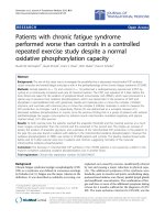
Differential expression of apoptotic genes in microglial cells obtained from wild-type and TLR2
-/-
miceFigure 2
Differential expression of apoptotic genes in microglial cells obtained from wild-type and TLR2
-/-
mice. Real-time PCR was per-
formed using RNA from uninfected and HSV-infected microglia with primers specific for the apoptotic genes indicated. β-actin
was used to normalize the values of apoptotic genes tested. Data presented are representative of three independent experi-
ments.
C57
co
ntr
ol
C57+HSV 8h p.i.
C
57+H
SV 16h p.
i
.
TLR
2KO+
H
SV
co
ntr
ol
TLR2KO+
HSV 8h p
.
i
.
T
L
R2K
O
+HSV 16
h
p.
i
.
Fold induction
0
10
20
30
40
Caspase-3
C57 control
C
57
+H
SV
8h p.
i
.
C
5
7+H
S
V 16h p.
i
.
T
LR2KO+HSV
c
ontro
l
T
L
R2K
O
+
H
SV
8h
p
.
i
.
TL
R
2
KO+HSV 1
6h
p
.
i
.
Fold induction
0
5
10
15
20
25
30
35
Dsip1
Caspase-2
C57 contr
o
l
C
57
+HSV 8h p.i.
C57+HSV 16h p.i.
TLR2
K
O
+
HSV co
n
t
r
ol
TLR2KO+HSV 8h p.i.
T
L
R
2KO+HSV
16h
p.i.
Fold induction
0
20
40
60
80
Cide-B
C
5
7 contr
o
l
C
57+
H
S
V8hp.
i
.
C
57+
H
SV
16h
p
.
i
.
T
LR
2K
O
+H
SV
c
o
n
t
r
o
l
TLR
2KO+
H
SV
8h
p
.
i
.
TL
R
2
K
O
+H
SV
16h p.
i
.
Fold induction
0
10
20
30
40
50
60
C
57 con
t
rol
C57+HSV 8h p.i
.
C57+HSV 16h p.i.
T
L
R2KO+HSV contr
o
l
T
LR2KO+H
SV
8
h
p
.
i.
TLR2K
O
+HSV 16h p.i.
0
20
40
60
80
100
120
140
160
Fold induction
Tnfrsf12a
C57
co
ntro
l
C57+HSV 8h
p
.i
.
C57
+HSV 16
h
p
.i
.
TLR
2
K
O+
H
SV
c
o
n
t
r
ol
T
L
R
2K
O+H
S
V
8h
p
.
i
.
TLR
2K
O
+
H
S
V 16h p.
i
.
Fold induction
0
10
20
30
40
RipK2
Publish with Bio Med Central and every
scientist can read your work free of charge
"BioMed Central will be the most significant development for
disseminating the results of biomedical research in our lifetime."
Sir Paul Nurse, Cancer Research UK
Your research papers will be:
available free of charge to the entire biomedical community
peer reviewed and published immediately upon acceptance
cited in PubMed and archived on PubMed Central
yours — you keep the copyright
Submit your manuscript here:
/>BioMedcentral
Journal of Neuroinflammation 2007, 4:11 />Page 7 of 7
(page number not for citation purposes)
Authors' contributions
RNA and JRL conceived the study and its design, and ana-
lyzed the data. RNA and SH performed the experiments.
RNA drafted the manuscript. All authors read and
approved the final manuscript.
Acknowledgements
This work was supported by United States Public Health Service Award
MH-066703.
References
1. Rock RB Gekker G, Hu S, Sheng WS, Cheeran M, Lokensgard JR,
Peterson PK: Role of microglia in central nervous system
infections. Clin Microbiol Rev 2004, 17(4):942-964.
2. Aloisi F: Immune function of microglia. Glia 2001, 36:165-179.
3. Aravalli RN Hu S, Rowen TN, Palmquist J, Lokensgard JR: Cutting
Edge: TLR2-mediated production of proinflammatory
cytokines and chemokines by microglial cells in response to
herpes simplex virus. J Immunol 2005, 175:4189-4193.
4. Kurt-Jones EA Chan M, Zhou S, Wang J, Reed G, Bronson R, Arnold
MM, Knipe D M, Finberg RW: Herpes simplex virus 1 interaction
with Toll-like receptor 2 contributes to lethal encephalitis.
Proc Natl Acad Sci USA 2004, 101(5):1315-1320.
5. Kurt-Jones EA BJ Yu C, Newburger PE, Wang J, Chan M, Knipe DM,
Finberg RW.: The role of toll-like receptors in herpes simplex
infection in neonates. J Infect Dis 2005, 191(5):746-748.
6. Town T Jeng D, Alexopoulou L, Tan J, Flavell RA: Microglia recog-
nize double-stranded RNA via TLR3. J Immunol 2006,
176(6):3804-3812.
7. Olson JK Miller SD: Microglia initiate central nervous system
innate and adaptive immune responses through multiple
TLRs. J Immunol 2004, 173:3916-3924.
8. Hochrein H Schlatter B, O'Keefe M, Wagner C, Schmitz F, Schiemann
M, Bauer S, Wagner H: Herpes simplex virus type-1 induces
IFN-a production via toll-like receptor 9-dependent and -
independent pathways. Proc Natl Acad Sci U S A 2004,
101:11416-11421.
9. Krug A Luker GD, Barchet W, Lieb DA, Akira S, Colonna M: Herpes
simplex virus type 1 activates murine natural interferon-pro-
ducing cells through Toll-like receptor 9. Blood 2004,
103:1433-1437.
10. Aravalli RN Hu S, Rowen TN, Gekker G, Lokensgard, JR: Differen-
tial apoptotic signaling in primary glial cells infected with
herpes simplex virus 1. J Neurovirol 2006, 12(6):501-510.
11. Aliprantis AO Yang RB, Mark MR, Suggett S, Devaux B, Radolf JD,
Klimpel GR, Godowski P, Zychlinsky A: Cell activation and apop-
tosis by bacterial lipoproteins through Toll-like receptor 2.
Science 1999, 285(5428):736-739.
12. Jung DY Lee H, Jung BY, Ock J, Lee MS, Lee WH, Suk K: TLR4, but
not TLR2, signals autoregulatory apoptosis of cultured
microglia: a critical role of IFN-beta as a decision maker. J
Immunol 2005, 174:6467-6476.
13. Chao CC Molitor TW, Hu S: Neuroprotective role of IL-4
against activated microglia. J Immunol 1993, 151:1473-1481.
14. Litvak KJ Schmittgen TD: Analysis of relative gene expression
data using real-time quantitative PCR and the 2(-Delta Delta
C(T)) method. Methods 2001, 25:402-408.