báo cáo hóa học: " ELISA measurement of specific non-antigenbound antibodies to Ab1-42 monomer and soluble oligomers in sera from Alzheimer’s disease, mild cognitively impaired, and noncognitively impaired subjects" pptx
Bạn đang xem bản rút gọn của tài liệu. Xem và tải ngay bản đầy đủ của tài liệu tại đây (787.44 KB, 11 trang )
RESEARCH Open Access
ELISA measurement of specific non-antigen-
bound antibodies to Ab1-42 monomer and
soluble oligomers in sera from Alzheimer’s
disease, mild cognitively impaired, and
noncognitively impaired subjects
Andrea C Klaver
1
, Mary P Coffey
2
, Lynnae M Smith
1
, David A Bennett
3,4
, John M Finke
5
, Loan Dang
6
and
David A Loeffler
1*
Abstract
Background: The literature contains conflicting results regarding the status of serum anti-Ab antibody
concentrations in Alzheimer’s disease (AD). Reduced levels of these antibodies have been suggested to contribute
to the development of this disorder. The conflicting results may be due to polyvalent antibodies, antibody
“masking” due to Ab binding, methodological differences, and/or small sample sizes. The objectives of this pilot
study were to compare serum anti-Ab antibody concentrations between AD, mild cognitive impairment (MCI), and
elderly noncognitively impaired (NCI) subjects while addressing these issues, and to perform power analyses to
determine appropriate group sizes for future studies employing this approach.
Methods: Serum antibodies to Ab1-42 monomer and soluble oligomers in AD, MCI, and NCI subjects (10/group)
were measured by ELISA, subtracting polyvalent antibody binding and dissociating antibody-antigen complexes.
Differences in mean antibody levels were assessed for significance with repeated measures ANOVA using restricted
maximum likelihood estimation, using Tukey-Kramer tests and confidence intervals for multiple comparisons.
Spearman’s rank correlation was used to determine associations between anti-monomer and anti-oligomer
antibody concentrations. Estimated sample sizes required to detect effects of various sizes were calculated.
Results: There were no significant differences between groups for mean anti-Ab antibody levels, although these
tended to be higher in AD than NCI specimens. Estimated group sizes of 328 and 150 for anti-Ab monomer and
oligomer antibodies, respectively, would have been required for 80% power for significance at 0.05 for a 25%
increase in the AD mean relative to the NCI mean. Serum antibody concentrations to Ab monomer and oligomers
were strongly associated (correlations: 0.798 for undissociated sera, 0.564 for dissociated sera). Antibody-antigen
dissociation significantly increased anti-Ab monomer but not anti-Ab oligomer antibody levels.
Conclusions: The findings in this pilot study are consistent with relatively similar concentrations of specific, non-
antigen-bound antibodies to Ab1-42 monomer and soluble oligomers in AD, MCI, and NCI sera. The differences
between groups for these antibodies would have required approximate group sizes of 328 and 150, respectively,
for a high probability for statistical significance. These findings do not support the hypothesis that reduced levels
of anti-Ab antibodies might contribute to AD’s pathogenesis.
* Correspondence:
1
Department of Neurology Research, William Beaumont Hospital Research
Institute, Royal Oak, MI 48073, USA
Full list of author information is available at the end of the article
Klaver et al . Journal of Neuroinflammation 2011, 8:93
/>JOURNAL OF
NEUROINFLAMMATION
© 2011 Klaver et al; licensee BioMed Central Ltd. This is an Open Access article distributed under the terms of the Creative Commons
Attribution Lic ense (http://creativec ommons.org/licenses/by/2.0), which permits unrestricted use, distribution, and rep roduction in
any medium, provided the original work is properly cited.
Background
Amyloid-beta (A b), the major plaque-associated protein
in the Alzheimer’s disease (AD) brain, has become the
main target for AD therapy since the formulation of the
“amyloid hypothesis” [1]. The significance of serum anti-
bodies to Ab in AD is unclear, because these antibodies
have been reported to be decreased [2-7], unaltered
[8-12], or increased [13-17] in this disorder. These stu-
dies are summarized in Table 1. Some investigators
have suggested that reduced levels of anti-Ab antibodies
may contribute to the pathogenesis of AD [18,19].
In previous studies [20,21] we used enzyme-linked
immunosorbent assay (ELI SA) to measure antibodi es to
Ab1-42 monomer and soluble oligomers in intravenous
immunoglobulin (IvIg) preparations. IvIg preparations
consist of pooled and purified plasma immunoglobulins
(> 95% IgG) from thousands of clinica lly normal indivi-
duals. These drugs are being evaluated as a possible
treatment for AD; encouraging results were obtained in
two clinical trials in which IvIg was administered to AD
patients [22,23] and a multi-site phase 3 trial is in pro-
gress. In our ELISA studies we found that in addition to
IvIg’sbindingtoAb-coated wells, it also bound exten-
sively to wells coated with buffer or with an irrelevant
protein, bovine serum albumin (BSA). We referred to
this as nonspecific binding [20,21] and co ncluded that it
should be subtracted from IvIg’sbindingtoAb-coated
wells to accurately calculate specific anti-Ab antibody
concentrations. A subsequent study [24] found this
binding to be mediated by IgG’ sFabfragmentsand
therefore referred to it as “polyvalent.” Among previous
studies comparing serum anti-Ab levels between AD
patients and aged normal controls, in only one study [3]
was this binding subtracted from total antibody binding
to Ab. The conflicting results for anti-Ab serum antibo-
dies in AD may be due in part to failure t o account for
this binding. Other reasons could include binding of
anti-Ab antibodies by serum Ab (antibody “masking”),
which could reduce ELISA detection of these antibodies
[25], incorrect diagnosis of some study subject s (clinical
diagnosis of AD is about 88-90% accurate [26,27]), dif-
ferences in preparation of the Ab conformations used to
detect antibody binding and/or other methodological
differences, and the small sample sizes used in some
studies. In previous ELISA studies comparing these anti-
bodies in AD subjects vs. norm al controls, only Moir et
al. [3], Gruden et al. [14,15], and Nath et al. [13] mea-
sured antibodies to Ab soluble oligomers, which are
thought to initiate AD-type patho logy [28], and only
Gustaw et al. [16] and Gustaw-Rothenberg et al. [17]
performed antibody-antigen complex dissociation. None
of the studies performed both subtraction of polyvalent
binding and dissociation of antibody-antigen complexes,
nor did any o f the studies confirm clinical diagnoses
with post-mortem examinations or p erform power
analyses.
Table 1 Summary of previous studies
Study Specimens Results
Hyman et al., 2001 Plasma: 82 AD, 271 NCI No differences between groups (ELISA)
Weksler et al., 2002 Serum: 19 AD, 33 NCI Decreased AD anti-Ab levels (ELISA)
Nath et al., 2003 Serum: 16 AD, 31 NCI Anti-Ab higher in AD patients
Gruden et al., 2004 Serum: 17 AD, 15 NCI Increased anti-Ab25-35 oligomer antibodies in AD patients (ELISA)
Baril et al., 2004 Serum: 36 AD, 34 NCI No differences between groups (ELISA)
Mruthinti et al., 2004 Plasma: 33 AD, 42 NCI Anti-Ab antibodies significantly (4-fold) increased in AD plasma (ELISA)
Moir et al., 2005 Plasma: 59 AD, 59 NCI No differences for anti-Ab monomer antibodies; decreased AD levels for
anti-Ab oligomer levels (ELISA)
Brettschneider et al.,
2005
Serum: 96 AD, 30 NCI Anti-Ab levels decreased in AD (immunoprecipitation assay)
Jianping et al., 2006 Serum: 20 AD, 20 NCI Decreased AD anti-Ab levels (ELISA) and avidity
Song et al., 2007 Serum: 153 AD, 193 NCI Decreased AD anti-Ab levels (ELISA)
Gruden et al., 2007 Serum: 48 AD, 28 NCI Increased anti-Ab25-35 oligomer antibodies in AD patients (ELISA, dot
blot)
Gustaw et al., 2008 Serum: 23 or 35 AD (assays performed in two
laboratories), 35 NCI
Anti-Ab levels consistently increased in AD vs. controls only after
dissociation
Xu et al., 2008 Plasma: 113 AD, 205 NCI No differences between groups (plaque immunoreactivity)
Britschgi et al., 2009 Plasma: 75 AD, 36 NCI No differences between groups (Ab microarrays)
Sohn et al., 2009 Serum: 136 AD, 210 NCI Anti-Ab decreased in AD patients (ELISA)
Gustaw-Rothenberg et
al., 2010
Serum: 25 AD < 1 year, 18 NCI, 27 AD > 1 year Anti-Ab increased in both AD groups (ELISA) vs. NCI, before and after
dissociation
Summary of previous studies in which serum anti-Ab antibodies have been measured. (AD = Alzheimer ’s disease; NCI = aged noncognitively impaired)
Klaver et al . Journal of Neuroinflammation 2011, 8:93
/>Page 2 of 11
The objectives of this pilo t study were t herefore to
compare serum antibody levels to Ab1-42 soluble con-
formations between AD patients, subjects with mild
cognitive impairment (MCI), and aged noncognitively
impaired (NCI) individuals, incorporating all of these
procedures, and to perform power analyses on the
resulting data to obtain estimates of appropriate group
sizes for future studies using this approach. Our findings
suggest that relatively similar levels of specific, non-anti-
gen-bound antibodies to soluble Ab1-42 conformations
are present in AD, MCI, and NCI sera. Large numbers
of samples (esti mated group sizes: 328 and 150 for anti-
Ab monomer and oligomer antibodies, respectively)
would be required for a high probability of achieving
statisti cal significance for the between-group differences
with this approach.
Methods
Serum samples
Serum samples were obtained from the R ush Alzhei-
mer’ s Disease Center (Chicago, IL) from individuals
whose diagnosis on the basis of post-mortem clinical
review was AD, MCI, or NCI. MCI subjects had only
one impaired cognitive domain and no other apparent
cause of cognitive impairment. AD patients had no
other apparent cause of cognitive impairment. These
individuals were partic ipants in the Rush Memory and
Aging Project, a community-b ased, longitudinal clinical-
pathologic study of aging and AD. Details of this project
were published previously [29]. The study was approved
by the Institutional Review Board of Rush University
Medical Center and was given exempt status by Beau-
mont’s Human Investigation Committee. Subject sum-
mary statistics are shown in Table 2.
Ab1-42 monomer and soluble oligomer preparations
Ab monomer was prepared as described previously
[20,21,30]. Ab1-42 (0.5 mg; AnaSpec, San Jose, CA) was
disaggregated by resuspending in 0.25 ml trifluoroacetic
acid (TFA, Sigma-Aldrich, Inc., St. Louis, MO) follo wed
by hexafluoro-2-pro panol (HFIP, Sigma-Aldrich). It was
aliquoted into eppitubes (20 μl/tube), dried overnight
(16-20hr)atroomtemperatureinafumehood,and
stored at -20°C. The Ab was resuspended in HPLC-
grade water adjusted to pH 3.0 with TFA (1 μl TFA per
10 ml HPLC H
2
O). 0.6 ml TFA water was added to an
Ab-containing eppitube, and after thorough vortexing,
this was put on ice in a separate tube. The procedure
was repeated twice more on the same eppitube, yielding
1.8 ml of Ab in TFA water. Tris base (21.8 mg) was
added to bring the Tris concent ration to 100 mM, and
3.8 μl of 12.1 N HCl was added to adju st the pH to 8.8.
The preparation was centrifuged (11,752 × g, 5 min),
passed through a 0.2 μm filter, and used immediately.
The protein concentration of the filtered preparation
was 6 μg/ml with the Bio-Rad Protein Assay (Bio-Rad
Laboratories, Hercules, CA).
Ab oligomers were also produced as described pre-
viously [20,30]. 4.8 μlof1%NH
4
OH (AnaSpec) was
added to an eppitube of disaggregated Ab,andafter
brief vortexing, the tube sat for one min. The contents
of the tube were then transferred sequentially to two
more A b eppitubes, followi ng thi s same procedure each
time. The preparation was water bath sonicated for 4
min, then incubated for one hr at room temperature.
After dilution in phosphate buffered saline (PBS; 0.01
M, pH 7.4, with 0.02% azide) to a final concentratio n of
58 μg/ml, it was used immediately or stored at 4°C for
up to one week.
Western blots of Ab conformations
Western blots of Ab monomer and soluble oligomer
preparations were performed under both reducing/dena-
turing and native conditions as described previously
[20,30] using 4-20% Tris-HCl Ready Gels (Bio-Rad
Laboratories, Hercules, CA). The molecular weight
Table 2 Subject summary statistics by group (based upon post-mortem clinical review).
Diagnosis Gender Age at Death (yrs) PMI (hrs:mins) ApoE Alleles Anti-Inflammatory Usage
NCI 2 male
8 female
89.46 ± 1.32 6:21
(3:40, 62:24)
E2E3: 2
E3E3: 6
E3E4: 1*
6 yes, 4 no
MCI 3 male
7 female
89.73 ± 1.41 4:43
(2:55, 20:30)
E2E2: 1
E2E3: 3
E3E3: 3
E3E4: 3
6 yes, 4 no
AD 8 male
2 female
89.55 ± 1.39 4:22
(1:30, 13:35)
E2E3: 1
E3E3: 5
E3E4: 4
8 yes, 2 no
Subject ages are reported as means ± SEM, while PMI values are shown as medians with minimum and maximum values in parentheses. Gender distribution was
significantly different between groups (chi square p = 0.020) with the AD group having more males than the other groups. There were no statistically significant
differences between groups for age, PMI, frequency of expression of the different apoE alleles, or use of anti- inflammatory medications. ApoE status was
unknown for one NCI subject. (AD = Alzheimer’s disease; NCI = aged noncognitively impaired; MCI = mild cognitive impairment; ApoE = apolipoprotein E; PMI =
post-mortem interval)
Klaver et al . Journal of Neuroinflammation 2011, 8:93
/>Page 3 of 11
standards for the native gels were from Sigma-Aldrich’s
Non-Denaturing Molecular Weight Kit (cat. #
MWND500). After electrophoresis, the proteins were
transferred to Westran S PVDF membranes (Whatman
International Ltd., Maidstone, UK). The membranes
were then blocked with 10% non-fat dry mi lk in 0.01 M
PBS, pH 7.4 for one hr at room temperature. Mem-
branes were incubated overnight at 4°C with agitation in
mouse monoclonal anti-Ab(1-16) 6E10 (Covance
Research Laboratories, Berkel ey, CA; 1:5,000 dilution).
After i ncubation in horseradish peroxidase (HRP)- con-
jugated anti-mouse IgG (Vector Laboratories, Inc., Bur-
lingame, CA; 1:10,000 dilution) for 1 hr at room
temperature, membranes were developed in SuperSignal
West Pico chemiluminescent substrate (Thermo Scienti-
fic, Rockford, IL). Bands were detected on CL-XPosure
film (Thermo Scientific).
Transmission electron microscopy (TEM)
TEM was performed as previously describe d [31]. Each
sample was spread on a Formvar coated grid (Electron
Microscopy Sciences, Fort Washington, PA) and incu-
bated for two hr at room temperature, then rinsed with
double distilled water. Samples were then fixed with 1%
glutaraldehyde in 100 mM phosphate buffer, pH 7.4 for
10 min, rinsed again with water, and stained with 1%
uranyl acetate for 10 min followed by alkaline lead
citrate for five min. Images were taken with a Morgagni
268 transmission electron m icroscope (FEI Company,
Hillsboro, OR) equipped with a Hamamatsu digital
camera.
ELISA measurement of serum antibodies to Ab1-42
monomer and soluble oligomers
Antibody concentrations to the A b1-42 monomer and
soluble oligomer preparations were measured by ELISA
in AD, MCI, and NCI serum samples. A separate ELISA
plate was required for each serum sample. The plate
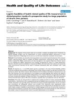
arrangement is shown in Figure 1. Samples were rando-
mized as to the order in which they were evaluated. A
volume of 100 μl was plac ed in ea ch well for each s tep
of the procedure. The Ab monomer and soluble o ligo-
mer preparations were incubated at 0.9 μg/ml in Tris
buffer (0.1 M, pH 8.8) overnight at 4°C on a 9 6-well
Nunc Maxisorp plate (Nalge Nunc International, Roche-
ster, NY). As a “specificity control” the same concent ra-
tion of bovine serum albumin (BSA, Sigma-Aldrich) in
Tris buffer was filtered and placed in adjacent wells.
After incubation overnight at 4°C, wells were washed
three times with PBS with 0.1% Tween-20 (Sigma-
Aldrich) (hereafter, PBS-T; this wash step was repeated
after all subsequent incubations). The plate was then
treated with SuperBloc k (SuperBlock Blocking Buffer in
PBS, Thermo Scientific) as per the manufacturer’ s
instructions, followed by addition of antibody-antigen
complex dissociated and undissociated serum samples.
These samples were diluted 1:100 in PBS (pH 7.2) with
0.1% Tween-20 and 1% BSA (hereafter, PBS-T-BSA) and
assayed in quadruplicate. Positive controls were disso-
ciated and undissociated preparations of an IvIg pro-
duct, Gamunex Immune Globulin Intravenous (Human),
10% (Talecris Bi otherapeutics, Inc., Research Triangle
Park,NC),diluted1:1,000.Anormalcontrolserum
sample from an individual not participating in the Rush
Memory and Aging Project was included on all plates to
allow data to be normalized between plates. Dissociation
of serum antibody-antigen complexes with pH 3.5 disso-
ciation buffer was performed as previously described
[20] using the procedure described by Li et al. [25] with
slight modifications. To produce the standard curve,
four-fold dilutions o f mouse monoclonal 6E10 anti-A b
antibody (1:4,0 00 [250 ng/ml], 1:16,000 [62.5 ng/ml],
1:64,000 [15.6 ng/ml], and 1:256,000 [3.9 ng/ml]) in
PBS-T-BSA were placed in wells previously coated with
Ab monomer, Ab oligomers, or BSA. Blank wells
received PBS-T-BSA at this step. Secondary antisera
were biotinylated goat anti-mouse IgG (Vector Labora-
tories, Inc., Burlingame, CA; 1:1,000 dilution) for the
wells previously receiving mouse 6E10 antibody and bio-
tinylated goat anti-human IgG (H + L) (Jackson Immu-
noResearch Laboratories, West Grove, PA; 1:1,000
dilution) for wells previ ously incubated with serum sam-
ples. After incubation with streptavidin-alkaline phos-
phatase (Zymed Laboratories, Invitrogen, Carlsbad, CA;
1:1,000 in PBS-T), para-nitrophenol phosphate (Sigma-
Aldrich) was added (5 mg in 40 ml o f 1 M diethanola-
mine buffer, pH 9.8). The plate was read at 405 nm
with a Vmax kinetic microplate reader (Molecular
Devices Corp., Sunnyvale, CA) until the standard curve
OD reached 1.0. Softmax Pro softwar e version 3.0
(Molecular Devices) was used to generate the best-fit
plot of the standard curve, using the log-logit option.
Calculation of serum antibody concentrations to Ab1-42
monomer and soluble oligomers
To calculate specific anti-Ab antibody concentrations,
the mean antibody concentration measured when each
serum sample was incub atedonBSA-coatedwellswas
subtracted from the antibody concentrations measured
on wells coated with the soluble Ab conformations.
Densitometric analysis of western blots indicated that
appr oximately 30% of the total band intensity in the Ab
oligomer preparation was du e to the Ab monomer band
[20]. Therefore, after calculating the mean anti-mono-
mer antibody concentration of each sampl e, 30% of this
was subtracted from its antibodies to the oligomer pre-
paration to determine its anti-oligomer antibody con-
centration. The antibody levels measured in each
Klaver et al . Journal of Neuroinflammation 2011, 8:93
/>Page 4 of 11
experiment were normalized for interassay variation by
multiplying them by the overall mean concentration
(from all 30 experiments) of anti-Ab oligomer antibodies
in antibody-antigen-dissociated serum from the normal
control sample, then dividing by the observed
concentration of the anti-Ab oligomer antibody in this
control sample in the experiment. This normalization
procedure was based on anti- Ab oligomer levels in dis-
sociated sera, rather than the other anti-Ab measure-
ments, because the most consistent findings across
Figure 1 ELISA plate configuration used to measure specific antibodies to Ab1-42 monomer and soluble oligomers. Antibodies to Ab1-
42 (both monomer and soluble oligomers) were measured on a separate ELISA plate for each serum sample. The plate layout for each sample
is shown. The mean antibody concentration measured when each serum sample was incubated on BSA-coated wells, representing polyvalent
antibody binding, was subtracted from the antibody concentrations measured on wells coated with the soluble Ab conformations. After
calculating the mean anti-monomer antibody concentration of each sample, 30% of this was subtracted from its antibodies to the oligomer
preparation to determine its anti- oligomer antibody concentration. An IvIg sample (Gamunex) was included on all plates as a positive control.
(CTL serum = normal control serum sample included on all plates to allow normalization of data between plates; Rush serum = experimental
serum sample whose anti-Ab antibody concentrations were being measured; GX = Gamunex Immune Globulin Intravenous (Human), 10%,
Talecris Biotherapeutics, Inc., Research Triangle Park, NC).
Klaver et al . Journal of Neuroinflammation 2011, 8:93
/>Page 5 of 11
experiments were detected for dissociated anti-Ab oligo-
mer antibody measurements.
Statistical Methods
Spearman’s correlation coefficient was used to assess the
association between antibody concentrations to A b
monomer and oligomeric Ab using pooled data from all
groups and also within each group. Differences in mean
antibody levels between groups and between sample
preparation methods (either dissociated or undisso-
ciated) were assessed with repeated measures ANOVA
using restricted maximum likelihood estimation with an
appropriate variance structure. Main effects models
were used w hen there was no ev idence of interaction.
Tukey-Kramer p-values and confidence inte rvals were
used for multiple comparisons as appropriat e. The sig-
nificance of differences between groups was evaluated
using one-way ANOVA (for subject age), the Kruskal-
Wallis test (for post-mortem intervals [PMI]), and exact
versions of Pearson’s chi-square tests (for gender, apoli-
poprotein E [apoE] status, and use of anti-inflammatory
medications). P-values ≤ 0.05 were considered statisti-
cally significant. All p-values were two-tailed. Statistical
analyses were performed using The SAS System for
Windows version 9.2.
Power and sample size analyses
All calculations were based on a s ignificance level of
0.05, with 80% power to detect specified differences
using the F test for the group effect from repeated mea-
sures ANOVA. The standard deviat ion of concentration
and the mean concentration of anti-Ab antibodies in
NCI sera (averaged between dissociated and undisso-
ciated samples) were estimated from the data. The
power analysis calculati ons specified that the mean anti-
Ab antibody concentration in AD subjects would be
increased by a given percentage (20%, 25%, 30%, 40%, or
50%) from the antibody concentration in the NCI
group. The calculations used NCPASS 2005 software
with equal group sample sizes.
Results
Western blots of Ab conformations
Western blots of the Ab conformations, performed on
gels run under both reducing/denaturing and native
conditions, were publishedpreviously[30].TheAb
monomer preparation produced a single band in both
blots. The blot of the reducing/denaturing gel of the oli-
gomer preparation contained bands corresponding to
Ab monomer, dimer, tetramer, pentamer, and higher-
order oligomers. Western blots of the this preparation
runonanativegelproducedaproteinsmearinwhich
individual bands were difficult to visualize.
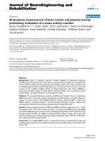
TEM imaging
Spherical structures were present in both the Ab mono-
mer and Ab oligomer preparations. The diameter of the
spherical structures in the oligomer preparation ranged
from 50 to 100 nm while the diameter of the largest sphe-
rical structure in the monomer preparation was approxi-
mately 20 nm. TEM images are shown in Figure 2.
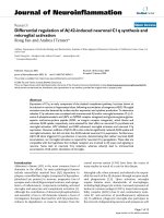
Serum anti-Ab monomer antibodies
There were no significant differences for serum antibody
concentrations to the Ab monomer preparation between
the three groups (p = 0.73 for combined data from
undissociated and dissociated serum samples), although
the mean concentrations of these antibodies tended to
be increased in AD vs. NCI sera (by 20% in undisso-
ciated samples and 29% in dissociated samples). 95%
Tukey confidence intervals for diff erences in the mean
antibody levels indicated that the possibility of large dif-
ferences between these groups could not be exc luded:
MCI - NCI: (-0.280, 0.431); AD - NCI: (-0.243, 0.468);
AD-MCI:(-0.318,0.392).Anti-Ab monomer antibody
levels were significantly increased after antibody-antigen
comp lex dissociation (pooled data from all subjects: p =
0.0011; 95% confidence interval for dissociated - undis-
sociated: [0.073, 0.258]), but none of the within-group
differences were statistically significant after Tukey-Kra-
mer adjustment of p-values. Data are shown in Figure 3.
Serum anti-Ab oligomer antibodies
Results were general ly similar to those for anti-A b
monomer antibodies. There were no significant differ-
ences between the levels of anti-Ab oligomer antibodies
beween AD, MCI, and NCI serum samples (p = 0.58 for
pooled data), although the mean levels again tended to
be increased in AD vs. NCI sera (30% increase in
Figure 2 Transmission electron microscope (TEM) results.
Typical TEM images are shown in Figures 2A and 2B for the Ab1-42
monomer and oligomer preparations, respectively. The diameters of
the spherical structures seen in the Ab monomer and oligomer
preparations were approximately 20 nm and 50-100 nm,
respectively.
Klaver et al . Journal of Neuroinflammation 2011, 8:93
/>Page 6 of 11
undissociated sera, 13% increase in dissociated sera), and
95% Tukey confidence intervals for the differences in
mean antibody levels indicated that the possibility o f
large differences between the groups could not be
excluded: MCI - NCI: (-0.161, 0.301); AD - NCI:
(-0.137, 0.325); and AD - MCI: (-0.207, 0.255). In con-
trast to the anti-monomer anti bodies, antibody- antigen
dissociation did not increase mean anti-Ab oligomer
antibody levels (p = 0.65; 95% confidence interval for
dissociated - undissociated = (-0.121, 0.072). Data a re
shown in Figure 4.
Power analyses
When the population means for serum anti-Ab mono-
mer antibody concentrations for NCI, MCI, and AD
subjects were modeled as 0.440 μg/ml, 0.495 μg/ml, and
0.550 μg/ml, specifying a 25% increase in anti-Ab mono-
mer antibody levels for AD vs. NCI subjects similar to
the findings in the present study, power analysis indi-
cated that 328 samples per group would have been
required for 80% probability of statistically significant
results at the 0.05 level. For anti-Ab oligomer antibo-
dies, when the population means for NCI, MCI, and AD
were modeled as 0.433 μg/ml, 0.487 μg/ml , and 0.541
μg/ml, resulting in a 25% increase in these antibodies
between AD and NCI subjects, 150 samples per group
woul d have been required for 80% probability of signifi-
cance at the 0.05 level. Tables 3 and 4 indicate the
approximate numbers of samples per group that would
have been required for 80% probability to achieve signif-
icance at the 0.05 level for specified increases in AD vs.
NCI antibodies to Ab monomer and oligomers, respec-
tively, between 20% and 50%.
Associations between anti-Ab monomer and oligomer
antibody concentrations
Antibody levels to Ab monomer and soluble Ab oligo-
mers were strongly associated. For pooled data from all
subjects, Spearman rank correlations were 0.798 for
undissociated serum preparations and 0.564 for disso-
ciated preparations. When evaluated for each group,
these associations remained positive (data not shown).
Evaluation of significance for differences between groups
for subject variables
There were no significant differences between groups
for subject age, apoE status, PMI, or use of anti-inflam-
matory medications. The gender differences between the
groups were statistically significant (p = 0.02) because
the majority of the AD group was male (8 males and 2
females) while the other two groups were predominantly
females (NCI, 2 males and 8 females; MCI, 3 m ales and
7 females).
Discussion
This study used ELISA, with subtraction of p olyvalent
antibody binding and dissociation of antibody-antigen
complexes, to compare the concentrations of serum
antibodies to soluble Ab1-42 conformations between
AD,MCI,andNCIsubjectswhoweregroupedonthe
basis of post-mortem clinical review. The between-
group differences for serum anti-Ab level s were not sta-
tistically significant. Although the mean levels of these
antibodies tended to be increased in AD vs. NCI speci-
mens, large group sizes (estimated at 328 for anti-Ab
monomer antibodies and 150 for anti-Ab oligomer
Figure 3 Serum anti-Ab1-42 monomer antibody
concentrations. No statistically significant differences were present
between group means. For pooled data from all subjects, the
antibody levels were significantly increased after antibody- antigen
complex dissociation (p = 0.0011), but none of the within-group
differences were significant after Tukey-Kramer adjustment of p-
values. Data shown are means ± SEM. (AD = Alzheimer’s disease;
NCI = aged noncognitively impaired; MCI = mild cognitive
impairment; Undissoc. = undissociated; Dissoc. = dissociated).
Figure 4 Serum anti-Ab1-42 soluble oligomer concen trations .
No statistically significant differences were found between groups
or between undissociated and dissociated serum preparations for
mean anti-oligomer antibody concentrations. Data shown are
means ± SEM. (AD = Alzheimer’s disease; NCI = aged
noncognitively impaired; MCI = mild cognitive impairment;
Undissoc. = undissociated; Dissoc. = dissociated).
Klaver et al . Journal of Neuroinflammation 2011, 8:93
/>Page 7 of 11
antibodies) would have been required for a high likeli-
hood that differences of this m agnitude would be statis-
tically significant. These sample sizes are conside red to
be approximate values because they are based on varia-
bility estimates from small numbers of samples. Previous
studies have suggested that anti-Ab antibodies may play
a protective role in AD, by preventing Ab’sneurotoxi-
city [32,33], inhibiting development of Ab soluble oligo-
mers [21], increasing phagocytic clearance of fibrillar Ab
[34], preventing Ab fibril development [35], and degrad-
ing preformed Ab fibrils [34]. Using procedures to mea-
sure specific, non-ant igen-bound anti-Ab antibodies, no
evidence was found in the present study for altered
levels of these antibodies in AD patients. Because the
secondary antibody used to detect anti-Ab antibodies in
the serum samples, biotinylated goat anti-human IgG (H
+ L), was not IgG-specific, the measurements in the pre-
sent study represent total serum anti-Ab an tibodies
rather than IgG. Our results do not support the hypoth-
esis that decreased concentrations of serum anti-Ab
antibodies may contribute to the pathogenesis of AD.
Some studies have suggested that human anti-Ab anti-
bodies may recognize conformational epitopes on aggre-
gated forms of Ab, while not recognizing linear epitopes
on monomeric Ab [12,33,36,37]. Howeve r, our IvIg
study[20]andthestudyofMoiretal.withADand
control plasma [3] suggested that these antibodies do
include those to Ab monomer as well as to Ab oligo-
mers. In the present study, specific antibodies were
foundinAD,MCI,andNCIseratobothAb monomer
and oligomer preparations. In an earlier study [30] we
evaluated our monomer preparation by western blot
after electrophoresis on native gels, immediately after
preparation and after storage at 4°C for more than two
months. Only one band was seen in each blot, suggest-
ing little, if any, oligomer contamination. The TEM
images in the present study also showed clear differ-
ences between the 10 nm structures seen in the mono-
mer preparation and the 50 - 100 nm structures
observed in the oligomer preparation. These findings
suggestthattheantibodiesmeasuredinthepresent
study to the Ab monomer preparation were directed to
monomer rather than to Ab oligomers. However,
because Ab monomer may exist in equilibrium with
low-order Ab oligomers [38], the possibility is not ruled
out that some of the antibody binding to the Ab mono-
mer preparation could have been to Ab oligo mers
whose concentrations were below the level of detection
of western blot.
A further difficulty with regard to differentiating
between antibodies to Ab monomer and oligomers is
that anti-monomer antibodies could also recognize Ab
oligomers. The strong association between anti-mono-
mer and anti-oligomer antibody levels in the serum
samples in this study raised the issue of whether the
two antibody measures may essentially be the same.
Depleting the samples of anti-monomer antibodies
would not necessarily resolve this issue because this
might also remove some anti-oligomer reactivity, if
some of the anti-Ab antibodies bind to both monomers
Table 3 Power analysis for anti-Ab1-42 monomer antibody levels
Specified % Difference Between Means NCI (μg/mL) AD (μg/mL) # Samples Required Per Group (80% power, p < 0.05)
20% 0.440 0.528 512
25% 0.440 0.550 328
30% 0.440 0.572 228
40% 0.440 0.616 129
50% 0.440 0.660 83
The mean concentrations for anti-Ab monomer antibodies in NCI specimens were determined for pooled data from undissociated and dissociated serum
samples. The mean anti-Ab antibody level in AD subjects was specified to be increased by a given percentage (20-50%) from this NCI antibody concentration,
and for each percentage the number of samples per group required to achieve 80% statistical power at a significance level of 0.05 was calculated. Approximately
328 samples per group would have been required to detect statistical significance for the observed differences of 25.7% in this study between NCI and AD
means. (AD = Alzheimer ’ s disease; NCI = aged noncognitively impaired)
Table 4 Power analysis for anti-Ab1-42 oligomer antibody levels
Specified % Difference Between Means NCI (μg/mL) AD (μg/mL) # Samples Required Per Group (80% power, p < 0.05)
20% 0.433 0.520 233
25% 0.433 0.541 150
30% 0.433 0.563 104
40% 0.433 0.606 59
50% 0.433 0.650 39
Approximately 150 samples per group would have been required to detect statistical significance for the observed differences of 21.8% in this study between
NCI and AD means. (AD = Alzheimer’s disease; NCI = aged noncognitively impaired)
Klaver et al . Journal of Neuroinflammation 2011, 8:93
/>Page 8 of 11
and oligomers. If, in fact, most of the anti-monomer
antibodies also recognize oligomers, then after subtract-
ing the ~30% of antibody reactivity to the oligomer pre-
parationwhichislikelytobeduetobindingto
monomers, little or no reactivity should remain. How-
ever, substantial reactivity was still detected. This sug-
gests that at least some of the reactivity was likely to be
oligomer-specific.
Previous studies reported that antibody-antigen com-
plex dissociation may allow detection of increased
levels of serum anti-Ab antibodies [16,17,39]. The Ab
conformation to which antibodies were measured in
those studies was not stated. In the present study, dis-
sociation increased the m easured concentrations of
antibodies to Ab mono mer but not to Ab oligomers.
The dissociation procedure used pH 3.5 dissociation
buffer to separate antibody-antigen complexes, fol-
lowed by passage through a 30 kDa molecular weight
cutoff filter to remove unbound Ab. Unlike antibody-
antigen dissociation with lower pH (2.5), dissociation
at pH 3.5 should not produce artifactual increases in
anti-Ab antibodies or inactivate authentic anti body
binding [25]. This procedure should allow removal of
Ab monomer (molecular weight 4.5 kDa) and Ab oli-
gomers no larger than hexamers (27 kDa), while larger
oligomers should be retained. A possible explanation
for the lack of an increase in detectable anti-Ab oligo-
mer antibodies after dissociation is that complexes
between anti-Ab antibodies and larger Ab aggregates
may have re-formed after dissociation, although
whether Ab oligomers are present in serum is unclear.
Detection of plasma Ab oligomers by ELISA was
reported by Xia et al. [40], but heterophilic antibodies
mayhaveresultedinafalsepositivesignalinthat
studybycrosslinkingcapture and reporter antibodies,
as noted by Sehlin et al. [41]. We found similar false
positive results (revealed as such when samples were
diluted 1:1 with ELISA Diluent from Mabtech, Inc.
[Mariemont, OH], stated by the manufacturer to pre-
vent heterophilic antibody-rel ated false positives) when
we attempted to measure total Ab1-42 in plasma sam-
ples from the subjects in this study (data not shown).
Surprisin gly, the actual concentrations of specific anti-
Ab antibodies in serum and pla sma are unclear. These
antibodies have been reported as OD units [5,13,16,24],
titers [2,6,9,10,15], and as relative or arbitrary units
[3,4,14]. An exception is the study by Storace et al. [39]
which reported anti-Ab antibody levels from dissociated
plasma samples from MCI patients and normal controls
as both concentrations and OD values. The levels
reported in that study ranged from 8.0 to 9.5 μg/ml,
higher than the range of 0.4 - 0.6 μg/ml in the present
study. The reasons for these differences are unclear.
One possibility for this discrepancy is that the
concentrations for anti-Ab antibody concentrations in
our study were calculated on the basis of a standard
curve using mouse anti-Ab antibody, whereas Storace et
al. used a purified human IgG reference standard. In
addition, Storace et al. did not subtract polyvalent anti-
body binding.
Conclusions
We report that when specific antibodies to Ab1-42
monomer a nd soluble oligomers were measured by
ELISA in serum specimens from subjects with post-
mortem clinical review diagnoses of AD, MCI, or NCI,
no significant differences in these antibody levels were
found between groups even after dissociation of anti-
body-antigen complexes to allow measurement of “free”
(non-antigen-bound) antibodies. Further, power analyses
on the data indicated that large group sizes (estimated
at 328 and 150 for measurements of anti-Ab monomer
and oligomer antibodies, respectively) would have been
necessary to achieve a high probability for the between-
group differences in these antibody concentrations to
achieve statistical significance. These results do not sup-
port the hypothesis that decreased levels of these antibo-
dies may contribute to AD pathogenesis.
List of abbreviations used
AD: Alzheimer’s disease; ApoE: apolipoprotein E; BSA: bovine serum albumin;
CTL: control; dissoc: dissociated; ELISA: enzyme-linked immunosorbent assay;
IvIg: intravenous immunoglobulin; MCI: mild cognitive impairment; NCI:
noncognitively impaired; PBS: phosphate-buffered saline; PMI: post-mortem
interval; undissoc: undissociated.
Acknowledgements
We thank the participants in the Rush Memory and Aging Project and their
families, as well as the staff of the Rush Alzheimer’s Disease Center. This
study was supported by an Oakland University-Beaumont Multi disciplinary
Grant Award, donations from the Erb family and the East Detroit Auxiliary of
the Fraternal Order of Eagles, and grant R01AG17917 from the National
Institute on Aging (to DAB).
Author details
1
Department of Neurology Research, William Beaumont Hospital Research
Institute, Royal Oak, MI 48073, USA.
2
Department of Biostatistics, William
Beaumont Hospital Research Institute, Royal Oak, MI 48073, USA.
3
Rush
Alzheimer’s Disease Center, Rush University Medical Center, Chicago, IL
60612, USA.
4
Department of Neurological Sciences, Rush University Medical
Center, Chicago, IL 60612, USA.
5
Department of Chemistry, Oakland
University, 2200 Squirrel Road, Rochester, MI 48309, USA.
6
Eye Research
Institute, Oakland University, 2200 Squirrel Road, Rochester, MI 48309, USA.
Authors’ contributions
ACK and LMS performed the experimental procedures, collected the data,
and assisted in manuscript preparation. MPC performed the data analyses
and assisted with manuscript preparation. DAB provided the serum samples
and assisted with manuscript preparation. JMF provided guidance with Aβ
monomer and oligomer preparation and assisted with manuscript
preparation. LD performed the transmission electron microscope studies.
DAL directed the research and wrote the manuscript. All authors read and
approved the final manuscript.
Competing interests
The authors declare that they have no competing interests.
Klaver et al . Journal of Neuroinflammation 2011, 8:93
/>Page 9 of 11
Received: 16 May 2011 Accepted: 9 August 2011
Published: 9 August 2011
References
1. Hardy J, Allsop D: Amyloid deposition as the central event in the
aetiology of Alzheimer’s disease. Trends Pharmacol Sci 1991, 12:383-388.
2. Weksler ME, Relkin N, Turkenich R, LaRusse S, Zhou L, Szabo P: Patients
with Alzheimer disease have lower levels of serum anti-amyloid peptide
antibodies than healthy elderly individuals. Exp Gerontol 2002, 37:943-948.
3. Moir RD, Tseitlin KA, Soscia S, Hyman BT, Irizarry MC, Tanzi RE:
Autoantibodies to redox-modified oligomeric Abeta are attenuated in
the plasma of Alzheimer’s disease patients. J Biol Chem 2005,
280:17458-17463.
4. Brettschneider S, Morgenthaler NG, Teipel SJ, Fischer-Schulz C, Bürger K,
Dodel R, Du Y, Möller HJ, Bergmann A, Hampel H: Decreased serum
amyloid beta(1-42) autoantibody levels in Alzheimer’s disease,
determined by a newly developed immuno-precipitation assay with
radiolabeled amyloid beta(1-42) peptide. Biol Psychiatry 2005, 57:813-816.
5. Jianping L, Zhibing Y, Wei Q, Zhikai C, Jie X, Jinbiao L: Low avidity and
level of serum anti-Abeta antibodies in Alzheimer disease. Alzheimer Dis
Assoc Disord 2006, 20:127-132.
6. Song MS, Mook-Jung I, Lee HJ, Min JY, Park MH: Serum anti-amyloid-beta
antibodies and Alzheimer’s disease in elderly Korean patients. J Int Med
Res 2007, 35:301-306.
7. Sohn JH, So JO, Hong HJ, Kim JW, Na DR, Kim M, Kim H, Nam E, Ha HJ,
Kim YH, Mook-Jung I: Identification of autoantibody against beta-amyloid
peptide in the serum of elderly. Front Biosci 2009, 14:3879-3883.
8. Hyman BT, Smith C, Buldyrev I, Whelan C, Brown H, Tang MX, Mayeux R:
Autoantibodies to amyloid-beta and Alzheimer’s disease. Ann Neurol
2001, 49:808-810.
9. Baril L, Nicolas L, Croisile B, Crozier P, Hessler C, Sassolas A, McCormick JB,
Trannoy E: Immune response to Abeta-peptides in peripheral blood from
patients with Alzheimer’s disease and control subjects. Neurosci Lett
2004, 355:226-230.
10. Mruthinti S, Buccafusco JJ, Hill WD, Waller JL, Jackson TW, Zamrini EY,
Schade RF: Autoimmunity in Alzheimer’s disease: increased levels of
circulating IgGs binding Abeta and RAGE peptides. Neurobiol Aging 2004,
25:1023-1032.
11. Xu W, Kawarabayashi T, Matsubara E, Deguchi K, Murakami T, Harigaya Y,
Ikeda M, Amari M, Kuwano R, Abe K, Shoji M: Plasma antibodies to
Abeta40 and Abeta42 in patients with Alzheimer’s disease and normal
controls. Brain Res 2008, 1219
:169-179.
12.
Britschgi M, Olin CE, Johns HT, Takeda-Uchimura Y, LeMieux MC, Rufibach K,
Rajadas J, Zhang H, Tomooka B, Robinson WH, Clark CM, Fagan AM,
Galasko DR, Holtzman DM, Jutel M, Kaye JA, Lemere CA, Leszek J, Li G,
Peskind ER, Quinn JF, Yesavage JA, Ghiso JA, Wyss-Coray T:
Neuroprotective natural antibodies to assemblies of amyloidogenic
peptides decrease with normal aging and advancing Alzheimer’s
disease. Proc Natl Acad Sci USA 2009, 106:12145-12150.
13. Nath A, Hall E, Tuzova M, Dobbs M, Jons M, Anderson C, Woodward J,
Guo Z, Fu W, Kryscio R, Wekstein D, Smith C, Markesbery WR, Mattson MP:
Autoantibodies to amyloid beta-peptide (Abeta) are increased in
Alzheimer’s disease patients and Abeta antibodies can enhance Abeta
neurotoxicity: implications for disease pathogenesis and vaccine
development. Neuromolecular Med 2003, 3:29-39.
14. Gruden MA, Davudova TB, Malisauskas M, Zamotin VV, Sewell RD,
Voskresenskaya NI, Kostanyan IA, Sherstnev VV, Morozova-Roche LA:
Autoimmune responses to amyloid structures of Abeta(25-35) peptide
and human lysozyme in the serum of patients with progressive
Alzheimer’s disease. Dement Geriatr Cogn Disord 2004, 18:165-171.
15. Gruden MA, Davidova TB, Malisauskas M, Sewell RD, Voskresenskaya NI,
Wilhelm K, Elistratova EI, Sherstnev VV, Morozova-Roche LA: Differential
neuroimmune markers to the onset of Alzheimer’s disease
neurodegeneration and dementia: autoantibodies to Abeta(25-35)
oligomers, S100b and neurotransmitters. J Neuroimmunol 2007,
186:181-192.
16. Gustaw KA, Garrett MR, Lee HG, Castellani RJ, Zagorski MG, Prakasam A,
Siedlak SL, Zhu X, Perry G, Petersen RB, Friedland RP, Smith MA: Antigen-
antibody dissociation in Alzheimer disease: a novel approach to
diagnosis. J Neurochem 2008, 106:1350-1356.
17. Gustaw-Rothenberg KA, Siedlak SL, Bonda DJ, Lerner A, Tabaton M, Perry G,
Smith MA: Dissociated amyloid-beta antibody levels as a serum
biomarker for the progression of Alzheimer’s disease: a population-
based study. Exp Gerontol 2010, 45:47-52.
18. Du Y, Dodel R, Hampel H, Buerger K, Lin S, Eastwood B, Bales K, Gao F,
Moeller HJ, Oertel W, Farlow M, Paul S: Reduced levels of amyloid beta-
peptide antibody in Alzheimer disease. Neurology 2001, 57:801-805.
19. Dodel R, Neff F, Noelker C, Pul R, Du Y, Bacher M, Oertel W: Intravenous
immunoglobulins as a treatment for Alzheimer’s disease: rationale and
current evidence. Drugs 2010, 70:513-528.
20. Klaver AC, Finke JM, Digambaranath J, Balasubramaniam M, Loeffler DA:
Antibody concentrations to Abeta1-42 monomer and soluble oligomers
in untreated and antibody-antigen-dissociated intravenous
immunoglobulin preparations. Int Immunopharmacol 2010, 10:115-119.
21. Klaver AC, Patrias LM, Coffey MP, Finke JM, Loeffler DA: Measurement of
anti- Aβ1- 42 antibodies in intravenous immunoglobulin with indirect
ELISA: the problem of nonspecific binding. J Neurosci Methods 2010,
187:263-269.
22. Dodel RC, Du Y, Depboylu C, Hampel H, Frölich L, Haag A, Hemmeter U,
Paulsen S, Teipel SJ, Brettschneider S, Spottke A, Nölker C, Möller HJ, Wei X,
Farlow M, Sommer N, Oertel WH: Intravenous immunoglobulins
containing antibodies against beta-amyloid for the treatment of
Alzheimer’s disease.
J Neurol Neurosurg Psychiatry 2004, 75:1472-1474.
23.
Relkin NR, Szabo P, Adamiak B, Burgut T, Monthe C, Lent RW, Younkin S,
Younkin L, Schiff R, Weksler ME: 18-Month study of intravenous
immunoglobulin for treatment of mild Alzheimer disease. Neurobiol
Aging 2009, 30:1728-1736.
24. Szabo P, Mujalli DM, Rotondi ML, Sharma R, Weber A, Schwarz HP,
Weksler ME, Relkin N: Measurement of anti-beta amyloid antibodies in
human blood. J Neuroimmunol 2010, 227:167-174.
25. Li Q, Gordon M, Cao C, Ugen KE, Morgan D: Improvement of a low pH
antigen- antibody dissociation procedure for ELISA measurement of
circulating anti-Abeta antibodies. BMC Neurosci 2007, 8:22.
26. Klatka LA, Schiffer RB, Powers JM, Kazee AM: Incorrect diagnosis of
Alzheimer’s disease. A clinicopathologic study. Arch Neurol 1996, 53:35-42.
27. Jellinger KA: Criteria for the neuropathological diagnosis of dementing
disorders: routes out of the swamp? Acta Neuropathol 2009, 117:101-110.
28. Walsh DM, Selkoe DJ: Oligomers on the brain: the emerging role of
soluble protein aggregates in neurodegeneration. Protein Pept Lett 2004,
11:213-228.
29. Bennett DA, Schneider JA, Buchman AS, Mendes de Leon C, Bienias JL,
Wilson RS: The Rush Memory and Aging Project: study design and
baseline characteristics of the study cohort. Neuroepidemiology 2005,
25:163-175.
30. Klaver AC, Patrias LM, Finke JM, Loeffler DA: Specificity and sensitivity of
the Abeta oligomer ELISA. J Neurosci Methods 2011, 195:249-254.
31. Patrias LM, Klaver AC, Coffey MP, Finke JM, Digambaranath JL, Dang L,
Martinez AA, Loeffler DA: Effects of external beam radiation on in vitro
formation of Abeta1-42 fibrils and preformed fibrils. Radiat Res 2011,
175:375-381.
32. Du Y, Wei X, Dodel R, Sommer N, Hampel H, Gao F, Ma Z, Zhao L,
Oertel WH, Farlow M: Human anti-beta-amyloid antibodies block beta-
amyloid fibril formation and prevent beta-amyloid-induced
neurotoxicity. Brain 2003, 126:1935-1939.
33. Dodel R, Balakrishnan K, Keyvani K, Deuster O, Neff F, Andrei-Selmer LC,
Röskam S, Stüer C, Al-Abed Y, Noelker C, Balzer-Geldsetzer M, Oertel W,
Du Y, Bacher M: Naturally occurring autoantibodies against {beta}-
amyloid: investigating their role in transgenic animal and in vitro
models of Alzheimer’s disease. J Neurosci 2011, 31:5847-5854.
34. Istrin G, Bosis E, Solomon B: Intravenous immunoglobulin enhances the
clearance of fibrillar amyloid-beta peptide. J Neurosci Res 2006,
84:434-443.
35. Legleiter J, Czilli DL, Gitter B, DeMattos RB, Holtzman DM, Kowalewski T:
Effect of different anti-Abeta antibodies on Abeta fibrillogenesis as
assessed
by atomic force microscopy. J Mol Biol 2004, 335:997-1006.
36. O’Nuallain B, Acero L, Williams AD, Koeppen HP, Weber A, Schwarz HP,
Wall JS, Weiss DT, Solomon A: Human plasma contains cross-reactive
Abeta conformer- specific IgG antibodies. Biochemistry 2008,
47:12254-12256.
Klaver et al . Journal of Neuroinflammation 2011, 8:93
/>Page 10 of 11
37. Balakrishnan K, Andrei-Selmer LC, Selmer T, Bacher M, Dodel R: Comparison
of intravenous immunoglobulins for naturally occurring autoantibodies
against amyloid-beta. J Alzheimers Dis 2010, 20:135-143.
38. Bitan G, Lomakin A, Teplow DB: Amyloid beta-protein oligomerization:
prenucleation interactions revealed by photo-induced cross-linking of
unmodified proteins. J Biol Chem 2001, 276:35176-35184.
39. Storace D, Cammarata S, Borghi R, Sanguineti R, Giliberto L, Piccini A,
Pollero V, Novello C, Caltagirone C, Smith MA, Bossù P, Perry G, Odetti P,
Tabaton M: Elevation of {beta}-amyloid 1-42 autoantibodies in the blood
of amnestic patients with mild cognitive impairment. Arch Neurol 2010,
67:867-872.
40. Xia W, Yang T, Shankar G, Smith IM, Shen Y, Walsh DM, Selkoe DJ: A
specific enzyme-linked immunosorbent assay for measuring beta-
amyloid protein oligomers in human plasma and brain tissue of patients
with Alzheimer disease. Arch Neurol 2009, 66:190-199.
41. Sehlin D, Söllvander S, Paulie S, Brundin R, Ingelsson M, Lannfelt L,
Pettersson FE, Englund H: Interference from heterophilic antibodies in
amyloid-beta oligomer ELISAs. J Alzheimers Dis 2010, 21:1295-1301.
doi:10.1186/1742-2094-8-93
Cite this article as: Klaver et al.: ELISA measurement of specific non-
antigen-bound antibodies to Ab1-42 monomer and soluble oligomers
in sera from Alzheimer’s disease, mild cognitively impaired, and
noncognitively impaired subjects. Journal of Neuroinflammation 2011
8:93.
Submit your next manuscript to BioMed Central
and take full advantage of:
• Convenient online submission
• Thorough peer review
• No space constraints or color figure charges
• Immediate publication on acceptance
• Inclusion in PubMed, CAS, Scopus and Google Scholar
• Research which is freely available for redistribution
Submit your manuscript at
www.biomedcentral.com/submit
Klaver et al . Journal of Neuroinflammation 2011, 8:93
/>Page 11 of 11