báo cáo hóa học: " Severe depression is associated with increased microglial quinolinic acid in subregions of the anterior cingulate gyrus: Evidence for an immune-modulated glutamatergic neurotransmission?" doc
Bạn đang xem bản rút gọn của tài liệu. Xem và tải ngay bản đầy đủ của tài liệu tại đây (1.17 MB, 9 trang )
RESEARCH Open Access
Severe depression is associated with increased
microglial quinolinic acid in subregions of the
anterior cingulate gyrus: Evidence for an
immune-modulated glutamatergic
neurotransmission?
Johann Steiner
1,2*†
, Martin Walter
1†
, Tomasz Gos
1,3
, Gilles J Guillemin
4
, Hans-Gert Bernstein
1
, Zoltán Sarnyai
5
,
Christian Mawrin
6
, Ralf Brisch
1
, Hendrik Bielau
1
, Louise Meyer zu Schwabedissen
1
, Bernhard Bogerts
1
and
Aye-Mu Myint
1,7
Abstract
Background: Immune dysfunction, including monocytosis and increased blood levels of interleukin-1, interleukin-6
and tumour necrosis factor a has been observed during acute episodes of major depression. These peripheral
immune processes may be accompanied by microglial activation in subregions of the anterior cingulate cortex
where depression-associated alterations of glutamatergic neurotransmission have been described.
Methods: Microglial immunoreactivity of the N-methyl-D-aspartate (NMDA) glutamate receptor agonist quinolinic
acid (QUIN) in the subgenual anterior cingulate cortex (sACC), anterior midcingulate cortex (aMCC) and pregenual
anterior cingulate cortex (pACC) of 12 acutely depressed suicidal patients (major depressive disorder/MDD, n = 7;
bipolar disorder/BD, n = 5) was analyzed using immunohistochemistry and compared with its expression in 10
healthy control subjects.
Results: Depressed patients had a significantly increased density of QUIN-positive cells in the sACC (P = 0.003) and
the aMCC (P = 0.015) compared to controls. In contrast, counts of QUIN-positive cells in the pACC did not differ
between the groups (P = 0.558). Post-hoc tests showed that significant findings were attributed to MDD and were
absent in BD.
Conclusions: These results add a novel link to the immune hypothesis of depression by providing evidence for an
upregulation of microglial QUIN in brain regions known to be responsive to infusion of NMDA antagonists such as
ketamine. Further work in this area could lead to a greater understanding of the pathophysiology of depressive
disorders and pave the way for novel NMDA receptor therapies or immune-modulating strategies.
Background
Recent studies have focused on the role of immune dys-
function in depression, and analogies to “ cytokine-
induced sickness behavior” have been established [1].
Sickness behavior is a coordinated set of adaptive beha-
vioral changes that develop in affected individuals during
the course of an infection. Disease symptoms include
lethargy, depression, failure to concentrate, anorexia,
sleep disturbances, reduction in personal hygiene or
social withdrawal, and are mediated by proinflammatory
cytokines, such as interleukin-1 (IL-1) , interleukin-6 (IL-
6) and tumor necrosis factor a (TNFa) [1].
Previous research has suggested that these specific
monocyte-derived cytokin es are increased in the periph-
eral blood of acutely depressed patients [2-7] along with
elevated monocyte counts [8,9]. Furthermore,
* Correspondence:
† Contributed equally
1
Department of Psychiatry, University of Magdeburg, Magdeburg, Germany
Full list of author information is available at the end of the article
Steiner et al. Journal of Neuroinflammation 2011, 8:94
/>JOURNAL OF
NEUROINFLAMMATION
© 2011 Steiner et al; licensee BioMed Central Ltd. This is an Open Access article distributed und er the terms of the Creative Commons
Attribution License ( which permits unrestricted use, distribution, and reproduction in
any me dium, provided the original work is properly cited.
lymphocyte and natural killer cell abnormalities have
been described [10-12]. It is not yet clear, whether these
changes in the peripheral blood are associated with cor-
responding neuroinflammatory responses and alterations
in neurotransmission. Peripheral immune proce sses may
be mirrored in the brains of patients with acute depres-
sion by micro glial cells which represent t he brain’ s
mononuclear phagocyte system (MPS) [2,13]. Indeed, an
increased density of microglia expressing human leuko-
cyte antigen (HLA)-DR has recently been observed in
the anterior midcingulate cortex (aMCC), t he dorsolat-
eral prefrontal cortex and the mediodorsal thalamus of
suicidal patients with affective disorde rs [14]. H owever,
thisstudyofthesurfacemarker HLA-DR did not sug-
gest a mechanism of how modulation of neurotransmis-
sion is accomplished.
Quinolinic acid (QUIN), an endogenous modulator
with agonistic properties on N-methyl-D -aspartate
(NMDA), which is produced by microglial cells, may
serve as a potential candidate for such a link between
immune and neurotransmitter changes in depression
[13]. This hypothesis is based on the observation that
the above mentioned proinflammatory cytokines induce
a shift from serotonin synthesis to tryptophan
metabolism via the kynurenine pathway in glial cells
[1,15-17], which may ultimately lead to serotonin deple-
tion and particularly an increased production of the
metabolite QUIN (Figure 1). MPS cells, such as micro-
glia, macrophages and monocytes, mainly produce the
NMDA receptor agonist QUIN, while astrocytes synthe-
size the NMDA receptor antagonist kynurenic acid
(KYNA) because they lack the enzyme kynurenine
monoxygenase (KMO) [18-20]. Analyses of blood and
cerebrospinal fluid revealed elevated QUIN levels in
cytokine-induced depression and major depressive disor-
der (MDD) [1,21,22], while an increase in KYNA pro-
duction was related to schizophrenia [23-25].
These findings may connect immune pathologies to
MPS activation in MDD. In addition to serotonin deple-
tion, a direct glutamatergic mechanism has been sug-
gested, which has recently been identified as an
important target of antidepressant treatment [26]. In
this context, the anterior cingulate cortex (ACC), with
its region-specific NMDA and a-amino-3-hydroxy-5-
methyl-4-isoxazolepropionic acid (AMPA) glutamate
receptor profiles that cover functionally segregated
areas, represents an important target region in the cen-
tral nervous system, although investigations must
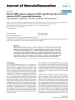
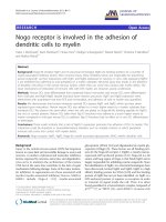
Figure 1 modifie d from [13]: Tryptophan is an essential amino acid and a precursor for the synthesis of serotonin. Alternatively,
tryptophan can be metabolized in glial cells via the kynurenine pathway to create kynurenic acid (synthesized by kynurenine aminotransferase,
KAT) or quinolinic acid (QUIN). These substances are endogenous modulators of NMDA glutamate receptors. A key enzyme of the kynurenine
pathway, indoleamine 2,3-dioxygenase (IDO), and the enzyme that catalyses the production of 3-OH-kynurenine, kynurenine monoxygenase
(KMO), are activated by proinflammatory cytokines, including interleukin-1 and -6 (IL-1, IL-6), tumor necrosis factor a (TNFa), or interferon g (IFNg).
These enzymes are inhibited by anti-inflammatory cytokines, including IL-4. Serotonin is normally broken down into 5-hydroxyindoleacetic acid
(5-HIAA), but the indole ring of serotonin can also be cleaved by IDO to form formyl-5-hydroxykynurenamine (f-5-KYM). Annotation: grey arrows:
activation; dotted grey lines with bar at the end: inhibition; black font: potentially neurotoxic; purple font: neutral or not known; bright blue:
potentially neuroprotective.
Steiner et al. Journal of Neuroinflammation 2011, 8:94
/>Page 2 of 9
account for the histo-architectural diversity of this
region [27]. The importance of the pregenual anterior
cingulate cortex (pACC) in MDD is supported by the
pronounced effects of the glutamate modulating NMDA
antagonist ketamine on the improvement of clinical
symptoms in treatment-resistant MDD patients [28], in
which ketamine leads to an increase in glutamate con-
centration precisely in this region [29].
Therefore, we hypothesized that brain region-specific
QUIN synthesis increases in depression and investigat ed
this idea by analyzing the cellular and regional focus of
QUIN immunoreactivity in the ACC of depressed suici-
dal MDD and bipolar disorder (BD) patients. An upre-
gulated production of QUIN by microglia in regions
with specific susceptibility to abnormal NMDA through-
put would support the hypothesis of an upregula ted
MPS, and would close the gap between neurochemical
imbalances and regional as well as functional in vivo
imaging findings in depression. Only acutely ill patients
were selected for the study, as previous studies of
peripheral blood indicate that MPS activation and
kynurenine pathway imbalances a re associated with
acute disease phases. In a postmortem study of chroni-
cally stable patients with MDD or BD, transient micro-
glial changes may be missed.
Methods
Human brain tissue
Postmortem brains were obtained from the Magdeburg
brain bank in accordance with the Declaration of Hel-
sinki and th e local institutional review board. Written
consent was obtained from the next of kin. The donors
were acutely depressed patients (n = 12) who had com-
mitted suicide (mean age 51 years; 6 males, 6 females)
and controls (n = 10) with no neuropsychiatric illness
(mean age 56 years; 5 males, 5 femal es). The cases were
matched with respect to age, gender, duration of disease
and autolysis time (Table 1). Patients had been diag-
nosed with either major depressive disorder (MDD; n =
7) or bipolar disorder (BD; n = 5).
Table 1 Demographic data of patients with depression (n = 12) and healthy control subjects (n = 10)
Case No. Diagnosis (DSM-IV) Gender Age (y) Autolysis time (h) Cause of death
1 Depression, MDD F 53 47 Suicide by electrocution
2 Depression, MDD F 46 48 Suicide by hanging
3 Depression, MDD F 53 46 Suicide by hanging
4 Depression, MDD F 60 24 Suicide by hanging
5 Depression, MDD F 68 78 Suicide by intoxication
6 Depression, MDD M 35 15 Suicide by wrist cutting
7 Depression, MDD M 36 42 Suicide by hanging
8 Depression, BD F 46 4 Suicide by intoxication
9 Depression, BD M 47 24 Suicide by wrist cutting
10 Depression, BD M 57 48 Suicide by strangulation
11 Depression, BD M 60 24 Suicide by hanging
12 Depression, BD M 53 24 Suicide by hanging
Depression (ratio/mean ± SD) 6F/6M 51 ± 9 35 ± 24
MDD (ratio/mean ± SD) 5F/2M 50 ± 12 45 ± 25
BD (ratio/mean ± SD) 1F/4MF 53 ± 6 19 ± 10
13 Control F 48 48 Status asthmaticus
14 Control F 50 72 Ruptured aortic aneurysm
15 Control F 61 8 Sudden death (reason unknown)
16 Control F 61 24 Heart failure (coronary heart disease)
17 Control F 63 24 Myocardial infarction
18 Control M 56 48 Retroperitoneal haemorrhage
19 Control M 47 24 Acute respiratory failure (aspiration)
20 Control M 54 35 Ruptured aortic aneurysm
21 Control M 63 48 Heart failure (after heart surgery)
22 Control M 54 24 Pulmonary embolism
Controls (ratio/mean ± SD) 5F/5M 56 ± 6 35 ± 18
Statistic (P value) 1.000
a
0.200
b
0.954
b
Control vs. Depression
Statistic (P value) 0.214
a
0.422
c
0.272
c
Control vs. MDD vs. BD
Abbreviations: BD bipolar disorder, MDD major depressive disorder, F female, M male, SD standard deviation,
a
chi-square test,
b
t-test (Control vs. Depression) and
c
ANOVA (Control vs. MDD vs. BD).
Steiner et al. Journal of Neuroinflammation 2011, 8:94
/>Page 3 of 9
The information used for clinical diagnoses was
obtained by carefully studying the patients’ clinical
records and by structured interviews with physicians
involved in patient treatment and with persons who
either lived with or had frequent contact with the sub-
ject s before death. The DSM-IV axis I diagnosis of MDD
and BD was established in consensus meetings of two
psychiatrists (JS and HB) using all available information
from interviews and clinical records [30]. Brains with life-
time reports o f substance abuse, dementia, neurological
illness, severe trauma, o r chronic terminal diseases
known to affect the brain were excluded. Additionally,
neuropathological changes due to neurodegenerative dis-
orders, tumors, inflammatory, vascular, or traumatic pro-
cesses identified by an experienced neuropathologist
(CM) were excluded. The determination of suicide was
made by a forensic pathologist (TG) and was verified
based on the individual records. As summarized in Table
2 the mean daily doses of psychotropic medication taken
by patients during the last 90 lifetime days were estab-
lished according to the clinical files [31-33].
Tissue preparation was performed as described pre-
viously [14,34]. Briefly, brains were fixed in 8% phosphate-
buffered formaldehyde (pH 7.0) for three months. Subse-
quently, after separation of the brainstem and the cerebel-
lum, the hemispheres were divided by coronal cuts into
three bi-hemispherical coronal blocks comprising the
frontal lobe anterior to the genu of the corpus callosum
("anterior” block), the fronto-temporo-parietal lobe
extending the entire length of the corpus callosum ("mid-
dle” block) and the occipital lobe ("posterior” block). After
embedding the brains in paraffin, serial coronal whole
brain sections were cut 20 μm in width and mounted.
Region selection
Sections for QUIN immunohistochemistry were anato-
mically selected corresponding to Brodmann’s area (BA)
24’ (anterior m idcingulate cortex, aMCC), BA 25 (sub-
genual anterior cingulate cortex, sACC) and B A 24/32
(pregenual anterior cingulate cortex, pACC) for QUIN
immunohistochemistry (Figure 2) [27,35]. We were able
to study both subgenual and supracallosal areas in the
same section. These two regions have similar receptor
architectonics, in contrast to a more pregenual region of
the ACC, which was covered by a second section. This
method was possible given the suitable angulation of the
coronal whole brain sections available in the Magdeburg
brain bank.
The exact thickness of each section was determined by
focusing on the upper and lower surfaces of the section
and subtracting the z-axis coor dinate of the lower sur-
face from that of the upper surface. The movements in
the z-axis were measured with a microcator, part of the
Leica DM RB microscope (Leica, Gießen, Germany).
The section thickness after histological procedures was
18.7 ± 1.1 μm (mean ± SD).
Immunohistochemistry
Formalin-fixed tissue sections were deparaffinized, and
antigen demasking was performed by boiling the sec-
tions for 4 min in 10 mM citrate buffer (pH 6.0). Prein-
cubation with 1.5% H
2
O
2
for 10 min to block
endogenous peroxidase activity was followed by blocking
non-specific binding sites with 10% normal goat serum
for 60 min and repeated washings with PBS. Next, a
polyclonal rabbit QUIN antibody was used (ab37106,
Abcam, Cambridge, UK) at a dilution of 1:150 for 72 h
at 4°C. S ections were then incubated with a biotinylated
goat anti-rabbit secondary antibody (Amersham, Little
Chalford, UK) for the streptavidin-biotin technique.
Chromogen 3,3’ -diaminobenzidine (DAB) and ammo-
nium nickel sulfate were used to visualize the reaction
product [36]. The specificity of the polyclonal rabbit pri-
mary antibody was confirmed by a loss of signal after
Table 2 Mean daily doses of psychotropic medication taken by patients during the last 90 lifetime days
Case No. Antidepressants
(amitriptyline equivalents, mg)
Neuroleptics
(chlorpromazine equivalents, mg)
Benzodiazepines
(diazepam equivalents, mg)
Carbamazepine
(mg)
Lithium
(mg)
167 0 000
2 124 109 0 0 0
30 0 000
4 100 400 0 0 0
5 100 50 7.5 0 0
60 0 000
70 0 000
8 133 327 3 0 558
920 0 000
10 n.a. n.a. n.a. n.a. n.a.
11 0 125 10 0 750
12 150 200 0 200 0
Annotations: none of these patients was treated with valproate or lamotrigine; n.a. not available.
Steiner et al. Journal of Neuroinflammation 2011, 8:94
/>Page 4 of 9
preabsorption of 2 ml of the primary antibody solution
(dilution 1:150) with 1 mg QUIN (Sigma-Aldrich,
Munich, Germany) for 24 h and by the supplier’s ELISA
competition experiments with QUIN, kynurenic acid
and phenylalanine.
Quantification
Immunopositive cells were counted in t he delineated
brain regions listed above at 200× magnification (Olym -
pus BH2, Olympus, Hamburg, Germany) by experimen-
ters blind to the donors’ diagnoses (TG and LMS).
Eval uati ons were performed in two c oronal sections per
brain region of interest. The counting area was mea-
sured with the graphical analysis software Digitrace v.
2.10a (Imatec, Miesbach, Germany) using a SZX12
stereomicroscope (Olympus,Hamburg,Germany).The
cytological classification of immunopositive cells as
microglia, astrocytes, oligodendrocytes or neurons was
performed according to established cytomorphological
criteria [37]. Cells visibly located inside v essels were
classifi ed as monocytes; only cells that were clearly out-
side the vessels and situated in tissue were evaluated.
Cell densities were calculated by dividing the cell num-
ber by the counting area multiplied by the section thick-
ness [cells/mm
3
].
Statistical analysis
Statistical analyses were performed with the SPSS 15.0
program (Statistical Product and Service Solutions, Chi-
cago, IL, USA). Demographic data were compared by
the chi-square test, t-test and analysis of variance
(ANOVA). QUIN data were not normally distributed, as
indicated by the Kolmogorov-Smirnov test. Therefore,
Spearman’ s rank correlation coefficient, the Kruskal-
Wallis H test and the Mann-Whitney U test were
employed. These non-parametric tests were further used
to explore potential confounds due to age, gender, dura-
tion of disease, method of suicide, autolysis or fixation
time, and medication dosage.
Results
Qualitative evaluation
Strong QUIN immunoreactivity was found exclusively in
vascular monocytes and microglial cells. In contrast,
faint staining was only occasionally observ ed in fibers
and other cell types, such as pyramidal neurons and
astroglia. The immunoreactive microglia revealed differ-
ent morphological features in healthy controls versus
patients. In control subjects, we found mostly a smooth,
ovoid or elongated cell form (Figure 2). In contrast, par-
ticularly in the aMCC and the sACC, the cortical grey
sACC
aMCC
pACC
Major depression
Healthy control
Figure 2 Illustrations of QUIN-immunoreactive cells from the left sACC of a depressed suicidal patient and a control case and the
locations of the analyzed regions of interest (sACC, aMCC and pACC). Depressed patients showed microglial formations with numerous
granular structure processes. Annotation: Scale bars represent 20 μm.
Steiner et al. Journal of Neuroinflammation 2011, 8:94
/>Page 5 of 9
matter of depressed patients revealed microglial forms
with numerous granular structure processes (Figure 2),
as previously demonstrated by Guillemin et al. in
human tissue [38].
Quantitative evaluation
Comparing QUIN-immunopositive microglia between
depressed patients and healthy controls revealed a
region-specific pattern with group effects only in the
aMCC and the sACC. Depressed patients had signifi-
cantly increased QUIN-positive cells in the sACC (P =
0.003) and the aMCC (P = 0.015). In contrast, cell
counts in the pACC did not differ between groups (P =
0.558) (Figure 3a).
Post-hoc tests of diagnostic subgroups identified
increased cell counts only for MDD patients. In these
patients, QUIN-immunopositive microglia was increased
compared to controls (sACC P = 0.003, aMCC P =
0.015) and compared to the subgroup of bipolar
depressed cases (sACC P =0.042,aMCCP = 0.028)
(Figure 3b). Notably, no significant increase was found
in the pACC in either comparison. Diagnostic specificity
of the increases in MDD was further supported by the
lack of any significant increase or decrease in QUIN-
immunopositive microglia cell counts in bipolar
depressed patients when compared to healthy controls.
The reported effects were controlled for the potential
confounding factors of age, gender, duration of disease,
method of suicide, autolysis or fixation time, and medi-
cation dosage.
Discussion
To our knowledge, this is the first report of microglial
QUIN expression in human brain during acute depres-
sive episodes. An increase in QUIN-immunopositive
microglia was specific to cingulate subregions with high
NMDA receptor densities, like the sACC and the
aMCC, but not the pACC, which shows a lower NMDA
receptor expression. This increase in QUIN-immunor-
eactive microglial cell densities was found particularly in
unipolar patients. With regard to BD less clear state-
ments can be given. We observed a significant difference
between MDD and BD, yet the BD group is also higher
than the controls, though this is apparently not signifi-
cant (Figure 3b). This could be due to the small number
of specimens studied. The numeric increase in QUIN-
immunopositive cell count s was paralleled by the pre-
sence of microglial forms that displayed numerous gran-
ular structure processes in the proximity of neurons in
the depressed group, supporting an interaction o f
inflammatory mechanisms and neurotransmission at the
time of acute depressive episodes. These findings thus
corroborate e vidence for acute inflammatory microglial
activation in depression, leading to increased levels of
the NMDA receptor agonist QUIN in regions with cor-
responding receptor profilesthathavebeenpreviously
revealed as key structures in non-invasive imaging
studies.
Increased levels of QUIN, which is also produced by
macrophages and monocytes, have already been found
in the blood and cerebrospinal fluid of subjects with
cytokine-induced depression or MDD [1,21,22]. Thus,
our result of increased microglial QUIN expression in
suicidal MDD patients is in line with the hypothesis of a
systemic MPS activation during acute disease phases of
depre ssion [2-9,14 ]. Due to the excitotoxic properties of
QUIN, our findings are also supporting the neurodegen-
eration hypothesis of d epression [15]. Therefore, our
study provides insight into why immune- and gluta-
mate- modulating therapies may be helpful for acutely ill
suicidal patients suffering from depression. Potential
candidate drugs include the tetracycline antibiotic mino-
cycline, which inhibits microglial activation by blocking
NF-kappa B nuclear translocation [39-42] or anti-
inflammatory inhibitors of cyclooxygenase-2 [43,44].
Furthermore, severely depressed suicidal patients may
Figure 3 Illustration of QUIN-immunopositiv e cell densities. a)
Depressed patients had increased QUIN-immunopositive cell
densities in the sACC and the aMCC but not in the pACC. b) MDD
patients showed the highest QUIN-immunoreactive cell counts in
the sACC and the aMCC compared to BD and control cases. No
diagnostic subgroup-dependent differences were observed in the
pACC. Annotation: The box plots show the median, interquartile
range, sample minimum and sample maximum, * P < 0.05, ** P <
0.01.
Steiner et al. Journal of Neuroinflammation 2011, 8:94
/>Page 6 of 9
benefit from the administration of glutamate-modulating
drugs, such as the NMDA receptor antagonist ketamine
[28,45,46].
It should be mentioned that Laugeray and colleagues
observed reduced levels of the QUIN precursor 3-OH-
kynurenine (3HK) in the cingulate cortex and increased
levels of 3HK in the stria tum and the amygdala of mice
using an unpredictable chronic mild-stress model for
the induction of depressive-like symptoms [47]. The
observation of reduced 3HK could be due to either
reduced formation of 3HK or increased degradation of
3HK to QUIN, which would result in reduced 3 HK
level. Since QUIN was not directly measured in this
study, a translational validation of these converging
results remains subj ect to future studies. A general
drawback of animal studies is that it is unclear if animal
models adequately reflect the pathophysiology of human
MDD or BD. Moreover, an analysis of ACC subregions
was not undert aken in this study, and direct correspon-
dence of subregions in primates and hu mans differ con-
siderably to those found in rodents. Therefore, the
implications on regional glutamatergic throughput in
depression, as a function of local NMDA and AMPA
receptor profiles, remain difficult to interpret in animal
studies.
We have shown that abnormal NMDA receptor func-
tion related to microglial activation is highly dependent
onthelocationintheACCinhumans.Non-invasive
studies have led to similar disti nctions of abnormal cin-
gulate cortex activation in MDD. While sACC hyperac-
tivity has been postu lated in a number of studies, the
pACC has been less consistently characterized. Grimm
et al. [48] found a reduced deactivation during a task
study, reflected in smaller negative BOLD responses in a
sample of severely depressed patients; this functional
deficit was accompanied by decreased pACC glutamate
and glutamine levels, which are correlated with the
severity of clinical depressive symptoms [49-51]. More-
over, these glutama tergic deficits have been related to
anhedonia and abnormal functional activations in the
pACC in humans [52]. Our finding of relatively
increased QUIN immunoreactivity, which is potentially
associated with serotonin depletion due to changes in
the kynurenine pathway, would thus be consistent with
the relative hyperactivation in the sACC. The sACC is
also a putative target of deep brain stimulation. Impor-
tantly, the metabolic activity after deep brain stimulation
in the sACC, as measured by positron emission tomo-
graphy, shows a reduction in hyperactivity similar to a
region bordering the aMCC and the pACC [53].
Specifically increased concentrations of the NMDA
receptor agonist QUIN in the aMCC and the sACC may
also directly contribute to the disturbed balance in glu-
tamatergic throughput, which could explain the rapid
onset of antidepressant effects after ketamine [28,46].
According to Salvadore et al. [54], activity bordering the
pACC does indeed predict the responsiveness towards
ketamine treatment; therefore , our finding may repre-
sent a histopathological surrogate. As shown by Vollen-
weider and Kometer [55], similar metabolic changes can
be found in the sACC and aMCC upon acute ketamine
administration. Therefore, the anatomical patterns of
such pharmacological challenges fit th e observed pattern
of microglial histopathology.
The present study has certain limitations that need to
be considered: (1) our findi ngs are based on a relatively
small number of MDD and BD cases and must be c on-
firmed in a larger sample size; (2) it was no t possible to
track data on drug exposure or the history of inflamma-
tion and infection across the patients’ entire life spans,
as we could only collect data on psychotropic medica-
tion in the three months prior to death; (3) the present
study enables us to draw conclusions about the cellular
QUIN content, but not released or secreted QUIN in
the extracellular space, which potential ly interferes with
glutamatergic neurotransmission; (4) it remains unclear
if increased QUIN immunoreactivity in microglial cells
is cause d by increased synthesis or reduced degrada tion
of QUIN. Future studies in frozen tissue may address
this question by measuring different kynurenine pathway
metabolites using high-performance liquid chromatogra-
phy (HPLC) or mass spectrometry (MS). (5) It is cur-
rently uncertain if drugs like glibencl amide, nifedipine,
metoprolol, o r theophylline which have been applied in
five of the control subjects may influence microglial
QUIN expression.
Conclusion
Here we present the first study providing evidence that
supports a disease-related upregulation of microglial
QUIN in depressive disorders, particularly in brain
regions known to be responsive to infusion of NMDA
antagonists such as ketamine [55]. These results add a
novel link to the immune [1,26] and neurodegeneration
[15] hypotheses of depression. Further work in this area
could lead to a greater understanding of the pathophy-
siology of depressive disorders and pave the way for
identification of novel biomarkers and therapeutic stra-
tegies targeting specific disease subtypes.
Acknowledgements
Pembroke College (University of Cambridge, Cambridge, UK) has invited JS
for a Visiting Scholarship. This work was supported in part by grants of the
Stanley Medical Research Foundation to BB and JS (Grant No. 07R-1832), the
Commission of European Communities 7th Framework Program
Collaborative Project “MOODINFLAME” to AMM (Grant No. 22963), and the
DFG-SFB 779 to BB and MW. We are grateful to Henrik Dobrowolny for his
skilful assistance in statistical analysis. Gabi Meyer-Lotz and Kathrin Paelchen
provided excellent technical assistance.
Steiner et al. Journal of Neuroinflammation 2011, 8:94
/>Page 7 of 9
Author details
1
Department of Psychiatry, University of Magdeburg, Magdeburg, Germany.
2
Pembroke College, University of Cambridge, Cambridge, UK.
3
Institute of
Forensic Medicine, Medical University of Gdańsk, Gdańsk, Poland .
4
Department of Pharmacology, University of New South Wales, Sydney,
Australia.
5
Department of Pharmacology, University of Cambridge,
Cambridge, UK.
6
Institute of Neuropathology, University of Magdeburg,
Magdeburg, Germany.
7
Department of Psychiatry, University of Munich,
Munich, Germany.
Authors’ contributions
The work presented here has been carried out in collaboration between all
authors. JS, MW, TG, GJG, HGB, BB and AMM have designed the study. CM
has done the routine neuropathological examination. DSM-IV axis I diagnosis
of MDD and BD was established in consensus meetings of JS and HB. JS, TG,
HGB and LMS carried out the laboratory experiments. JS, TG, GJG, LMS and
AMM analyzed the data and interpreted the results. RB was involved in the
creation of figures. JS, MW, TG, ZS, BB and AMM wrote the manuscript. All
authors have read and approved the final version of the manuscript.
Competing interests
The authors declare that they have no competing interests.
Received: 30 June 2011 Accepted: 10 August 2011
Published: 10 August 2011
References
1. Dantzer R, O’Connor JC, Freund GG, Johnson RW, Kelley KW: From
inflammation to sickness and depression: when the immune system
subjugates the brain. Nat Rev Neurosci 2008, 9:46-56.
2. Drexhage RC, Knijff EM, Padmos RC, Heul-Nieuwenhuijzen L, Beumer W,
Versnel MA, Drexhage HA: The mononuclear phagocyte system and its
cytokine inflammatory networks in schizophrenia and bipolar disorder.
Expert Rev Neurother 2010, 10:59-76.
3. Myint AM, Leonard BE, Steinbusch HW, Kim YK: Th1, Th2, and Th3
cytokine alterations in major depression. J Affect Disord 2005, 88:167-173.
4. Kaestner F, Hettich M, Peters M, Sibrowski W, Hetzel G, Ponath G, Arolt V,
Cassens U, Rothermundt M: Different activation patterns of
proinflammatory cytokines in melancholic and non-melancholic major
depression are associated with HPA axis activity. J Affect Disord 2005,
87:305-311.
5. Miller AH, Maletic V, Raison CL: Inflammation and its discontents: the role
of cytokines in the pathophysiology of major depression. Biol Psychiatry
2009, 65:732-741.
6. Zorrilla EP, Luborsky L, McKay JR, Rosenthal R, Houldin A, Tax A, McCorkle R,
Seligman DA, Schmidt K: The relationship of depression and stressors to
immunological assays: a meta-analytic review. Brain Behav Immun 2001,
15:199-226.
7. Padmos RC, Hillegers MH, Knijff EM, Vonk R, Bouvy A, Staal FJ, de Ridder D,
Kupka RW, Nolen WA, Drexhage HA: A discriminating messenger RNA
signature for bipolar disorder formed by an aberrant expression of
inflammatory genes in monocytes. Arch Gen Psychiatry 2008, 65:395-407.
8. Seidel A, Arolt V, Hunstiger M, Rink L, Behnisch A, Kirchner H: Major
depressive disorder is associated with elevated monocyte counts. Acta
Psychiatr Scand 1996, 94:198-204.
9. Maes M, Van der Planken M, Stevens WJ, Peeters D, DeClerck LS, Bridts CH,
Schotte C, Cosyns P: Leukocytosis, monocytosis and neutrophilia:
hallmarks of severe depression. J Psychiatr Res 1992, 26:125-134.
10. Irwin M, Smith TL, Gillin JC: Low natural killer cytotoxicity in major
depression. Life Sci 1987, 41:2127-2133.
11. Kronfol Z, House JD: Depression, hypothalamic-pituitary-adrenocortical
activity, and lymphocyte function. Psychopharmacol Bull 1985, 21:476-478.
12. Maes M, Lambrechts J, Suy E, Vandervorst C, Bosmans E: Absolute number
and percentage of circulating natural killer, non-MHC-restricted T
cytotoxic, and phagocytic cells in unipolar depression.
Neuropsychobiology 1994, 29:157-163.
13. Steiner J, Bogerts B, Sarnyai Z, Walter M, Gos T, Bernstein HG, Myint AM:
Bridging the gap between the immune and glutamate hypotheses of
schizophrenia and major depression: Potential role of glial NMDA
receptor modulators and impaired blood-brain barrier integrity. World J
Biol Psychiatry 2011.
14. Steiner J, Bielau H, Brisch R, Danos P, Ullrich O, Mawrin C, Bernstein HG,
Bogerts B: Immunological aspects in the neurobiology of suicide:
Elevated microglial density in schizophrenia and depression is
associated with suicide. J
Psychiatr Res 2008, 42:151-157.
15. Myint AM, Kim YK: Cytokine-serotonin interaction through IDO: a
neurodegeneration hypothesis of depression. Med Hypotheses 2003,
61:519-525.
16. Connor TJ, Starr N, O’Sullivan JB, Harkin A: Induction of indolamine 2,3-
dioxygenase and kynurenine 3-monooxygenase in rat brain following a
systemic inflammatory challenge: a role for IFN-gamma? Neurosci Lett
2008, 441:29-34.
17. Hu B, Hissong BD, Carlin JM: Interleukin-1 enhances indoleamine 2,3-
dioxygenase activity by increasing specific mRNA expression in human
mononuclear phagocytes. J Interferon Cytokine Res 1995, 15:617-624.
18. Guillemin GJ, Smythe G, Takikawa O, Brew BJ: Expression of indoleamine
2,3-dioxygenase and production of quinolinic acid by human microglia,
astrocytes, and neurons. Glia 2005, 49:15-23.
19. Guillemin GJ, Kerr SJ, Smythe GA, Smith DG, Kapoor V, Armati PJ, Croitoru J,
Brew BJ: Kynurenine pathway metabolism in human astrocytes: a
paradox for neuronal protection. J Neurochem 2001, 78:842-853.
20. Guillemin GJ, Smith DG, Kerr SJ, Smythe GA, Kapoor V, Armati PJ, Brew BJ:
Characterisation of kynurenine pathway metabolism in human
astrocytes and implications in neuropathogenesis. Redox Rep 2000,
5:108-111.
21. Myint AM, Kim YK, Verkerk R, Scharpe S, Steinbusch H, Leonard B:
Kynurenine pathway in major depression: evidence of impaired
neuroprotection. J Affect Disord 2007, 98:143-151.
22. Raison CL, Dantzer R, Kelley KW, Lawson MA, Woolwine BJ, Vogt G,
Spivey JR, Saito K, Miller AH: CSF concentrations of brain tryptophan and
kynurenines during immune stimulation with IFN-alpha: relationship to
CNS immune responses and depression. Mol Psychiatry 2010, 15:393-403.
23. Erhardt S, Blennow K, Nordin C, Skogh E, Lindstrom LH, Engberg G:
Kynurenic acid levels are elevated in the cerebrospinal fluid of patients
with schizophrenia. Neurosci Lett 2001, 313:96-98.
24. Nilsson LK, Linderholm KR, Engberg G, Paulson L, Blennow K, Lindstrom LH,
Nordin C, Karanti A, Persson P, Erhardt S: Elevated levels of kynurenic acid
in the cerebrospinal fluid of male patients with schizophrenia. Schizophr
Res 2005, 80:315-322.
25. Linderholm KR, Skogh E, Olsson SK, Dahl ML, Holtze M, Engberg G,
Samuelsson M, Erhardt S: Increased Levels of Kynurenine and Kynurenic
Acid in the CSF of Patients With Schizophrenia. Schizophr Bull 2010.
26. Sanacora G, Zarate CA, Krystal JH, Manji HK: Targeting the glutamatergic
system to develop novel, improved therapeutics for mood disorders. Nat
Rev Drug Discov 2008, 7:426-437.
27. Palomero-Gallagher N, Vogt BA, Schleicher A, Mayberg HS, Zilles K:
Receptor architecture of human cingulate cortex: evaluation of the four-
region neurobiological model. Hum Brain Mapp 2009, 30:2336-2355.
28. Zarate CA, Singh JB, Carlson PJ, Brutsche NE, Ameli R, Luckenbaugh DA,
Charney DS, Manji HK:
A randomized trial of an N-methyl-D-aspartate
antagonist
in treatment-resistant major depression. Arch Gen Psychiatry
2006, 63:856-864.
29. Rowland LM, Bustillo JR, Mullins PG, Jung RE, Lenroot R, Landgraf E,
Barrow R, Yeo R, Lauriello J, Brooks WM: Effects of ketamine on anterior
cingulate glutamate metabolism in healthy humans: a 4-T proton MRS
study. Am J Psychiatry 2005, 162:394-396.
30. APA: Diagnostic and Statistical Manual of Mental Disorders, 4th revised edition
(DSM-IV-TR). 4 edition. Washington, DC: American Psychiatric Press; 2000.
31. Bollini P, Pampallona S, Tibaldi G, Kupelnick B, Munizza C: Effectiveness of
antidepressants. Meta-analysis of dose-effect relationships in
randomised clinical trials. Br J Psychiatry 1999, 174:297-303.
32. Rey MJ, Schulz P, Costa C, Dick P, Tissot R: Guidelines for the dosage of
neuroleptics. I: Chlorpromazine equivalents of orally administered
neuroleptics. Int Clin Psychopharmacol 1989, 4:95-104.
33. Perry PJ, Alexander B: Sedative/hypnotic dependence: patient
stabilization, tolerance testing, and withdrawal. Drug Intell Clin Pharm
1986, 20:532-537.
34. Steiner J, Mawrin C, Ziegeler A, Bielau H, Ullrich O, Bernstein HG, Bogerts B:
Distribution of HLA-DR-positive microglia in schizophrenia reflects
impaired cerebral lateralization. Acta Neuropathol 2006, 112:305-316.
35. Mai JK, Assheuer J, Paxinos G: Atlas of the Human Brain. 2 edition. San
Diego: Academic Press; 2003.
Steiner et al. Journal of Neuroinflammation 2011, 8:94
/>Page 8 of 9
36. Hsu SM, Soban E: Color modification of diaminobenzidine (DAB)
precipitation by metallic ions and its application for double
immunohistochemistry. J Histochem Cytochem 1982, 30:1079-1082.
37. Polak M, Haymaker W, Johnson JE, D’Amelio F: Neuroglia and their
reactions. In Histology and Histopathology of the Nervous System. Volume 1.
Edited by: Haymaker W, Adams RD. Springfield: Charles C. Thomas
Publishing; 1982:363-480.
38. Guillemin GJ, Kerr SJ, Brew BJ: Involvement of quinolinic acid in AIDS
dementia complex. Neurotox Res 2005, 7:103-123.
39. Miyaoka T, Yasukawa R, Yasuda H, Hayashida M, Inagaki T, Horiguchi J:
Possible antipsychotic effects of minocycline in patients with
schizophrenia. Prog Neuropsychopharmacol Biol Psychiatry 2007, 31:304-307.
40. Molina-Hernandez M, Tellez-Alcantara NP, Perez-Garcia J, Olivera-Lopez JI,
Jaramillo-Jaimes MT: Antidepressant-like actions of minocycline
combined with several glutamate antagonists. Prog
Neuropsychopharmacol Biol Psychiatry 2008, 32:380-386.
41. Pae CU, Marks DM, Han C, Patkar AA: Does minocycline have
antidepressant effect? Biomed Pharmacother 2008, 62:308-311.
42. Levkovitz Y, Mendlovich S, Riwkes S, Braw Y, Levkovitch-Verbin H, Gal G,
Fennig S, Treves I, Kron S: A double-blind, randomized study of
minocycline for the treatment of negative and cognitive symptoms in
early-phase schizophrenia. J Clin Psychiatry 2010, 71:138-149.
43. Akhondzadeh S, Tabatabaee M, Amini H, Ahmadi Abhari SA, Abbasi SH,
Behnam B: Celecoxib as adjunctive therapy in schizophrenia: a double-
blind, randomized and placebo-controlled trial. Schizophr Res 2007,
90:179-185.
44. Müller N: COX-2 inhibitors as antidepressants and antipsychotics: clinical
evidence. Curr Opin Investig Drugs 2010, 11:31-42.
45. Price RB, Nock MK, Charney DS, Mathew SJ: Effects of intravenous
ketamine on explicit and implicit measures of suicidality in treatment-
resistant depression. Biol Psychiatry 2009, 66:522-526.
46. Diazgranados N, Ibrahim L, Brutsche NE, Newberg A, Kronstein P, Khalife S,
Kammerer WA, Quezado Z, Luckenbaugh DA, Salvadore G, Machado-
Vieira R, Manji HK, Zarate CA Jr: A randomized add-on trial of an N-
methyl-D-aspartate antagonist in treatment-resistant bipolar depression.
Arch Gen Psychiatry 2010, 67:793-802.
47. Laugeray A, Launay JM, Callebert J, Surget A, Belzung C, Barone PR:
Peripheral and cerebral metabolic abnormalities of the tryptophan-
kynurenine pathway in a murine model of major depression. Behav Brain
Res 2010, 210:84-91.
48. Grimm S, Ernst J, Boesiger P, Schuepbach D, Boeker H, Northoff G: Reduced
negative BOLD responses in the default-mode network and increased
self-focus in depression. World J Biol Psychiatry 2011.
49. Auer DP, Putz B, Kraft E, Lipinski B, Schill J, Holsboer F: Reduced glutamate
in the anterior cingulate cortex in depression: an in vivo proton
magnetic resonance spectroscopy study. Biol Psychiatry
2000, 47:305-313.
50. Rosenberg DR, Macmaster FP, Mirza Y, Smith JM, Easter PC, Banerjee SP,
Bhandari R, Boyd C, Lynch M, Rose M, Ivey J, Villafuerte RA, Moore GJ,
Renshaw P: Reduced anterior cingulate glutamate in pediatric major
depression: a magnetic resonance spectroscopy study. Biol Psychiatry
2005, 58:700-704.
51. Horn DI, Yu C, Steiner J, Buchmann J, Kaufmann J, Osoba A, Eckert U,
Zierhut KC, Schiltz K, He H, Biswal B, Bogerts B, Walter M: Glutamatergic
and resting-state functional connectivity correlates of severity in major
depression-the role of pregenual anterior cingulate cortex and anterior
insula. Front Syst Neurosci 2010, 4:10.
52. Walter M, Henning A, Grimm S, Beck J, Schulte RF, Dydak U, Boeker H,
Boesinger P, Northoff G: The relationship between aberrant neuronal
activation patterns in the pregenual anterior cingulate, altered
glutamatergic metabolism and anhedonia in Major Depression. Arch Gen
Psychiatry 2009, 40:1482-1494.
53. Ressler KJ, Mayberg HS: Targeting abnormal neural circuits in mood and
anxiety disorders: from the laboratory to the clinic. Nat Neurosci 2007,
10:1116-1124.
54. Salvadore G, Cornwell BR, Sambataro F, Latov D, Colon-Rosario V, Carver F,
Holroyd T, DiazGranados N, Machado-Vieira R, Grillon C, Drevets WC,
Zarate CA Jr: Anterior cingulate desynchronization and functional
connectivity with the amygdala during a working memory task predict
rapid antidepressant response to ketamine. Neuropsychopharmacology
2010, 35:1415-1422.
55. Vollenweider FX, Kometer M: The neurobiology of psychedelic drugs:
implications for the treatment of mood disorders. Nat Rev Neurosci 2010,
11:642-651.
doi:10.1186/1742-2094-8-94
Cite this article as: Steiner et al.: Severe depression is associated with
increased microglial quinolinic acid in subregions of the anterior
cingulate gyrus: Evidence for an immune-modulated glutamatergic
neurotransmission? Journal of Neuroinflammation 2011 8:94.
Submit your next manuscript to BioMed Central
and take full advantage of:
• Convenient online submission
• Thorough peer review
• No space constraints or color figure charges
• Immediate publication on acceptance
• Inclusion in PubMed, CAS, Scopus and Google Scholar
• Research which is freely available for redistribution
Submit your manuscript at
www.biomedcentral.com/submit
Steiner et al. Journal of Neuroinflammation 2011, 8:94
/>Page 9 of 9