báo cáo hóa học: " A role for DNA-dependent activator of interferon regulatory factor in the recognition of herpes simplex virus type 1 by glial cells" pot
Bạn đang xem bản rút gọn của tài liệu. Xem và tải ngay bản đầy đủ của tài liệu tại đây (2.25 MB, 12 trang )
RESEARC H Open Access
A role for DNA-dependent activator of interferon
regulatory factor in the recognition of herpes
simplex virus type 1 by glial cells
Samantha R Furr, Vinita S Chauhan, Megan J Moerdyk-Schauwecker and Ian Marriott
*
Abstract
Background: The rapid onset of potentially lethal neuroinflammation is a defining feature of viral encephalitis.
Microglia and astrocytes are likely to play a significant role in viral encephalitis pathophysiology as they are ideally
positioned to respond to invading central nervous system (CNS) pathogens by producing key inflammatory
mediators. Recently, DNA-dependent activator of IFN regulatory factor (DAI) has been reported to function as an
intracellular sensor for DNA viruses. To date, the expression and functional role of DAI in the inflammatory
responses of resident CNS cells to neurotropic DNA viruses has not been reported.
Methods: Expression of DAI and its downstream effector molecules was determined in C57BL/6-derived microglia
and astrocytes, either at rest or following exposure to herpes simplex virus type 1 (HSV-1) and/or murine
gammaherpesvirus-68 (MHV-68), by immunoblo t analysis. In addition, such expression was studied in ex vivo
microglia/macrophages and astrocytes from uninfected animals or mice infected with HSV-1. Inflammatory cytokine
production by glial cultures following transfection with a DAI specific ligand (B-DNA), or following HSV-1 challenge
in the absence or presence of siRNA directed against DAI, was assessed by specific capture ELISA. The production
of soluble neurotoxic mediators by HSV-1 infected glia following DAI knockdown was assessed by analysis of the
susceptibility of neuron-like cells to conditioned glial media.
Results: We show that isolated microglia and astrocytes constitutively express DAI and its effector molecules, and
show that such expression is upregulated following DNA virus challenge. We demonstrate that these resident CNS
cells express DAI in situ, and show that its expression is similarly elevated in a murine model of HSV-1 encephalitis.
Importantly, we show B-DNA transfection can elicit inflammatory cytokine production by isolated glial cells and DAI
knockdown can significantly reduce microglial and astrocyte responses to HSV-1. Finally, we demonstrate that
HSV-1 challenged microglia and astrocyte s release neurotoxic mediators and show that such production is
significantly attenuated following DAI knockdown.
Conclusions: The functional expression of DAI by microglia and astrocytes may represent an important innate
immune mechanism underlying the rapid and potentially lethal inflammation associ ated with neurotropic DNA
virus infection.
Keywords: DNA-dependent activator of IFN regulatory factor, microglia, astrocytes, neuroinflammation, innate
immunity, encephalitis, herpes simplex virus-1
* Correspondence:
Department of Biology, University of North Carolina at Charlotte, Charlotte,
NC 28223, USA
Furr et al. Journal of Neuroinflammation 2011, 8:99
/>JOURNAL OF
NEUROINFLAMMATION
© 2011 Furr et al; licensee BioMed Centr al Ltd. This is an Open Access article distribut ed under the terms of the Creative Commons
Attribution License ( which permits unrestricted use, distribution, and reproduction in
any medium, provided the original work is properly cited.
Background
The neurotropic DNA virus herpes simplex virus type 1
(HSV-1) is capable of causing severe necrotizing encepha-
litis and accounts for 95% of all fatal cases of sporadic viral
encephalitis [1]. Untreated HSV-1 encephalitis has a 70%
mortality rate and patients who receive early treatment
have only a 40% chance of recovery without significant
neurological deficits. Furthermore, the overall mortality
rate of HSV-1 encephalitis in the US remains at 30%
despite improvements in diagnosis and therapy [2]. It has
recently been shown that HSV-1-mediated cytokine and
chemokine production contributes to CNS damage follow-
ing in vivo infection suggesting that an overzealous host
response is a major contributor to the neuropathology
associated with acute viral encephalitis [3-7]. However, the
mechanisms underlying the onset of such damaging neu-
roinflammation have not been defined.
The rapid onset of inflammation following CNS infec-
tion suggests that resident glial cells play a pivotal role in
the initiation and progression of encephalitis. Microglia
and astrocytes are resident cells of the CNS cells and are
susceptible to HSV-1 infection [8]. Both of these cell
types are now recognized to have innate immune func-
tions and respon d to invading pathogens by producing
soluble mediators that can promote inflammation and
leukocyte recruitment across the blood-brain barrier
[9-11]. Importantly, microglia have been shown to pro-
duce significant levels of the proinflammatory cytokines
TNF-a and IL-6 in response to HSV-1 infection [12].
While host immune cells have been shown to recognize
HSV-1 or HSV-2 via cell-surface/endosomal pattern
recognition receptors including Toll-like rec eptor (TLR)
2, and TLR9 [13,14], the means by which resident CNS
cells perceive DNA virus infection and initiate inflamma-
tory cytokine production have not been defined.
Recently, the cytosolic protein, DNA-dependent activa-
tor of interferon-regulatory factors (DAI; also known as Z-
DNA-binding protein 1 (ZBP1)), has been reported to
function as an innate sensor of intracellular viral DNA
[15-18]. This molecule has been shown to recognize dou-
ble-stranded DNA in its canonical B helical form (B-
DNA) [17,18] and elicit type-I IFN production in a
TANK-binding kinase 1 and interferon regulatory factor 3
dependent, but TLR9-independent, manner [16,19].
Importantly, this cytosolic sensor has been reported to
mediate type I interferon and inflammatory cytokine pro-
duction by HSV-1-infected murine fibroblasts [17]. To
date, DAI expression has not been reported in microglia
or astrocytes, and a role for this putat ive viral sensor in
theinnateimmuneresponsesofresidentCNScellsto
viral challenge has not been explored. In the present
study, we provide the first demonstration of constitutive
and inducible expression of DAI and its downstream
effector molecules by glial cells both in vitro and in situ.
Importantly, we confirm the functional status of DAI in
primary microglia and astro cytes and demonstrate that
this viral sensor plays a significant role in the inflamma-
tory responses of resident CNS cells to HSV-1 challenge.
Materials and methods
Isolation of primary murine microglia and astrocytes
Murine neonatal brain microglia and astrocytes were iso-
lated as described previously [20-22]. Microglia were cul-
tured in RPMI 1640 w ith 10% FBS and 20% conditioned
medium from LADMAC cells (ATCC, CRL-2420) as a
source of colony stimulating factor-1 while astrocytes
were cultured in RPMI 1640 containi ng 10% F BS. Cells
isolated in this manner were >95% and >97% authentic
microglia and astrocytes, respectively, as assessed by their
characteristic morphology and by expression of the
microglial/macrophag e markers CD11b and F4/80 or the
astrocyte marker glial fibrillary acidic protein (GFAP) as
determined by immunofluorescent microscopy.
Preparation of viral stocks
HSV-1 viral stocks were prepared by infecting mono-
layer cultures of Ve ro cells (ATCC; CCL-81) with HSV-
1 (MacIntyre strain from a patient with encephalitis;
ATCC, VR-539) at a low multiplicity of infection (MOI)
of 0.05 plaque-forming units (PFU) per cell and incu-
bated for 48 hours at 34°C in SFM4MegaVir protein-
free medium (Thermo Scientific Hyclone, Waltham,
MA). Cells were remov ed with trypsin and pulse soni-
cated (Vibra Cell; Sonics & Materials Inc., Newtown,
CT) to release intact virions. The sonicated material was
centrifuged to remove unwanted cellular debris and the
viral titers in the cell-free supernatant was quantified
using a standard plaque assay of serial dilutions of HSV-
1 on Vero cells at 34°C. MHV-68 viral stocks were pre-
pared by infecting monolayer cultures of BHK-21 cells
(ATCC, CCL-10) at a low viral MOI of 0.1 PFU per cell.
After 24 hour, the cells were removed with trypsin and
pulse sonicated to release intact virions. The sonicated
material was centrifuged to remove unwanted cellular
debris and the supernatant containing virus was ali-
quoted and stored at -80°C. The medium containing
released virus was collect ed and vira l titers were quanti-
fied using a standard plaque assay of t hreefold serial
dilutions on NIH-3T3 cells at 34°C.
In vitro infection with HSV-1 and MHV-68
Isolated primary microglia and astrocytes were infected
with HSV-1 or MHV-68 at MOIs of 0.01, 0.1, 1, and 10
PFU per cell and the viruses were allowed to adsorb for
1 hour prior to washing to remove nonadherent viral
particles. Cultures were maintained for 12 o r 24 hours
Furr et al. Journal of Neuroinflammation 2011, 8:99
/>Page 2 of 12
prior to collectio n of culture supernatants or prepara-
tion of whole cell protein isolates.
Western blot analyses for DAI, RIP3, STING, and a HSV-1
gene product
Western blot analyses for the presence of DAI, receptor-
interacting protein 3 (RIP3), stimulator of interferon genes
(STING), and the HSV-1 gene product glycoprotein G-1
(gG1) in microglia, astrocytes, and whole brain samples
were performed as described previously by our laboratory
[21,23,24]. After incubation with a rabbit polyclonal anti-
body against DAI (Abcam, Cambridge, MA), RIP3
(Abcam, Cambridge, MA), STING (Abcam, Cambridge,
MA) or a mouse monoclonal antibody against gG1
(Abnova, Taipei, Taiwan) for 24 hours at 4°C, blots were
washed and incubated in the presence of an horseradish
peroxidase (HRP)-conjugated anti-rabbit antibody (Cell
Signaling, Danvers, MA) or an HRP-conjugated anti-
mouseantibody(CellSignaling,Danvers,MA),respec-
tively. Bound enzyme was detected with the Super Signal
system (Thermo Scientific, Rockford, IL). The expression
level of an unidentified non-specific protein present in
Coomassie blue stained membranes was determined to
confirm equal protein loading in each lane (loading con-
trol: lc). Immunoblots shown are representative of at least
three separate experiments.
Densitometric analyses
Densitometric analysis of each immunoblot was per-
formed using ImageJ obtained from the NIH Web site
http:/ /rsb web.nih.gov/ij/download.html. Results are pre-
sented as fold increases in the number of arbitrary den-
sitometric units, corrected for background intensity and
normalized to the expression of the loading control pro-
tein, over those in unstimulated cells.
In vivo HSV-1 infection
HSV-1 (MacIntyre strain) was administered to 4-6 week-
old female C57BL/6 mice (Jackson Laboratories) via intra-
nasal (i.n.) infection essentially as described by our labora-
tory with other viral pathogens [23,25]. Anesthetized
animals were untreated or received i.n. HSV-1 administra-
tion (1 × 10
5
-1 × 10
6
PFU) in PBS (final volume of 20 μl).
Animals were euthanized at 4 days post-infection and pro-
tein isolates were prepared from whole brain tissue homo-
genates or glial cells isolated from infected and uninfected
animals by flow cytometry as described below. All studies
were performed in accordance with relevant federal guide-
lines and institutional policies regarding the use of animals
for research purposes.
Isolation and cytometric analysis of ex vivo CNS cells
Mixed CNS cells were isolated from infected and unin-
fected animals using a protocol modified from Campanella
and coworkers [26] as previously described [27]. Whole
brains were rapidly removed and mechani cally disrupted
in a glass homogenizer, washed, and resuspended in PBS/
30% Percoll (Fluka, Sigma Aldrich, St. Louis, MO) solu-
tion. This was overlaid on a gradient containing 37% and
70% Percoll solutions and centrifuged at 600 × G for
20 minutes at room temperature. Glial cells were then col-
lected from the interface and washed with PBS. Microglia/
macrophages and astrocytes were isolated from the mixed
glial preparation by flow cytometry using an R-phycoery-
thrin conjugated monoclonal antibody directed against
mouse CD11b (BD Biosciences, clone M1-70) and an
AlexaFluor488-conjugated monoclonal antibody directed
against mouse GFAP (Invitrogen, Eugene, OR, clone 131-
17719), respectively. Protein isolates were prepared and
analyzed for the presence of DAI, STING, RIP3, and viral
gG1 by immunoblot analysis.
In vitro stimulation of microglia and astrocytes with the
DAI ligand, B-DNA
Poly(dA:dT) double-stranded B-DNA (InvivoGen, San
Diego, CA) was directly introduced into microglia and
astrocytes at concentrations of 3 μg/ml and 6 μg/ml
using FuGENE HD Transfection Reagent (Promega,
Madison, WI) according to the manufacturer’ sinstruc-
tions. At 6 and 12 hours following transfection, culture
supernatants and whole cell prote in isolates were col-
lected for analysis. For comparison purposes, cells were
exposed to transfection reagent alone or HSV-1 (MOI
of 1 and 10) for 24 hours.
Quantification of IL-6 and TNF-a secretion in glial cell
culture supernatants
Specific capture ELISAs were performed to quant ify IL-6
and TNF-a as described previously by our laboratory
[20-24]. A commercially available ELISA kit was used to
measure TNF-a (R&D Systems, Minneapolis, MN) secre-
tion while IL-6 secretion was measured using a rat anti-
mouse IL-6 capture antibody (Clone MP5-20F3) and a
biotinylated rat anti-mouse IL-6 detection antibody (Clone
MP5-C2311) (BD Pharmingen, San Diego, CA).
siRNA-mediated DAI knockdown
Three validated Stealth RNAi™ small interfering (si)RNA
duplexes targeting murine DAI, in addition to a universal
negative control siRN A that was not homologous to any-
thing in the vertebrate transcriptome, were purchased
from Invitrogen (Carlsbad, CA). Microglia and astrocytes
were transfected with a combination of the siRNA
duplexes targeting DAI or the negative control siRNA
using FuGENE HD transfection reagent as previously
described by our laboratory [24]. Antibiotic-free media
was replaced with complete media at 6 hours following
transfection. Maximal DAI knockdown was achieved in
Furr et al. Journal of Neuroinflammation 2011, 8:99
/>Page 3 of 12
both cell types at 72 hours post-transfection and our abil-
itytomarkedlyreduceDAIexpressionfollowingHSV-1
infected glial cells using this protocol was confirmed in
whole cell lysates by immunoblot analysis (Figure 1).
This knockdown protocol was also employed in some
studies prior to HSV-1 infection (MOI of 0.01, 0.1, 1, and
10).
Assessment of soluble neurotoxic mediator production by
infected microglia and astrocytes
Transfected or untransfected microglia and astrocytes
were uninfected or infected with HSV-1 (MOI of 1 PFU
percell)for1hourpriortowashingtoremovenon-
adherent viral particles. At 24 hours following infection,
the conditioned medium was collected and filtered using
a0.1-μm syringe filter (Sterlit ech, Kent, WA) to remove
residual HSV-1 particles (180 - 200 nm in diameter)
prior to addition to resting CATH.a murine neuron-like
cell cultures (ATCC #CRL-11179). Our ability to remove
all infectious viral particles from the conditioned medium
was verified in parallel experime nts by demonstrating the
exclusion of the smaller vesicular stomatitis virus (0.1-0.4
nm in length) engineered to express green fluorescent
protein as desc ribed previously [23, 24]. At 4, 8, and
12 hours following filtered conditioned medium addition,
the numbers of adherent CATH.a cells were counted in
ten microscopy fields and viability was assessed by trypan
blue exclusion.
Statistical analyses
Results of the present studies were tested statistic ally by
one-way ANOVA and Tukey’ s post hoc test using com-
mercially available software (SAS Institute, Cary, NC).
Results were considered statistically significant when a p
value of less than 0.05 was obtained.
Results
Primary glial cells express DAI and its associated
downstream effector molecules
To determine whether resident CNS cells express DAI we
have assessed the expression of this cytoplasmic viral sen-
sor in isolated primary murine microglia and astrocyte cul-
tures. As shown in Figure 2, both microglia and astrocytes
constitutively express detectable levels of DAI as deter-
mined by immunoblot analysis although it is noteworthy
that resting astrocytes demonstrated higher expression
levels than that seen in an equal number of microglia.
HSV-1 elicited an upregulation in microglial DAI expres-
sion by up to 23.8 fold over that seen in resting cells
(Figure 2A). Despite robust constitutive expression, HSV-1
exposure was also able to further increase DAI expression
in astrocytes with a maximal increase of 2.2 fold over that
seen in unstimulated cells (Figure 2B). Interestingly, the
ability of viral challenge to augment DAI expression by
primary glial cells was not limited to this neurotropic
alphaherpesvirus as the lymphotropic [28] gammaherpes-
virus, MHV-68, w as also capable of eliciting robust
increases in microglial DAI expression (Figure 2A) and
causing modest increases in the expression of this mole-
cule in astrocytes (7.3-fold) (Figure 2B).
Finally, to begin to determin e whether DAI i s func-
tional in glial cells we have investigated whether t hese
cells express RIP3 and STING, two critical downstream
effector molecules in DAI signaling [29-31]. As shown in
Figure 2C, both microglia and astrocytes constitutively
express robust levels of RIP3 and STING. In contrast to
DAI expression, STING levels were essentially unalte red
following viral challenge (Figure 2C) and while RIP3
expression tended to decrease in microglia following
HSV-1 challenge (maximal decrease of 1.5 +/- 0.3 fold;
n = 3) and increase in astrocytes (maximal increase of
1.4 +/- 0.2 fold, n = 3) suc h effects were not statistically
significant.
In vivo HSV-1 infection induces DAI expression by
microglia/macrophages and astrocytes
To determine if glial cells express DAI in situ,wefirst
assessed the expression of this viral sensor in ex vivo
microglia/macrophages and astrocytes isolated from
uninfected mouse brain tissue. As shown in Figure 3A,
whole brain homogenates constitutively expressed
robust levels of DAI. Interestingly, both GFAP+ astro-
cytes and CD11b+ microglia/macrophages isolated from
uninfected brain tissue demonstrated only very low DAI
expression levels (Figure 3B). This apparent discrepancy
suggested that other major CNS cell types, such as neu-
rons, might constitutiv ely express DAI and this hypoth-
esis was supported by the observation that resting
CATH.a neuron-like cells expressed high levels of this
molecule (Figure 3A).
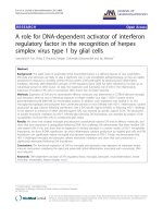
Figure 1 Efficiency of DAI knockdown in primary murine
microglia and astrocytes by small-interfering RNA. Microglia
(Mg) and astrocytes (Ast) were untreated (Con) or transfected with a
combination of three different siRNA duplexes targeting murine DAI
using FuGENE HD transfection reagent. At 72 hours post-
transfection, untreated and transfected cells were exposed to HSV-1
(MOI of 10) for 24 hours and DAI knockdown was confirmed in
whole cell lysates by immunoblot analysis. Expression of a non-
specific protein band observed following Coomassie blue staining is
shown as a loading control (lc). For comparison purposes, DAI
expression in murine small intestine tissue is shown (+).
Furr et al. Journal of Neuroinflammation 2011, 8:99
/>Page 4 of 12
Furthermore, we assessed glial DAI expression follow-
ing in vivo HSV-1infection.Toconfirmthati.n.HSV-1
administration infects glial cells in situ we determined
the expression of HSV-1 glycoprotein G1 (gG1) in micro-
glia and astrocytes isolated from infected and sham-
infected mice. As shown in Figure 3C, this HSV-1 gene
productwasabsentincellsderivedfromshaminfected
mice but was readily detectable in glial cells isolated from
the brains of infected animals with astrocytes exhibiting
higher levels than microglia/macrophages. Importantly,
in vivo HSV-1 challenge elicited marked increases in DAI
expression in both microglia/macrophages (24.8 fold)
and astrocytes (8.5 fold) isolated from the brains o f
infected animals (Figure 3B) in the absence of significant
increases in levels of this viral sensor in total brain pro-
tein isolates (Figure 3A). Finally, we assessed the expres-
sion of the DAI downstream effector molecules RIP3 and
STING in glia isolated from HSV-1 infected and sham
infected mice. As shown in Figure 3D, microglia/macro-
pha ges and astrocytes isolated from uninfected brain tis-
sue showed detectable levels of RIP3 and STING.
Consistent with our in vitro findings, in vivo HSV-1
infection failed to elicit significant changes in RIP3
expression by either cell type. Interestingly, and in con-
trast to our studies in cultured cells, STING expression
was significantly elevated in both microglia/macrophages
and astrocytes following in vivo HSV-1 infection (Figure
3D). While it is likel y that STING expression is regulated
invivobyasyetunidentifiedfactors,itmustbenoted
that resting cultured glial cells express high levels of this
molecule (Figure 2C), perhaps due to in vitro culture
conditions and/or adherence to plastic, and so further
increases in STING expression may not be possible in
such cells.
B-DNA induces inflammatory mediator production by
primary murine microglia and astrocytes
To begin to deter mine whether DAI is functiona l in glial
cells, we have assessed the sensitivity of cultured astrocytes
and microglia to intracellular administration of poly(dA:
dT) double stranded B-DNA. This molecule is a synthetic
DNA that displays a helical B-form configuration in solu-
tion. Therefore, poly(dA:dT) double stranded B-DNA
resembles the form of HSV-1 genomic DNA in infected
cells, if not the specific sequence. Importantly, this mole-
cule has previously been demonstrated to strongly elicit
innate immune responses in a DAI-dependent manner in
other cell types [17,18]. As shown in Figure 4, B-DNA
administration induced production of the inflammatory
cytokines TNF-a and IL-6 by prima ry microglia or astro-
cytes as rapidly as 6 hours post-transfection at levels that
matched or exceeded those elicited following 24-hour
HSV-1 challenge.
DAI knockdown attenuates HSV-induced inflammatory
cytokine production by primary glia
To confirm the functional status of DAI in glial cells
and to begin to determine the relative importance of
this innate immune sensor in their responses to neuro-
tropic DNA viruses we have assessed the effect of DAI
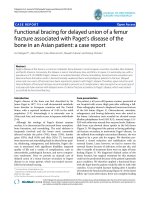
Figure 2 Cultured primary murine astrocytes and microglia constitutively express the cytoplasmic viral sensor DAI and associated
effector molecules, and its expression is elevated following viral challenge. Cultured microglia (Panels A and C) or astrocytes (Panels B and
C) were untreated (0) or infected with HSV-1 (MOI of 0.1, 1 and 10) or MHV-68 (MOI of 1 and 10). At 24 hours post-infection expression of DAI
(Panels A and B), RIP3 and STING (Panel C) were determined in whole cell lysates by immunoblot analysis. Expression of a non-specific protein
band observed following Coomassie blue staining is shown as a loading control (lc) for each blot. For comparison purposes, DAI expression in
murine small intestine tissue is shown (+). Representative results are shown for one of three separate experiments.
Furr et al. Journal of Neuroinflammation 2011, 8:99
/>Page 5 of 12
knockdown on inflammatory cytokine production by
HSV-1 challenged microglia and astrocytes. As shown in
Figure 5, siRNA directed against DAI significantly atte-
nuated HSV-1 induced TNF-a and IL-6 production by
murine microglia. Such an approach also markedly
reduced IL-6 production by HSV-1 infected astrocytes
but was not as effective in reducing TNF-a production
by these cells, where a statistically significant reduction
was only observed at the highest viral MOI used
(Figure 5).
DAI is required for virally-induced production of
neurotoxic mediators by microglia and astrocytes
To begin to establish a role for DAI in the inflammatory
CNS damage associated with neurotropic DNA viral
infections,wehaveassessedtheeffectofDAIknock-
down on the production of soluble neurotoxic mediators
by microglia and astrocytes following HSV-1 infection.
As shown in Figure 6, HSV-1 induced the production of
soluble mediators by microglia that decreased CATH.a
neuron-like cell viability as assessed by changes in cell
Figure 3 Ex vivo glial cells express DAI constitutively and/or inducibly following in vivo HSV-1 infection. Mice were sham-infected (0) or
infected with HSV-1 (HSV: 1 × 10
5
PFU, i.n.). At 4 days post-infection protein isolates were prepared from whole brain tissue (Brain), or microglia
(Mg) or astrocytes (Ast) acutely isolated by flow cytometry, and analyzed for the presence of DAI (Panels A and B), viral gG1 (Panel C), RIP3 and
STING (Panel D) by immunoblot analysis. For comparison purposes, DAI expression in the murine neuron-like cell line CATH.a (Panel A: Cath) and
murine small intestine tissue (Panels A and B: +), or viral gG1 expression in HSV-1 infected Vero cells (Panel C: +) is shown. Expression of a non-
specific protein band observed following Coomassie blue staining is shown as a loading control (lc) for each blot. Immunoblots shown are
representative of three separate experiments.
Furr et al. Journal of Neuroinflammation 2011, 8:99
/>Page 6 of 12
Figure 4 A specific ligand for DAI induces inflammatory cytokine production by isolated cultures of microglia and astrocytes. Microglia
and astrocytes were untreated (0), treated with transfection reagent alone (FG), or transfected with the DAI ligand B-DNA (3 or 6 ug/ml). At 6
and 12 hours following transfection culture supernatants were collected and IL-6 and TNF-a content was assessed by specific capture ELISA. For
comparison purposes inflammatory cytokine production was assessed at 24 hours following HSV-1 infection (MOI of 1 and 10). Data is expressed
as mean +/- SEM (n = 6) and an asterisk indicates a statistically significant difference from levels produced by unstimulated cells (p < 0.05).
Figure 5 DAI knockdown attenuates HSV-1-induced inflammatory cytokine production by murine glial cells. Microglia and astrocytes
were untreated (0), or transfected with siRNA targeting DAI (siDAI) or scrambled siRNA (siCon). At 72 hours following transfection, cells were
exposed to HSV-1 (MOI of 0.01, 0.1, 1, and 10) and levels of TNF-a and IL-6 in the culture supernatants were assessed at 24 hours post-infection.
Data is expressed as mean +/- SEM (n = 3) and an asterisk indicates a statistically significant difference from levels produced by cells treated
with scrambled siRNA (p < 0.05).
Furr et al. Journal of Neuroinflammation 2011, 8:99
/>Page 7 of 12
attachment and trypan blue e xclusion in an MOI and
time dependent manner . Importantly, virally-induced
neurotoxic mediator production was significantly attenu-
ated in cells transfected with siRNA targeting DAI
(siDAI) as compared to control cells that were trans-
fected with scrambled non-specific siRNA ( siCon)
(Figure 6). Interestingly, HSV-infected primary astro-
cytes also produced a substance that elicited neuronal
detachment and death, and production of such neuro-
toxic mediators was similarly attenuated in cells trans-
fected with siRNA targeting DAI (Figure 6). Toget her,
these data point to a key role for DAI in the neurotoxic
immune responses of microglia and astrocytes to DNA
virus infection and may represent an important compo-
nent in the inflammation and damage associated with
viral encephalitis.
Discussion
Several members of the herpesvirus family including
human herpesvirus 6, HSV-1, and HSV-2 can elicit
damaging CNS inflammation [2,32]. Acute HSV -1 infec-
tion or the reactivation of latent virus in the trigeminal
ganglion can lead to the development of severe encepha-
litis that is associated with a high degree of morbidity
and mortality. While acyclovir is currently employed in
the treatment of HSV encephalit is, drug-resistant strains
of HSV-1 are beginning to emerge [33,34]. Furthermore,
despite improvements in diagnosis such infections are
associated with a 30% mortality rate and 62% of survivors
recover with severe neurological deficits [33,35,36]. The
treatment of HSV-1 associated encephalitis is especially
challenging due to the rapid onset of disease and devel-
opment of irreversible neurological damage in otherwise
Figure 6 HSV-1 induces the production of soluble factor(s) by glial cells that elicits neuronal cell damage in a DAI dependent manner.
Primary microglia or astrocytes were untreated (0) or transfected with siRNA targeting DAI (siDAI) or scrambled siRNA (siCon). At 72 hours
following the transfection protocol, cells were uninfected or infected with HSV-1 (MOI of 1) and cultured for a further 24 hours. Filtered
conditioned media from these cells was placed on CATH.a cells and the number of attached neuronal cells was monitored at 4, 8 and 12 hours
post-infection prior to assessment of neuronal cell death by trypan blue exclusion at 12 hours post-infection. For comparison purposes neuronal
cell attachment and death was assessed following addition of media spiked with recombinant TNF-a (65 pg/ml: +). Data is expressed as mean
+/- SEM (n = 3). Asterisks indicate a statistically significant difference in the number of attached cells or degree of cell death from that seen in
unstimulated cells while pound symbols indicate a statistically significant difference in these parameters between cells treated with siRNA
directed against DAI and those that received scrambled siRNA (p < 0.05).
Furr et al. Journal of Neuroinflammation 2011, 8:99
/>Page 8 of 12
healthy individuals. These characteristics suggest that the
innate immune responses of resident CNS cells play a
pivotal role in disease progression, a notion that is sup-
ported by the ability of human microglia to produce key
inflammatory mediators in response to in vitro HSV chal-
lenge or following in vivo infection [37-40]. However,
muc h of our current understanding of HSV-1 enceph ali-
tis pathogenesis comes from rodent models in whic h
intranasal HSV-1 administration results in an acute
necrotizing encephalitis that closely resembles human
disease [41-45]. In these models, HSV-1 infects both neu-
rons and glial cells and elicits inflammatory me diator
production that precedes leukocyte infiltration into the
CNS [41,46]. Such findings are consistent with the recog-
nized ability of other viral pathogens to induce inflamma-
tory cytokine production by microglia [47] and the
susceptibility and responsiveness of astrocytes to produc-
tive HSV-1 infection [8,48]. However, the mechanisms by
which resident CNS cells recognize DNA v iral pathogens
such as HSV-1 have not been fully defined.
We and others have demonstrated that microglia and
astrocytes express an array of cell surface and endosomal
innate pattern recognition receptors, including TLR2
[9,21], TLR3 [49 ,50], TLR7 [50], and TLR9 [50], that are
capable of recognizing viral motifs [51]. Importantly, each
of these cell surface sensors has been implicated in the
perception of HSV by a variety of cell types [13,14,51,52].
However, it is becoming increa singly appar ent that cells,
including glia, possess intracellular sensors that can detect
compromise of the cytosolic compartment. We have
rece ntly demonstrated that murine and human glial cells
functionally express retinoic acid-inducible gene I (RIG-I)
and melanoma differentiation-associated antigen 5
(MDA5) [23,24], two members of the RIG-I-like family of
helicases that have been shown to function as intracellular
pattern recognition receptors for replicative viral RNA
motifs [51,53]. It is possible that such rece ptors may also
indirectly serve as sensors for viral and/or bacter ial DNA
via the actions of RNA polymerase III [54] although a role
for this pathway in HSV recognition by immune cells
remains controversial [55]. Interestingly, a number of cyto-
solic proteins including DAI have been described that can
directly mediate cellular responses to dsDNA [17,18,53]. It
has therefore been suggested that such sensors could play
a critical role in the per ception of viral DNA and this
notion has been supported by the report that DAI med-
iates immune molecule production by HSV-1-infected
murine fibroblasts [17].
In the present study, we provide the first evidenc e that
glial cells express DAI . Resting cultures of pri mary
microglia expressed low levels of this intracellular v iral
sensor, a finding that is consistent with the very low DAI
expression observed in ex vivo microglia. In contrast, cul-
tured astrocytes constitutively expressed robust levels of
this molecule, although it should be noted that this
might be attributable, in part, to our in vitro culture con-
ditions, as astrocytes acutely isolated from uninfected
mice express ed somewhat lower DAI levels. Importantly,
DAI expression was significantly elevated in micro glia
and astrocytes following either in vitro or in vivo HSV-1
challenge. Such upregulation was not specific to this neu-
rotropic alphaherpesvirus as the leukotropic gammaher-
pesvirus, MHV-68, was also capable of elevating DAI
expression by both cell types. As such, it is possible that
glial perception of DNA viruses via this sensor could pro-
mote further DAI expression in a feed-forward manner.
The constitutive expression of this viral sensor by glial
cells and its upregulation following viral challenge pro-
vide circumstantial evidence of a role for DAI in glial
responses to DNA viral pathogens. This notion is further
supported by the robust constitutive expression by
microglia and astrocytes of IPS-1 [23], STING, and R IP3,
reportedly critical downstream effector molecules for
DAI [29-31]. Importantly, we have confirmed that DAI is
functional in primary murine glia by showing that cyto-
plasmic administratio n of the DAI ligand, B-DNA, is a
potent stimulus for the production of key inflammatory
mediators by both microglia and astrocytes. Finally, we
have demonstrated a major role for this intracellular viral
sensor in the immune responses of primary murine glia
to a clinically relevant neurotropic DNA virus by demon-
strating that DAI knockdown significantly and specifi-
cally inhibits HSV-1-induced inflammatory cytokine
production by these cells.
Host responses to viral CNS infections are increasingly
recognized to play a major role in disease pathology and
the neuroinflammation elicited by HSV-1 has been sug-
gested to underl ie the neurological damage associated
with infection [3-7]. However, it not clear whether this
inflammatory damage is mediated primarily by infiltrat-
ing leukocyt es or by the responses of the resident cells of
the CNS themselves. In support of the latter possibility,
activate d glial cells are known to be capable of pro ducing
toxic mediators that can cause widespread CNS damage
[39]. Furthermore, the rapidity with which HSV-1 travels
from the initial site o f infection to the brain all but
assures escape from recognition by adaptive immune
cells. Based on these observations, it appears li kely that
the production of cytotoxic substances by HSV-1 chal-
lenged glial cells could play a significant role in ne uronal
cell dysfunction and/or loss and contribute to the neuro-
pathology associated with HSV-1 encephalitis. In the pre-
sent study, we have demonstrated that soluble factor(s)
released by HSV-infected microglia and astrocytes elicit
neuronal cell damage/death and that the production of
this factor(s) is dependent, at least in part, on the expres-
sion of DAI. While the identification of this/these neuro-
toxic factor(s) is ongoing in our laboratory, we have
Furr et al. Journal of Neuroinflammation 2011, 8:99
/>Page 9 of 12
shown that TNF-a is released by HSV-1 infected glia
(Figure 4) and this cyto kine can eli cit neuronal cell death
(Figure 6). In contrast, we have found that another likely
candidate, nitric oxide, is not released by either microglia
or astrocytes following HSV-1 infection as assessed by
culture medium nitrite content (data not shown) and is
therefore unlikely to be a contri buting factor to neuronal
cell death. Irrespective of the mediator(s) involved, our
findings directly implicate DAI in the initiation of inflam-
matory immune responses by glial cells and suggest a
novel mec hanism underlying the neuropath ology asso-
ciated with acute DNA viral infections of the CNS.
Conclusions
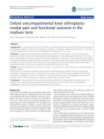
Based upon our results we propose the model shown in
Figure 7. We suggest that neurotropic double-stranded
DNA viruses such as HSV-1 infect microglia and astro-
cytes and replicate within them, generating genomic DNA
within the cytoplasm of each cell type. These viral DNA
motifs are then recognized by DAI, leading to the activa-
tion of downstream effector molecules including IPS-1,
STING, and RIP3. Their activation ultimately liberates the
RelA subunit of NF-kB allowing it to translocate to the
nucleus and to init iate the de novo production of soluble
inflammatory mediators. In addition, it is possible that
RNA polymerase III in the cytosol could transcribe the
viral DNA templates into double stranded RNA contain-
ing 5’-triphosphate which, in turn, could be recognized by
RIG-I and similarly activate NF-kB via the adap tor mole-
cules IPS-1 and STING [54]. However, it should be noted
that at least one study suggests that such a pathway does
not appear to play a significant role in the responses of
Figure 7 Proposed model by whi ch glia recognize neurotropic DNA viruses such as HSV-1 and elicit inflammatory CNS damage.
Replicating DNA viruses generate genomic DNA that serves as a ligand for DAI. DAI then associates with IPS-1 and STING, subsequently
activating NF-kB via the actions of RIP1 and RIP3. In addition, it is possible that RNA polymerase III (Pol III) in the cytosol could transcribe viral
DNA into dsRNA containing 5’-triphosphate (ppp-dsRNA), that could be recognized by RIG-I and similarly activate NF-kB via IPS-1 and STING.
Activated NF-kB subunits then translocate to the nucleus and initiate the production of inflammatory mediators. These soluble mediators could
then promote inflammation, increase blood-brain-barrier permeability, recruit leukocytes into the CNS, and directly or indirectly initiate neuronal
cell damage and/or death.
Furr et al. Journal of Neuroinflammation 2011, 8:99
/>Page 10 of 12
some cell types to HSV infection [55]. Once production is
initiated, cytokines such as TNF-a and IL-6 would be
anticipated to promote inflammation and to increase
blood-brain barrier permeability, facilitating leukocyte
recruitment i nto the CNS. In addition, these soluble
inflammatory mediators could also initiate neuronal cell
loss, either directly or via activation of resident/infiltrating
myeloid cells. As such , the functional expression of DAI
by glial cells may represent an important innate immune
mechanism underlying the rapid and potentially lethal
inflammation associated with neurotropic DNA virus
infection.
Acknowledgements
This work was supported by grant NS050325 to IM from the National
Institutes of Health.
Authors’ contributions
SRF helped to conceive the study, prepared cell cultures and carried out the
in vitro and in vivo experiments, performed data analysis, and drafted the
manuscript. VSC participated in the performance of the in vivo viral
infections and the isolation of CNS cells. MM participated in the preparation
of viral stocks and determination of viral titers. IM helped to conceive the
study, contributed to the experimental design, and edited the final
manuscript. All authors read and approved the final version of the
manuscript.
Competing interests
The authors declare that they have no competing interests.
Received: 14 April 2011 Accepted: 12 August 2011
Published: 12 August 2011
References
1. Xu F, Sternberg MR, Kottiri BJ, McQuillan GM, Lee FK, Nahmias AJ,
Berman SM, Markowitz LE: Trends in herpes simplex virus type 1 and
type 2 seroprevalence in the United States. JAMA 2006, 296:964-973.
2. Baringer JR: Herpes Simplex Infections of the Nervous System. Neurol Clin
2008, 26:657-674.
3. Nakajima H, Kobayashi M, Pollard RB, Suzuki F: Monocyte chemoattractant
protein-1 enhances HSV-induced encephalomyelitis by stimulating Th2
responses. J Leukoc Biol 2001, 70:374-380.
4. Rosler A, Pohl M, Braune HJ, Oertel WH, Gemsa D, Sprenger H: Time course
of chemokines in the cerebrospinal fluid and serum during herpes
simplex type 1 encephalitis. J Neurol Sci 1998, 157:82-89.
5. Wildemann B, Ehrhart K, Storch-Hagenlocher B, Meyding-Lamade U,
Steinvorth S, Hacke W, Haas J: Quantitation of herpes simplex virus type 1
DNA in cells of cerebrospinal fluid of patients with herpes simplex virus
encephalitis. Neurology 1997, 48:1341-1346.
6. Trifilo MJ, Lane TE: Adenovirus-mediated expression of CXCL10 in the
central nervous system results in T-cell recruitment and limited
neuropathology. J Neurovirol 2003, 9:315-324.
7. Conrady CD, Drevets DA, Carr DJ: Herpes simplex type I (HSV-1) infection
of the nervous system: is an immune response a good thing? J
Neuroimmunol 2010, 220:1-9.
8. Aravalli RN, Hu S, Rowen TN, Gekker G, Lokensgard JR: Differential
apoptotic signaling in primary glial cells infected with herpes simplex
virus 1. J Neurovirol 2006, 12:501-510.
9. Konat GW, Kielian T, Marriott I: The role of Toll-like receptors in CNS
response to microbial challenge. J Neurochem 2006, 99:1-12.
10. Carpentier PA, Duncan DS, Miller SD: Glial toll-like receptor signaling in
central nervous system infection and autoimmunity. Brain Behav Immun
2008, 22:140-147.
11. Falsig J, van Beek J, Hermann C, Leist M: Molecular basis for detection of
invading pathogens in the brain. J Neurosci Res 2008, 86:1434-1447.
12. Lokensgard JR, Hu S, Sheng W, vanOijen M, Cox D, Cheeran MC,
Peterson PK: Robust expression of TNF-alpha, IL-1beta, RANTES, and IP-
10 by human microglial cells during nonproductive infection with
herpes simplex virus. J Neurovirol 2001, 7:208-219.
13. Lund J, Sato A, Akira S, Medzhitov R, Iwasaki A: Toll-like receptor 9-
mediated recognition of herpes simplex virus-2 by plasmacytoid
dendritic cells. J Exp Med 2003, 198:513-520.
14. Kurt-Jones E, Mandell L, Cerny A, Chan M, Zhou S, Reed G, Bronson R,
Arnold MM, Knipe DM, Finberg RW: Neonatal herpes simplex infection:
Herpes simplex virus I interaction with TLR2 contributes to lethal
encephalitis. Proc
Natl Acad Sci 2004, 101:1315-1320.
15. Fu Y, Comella N, Tognazzi K, Brown LF, Dvorak HF, Kocher O: Cloning of
DLM-1, a novel gene that is up-regulated in activated macrophages,
using RNA differential display. Gene 1999, 240:157-163.
16. Ishii KJ, Coban C, Kato H, Takahashi K, Torii Y, Takeshita F: A Toll-like
receptor-independent antiviral response induced by double-stranded B-
form DNA. Nat Immunol 2006, 7:40-48.
17. Takaoka A, Wang Z, Choi M, Yanai H, Negishi H, Ban T, Lu Y, Miyagishi M,
Kodama T, Hondra K, Ohba Y, Taniguchi T: DAI (DLM-1/ZBP1) is a cytosolic
DNA sensor and an activator of innate immune response. Nature 2007,
448:501-506.
18. Wang Z, Choi MK, Ban T, Yanai H, Negishi H, Lu Y, Tamura T, Takaoka A,
Nishikura K, Taniguchi T: Regulation of innate immune responses by DAI
(DLM-1/ZBP1) and other DNA-sensing molecules. Proc Natl Acad Sci 2008,
105:5477-5482.
19. Stetson DB, Medzhitov R: Recognition of cytosolic DNA activates an IRF3-
dependent innate immune response. Immunity 2006, 24:93-103.
20. Rasley A, Anguita J, Marriott I: Borrelia burgdorferi induces inflammatory
mediator production by murine microglia. J Neuroimmunol 2002,
130:22-31.
21. Bowman CC, Rasley A, Tranguch SL, Marriott I: Cultured astrocytes express
toll-like receptors for bacterial products. Glia 2003, 43:281-291.
22. Rasley A, Bost KL, Marriott I: Murine gammaherpesvirus-68 elicits robust
levels of interleukin-12 p40, but not interleukin-12 p70 production, by
murine microglia and astrocytes. J Neurovirol 2004, 10:171-180.
23. Furr SR, Chauhan VS, Sterka D Jr, Grdzelishvili VZ, Marriott I:
Characterization of retinoic acid-inducible gene-I expression in primary
murine glia following exposure to vesicular stomatitis virus. J Neurovirol
2008, 7:1-11.
24. Furr SR, Moedyck-Schauwecker M, Grdzelishvili VZ, Marriott I: RIG-I mediates
nonsegmented single-stranded RNA virus-induced inflammatory
immune responses of primary human astrocytes. Glia 2010, 58:1620-1629.
25. Gasper-Smith N, Marriott I, Bost KL: Murine gamma-herpesvirus 68 limits
naturally occurring CD4+CD25+T regulatory cell activity following
infection. J Immunol 2006, 177:4670-4678.
26. Campanella M, Sciorati C, Tarozzo G, Beltramo M: Flow cytometric analysis
of inflammatory cells in ischemic rat brain. Stroke 2003, 33:586-592.
27. Chauhan VS, Sterka DG Jr, Gray DL, Bost KL, Marriott I: Neurogenic
exacerbation of microglial and astrocyte responses to Neisseria
meningitidis and Borrelia burgdorferi. J Immunol 2008, 180:8241-8249.
28. Flano E, Woodland DL, Blackman MA:
A mouse model for infectious
mononucleosis. Immunol
Res 2002, 25:201-217.
29. Rebsamen M, Heinz LX, Meylan E, Michallet MC, Schroder K, Hofmann K,
Vazquez J, Benedict CA, Tschopp J: DAI/ZBP1 recruits RIP1 and RIP3
through RIP homotypic interaction motifs to activate NF-kappaB. EMBO
2009, 10:916-922.
30. Ishikawa H, Barber GN: STING is an endoplasmic reticulum adaptor that
facilitates innate immune signalling. Nature 2008, 455:674-678, Erratum in:
Nature, 2008 456:274.
31. Ishikawa H, Ma Z, Barber GN: STING regulates intracellular DNA-mediated,
type I interferon-dependent innate immunity. Nature 2009, 461:788-792.
32. Isaacson E, Glaser CA, Forghani B, Amad Z, Wallace M, Armstrong RW,
Exner MM, Schmid S: Evidence of human herpesvirus 6 infection in 4
immunocompetent patients with encephalitis. Clin Infect Dis 2005,
40:890-893.
33. Whitley RJ, Alford CA, Hirsch MS, Schooley RT, Luby JP, Aoki FY, Hanley D,
Nahmias AJ, Soong SJ: Vidarabine versus acyclovir therapy in herpes
simplex encephalitis. N Engl J Med 1986, 314:144-149.
34. Kimberlin DW: Management of HSV encephalitis in adults and neonates:
diagnosis, prognosis and treatment. Herpes 2007, 14:11-16.
Furr et al. Journal of Neuroinflammation 2011, 8:99
/>Page 11 of 12
35. Raschilas F, Wolff M, Delatour F, Chaffaut C, De Broucker T, Chevret S,
Lebon P, Canton P, Rozenberg F: Outcome of and prognostic factors for
herpes simplex encephalitis in adult patients: results of a multicenter
study. Clin Infect Dis 2002, 35:254-260.
36. Hjalmarsson A, Blomqvist P, Sköldenberg B: Herpes simplex encephalitis in
Sweden, 1990-2001: incidence, morbidity, and mortality. Clin Infect Dis
2007, 45:875-880.
37. Hu S, Sheng W, vanOijen M, Cox D, Cheeran MC, Peterson PK: Robust
expression of TNF-alpha, IL-1beta, RANTES, and IP-10 by human
microglial cells during nonproductive infection with herpes simplex
virus. J Neurovirol 2001, 7:208-219.
38. Marques CP, Hu S, Sheng W, Cheeran MC, Cox D, Lokensgard JR:
Interleukin-10 attenuates production of HSV-induced inflammatory
mediators by human microglia. Glia 2004, 47:358-366.
39. Marques CP, Cheeran MC, Palmquist JM, Hu S, Urban SL, Lokensgard JR:
Prolonged microglial cell activation and lymphocyte infiltration
following experimental herpes encephalitis. J Immunol 2008,
181:6417-6426.
40. Marques CP, Hu S, Sheng W, Lokensgard JR: Microglial cells initiate
vigorous yet non-protective immune responses during HSV-1 brain
infection. Virus Research 121:1-10.
41. Mori I, Goshima F, Ito H, Koide N, Yoshida T, Yokochi T, Kimura Y,
Nishiyama Y: The vomeronasal chemosensory system as a route of
neuroinvasion by herpes simplex virus. Virology 2005, 334:51-58.
42. Ikemoto K, Pollard R, Fukumoto T, Morimatsu M, Suzuki F: Small amounts
of exogenous IL-4 increase the severity of encephalitis induced in mice
by the intranasal infection of herpes simplex virus type 1. J Immunol
1995, 155:1326-1333.
43. Esiri MM, Drummond CWE, Morris CS: Macrophages and microglia in HSV-
1 infected mouse brain. J Neuroimmunol 1995, 62:201-205.
44. Mansur DS, Kroon EG, Nogueira ML, Arantes RME, Rodrigues SCO, Akira S,
Gazzinelli RT, Campos MA: Lethal encephalitis in myeloid differentiation
factor 88-deficient mice infected with herpes simplex virus 1. Am J
Pathol 2005, 166:1419-1426.
45. Nair A, Hunzeker J, Bonneau RH: Modulation of microglia and CD8+ T cell
activation during the development of stress-induced herpes simplex
virus type-1 encephalitis. Brain Behav Immun 2007, 21:791-806.
46. Beers DR, Henkel JS, Kesner RP, Stroop WG: Spatial recognition memory
deficits without notable CNS pathology in rats following herpes simplex
encephalitis. J Neurol Sci 1995, 131:119-127.
47. Mariani MM, Kielian T: Microglia in infectious diseases of the central
nervous system. J Neuroimmune Pharmacol 2009, 4:448-461.
48. Dix RD, Hurst LI, Keane RW: Herpes simplex virus type 1 infection of
mouse astrocytes treated with basic fibroblast growth factor. Journal of
General Virology 1992, 73:1845-1848.
49. Bsibsi M, Persoon-Deen C, Verwer RW, Meeuwsen S, Ravid R, Van Noort JM:
Toll-like receptor 3 on adult human astrocytes triggers production of
neuroprotective mediators. Glia 2006, 53:688-695.
50. Jack CS, Arbour N, Manusow J, Montgrain V, Blain M, McCrea E, Shapiro A,
Antel JP: TLR signaling tailors innate immune responses in human
microglia and astrocytes. J Immunol 2005, 175:4320-4330.
51. Kumar H, Kawai T, Akira S: Pathogen recognition by the innate immune
system. Int Rev Immunol 2011, 30:16-34.
52. Zhang SY, Jouanguy E, Ugolini S, Smahi A, Elain G, Romero P, Segal D,
Sancho-Shimizu V, Lorenzo L, Puel A, Picard C, Chapgier A, Plancoulaine S,
Titeux M, Cognet C, von Bernuth H, Ku CL, Casrouge A, Zhang XX,
Barreiro L, Leonard J, Hamilton C, Lebon P, Héron B, Vallée L, Quintana-
Murci L, Hovnanian A, Rozenberg F, Vivier E, Geissmann F, Tardieu M,
Abel L, Casanova JL: TLR3 deficiency in patients with herpes simplex
encephalitis. Science 2007, 317:1522-1527.
53. Wilkins C, Gale M Jr: Recognition of viruses by cytoplasmic sensors. Curr
Opin Immunol 2010, 22:41-47.
54. Ablasser A, Bauernfeind F, Hartmann G, Latz E, Fitzgerald KA, Hornung V:
RIG-I-dependent sensing of poly(dA:dT) through the induction of an
RNA polymerase III-transcribed RNA intermediate. Nat Immunol 2009,
10:1065-1072.
55. Melchjorsen J, Rintahaka J, Søby S, Horan KA, Poltajainen A, Østergaard L,
Paludan SR, Matikainen S: Early innate recognition of herpes simplex virus
in human primary macrophages is mediated via the MDA5/MAVS-
dependent and MDA5/MAVS/RNA polymerase III-independent pathways.
J Virol 2010, 84:11350-11358.
doi:10.1186/1742-2094-8-99
Cite this article as: Furr et al.: A role for DNA-dependent activator of
interferon regulatory factor in the recognition of herpes simplex virus
type 1 by glial cells. Journal of Neuroinflammation 2011 8:99.
Submit your next manuscript to BioMed Central
and take full advantage of:
• Convenient online submission
• Thorough peer review
• No space constraints or color figure charges
• Immediate publication on acceptance
• Inclusion in PubMed, CAS, Scopus and Google Scholar
• Research which is freely available for redistribution
Submit your manuscript at
www.biomedcentral.com/submit
Furr et al. Journal of Neuroinflammation 2011, 8:99
/>Page 12 of 12