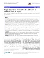
báo cáo hóa học: " Nogo receptor is involved in the adhesion of dendritic cells to myelin" ppt
Bạn đang xem bản rút gọn của tài liệu. Xem và tải ngay bản đầy đủ của tài liệu tại đây (1.59 MB, 12 trang )
RESEARC H Open Access
Nogo receptor is involved in the adhesion of
dendritic cells to myelin
Claire L McDonald
1
, Karin Steinbach
2
, Florian Kern
3
, Rüdiger Schweigreiter
3
, Roland Martin
4
, Christine E Bandtlow
3
and Markus Reindl
1*
Abstract
Background: Nogo-66 receptor NgR1 and its structural homologue NgR2 are binding proteins for a number of
myelin-associated inhibitory factors. After neuronal injury, these inhibitory factors are responsible for preventing
axonal outgrowth via their interactions with NgR1 and NgR2 expressed on neurons. In vitro, cells expressing NgR1/
2 are inhibited from adhering to and spreading on a myelin substrate. Neuronal injury also results in the presence
of dendritic cells (DCs) in the central nervous system, where they can come into contact with myelin debris. The
exact mechanisms of interaction of immune cells with CNS myelin are, however, poorly understood.
Methods: Human DCs were differentiated from peripheral blood monocytes and mouse DCs were differ entiated
from wild type and NgR1/NgR2 double knockout bone marrow precursors. NgR1 and NgR2 expression were
determined with quantitative real time PCR and immunoblot, and adhesion of cells to myelin was quantified.
Results: We demonstrate that human immature myeloid DCs express NgR1 and NgR2, which are then down-
regulated upon maturation. Human mature DCs also adhere to a much higher extent to a myelin substrate than
immature DCs. We observe the same effect when the cells are plated on Nogo-66-His (binding peptide for NgR1),
but not on control proteins. Mature DCs taken from Ngr1/2 knockout mice adhere to a much higher extent to
myelin compared to wild type mouse DCs. In addition, Ngr1/2 knockout had no effect on in vitro DC differentiation
or phenotype.
Conclusions: These results indicate that a lack of NgR1/2 expression promotes the adhesion of DCs to myelin. This
interaction could be important in neuroinflammatory disorders such as multiple sclerosis in which peripheral
immune cells come into contact with myelin debris.
Keywords: Nogo receptor, NgR1, NgR2, Nogo-66, myelin associated glycoprotein, MAG, myelin, dendritic cells
Background
Injury to the central nervous system (CNS) has long been
known to cause fatal and irreversible damage to axons and
neurons. A number of physi cal and molecular inh ibitory
factors expressed by neurons, astrocytes, and oligodendro-
cytes serve to maintain the architecture of the mature
CNS, but at the same time contribute to the lack of repair
mechanisms following da mage. Some of the major molecu-
lar inhibitors to regeneration are those associated with
myelin (myelin-associated inhibitory factors, MAIFs).
MAIFs include Nogo-A [1,2], myelin-associated
glycoprotein (MAG) [3,4] and oligodendrocyte-myelin gly-
coprotein (OMgp) [5]. These factor s are all binding part-
ners for the Nogo-66 receptor-1 (NgR1), a mainly neuron-
expressed, GPI-anchored protein [6-8]. Nogo-66 is a 66
amino acid long region of Nogo-A that binds NgR1 and is
largely responsible for inhibiting neurite outgrowth. Since
the identification of NgR1, two structural homologues have
been discovered, termed NgR2 and NgR3. NgR2 is a high
affinity binding protein for MAG [9,10] and the binding
protein of NgR3 has not yet been identified. As NgR1 is a
GPI-anchored protein, it requires co-receptors in order
to transmit its signal inside the cell. Thus, it is o ften
found assembled in a heterotrimeric complex composed of
p75
NTR
[7]orTROY[11],andLINGO-1(Leucinerich
* Correspondence:
1
Clinical Department of Neurology, Innsbruck Medical University,
Anichstrasse 35, A-6020 Innsbruck, Austria
Full list of author information is available at the end of the article
McDonald et al. Journal of Neuroinflammation 2011, 8:113
/>JOURNAL OF
NEUROINFLAMMATION
© 2011 McDonald et al; licensee BioMed Central Ltd. This is an Open Access article distribute d under the terms of the Creative
Commons Attribution License (http://c reativecommons.org/licenses/by/2.0), which permits unrest ricted use, distribution, and
reproduction in any medium, provided the original work is properly cited.
repeat and Ig domain-containing, Nogo receptor-interact-
ing protein) [12]. However, due to the findings of NgR1
expression without LINGO-1 [13], or without both TROY
and p75
NTR
[14], it is likely that more signal tra nsducing
subunits of the NgR1 complex remain to be identified.
Binding of the NgR1 inhibitory complex by MAIFs leads to
activation of intracellular RhoA, thereby resulting in axonal
outgrowth inhibition, or modulation of cell adhesion and
motility [15].
NgR1 expression has been identified in a few non-
neuronal cell types, where it mediates adhesion of
these cells to MAIFs. For example, fibroblasts, glioma
cells, macrophages, and some human immune cells
have all bee n found to express NgR1 and to be inhib-
ited from adhering to myelin substrates [13,16-18]. Our
aim was to expand on this data and to further clarify
the r ole of NgRs i n human immune cells. In this paper
we focus on dendritic cells (DCs) due to their impor-
tance in a number of neuroinflammatory situations and
due to the high NgR1 ex pression we found in imma-
ture DCs. DCs in the immature state are tissue resident
and are responsible for surveying the tissue for possible
insults. Upon activation by defined factors (cytokines,
bacterial or viral molecules), DCs become mature and
travel to lymph nodes to present antigen to T cells
[19]. T his change in func tion is reflected in the up-reg-
ulation of the antigen presenting molecules HLA-DR,
CD86 and CD83, as well as the chemokine receptor
CCR7 to aid cellular migration.
DCs are usually not present in the healthy brain, how-
ever, they have been found to accumulate in the CNS
parenchyma during a wide range of inflammatory insults
[20-22] and they are emerging as important players in
CNS autoimmunity, speci fically in multiple sclerosis
(MS) [23]. Indeed, mature DC markers have been con-
sistently found in the inflamed meninges and perivascu-
lar cuffs of most active MS lesions examined [24]. Thus,
it would be valuable to further understand the role of
DCs within the inflammatory milieu of CNS myelin
debris.
In the current study, we demonstrate that NgR1 and
NgR2 (referred to jointly as NgR1/2) are expressed to a
higher extent by human immature myeloid DCs
(immDCs) compare d to mature myeloid DCs (matDCs).
DCsthatdonotexpressNgR1/2aremoreadherent
when plated on a myelin substrate compa red to those
that express NgR1/2. Promotion of adhesion could also
be demonstrated in mouse DCs genetically lacking
NgR1/2. The interaction of DCs with myelin debris pro-
posed here c ould have important implications for our
understanding of how immune cells act within CNS
inflammatory lesions.
Methods
Generation of human monocyte-derived dendritic cells
Whole human blood was obtained by venous puncture
into EDTA tubes with informed, written consent from 9
healthy donors with approval from the local institutional
review board of Innsbruck Medical University. Myeloid
DCs were generated according to established standard
procedures [25,26]. Firstly, peripheral blood mononuc-
lear cells (PBMCs) were isolated from the blood by den-
sity gradient centrifugation using Ficoll™-based
lymphocyte separation medium (PAA, Pasching, Aus-
tria). PBMCs were washed with 0.9% saline solution
(FreseniusKabi,Graz,Austria)andseededatadensity
of 3.3 × 10
6
cells/ml in a 6-well plate in serum-free
medium (Lonza x-vivo chemically-defined medium,
Cologne, Germany). After two h ours of incubation at
37°C, with 5% CO
2
, monocytes selectively adhered to
the cell culture-treated plastic. At this stage, all non-
adherentcellpopulationswerewashedawaybyrinsing
three times with RPMI1640 medium (Gibco, Invitrogen,
Carlsbad, CA, USA). After the washing steps, adherent
monocytes were cultured for 8 days in serum-free med-
ium supplemented with 1% penicillin streptomycin
(PenStrep, Invitrogen, Carlsbad, CA, USA), 800 U/ml
granulocyte/monocyte colony sti mulating factor (GM-
CSF, Novartis, Leukomax, Basel, Switzerland) and 40
ng/ml interleukin-4 (human recombinant IL-4, Invitro-
gen). Every two days, cells were fed with fresh medium,
PenStrep, GM-CSF and IL-4. By day 6, the human
monocytes had differentiated into loosely adherent
immature dendritic cells (immDCs). Addition of a
defined maturation cocktail for the last two days of cul-
ture resulted in generation of mature DCs (matDCs).
Maturation cocktail (MC) consisted of interleukin 1b
(2 ng/ml, Invitrogen), IL-6 (10 ng/ml, Invitrogen),
tumour necrosis factor-a (TNF-a, 10 ng/ml, Invitrogen)
and prostaglandin E2 (PGE2, 1 μg/ml, Sigma-Aldrich,
St.Louis,MO,USA).Onday8ofculture,immature
and mature DCs were harvested for flow cytometric
analysis, RNA extraction, and adhesion assay.
Isolation of human immune cell subsets
T cells were isolated from human PBMCs using a com-
mercially available magnetic cell separator T cell deple-
tion kit (Miltenyi Biotec GmbH, Bergisch Gladbach,
Germany) to produce ex vivo T cells (Tex). Cells were
cultured in serum-free medium in the presence of anti-
CD3antibody(Tcont),andinthepresenceorabsence
of T cell activator phytohaem agglutinin (T PHA+ and T
PHA-, respectively) for 2 days. Epstein Barr virus-trans-
formed B lymphocytes were used as a B cell line (BCL).
Monocytes were isolated from PBMCs by magnetic cell
McDonald et al. Journal of Neuroinflammation 2011, 8:113
/>Page 2 of 12
separator monocyte depletion kit (Miltenyi Biotec
GmbH) to produce ex vivo monocytes. They were then
maintained in serum-fr ee medium for 7 days, and given
either interferon gamma (IFN-g, 100 ng/ml, Invitrogen)
or lipopolysaccharide (LPS, 100 ng/ml, Si gma-Aldrich)
for the last two days of culture.
Generation of mouse bone marrow-derived dendritic cells
Wild type male C57BL/6J mice were obtained from
Jackson Laboratories and hous ed in the animal house of
Innsbruck Medical University. Ngr1/2 double knockout
mice (Ngr1/2-/-) were generated by crossing Ngr1-/-
mice [27] with Ngr2-/- mice as previously described
[10]. Bone marrow derived myeloid DCs were prepared
according to established standard procedures as
describedbyLutzetal.[28].Miceweresacrificedby
cervical dislocation and the tibi ae and femurs were
removed. The bones were cleaned of all muscle tissue
and sterilised with 70% ethanol. The bone marrow was
flushed out with cold RPMI1640 containing 10% foetal
calf serum (FCS, Sigma-Aldrich) and b-mercaptoethanol
(b-ME, 50 μM, Sigma-Aldrich). The marrow was sepa-
rated into a single cell sus pension by repeated pipetting
and passed through a nylon mesh to remove bone and
debris. Contaminating erythrocytes were removed by
lysis on ice using erythrocyte lysis buffer (containing
0.15 M ammonium chloride, 10 mM potassium bicarbo-
nate, 0.1 mM EDTA (all from Roth, Karls ruhe, Ger-
many), with pH adjusted to 7.0-7.2) and cells were
counted. 20 × 10
6
bone marrow precursor cells were
seeded in RPMI containing 10% FCS, 50 μM b-ME, and
20 ng/ml GM-CSF (ImmunoTools, Friesoythe, Ge r-
many) in 75 cm
3
flasks. After two days, flasks were
gently swirled and 75% of medium was removed. The
same volume of fresh medium was added back, contain-
ing 40 n g/ml GM-CSF. On day 4, the culture is made
up of firmly attached stromal cells covered in clusters of
loosely attached DCs, and non-adherent granulocytes.
The granulocytes were washed away and DCs were sub-
cultured at a concentrati on of 1 × 10
6
cells/ml/well in a
24-well plate, with 20 ng/ml GM-CSF. On day 6, cells
were fed by removal of 75% of the medium and adding
back the same volume containing GM-CSF. For the gen-
eration of mature DCs, maturation cocktail containing a
final concentration of 2 ng/ml IL-1b (Invitrogen), 10 ng/
ml IL-6 (ImmunoTools), 10 ng/ml TNF-a (Immuno-
Tools), and 1 μg/ml PGE-2 ( Sigma-Aldrich) was add ed
for the last two days of cu lture. Cells were harvested for
flow cytometric analysis, RNA extraction, and adhesion
assay on day 8.
Flow cytometric analysis
In order to define and compare the phenotype of in
vitro-generated human and mouse DCs, cells were
characterised by flow cytometry. Briefly, cells were
washed with FACS Cell Wash solution (Becton Dickin-
son Biosciences, San Jose, CA, USA) and 200000 cells in
100 μl Cell Wash solution were used per staining. Each
staining consisted of a fluorescein isothiocyanate
(FITC)-labelled antibody and phycoerythrin (PE)-labelled
antibody, occasionally in combination with a peridinin
chlorophyll protein complex (perCP)-labelled antibo dy.
The following fluorescently labelled antibodies were
used for detection of human DC antigens: HLA-DR-
PerCP (BD Biosciences), CD86-FITC (BD Biosciences),
CCR7-PE (R&D Systems, Minneapolis, MN, USA),
CD83-FITC (BD Bioscien ces), CD11b-PE (BD Bios-
ciences), CD1a-FITC (BD Biosciences), and CD11c-PE
(BioLegend, San Diego, CA, USA). Fluorescently labelled
mouse antibodies were: MHC II-PE (BD Biosciences),
CD86-FITC (BioLegend), CCR7-PE (BioLegend), CD83-
FITC (BioLegend), CD11b-PE (BioLegend), CD11c-PE
(BioLegend), and CD14-FITC (BD Biosciences). First,
human or mouse DCs were blocked for 15 minutes with
2 μg/200000 cells human or mouse IgG, respectively.
Fluorescently labelled antibodies were adde d to the cells
at the concentration suggested by the manufacturer, and
incubated for 20 minutes at room temperature in the
dark. Cells were washed and resuspended in 300 μl Cell
Wash before being analysed by flow cytometry with a
BD FACScan instrument using Cell Quest Pro Software
(BD Biosciences).
Determination of mouse supernatant cytokine
concentrations
The following cytokines were measured in cell culture
supernatant from mouse immature and mature DCs
from WT and Ngr1/2- /- mice: 6Ckine , CTACK, Eotaxin,
GCSF, GM-CSF, IL-2, IL-3, IL-4, IL-5, IL-9, IL-10, IL-
12p40p70, IL-12p70, IL-13, IL-17, IFN-g, KC, Leptin,
MCP-1, MCP-5, MIP-1a,MIP-2,MIP-3b,RANTES,
SCF,sTNFRI,TARC,TIMP-1,Thrombopoietin,and
VEGF. Cytokine levels were determined as per the pro-
tocol using the Ray Biotech mouse cytokine antibody
array G2 (AAM-CYT-G2-8, RayBiotech, Norcross, GA,
USA). The array consists of antibody-coat ed glass slides
that were pre-treated according to the manufacturer’s
instructions and incubated with cell culture superna-
tants for 2 hours. All sample measurements were per-
formed in duplicate. The glass slides were then washed,
incubated with a biotin-conjugated anti-cytokine mix for
2hours,washedagain,anddevelopedfor2hourswith
Cy3-conjugated streptavidin. The arrays were scanned
for fluorescent signals with a GenePix 4000B scanner
(Axon Instruments, GenePix version 5.0) and analysed
with the Ray biotech analysis tool, a data analysis pro-
gram based on Microsoft Excel technology specifically
designed to analyse Ray biotech G Series Antibody
McDonald et al. Journal of Neuroinflammation 2011, 8:113
/>Page 3 of 12
Arrays. Signals were normalised using positive and nega-
tive controls included on the array.
RNA isolation and real time quantitative PCR
Cells (a minimum of 10
6
) were washed with PBS and
homogenised in 1 ml TRIzol reagent (Invitrogen). RNA
was extracted as per the manufacturer’sprotocol,dis-
solved in diethylpyrocarbonate (DEPC)-treated water and
the concentration was determined using a spectrophot-
ometer (NanoDrop 1000, peqlab, P olling, Austria). 1 μg
of RNA from each sample was reverse transcribed with
the High Capacity cDNA Reverse Transcription Kit
(Applied Biosystems, Carlsbad, CA, USA) using random
primers. The protocol was followed as described in the
kit. cDNA was diluted 1:4 and immediately used f or
RT qPCR. Levels of Nogo receptor component mRNAs
in human and mouse DCs were determined using
TaqMan RT qPCR Assays (Applied Biosystems).
Assays used for human mRNA detection were: NgR1
(Hs00368533_m1), NgR2 (Hs00604888_m1), LINGO-1
(Hs01072978_m1), TROY (Hs00218634_m1) and p75
NTR
(Hs00609977_m1). Assays used for mouse mRNA detec-
tion were as follows: NgR1 (Mm00452228_m1), NgR2
(Mm01336368_g1), LINGO-1 (Mm01173306_m1), TROY
(Mm00443506 _m1), and p75
NTR
(Mm00446296_m1). 18
s rRNA was measured in each sample as an endogenous
control in order to control for varying cDNA concentra-
tions and human or mouse brain cDNA were used as a
positive control for all assays (commercially available
human foetal brain RNA was used from Clontech
Laboratories,Inc.,MountainView,CA,USA).Assays
were performed as described by the manufacturer, with a
final assay volume of 25 μl. Experiments were performed
in duplicate wells a nd all assays were f irst screened for
detection of genomic DNA. Data were collected using
the 7300 Real-Time PCR System (Applied Biosystems)
and analysed by the comparative Ct method, where; ΔCt
= target Ct - endogenous Ct; and ΔΔCt = ΔCt
matDC
-
ΔCt
immDC
; relative mRNA expression = 2
-ΔΔCt
. Immature
DCs were assigned as the calibrator for all relative quan-
tifications, except where otherwise stated.
Western Blot
Human brain and human immature and mature DCs
were lysed in buffer containing 150 mM NaCl, 1% Tri-
ton X-100, 10% glycerol, 50 mM Hepes pH 7.40, and
protease inhibitor cocktail (Roche Applied Sciences,
Mannheim, Germany). Protein concentration of all
lysates was determined with the bicinchoninic acid pro-
tein assay (BCA, Sigma-Aldrich). 22 μ g of human brain,
and 70 μg each of immDC and matDC protein were
denatured and loaded onto a 10% Bis/Tris gel (Invitro-
gen). After separation, protein was blotted onto a
Hybond membrane (Amersham, GE Healthcare,
Buckinghamshire, UK) and probed with NgR11-A anti-
body (diluted 1:3000, Alpha Diagnostic, San Antonio,
TX, USA). Detection was performed with horseradish
peroxidase-conjugated secondary antibodies and
enhanced chemiluminescence detection on film (Amer-
sham, GE Healthcare). To conf irm antibody specificit y,
NgR11-A was first blocked with the immunising peptide
(NgR11-P, Alpha Diagnostic) at 5 times the weight of
NgR11-A used. In order to probe the same membranes
for actin, they were stripped at 60°C with buffer contain-
ing 2% SDS, 100 mM beta-mercaptoethanol and 62.5
mM Tris-HCl, pH 6.8, then washed, blocked and incu-
bated with anti-actin monoclonal antibody (dil uted
1:20000, BD Biosciences).
Myelin extraction from brain
Myelin was isolated from central nervous system tissue
by the density gradient centrifugation method, as
described previously [29]. Briefly, a segment of human
brain, or whole mouse CNS was shock frozen in liquid
nitrogen and stored at -80°C until needed. Tissue was
thawed on ice and cut into smal ler piec es and homoge-
nised in a 0.32 M sucrose solution. The homogenized
tissue was washed three times in 0.32 M sucrose before
being layered over a 0.85 M sucrose sol ution. After cen-
trifugation at 26000 × g for 60 minutes at 4°C, myelin
was contained in the interphase between the high and
low sucrose solutions. The myelin was subjected to
osmotic shock by stirring with distilled water fo r 30
minutes at 4°C, before being washed and ultracentri-
fuged again with 0.32 M sucrose. Finally, the myelin was
washed three times with, and resuspended in distilled
water. The protein concentration of the myelin extract
was determined with BCA protein assay.
MBP extraction
Human myelin derived MBP was purified from normal
human brain according to the procedure of Eylar et al.
[30]. SDS-PAGE and Western blot with a monoclonal
antibody to MBP 130-137 (Millipore GmbH, Vienna,
Austria) was used to confirm the purity of the MBP
preparation.
Cloning and production of recombinant proteins
A DNA fragment encoding the mouse Nogo-66 loop
was amplified from the mouse Nogo-A clone IRAV-
p968A04133D (ImaGene, Berlin, Germany) with the pri-
mers mM_RTN4-66-s (5’ -CTA CCA TGG GCA GGA
TAT ATA AGG GTG TGA TCC-3’ )andmM_RTN4-
66-as (5’-GCT TGC GGC ACC CTT CAG GGA ATC
AAC TAA ATC-3’). The fragment was digested with
NcoI and ligated into pET28a(+) vector using the NcoI
and a blunted NotI site. The sequence was verified by
sequencing at LGC Genomics (Berlin).
McDonald et al. Journal of Neuroinflammation 2011, 8:113
/>Page 4 of 12
E. coli Rosetta were transformed with Nogo-66 pET28
and induced in 1 litre of LB culture medium with 1 mM
IPTG at an OD600 of 0.6 and a temperature of 30°C.
After 3 hours the bacteria were harvested by centrifuga-
tion at 4000 × g for 5 minutes. The p ellet was resus-
pended in PBS substituted with 1% Triton X-100 and
protease inhibitors and sonicated. After 30 minutes of
incubation on ice the lysate was centrifuged and the pel-
let dissolved in 8 M urea. Recombinant Nogo-66-His
was purified with TALON
®
Cobalt resin (Clontech
Laboratories) under denaturing conditions and eluted
with elution buffer (50 mM Hepes pH 4, 300 mM imi-
dazol, 150 mM NaCl). Finally, the eluate was dialysed
against DMEM adjusted to p H 4. Protein concentration
was determined by BCA assay (Thermo Scientific, Rock-
ford, IL, USA).
MAG-Fc was produced as described previously [31].
Briefly, conditioned medium of transiently transf ected
CHO-K1 cells was harvested and re combin ant protein
was purified using Protein A/G Agarose (Thermo Scien-
tific, Rockford, IL, USA). Purity and concent ration were
confirmed by comparing band intensity o n SDS-PAGE
to BSA standard.
DC adhesion assay
Adh esion assay s for human DCs were conduc ted in 96-
well plates. All cell types and conditions were assayed in
triplicate. The following substrates were all used at con-
centrations of 100 and 10 μg/ml for adhesion assay:
human myelin, human MBP, His-tagged mouse Nogo-
66 (Nogo-66-His, amino acids 1025-1090). 50 μlofeach
substrate w as added to the 96-well plate and incubated
for 4 hours at 37°C. MAG-Fc was added at 10 μg/ml
and was coa ted on human IgG to aid clustering of the
protein. First, 15 μg/ml human IgG (Sigma-Aldrich) was
added to wells in 50 mM bicarbonate buffer (pH 9) and
incubated over night at 4°C. The next da y, wells were
washed and MAG-Fc was added along with the other
adhesion substrates to their respective wells and the
plate was incubated for 4 hours at 37°C, 5% CO
2
. Excess
substrate was removed and all wells were washed once
with medium before addition of cells. Human mono-
cyte-derived DCs were prepared as described above and
harvested on day 8. Cells were collecte d, counted and
plated at 200000 cells/ml in the 96-well plate in se rum-
free medium. Cells were allowed to adhere for 30 min-
utes at 37°C, 5% CO
2
. Non-adherent cells were gently
removed and wells were washed three times with med-
ium. In order to detect and count CD11b
+
DCs, cells
were fixed and fluorescently stained as follows. Cells
were fixed with 4% paraformaldehyde (PFA) for 30 min-
utes at room temperature. After the PFA w as washed
away, nonspecific antibody binding was blocked by addi-
tion of 20 μg/ml human IgG in PBS containing 5%
normal goat serum (NGS, Invitrogen) and 1% bovine
serum alb umin (BSA, Sigma-Aldrich) for 1 hour at
room temperature. In order to detect adherent DCs,
CD11b antibody ( BD Biosciences) diluted 1:100 in 1%
NGS, 1% BSA was added to the cells and incubated
over night at 4°C with gentle shaking. The following
day, the cells were washed three times with PBS and
visualised at 10 × magnification with a fluorescent
microscope (Leica Microsystems, Camb ridge, UK). Four
digital photos were taken per well and cells were
counted using the particle analysis tool from ImageJ
[32].
Adhe sion of mouse DCs was assayed in 96-well plates
and based on the method described by Kueng et al [33].
10 μg/ml mouse myelin was added to the respective
wells and incubated for 4 hours at 37°C. Myelin was
removed and wells were washed once with medium
before addition of cells. Mouse bone marrow-derived
DCs were prepared as described above and harvested on
day 8. Cel ls were collected, counted and plated at
200000 cells/ml in tripli cate in the 96-well plate in
RPMI 1640. Cells were allowed to adhere for 30 minutes
at 37°C, 5% CO
2
. Non-adherent cells were gently
removed and wells were washed three times with med-
ium. In order t o quantify the number of remaining
adherent cells, they were stained with cresyl violet and
absorbance was measured as follows. Cells were fixed
with 4% PF A for 30 minutes at room temperature. Cells
were staine d with 0.04% cresyl violet (Sigma -Aldrich) in
20% methanol for 30 minutes. The dye was then
extracted with 0.1 M citric acid in 50% ethanol for 30
minutes on a rotating shaker. Absorbance of eac h well
was measured at 570 nm using the DTX 880 Multimode
Detector with Multimode Analysis Software (Beckman
Coulter, Krefeld, Germany).
Statistical analysis
All statistical analy ses were carried out using GraphPad
Prism 5 software (GraphPad Software Inc., San Diego,
CA, USA). For flow cytometry data, an unpaired, two-
tailed student’s t test was used to compare the means of
each marker in immature versus mature DCs. Microar-
ray data was analysed using TIGR MeV_4_5 (Multiple
Experiment Viewer), a Java tool for genomic data analy-
sis [34] which measures
significance of microarray (SAM). Multi-class SAM was
used to identify significant cytokines based on differen-
tial expression between the four groups at a false discov-
ery rate (FDR, proportion of genes likely to have been
identified by chance as being significant) of 0%. To
determine significance of RT qPCR data, ΔCt values
were compared using the Wilcoxon matched-pairs
signed rank test, as per Yuan et al. [35]. Human DC
adhesion to myelin was measured using a two-way
McDonald et al. Journal of Neuroinflammation 2011, 8:113
/>Page 5 of 12
repeated measure ANOVA. The association of human
RT qPCR ΔCt values with adhesion was calculated
using Spearman correlation. Mouse immature WT vs.
Ngr1/2-/- and mature WT vs. Ngr1/2-/- adhesion to
myelin were analysed using the Wilcoxon matched-pairs
signed rank test.
Results
Expression of NgRs in human and mouse DCs
As our aim was to expand on current knowledge of the
role of NgRs in non-CNS cells, we began the study as a
screen for NgR1 expression in human peripheral
immune cells. Expression of NgR1 mRNA was measured
in a panel of human immune cells (un-stimulated and
stimulated T cells, B cell line, monocytes, immature and
mature DCs) using TaqMan real time quantitative PCR
(RT qPCR). Expression of NgR1 mRNA was five times
higher in i mmature DCs compared to all other immune
cells tested (Figure 1A). We thus concentrated on
further examining NgR expression in DCs. Human
monocyte derived myeloid DCs were generated as
described and first characterised by flow cytometry (Fig-
ure 1B). More than 95% of the cells expressed CD11b,
indicating high purity of monocyte-derived DCs. The
DC phenotype was confirmed by high CD11 c expres-
sion (Figure 1B). Only 2.8 ± 0.6% of untreated DCs
expressed CD83. The percentage of cells expressing
CD83 increased significantly to 90.0 ± 2.2% after addi-
tion of maturation cocktail, indicating successful DC
maturation. Significant increases in expression of human
leukocyte antigen (HLA), CD86, CCR7 and a decrease
in CD1a provide further evidence of successful matura-
tion (Figure 1B). Having verified the in vitro generation
of myeloid DCs, we went on to examine the regulation
of NgR expression in these cells.
NgR1 expression was increased in comparison to the
monocytes from which the immDCs were generated
(Figure 1A). It is then down-regulated upon maturation.
The increased transcription was confirmed by higher
protein expression of NgR1 in human immature DCs, as
determined by western blot (Figure 1C). This regulation
of NgR1 expression between immature and mature DCs
prompted us to also measure the expression of NgR1’s
co-receptors , as well as of NgR2. NgR2 mRNA is down-
regulated in the same manner upon maturation of
human DCs (Figure 1D). Expression of NgR1 co-recep-
tors LINGO-1, TROY and p75
NTR
was not down-regu-
lated upon maturation in the same way as NgR1.
Due to the fact that we later used mouse DCs in our
functional analysis of Ngr1/2 knockout, we also charac-
terised mouse bone marrow derived DCs and analysed
expression of NgR1, NgR2 and co-receptors. The char-
acterisation of WT mouse DC is shown in Figure 2A.
Like human DC, more than 95% of mouse in vitro-
generated DCs express CD11b, suggesting a high purity
of monocyte-derived DCs, whereas CD14 expression
was low. There is a tre nd towards higher CD86 and
lower CD11 c upon maturation, in concurrence with
Lutz et al. [28].
In contrast to human DCs, mouse NgR1 expression
does not change significantly upon maturation; however
there is a trend towards up-regulation in mature DCs
(Figure 2B). NgR2 expression on the other hand, is sig-
nificantly down-regulated upon addition of maturation
cocktail, however not to the same extent as in human
mature DCs. LINGO-1 and p75
NTR
are expressed at
similar levels as NgR1 in mouse immature and mature
DCs but also do not follow the same expression pattern
observed in human DCs. Discrepancies in expression of
NgRs between human and mouse DCs are also reflected
in expressio n of DC cell surf ace markers, suggesting we
are not dealing with directly comparable cell types.
Furthermore, in both the human and mouse systems,
we did not observe NgR1’sco-receptorsbeingregulated
in the same way as NgR1. However, as it has previousl y
been demonstrated that NgR1 can function without the
full complement of identified co-receptors [13,14,36], we
went on to determine the function al role of NgR1 and
NgR2 in human and mouse DCs.
Myelin promotes adhesion of DCs lacking NgR1 and NgR2
expression
NgR1/2 have been found to mediate the inhibition of
cellul ar adhesion to myelin. Thus, in order to determine
the possible function of NgR1/2 down-regulation in
human mature DCs, adhesion of immature and mature
DCs to a myelin substrate was quantified. Immature and
mature DCs were plated on human myelin and adhesion
was then calculated as the fold change in adhesion com-
pared to on plast ic control. Human mature DCs were
found to adhere significantly more to human CNS mye-
lin compared to immature DCs (Figure 3A). As men-
tioned above, human mature DCs down-regulate NgR1
and NgR2 (Figure 1D). Figure 3B depicts the correlation
between NgR1/2 mRNA expression (graphed as the raw
value for both, 1/ΔCt) in all DCs and their adhesion to
myelin, showing that increased NgR1/2 expression cor-
relates significantly with decreased adhesion to myelin.
In orde r to identify which protein fraction of myelin is
responsible for promoting adhesion of matD Cs, we iso-
lated various components of myelin that do or do not
interact with NgRs, and measured adhesion of the ce lls
in comparison to plastic control. His-tagged Nogo-66
recombinant peptide was plated at the same concentra-
tions as myelin and used as a positive control for NgR1.
As a positive control for NgR2, MAG-Fc was used. As
MAG-Fc needs to be clustere d in order to function cor-
rectly, the plate was first coated with IgG and then
McDonald et al. Journal of Neuroinflammation 2011, 8:113
/>Page 6 of 12
MAG-Fc was added. Myelin basic protein (MBP) was
isolated from human brain white matter and used as a
negative control as it is not known to bind or activate
NgR1 or NgR2. Nogo-66-His demonstrated the same
effect on adhesion of immature and mature DC as seen
with myelin (Figure 3C). That is, mature DCs (which
express less N gR1/2) were found to adhere to a much
higher extent to Nogo-66-His than immature DCs.
Adhesion of mature DCs to MBP and MAG-Fc was
found to remain at a background level and not reach
statistical significance. To ensure that the adhesion
observed with Nogo-66-His was not due to side effects
of the His-tag or bacterial contamination, we performed
a control adhesion assay with two His-tagged and bacte-
rially expressed peptides that do not bind NgR1/2.
Neither NiR-His (amino acids 1 - 172 of rat Nogo-A)
nor the 66-amino acid loop domain of RTN1 had any
effect on adhesio n of DCs (data not shown), thus sug-
gesting that increased adhesion of matDCs to Nogo-66-
His is indeed specific. This confirms tha t Nogo con-
tained in the myelin preparation mediates promotion of
matDC adhesion. Generally, mat DCs adhere better to
any substrate, however, the increase is most significant
for myelin and Nogo-66-His. This indicates that Nogo
might mediate this effect (prob ably due to the loss of
NgR1).
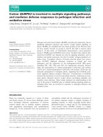
Figure 1 NgR expression in human immune cells.(A) Expression of NgR 1 mRNA in a panel of human immune cells, as determined by
TaqMan RT qPCR. Expression relative to human foetal brain is depicted. Tex: ex vivo T cells; T cont: T cells cultured with anti-CD3 antibody for 2
days; T PHA-: T cells cultured with anti-CD3 and without PHA; T PHA+: T cells cultured with anti-CD3 and stimulated with PHA; BCL: B cell line;
Mono ex: ex vivo monocytes; mono 7d: monocytes maintained in serum-free medium for 7 days; mono 7d IFN: 7d mono’s treated with
interferon gamma for the last 2 days; mono 7d LPS: 7d mono’s treated with lipopolysaccharide for the last 2 days; Brain: commercially available
human foetal brain RNA. (B) Expression of cell surface markers on human monocyte-derived DCs, quantified with flow cytometry. Bars represent
mean of percentage of positive cells for the indicated marker, with SEM of 8 experiments, each representing a different donor. *P < 0.05, **P <
0.01, ***P < 0.001. (C) A representative western blot of NgR1 protein expression in human brain, immature and mature DC. (D) Relative mRNA
expression of NgR1, NgR2, LINGO-1, TROY and p75
NTR
were determined with TaqMan RT qPCR. Mean values relative to immDC with SEM from 8
donors are shown. Wilcoxon matched-pairs signed rank test was used with delta Ct values to determine significance. *P < 0.05, **P < 0.01.
McDonald et al. Journal of Neuroinflammation 2011, 8:113
/>Page 7 of 12
Having found that Nogo-66 promotes adhesion of
human matDC, we wanted to further clarify if it is indeed
the loss of NgR1 expression that is the functional cause
for increased adh esion of matDCs to myelin. To this end,
we took DCs from NgR1/2 double knockout (Ngr1/2-/-)
mice and compared them to wild ty pe (WT) before mea-
suring how they adhere to a myelin substrate. The double
knockout mice were used rather than the single knockouts
in order to exclude an effect resulting f rom the possible
compensatory up-regulation of NgR2 in Ngr1-/- DCs.
DCs generated in vitro from WT and Ngr1/2-/- mice
show similar phenotypes (Figure 2A and 2C). Further-
more, 32 cytokines released from WT and Ngr1/2-/-
DCs were compared in cell culture supernatants using a
glass chip protein array system (Figure 2D). We found
no significant changes in secreted cytokines from mouse
WT and Ngr1/2-/- DCs, thus indicating that the dele-
tion of NgR1 and NgR2 had no influence on the differ-
entiation and phenotype of DC. However, in the
adhesionassaywedidobservethatmatureDCsfrom
Ngr1/2 -/- mice adhere significantly more to myelin than
mature DCs from WT mice (p = 0.02, Figure 3D). This
indicates that a lack of NgR1/2 in mouse mature DCs
promotes their adhesion to a myelin substrate.
Discussion
We describe here the e nhanced adhesion of human
mature DCs to human CNS myelin, and that this
enhanced adhesi on is mediated by a down-regulation
of NgR1 expression. We propose that high NgR1
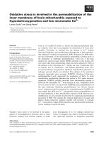
Figure 2 Mouse WT and Ngr1/2-/- DC characterisation and NgR expression . Expression of cell surface markers on mouse WT (A) and Ngr1/
2-/- (KO) (C) bone marrow derived DCs, as quantified with flow cytometry. Bars represent mean of percentage of positive cells for the indicated
marker, with SEM of at least 6 experiments (6 of each WT and Ngr1/2-/- mice). *P < 0.05, **P < 0.01. (B) Relative mRNA expression of NgR1,
NgR2, LINGO-1, TROY and p75
NTR
were determined with TaqMan RT qPCR. Mean values relative to immDC with SEM from 9 experiments,
representing 9 mice, are shown. Wilcoxon matched-pairs signed rank test was used with delta Ct values to determine significance. *P < 0.05, **P
< 0.01. (D) A panel of 32 cytokines were measured in cell culture supernatant from 3 mice of each: WT immDC (WT iDC) and matDC (WT mDC),
and Ngr1/2-/- immDC (KO iDC) and matDC (KO mDC). The relative concentrations (as a ratio to the positive control of the assay) of these
cytokines are shown as a heatmap. Low concentrations are shown in blue, median concentrations in green and high concentrations in red.
McDonald et al. Journal of Neuroinflammation 2011, 8:113
/>Page 8 of 12
expression in human immature DCs prevents their
adhesion to a myelin substra te and that reduced NgR1
expression in mature DCs promotes the adhesion of
those cells to myelin.
Previous studies on NgR1/2 expression in immune
cells have also shown that where NgR1/2 are expressed,
there is an inhibition of adhesion to myelin. This was
showninaratperipheralnervelesionmodelinwhich
macrophages invading the lesion site began to express
NgR1 and NgR2 7 days after injury [18]. At this stage,
the macrophages were inhibited from adhering to mye-
lin and to MAG, and indeed were found to migrate
away from the lesion site as soon as healthy myelin
began to regenerate. This effect was not observed
both in MAG knockout mice and when NgR1/2 were
down-regulated in macrophages with siRNA [18]. As
peripheral nervous system myelin contains higher
concentrations of MAG and very little Nogo, it is most
likely the interaction of MAG with NgR2 that is being
described. Another publication to describe NgR1 expres-
sion in immune cells demonstrates that NgR1 is up-
regulated in a ctivated human T cells in vitro and that
these cells show a reduced adhesion to myelin [13].
However, this effect was shown to be unaffected by the
NgR1-specific antagonist N EP1-40. DCs w ere also not
analysed as part of this study. We were able to advance
these findings by using highly sensitive TaqMan RT
qPCR to measure regulation of NgR1 gene expression in
human DCs. Although the expression of NgR1’sidenti-
fied co-receptors was not regulated in the same way as
NgR1, we went on to study the functional relevance of
NgR1 expression in human DCs. This is due to previous
findings of functioning NgR1 in the absence of LINGO-
1and/orp75
NTR
and TROY [11,13,14,37], which leads
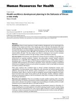
Figure 3 Adhesion of human and mouse DCs to myelin .(A) Adhesion of human DCs (8 healthy donors) to human myelin. Values were
calculated as the fold change in adhesion on myelin compared to adhesion of the same cells to plastic control (plastic = 1). (B) Human
immature and mature DC (grouped) NgR1 and NgR2 mRNA expression (expressed as 1/delta Ct, 1/ΔCt) correlate with adhesion to a myelin
substrate (at 100 μg/ml), expressed as the fold increase in adhesion on myelin compared to plastic. NgR1, Spearman r = -0.7818, P = 0.0105.
NgR2, Spearman r = -0.7697, P = 0.0126. (C) Adhesion of human DCs (3 donors) to myelin, Nogo-66-His, MAG-Fc, MBP (all at 10 μg/ml), IgG
(15 μg/ml), expressed as fold increase in adhesion compared to on plastic. (D) Adhesion of mouse WT and Ngr1/2-/- (KO) DCs (9 mice of each
genotype) to mouse myelin (10 μg/ml), expressed as fold increase in adhesion to myelin compared to plastic. Bars represent mean with SEM of
the fold change in adhesion on myelin compared to adhesion of the same cells to plastic (plastic = 1). *P < 0.05, **P < 0.01.
McDonald et al. Journal of Neuroinflammation 2011, 8:113
/>Page 9 of 12
us to suggest that there are as yet unidentified co-recep-
tors which can act as the signal transducing subunit of
the NgR1 complex. We went on to conclusively demon-
strate that in human matDCs, which lack NgR1 expres-
sion, there is an increased adhesion to myelin. This is
supported by our demonstration of increased adhesion
to myelin of mouse matDC genetically lacking NgR1/2.
Taking a closer look at adhesion of mouse WT DCs to
myelin, we see a marke d difference in how they adhere
to myelin when compared with human DCs. These cells
also show different patterns of expression of NgR1/2
and co-receptors. Furthermore, when comparing the
expression of the various DC surface markers, it
becomes obvious that human and mouse in vitro gener-
ated DCs demonstrate phenotypic al differences. An
explanation for the variation between the two species
could be that the cells undergo distinct differentiation
procedures. As mentioned, mouse DCs are differentiated
directly from precursor cells present in the bone mar-
row. Human DCs, on the other hand, are differentiated
from blood-bo rne monocytes. This variation in pr epara-
tion could lead to differences in phenotype of this highly
heterogeneous cell family. A number of publications
have also addressed the issue of dissimilarities not only
between human and mouse DCs but al so in the func-
tions of the various populations of DCs found in vivo
when compared to those that are generated in vitro
[38-40]. In both humans and mice, several DC subsets
have been identified based on differences in phenotypes,
anatomic al locations or functions [41,42]. These subsets
are generated in vivo with very complex and specific
environmental influences, which have not yet been repli-
cated in culture. Thus, both the diff erent experimental
preparations of DCs and endogenous inter-species varia-
tion could contribute to the observed variations in cell
types.
Our results further suggest that NgR1 and/or NgR2
may not play such a significant role in DC interaction
with myelin in the mouse WT system. This is in line
with the observation of no change in immune response
after EAE was induced in Ngr1/2-/- mice,aswellasno
difference in the number of CNS invading DCs after
EAE was induced in Ngr1/2-/- mice compared to WT
[43].
The described regulation of human NgR1/2 expression
could have a number of implications for DCs both dur-
ing the normal immune response and in autoimmune
diseases. The results presented here show a decrease in
NgR1 and NgR2 expression upon mat uration of human
DCs. Immature DCs have been well described as phago-
cytic cells expressing low levels of chemokine receptors.
Upon maturation, DCs are no longer phagocytic, they
up-regulate the chemokine receptor CCR7 and are
highly migratory [44]. The complex processes of matDC
migration involve adhesion to and transmigration across
a number of different cell types and extracellular
matrices, and are mediated in large part by chemokine
receptors (such as CCR7) and Rho GTPases [45,46].
Our results indicate that NgR1/2 are up-regulated in
the tissue resident cells, and are down -regulated when
the cells are activated and required to migrate. This
could indicate a possible role for NgR1/2 outside the
CNS, perhaps in the activation of DCs or in homing of
DCs to specific tissues. This is supported by the findings
of the non-myel in associated proteins B cell acti vating
factor (BAFF) and leucine-rich glioma activated (LGI1)
as functional ligands for NgR1 [47,48]. BAFF is a TNF-
like cytokine that supports survival and differentiation of
B cells. It is expressed in many cell types, in cluding
monocytes, DCs, neutrophils, stromal cells, activated T
cells, B cells, B cell tumours and epithelial cells [49].
Thus, a wide variety of cells have the ability to produce
BAFF and might potentially act on immDCs via NgR1.
The possibility that NgR1 plays a functional role in
mediating adhesion of DCs to myelin could become
important in situations where peripheral immune cells
come into contact with myelin debris, such as after neu-
rodegenerative events. Expression of NgR1, TROY and
LINGO-1 was found in CD68
+
cells (i.e. macrophages,
microglia, and a subset of DCs) within chronic, active
demyelinating MS lesions and ischemic lesions of acute
and old cerebral infarctions [50,51]. DCs are emerging
as important players in CNS autoimmunity, specifically
in MS [23]. The finding of mature DC markers in the
inflamed meninges and perivascular cuffs of active MS
lesions has lead to the suggestion that DCs are recruited
to and mature within MS lesions [24]. Here, self-anti-
gens are continuously made available by myelin destruc-
tion, thus mature DCs can contribute t o the local
activation and expansion of pathogenic T cells. This
model is conducive to our findings of increased adhe-
sion of matDC to myelin, and pr ovides a possible phy-
siological role for the down-regulat ion of NgR1 in
matDC. That is, down-regulatio n of NgR1 in matDCs
promotes their adhesion to myelin, resulting in the
selective accumulation of matDCs rather than immDC
in the myelin debris-containing lesion. This would result
in further antigen presentation and activation of myelin-
reactive T cells, potentially aggravating the disease.
Conclusions
Our study documents the differential expression and
function of NgR1 and NgR2 in human DCs. We
describe the increased expression of NgR1 and NgR2 in
human immature DCs, which are then down-regulated
upon maturation. Since human mature DCs adhere to a
much higher extent to myelin than immature DCs, we
hypothesise that this effect is mediated by NgR1. T his
McDonald et al. Journal of Neuroinflammation 2011, 8:113
/>Page 10 of 12
finding was corroborated by using mature DCs from
Ngr1/2-/- mice, which adhere significantly more to a
myelin substrate compared to WT mature DCs. The
interaction of DCs with myelin provides insight into
how DCs act when in the presence of CNS myelin, such
as during neurodegeneration and/or neuroinflammation.
The down-regulation of myelin-associated inhibitory fac-
tor receptors NgR1 and NgR2 on mature DCs may facil-
itate their initiation of local antigen presentation
function during physiological and pathological immune
responses in the CNS.
Acknowledgements
CMD is enrolled in the graduate programme SPIN, supported by the
Austrian Science Fund (FWF): project number W1206. KS is supported by a
research grant from the Gemeinnuetzige Hertie-Stiftung (Grant No. 1.01.1/
08/001). FK is supported by a grant from the Austrian Research Foundation
(FWF P 19908-B05) to RS. The authors wish to thank Sandra Trojer and
Kathrin Schanda for excellent technical assistance in purifying MAG-Fc and
MBP, respectively, Prof. Thomas Berger for providing human blood samples
and Dr. Johannes Rainer for his help in analysing the cytokine microarray
experiment.
Author details
1
Clinical Department of Neurology, Innsbruck Medical University,
Anichstrasse 35, A-6020 Innsbruck, Austria.
2
Centre for Molecular
Neurobiology, Institute for Neuroimmunology and Clinical MS Research
(inims), Falkenried 94, D-20251 Hamburg, Germany.
3
Division of
Neurobiochemistry, Innsbruck Medical University, Biocenter, Fritz-Pregl-
Strasse 3, A-6020 Innsbruck, Austria.
4
Department of Clinical
Neuroimmunology and MS Research, Neurology Clinic, University Hospital
Zürich, Frauenklinikstrasse 26, CH-8091 Zürich, Switzerland.
Authors’ contributions
CMD, KS, CB, and MR conceived and designed the experiments. CB and RS
generated and provided Ngr1/2-/- mice. CMD and FK carried out all
experiments. CMD, KS, FK, RS and MR analysed and interpreted the data.
CMD, KS, and MR wrote the manuscript. All authors read and approved the
final manuscript.
Competing interests
The authors declare that they have no competing interests.
Received: 21 June 2011 Accepted: 9 September 2011
Published: 9 September 2011
References
1. Chen MS, Huber AB, van der Haar ME, Frank M, Schnell L, Spillmann AA,
Christ F, Schwab ME: Nogo-A is a myelin-associated neurite outgrowth
inhibitor and an antigen for monoclonal antibody IN-1. Nature 2000,
403:434-439.
2. Prinjha R, Moore SE, Vinson M, Blake S, Morrow R, Christie G, Michalovich D,
Simmons DL, Walsh FS: Inhibitor of neurite outgrowth in humans. Nature
2000, 403:383-384.
3. McKerracher L, David S, Jackson DL, Kottis V, Dunn RJ, Braun PE:
Identification of myelin-associated glycoprotein as a major myelin-
derived inhibitor of neurite growth. Neuron 1994, 13:805-811.
4. Mukhopadhyay G, Doherty P, Walsh FS, Crocker PR, Filbin MT: A novel role
for myelin-associated glycoprotein as an inhibitor of axonal
regeneration. Neuron 1994, 13:757-767.
5. Wang KC, Koprivica V, Kim JA, Sivasankaran R, Guo Y, Neve RL, He Z:
Oligodendrocyte-myelin glycoprotein is a Nogo receptor ligand that
inhibits neurite outgrowth. Nature 2002, 417:941-944.
6. Fournier AE, GrandPre T, Strittmatter SM: Identification of a receptor
mediating Nogo-66 inhibition of axonal regeneration. Nature 2001,
409:341-346.
7. Wang KC, Kim JA, Sivasankaran R, Segal R, He Z: P75 interacts with the
Nogo receptor as a co-receptor for Nogo, MAG and OMgp. Nature 2002,
420:74-78.
8. Liu BP, Fournier A, GrandPre T, Strittmatter SM: Myelin-associated
glycoprotein as a functional ligand for the Nogo-66 receptor. Science
2002, 297:1190-1193.
9. Venkatesh K, Chivatakarn O, Lee H, Joshi PS, Kantor DB, Newman BA,
Mage R, Rader C, Giger RJ: The Nogo-66 receptor homolog NgR2 is a
sialic acid-dependent receptor selective for myelin-associated
glycoprotein. J Neurosci 2005, 25:808-822.
10. Worter V, Schweigreiter R, Kinzel B, Mueller M, Barske C, Bock G, Frentzel S,
Bandtlow CE: Inhibitory activity of myelin-associated glycoprotein on
sensory neurons is largely independent of NgR1 and NgR2 and resides
within Ig-Like domains 4 and 5. PLoS One 2009, 4:e5218.
11. Park JB, Yiu G, Kaneko S, Wang J, Chang J, He XL, Garcia KC, He Z: A TNF
receptor family member, TROY, is a coreceptor with Nogo receptor in
mediating the inhibitory activity of myelin inhibitors. Neuron 2005,
45:345-351.
12. Mi S, Lee X, Shao Z, Thill G, Ji B, Relton J, Levesque M, Allaire N, Perrin S,
Sands B, et al: LINGO-1 is a component of the Nogo-66 receptor/p75
signaling complex. Nat Neurosci 2004, 7:221-228.
13. Pool M, Niino M, Rambaldi I, Robson K, Bar-Or A, Fournier AE: Myelin
regulates immune cell adhesion and motility. Exp Neurol 2009,
217:371-377.
14. Barrette B, Vallieres N, Dube M, Lacroix S: Expression profile of receptors
for myelin-associated inhibitors of axonal regeneration in the intact and
injured mouse central nervous system. Mol Cell Neurosci 2007, 34
:519-538.
15.
Yamashita T, Tohyama M: The p75 receptor acts as a displacement factor
that releases Rho from Rho-GDI. Nat Neurosci 2003, 6:461-467.
16. Schwab ME, Caroni P: Oligodendrocytes and CNS myelin are
nonpermissive substrates for neurite growth and fibroblast spreading in
vitro. J Neurosci 1988, 8:2381-2393.
17. Liao H, Duka T, Teng FY, Sun L, Bu WY, Ahmed S, Tang BL, Xiao ZC: Nogo-
66 and myelin-associated glycoprotein (MAG) inhibit the adhesion and
migration of Nogo-66 receptor expressing human glioma cells.
J Neurochem 2004, 90:1156-1162.
18. Fry EJ, Ho C, David S: A role for Nogo receptor in macrophage clearance
from injured peripheral nerve. Neuron 2007, 53:649-662.
19. Steinman RM: The dendritic cell system and its role in immunogenicity.
Annu Rev Immunol 1991, 9:271-296.
20. Matyszak MK, Perry VH: The potential role of dendritic cells in immune-
mediated inflammatory diseases in the central nervous system.
Neuroscience 1996, 74:599-608.
21. Pashenkov M, Link H: Dendritic cells and immune responses in the
central nervous system. Trends Immunol 2002, 23:69-70, author reply 70.
22. Newman TA, Galea I, van Rooijen N, Perry VH: Blood-derived dendritic
cells in an acute brain injury. J Neuroimmunol 2005, 166:167-172.
23. Zozulya AL, Clarkson BD, Ortler S, Fabry Z, Wiendl H: The role of dendritic
cells in CNS autoimmunity. J Mol Med 2010, 88:535-544.
24. Serafini B, Rosicarelli B, Magliozzi R, Stigliano E, Capello E, Mancardi GL,
Aloisi F: Dendritic cells in multiple sclerosis lesions: maturation stage,
myelin uptake, and interaction with proliferating T cells. J Neuropathol
Exp Neurol 2006, 65:124-141.
25. Bender A, Sapp M, Schuler G, Steinman RM, Bhardwaj N: Improved
methods for the generation of dendritic cells from nonproliferating
progenitors in human blood. J Immunol Methods 1996, 196:121-135.
26. Romani N, Reider D, Heuer M, Ebner S, Kampgen E, Eibl B, Niederwieser D,
Schuler G: Generation of mature dendritic cells from human blood. An
improved method with special regard to clinical applicability. J Immunol
Methods 1996, 196:137-151.
27. Zheng B, Atwal J, Ho C, Case L, He XL, Garcia KC, Steward O, Tessier-
Lavigne M: Genetic deletion of the Nogo receptor does not reduce
neurite inhibition in vitro or promote corticospinal tract regeneration in
vivo. Proc Natl Acad Sci USA 2005, 102:1205-1210.
28. Lutz MB, Kukutsch N, Ogilvie AL, Rossner S, Koch F, Romani N, Schuler G: An
advanced culture method for generating large quantities of highly pure
dendritic cells from mouse bone marrow. J Immunol Methods 1999,
223:7
7-92.
29. Norton WT, Poduslo SE: Myelination in rat brain: method of myelin
isolation. J Neurochem 1973, 21:749-757.
30. Eylar EH, Hashim GA: The isolation and properties of large basic peptides
from bovine spinal cord. J Neurochem 1974, 23:973-979.
McDonald et al. Journal of Neuroinflammation 2011, 8:113
/>Page 11 of 12
31. Niederost B, Oertle T, Fritsche J, McKinney RA, Bandtlow CE: Nogo-A and
myelin-associated glycoprotein mediate neurite growth inhibition by
antagonistic regulation of RhoA and Rac1. J Neurosci 2002,
22:10368-10376.
32. Abramoff MD, Magelhaes PJ, Ram SJ: Image Processing with ImageJ.
Biophotonics International 2004, 11:36-42.
33. Kueng W, Silber E, Eppenberger U: Quantification of cells cultured on 96-
well plates. Anal Biochem 1989, 182:16-19.
34. Saeed AI, Sharov V, White J, Li J, Liang W, Bhagabati N, Braisted J, Klapa M,
Currier T, Thiagarajan M, et al: TM4: a free, open-source system for
microarray data management and analysis. Biotechniques 2003,
34:374-378.
35. Yuan JS, Reed A, Chen F, Stewart CN Jr: Statistical analysis of real-time
PCR data. BMC Bioinformatics 2006, 7:85.
36. Mi S: Troy/Taj and its role in CNS axon regeneration. Cytokine Growth
Factor Rev 2008, 19:245-251.
37. Shao Z, Browning JL, Lee X, Scott ML, Shulga-Morskaya S, Allaire N, Thill G,
Levesque M, Sah D, McCoy JM, et al: TAJ/TROY, an orphan TNF receptor
family member, binds Nogo-66 receptor 1 and regulates axonal
regeneration. Neuron 2005, 45:353-359.
38. Geissmann F, Manz MG, Jung S, Sieweke MH, Merad M, Ley K:
Development of monocytes, macrophages, and dendritic cells. Science
2010, 327:656-661.
39. Robbins SH, Walzer T, Dembele D, Thibault C, Defays A, Bessou G, Xu H,
Vivier E, Sellars M, Pierre P, et al: Novel insights into the relationships
between dendritic cell subsets in human and mouse revealed by
genome-wide expression profiling. Genome Biol 2008, 9:R17.
40. Mestas J, Hughes CC: Of mice and not men: differences between mouse
and human immunology. J Immunol 2004, 172:2731-2738.
41. Crozat K, Guiton R, Guilliams M, Henri S, Baranek T, Schwartz-Cornil I,
Malissen B, Dalod M: Comparative genomics as a tool to reveal functional
equivalences between human and mouse dendritic cell subsets.
Immunol Rev 2010, 234:177-198.
42. Shortman K, Liu YJ: Mouse and human dendritic cell subtypes. Nat Rev
Immunol 2002, 2:151-161.
43. Steinbach K, McDonald CL, Reindl M, Bandtlow C, Martin R: The roles of
Nogo receptor-mediated interactions in autoimmune inflammation in
experimental autoimmune encephalomyelitis. 10th International Congress
of Neuroimmunology 26th October, 2010; Barcelona Elsevier/North-Holland;
2010, 1-219.
44. Banchereau J, Steinman RM: Dendritic cells and the control of immunity.
Nature 1998, 392:245-252.
45. Quast T, Tappertzhofen B, Schild C, Grell J, Czeloth N, Forster R, Alon R,
Fraemohs L, Dreck K, Weber C, et al: Cytohesin-1 controls the activation of
RhoA and modulates integrin-dependent adhesion and migration of
dendritic cells. Blood 2009, 113:5801-5810.
46. Swetman CA, Leverrier Y, Garg R, Gan CH, Ridley AJ, Katz DR, Chain BM:
Extension, retraction and contraction in the formation of a dendritic cell
dendrite: distinct roles for Rho GTPases. Eur J Immunol 2002,
32:2074-2083.
47. Zhang L, Zheng S, Wu H, Wu Y, Liu S, Fan M, Zhang J: Identification of
BLyS (B lymphocyte stimulator), a non-myelin-associated protein, as a
functional ligand for Nogo-66 receptor. J Neurosci 2009, 29:6348-6352.
48. Thomas R, Favell K, Morante-Redolat J, Pool M, Kent C, Wright M,
Daignault K, Ferraro GB, Montcalm S, Durocher Y, et al: LGI1 is a Nogo
receptor 1 ligand that antagonizes myelin-based growth inhibition.
J Neurosci 2010, 30:6607-6612.
49. Davidson A: Targeting BAFF in autoimmunity. Curr Opin Immunol 2010,
22:732-739.
50. Satoh J, Onoue H, Arima K, Yamamura T: Nogo-A and nogo receptor
expression in demyelinating lesions of multiple sclerosis. J Neuropathol
Exp Neurol 2005, 64:129-138.
51. Satoh J, Tabunoki H, Yamamura T, Arima K, Konno H: TROY and LINGO-1
expression in astrocytes and macrophages/microglia in multiple sclerosis
lesions. Neuropathol Appl Neurobiol 2007, 33:99-107.
doi:10.1186/1742-2094-8-113
Cite this article as: McDonald et al.: Nogo receptor is involved in the
adhesion of dendritic cells to myelin. Journal of Neuroinflammation 2011
8:113.
Submit your next manuscript to BioMed Central
and take full advantage of:
• Convenient online submission
• Thorough peer review
• No space constraints or color figure charges
• Immediate publication on acceptance
• Inclusion in PubMed, CAS, Scopus and Google Scholar
• Research which is freely available for redistribution
Submit your manuscript at
www.biomedcentral.com/submit
McDonald et al. Journal of Neuroinflammation 2011, 8:113
/>Page 12 of 12