báo cáo hóa học: " LPS preconditioning redirects TLR signaling following stroke: TRIF-IRF3 plays a seminal role in mediating tolerance to ischemic injury" doc
Bạn đang xem bản rút gọn của tài liệu. Xem và tải ngay bản đầy đủ của tài liệu tại đây (2.39 MB, 12 trang )
RESEARC H Open Access
LPS preconditioning redirects TLR signaling
following stroke: TRIF-IRF3 plays a seminal role in
mediating tolerance to ischemic injury
Keri B Vartanian, Susan L Stevens, Brenda J Marsh, Rebecca Williams-Karnesky, Nikola S Lessov and
Mary P Stenzel-Poore
*
Abstract
Background: Toll-like receptor 4 (TLR4) is activated in response to cerebral ischemia leading to substantial
brain damage. In contrast, mild acti vation of TLR4 by preconditioning with low dose exposure to
lipopolysaccharide (LPS) prior to cerebral ischemia dramatically improves ou tcome by reprogramming the
signaling response to injury. This suggests that TLR4 signaling can be altered to induce an endogenously
neuroprotective phenotype. However, the TLR4 signaling events invol ved in this neuroprotective response are
poorly understood. Here we define several molecular mediators of the primary signaling cascades induced by
LPS preconditioning that give rise to the reprogrammed response to cerebral ischemia and confer the
neuroprotective phenotype.
Methods: C57BL6 mice were preconditioned with low dose LPS prior to transient middle cerebral artery occlusion
(MCAO). Cortical tissue and blood were collected following MCAO. Microarray and qtPCR were performed to
analyze gene expression associated with TLR4 signaling. EMSA and DNA binding ELISA were used to evaluate
NFB and IRF3 activity. Protein expression was determined usin g Western blot or ELISA. MyD88-/- and TRIF-/- mice
were utilized to evaluate signaling in LPS preconditioning-induced neuroprotection.
Results: Gene expression analyses revealed that LPS preconditioning resulted in a marked upregulation of anti-
inflammatory/type I IFN-associated genes following ischemia while pro-inflammatory genes induced following
ischemia were present but not differentially modulated by LPS. Interestingly, although expression of pro-
inflammatory genes was observed, there was decreased activity of NFB p65 and increased presence of NFB
inhibitors, including Ship1, Tollip, and p105, in LPS-preconditioned mice following stroke. In contrast, IRF3 activity
was enhanced in LPS-preconditioned mice following stroke. TRIF and MyD88 deficient mice revealed that
neuroprotection induced by LPS depends on TLR4 signaling via TRIF, which activates IRF3, but does not depend
on MyD88 signaling.
Conclusion: Our results characterize several critical mediators of the TLR4 signaling events associated with
neuroprotection. LPS preconditioning redirects TLR4 signaling in response to stroke through suppression of NFB
activity, enhanced IRF3 activity, and increased anti -inflammatory/type I IFN gene expression. Interestingly, this
protective phenotype does not require the suppression of pro-inflammatory mediators. Furthermore, our results
highlight a critical role for TRIF-IRF3 signaling as the governing mechanism in the neuroprotective response to
stroke.
Keywords: Toll-like receptors, stroke, NFκB, inflammation, preconditioning, neuroprotection
* Correspondence:
Department of Molecular Microbiology & Immunology, Oregon Health &
Science University, Portland, OR 97239, USA
Vartanian et al. Journal of Neuroinflammation 2011, 8:140
/>JOURNAL OF
NEUROINFLAMMATION
© 2011 Vartanian et al; licensee BioMed Central Ltd. This is an Open Access article distributed under the terms of the Creative
Commons Attribution License (http://creative commons.org /licenses/by/2.0), which permits unrestricte d use, distribution, and
reproduction in any mediu m, provided the original work is properly cited.
Background
Stroke is one of the leading causes of death and the
leading cause of morbidity in the United Stat es [1]. The
inflammatory response to stroke substantially exacer-
bates ischemic damage. The acute activation of the
NFB transcription factor has been linked to the inflam-
matory response to stroke [2] and suppression of NFB
activity following stroke has been found to reduce
damage [3]. NFB activation can lead to the dramatic
upregulation of inflammatory molecules and cytokines
including TNFa,IL6,IL1b, and COX2 [2]. The sourc e
of these inflammatory molecules in the acute response
to stroke appears to stem from the cells of the central
nervous system (CN S), including neurons and glial cells
[2]. The cells in the CNS play a particularly dominant
role early in the response to ischemia because infiltrat-
ing leukocytes do not a ppear in substantial numbers in
the brain until 24 hr following injury [4]. Stroke also
induces an acute inflammatory response in the circulat-
ing blood. Inflammatory cytokine and chemokine levels,
including IL6, IL1 b,MCP-1andTNFa are elevated in
the circulation following stroke [5]. This suggests there
is an intimate relationship between responses in the
brain and blood following stroke– responses that result
in increased inflammation.
Toll-like receptors (TLRs), traditionally considered
innate immune receptors, signal through the adaptor
proteins MyD88 and TRIF to activate NFBandinter-
feron regulatory factors (IRFs). It has been shown
recently that TLRs become activated in response to
endogenous ligands, known as damage associ ated mole-
cular patterns (DAMPs), released during injury. Interest-
ingly, animals deficient in TLR2 or TLR4 have
significantly reduced infarct sizes in several models of
stroke [6-11]. This suggests that TLR2 and TLR4 activa-
tion in response to isc hemic injury exacerbates damage.
In addition, a recent investigation in humans showed
that the inflammatory responses to stroke in the blood
were linked to increased TLR2 and TLR4 expression on
hematopoetic cells and associated with worse outcome
in stroke [12]. The detrimental effect of TLR signaling is
associated with the pathways that lead to NFBactiva-
tion and pro-inflammatory responses. In contrast, TLR
signaling pathways that a ctivate IRFs can induce anti-
inflammatory mediators and type I I FNs that have been
associated with neuroprotection [13,14]. Thus, in TLR
signaling there is a fine balance between pathways lead-
ing to injury or protection.
TLR ligands have been a major source of interest as
preconditioning agents for prophylactic therapy against
ischemic injury. Such therapies would target a popula-
tion of patients that are at risk of ischemia in the setting
of surgery [15-18]. Preconditioning with low doses of
ligands for TLR2, TLR4, and TLR9 all successfully
reduce infarct size in experimental models of stroke
[19-21], including a recent study s howing that a TLR9
ligand is neuroprotec tive in a nonhuman primate model
of stroke [22]. Emerging evidence suggests that TLR-
induced neuroprotection occurs by reprogramming the
genomic response to the DAMPs, which are produced
in response to ischemic injury. In this reprogrammed
state, the resultant pathway activation of TLR4 signaling
preferentially leads to IRF-mediated gene expression
[13,14]. However, whether TLR preconditioning affects
NFB activity and pro-inflammatory signaling is
unknown. As yet, a complete analysis of the characteris-
tic TLR signaling responses to stroke following precon-
ditioning has not been reported. The objective of this
study is to utilize LPS preconditioning followed by tran-
sient middle cerebral artery occlusion (MCAO) to eluci-
date the reprogrammed TLR response to stroke and to
determine the major pathways involved in producing
the neuroprotective phenotype.
Here we show that preconditioning against ischemia
using LPS leads to suppressed NFB activity–although
pro-inflammatory gene expression does not appear to be
attenuated. We also demonstrate that LPS-precondi-
tioned mice have enhanced IRF3 activity and anti-
inflammatory/type I IFN gene expression in the
ischemic brain. This expression pattern was recapitu-
lated in the blood where plasma l evels of pro-inflamma-
tory cytokine proteins were comparable in LPS-
preconditioned and control mice while IRF-associated
proteins were enhanced in LPS preconditioned mice. To
our knowledge, we provide the first evidence that pro-
tection due to LPS preconditioning stems from TRIF
signaling, the cascade that is associated with IRF3 acti-
vation, and is independent of MyD88 signaling. These
molecular features suggest that, following stroke, signal-
ing is directed away from NFB activity and towards a
dominant TRIF-IRF3 response. Understanding the endo-
genous signaling events that promote protection against
ischemic injury is integral to the identification and
development of novel stroke therapeutics. In particular,
the evidence presented here further highlights a key role
for IRF3 activity in the protective response to stroke.
Methods
Animals
C57Bl/6J mice (male, 8-12 weeks) were purchased from
Jackson Laboratories (West Sacramento, CA). C57B l/6J-
Ticam1
LPS2
/J (TRIF-/-) mice were also obtained from
Jackson Laboratories. MyD88-/- mice were a kind gift of
Dr. Shizuo Akira (Osaka University, Os aka Japan) and
were bred in our faci lity. All mice were housed in an
American Association for Laboratory Animal Care-
approved facility. Procedures were conducted according
to Oregon Health & Science University, Institutional
Vartanian et al. Journal of Neuroinflammation 2011, 8:140
/>Page 2 of 12
Animal Care and Use Committee, and National Insti-
tutes of Health guidelines.
LPS treatment
Mice were preconditioned with LPS (0.2 or 0.8 m g/kg,
Escherichia coli serotype 0111:B4; Sigma) or saline by
one subcutaneous injection , unless otherw ise indicated,
72 hr prior to MCAO. Each new lot of LPS was titrated
for the optimal dose that confers neuroprotection. No
differences were observed in the gen omic responses to
LPS for each dose used and route of administration
(subcutaneous or intraperitoneal, data not shown).
Middle Cerebral Artery Occlusion (MCAO)
Mice were anesthetized with isoflurane (1.5-2%) and
subjected to MCAO using the monofilament suture
method described previously [23]. Briefly, a silicone-
coated 7-0 monofilament nylon surgical suture was
threaded through the external carotid artery to the
internal carotid artery to block the middle cerebral
artery, and maintained intraluminally for 40 to 60 min.
The suture was then removed to restore blood flow.
The duration of occlusion was optimized based on the
specific surgeon who performed the MCAO to yield
comparable infarct sizes in the saline treated control
animals (~35-40%). The s elected duration of MCAO
was held constant within experiments. Cerebral blood
flow (CBF) was monitored throughout surgery by laser
doppler flowmetry. Any mouse that did not maintain a
CBF during occlusion of <25% of baseline was excluded
from the study. The reduction of CBF was comparable
in LPS and saline preconditioned mice in response to
MCAO. Body temperature was monitored and main-
tained at 37°C with a thermostat-controlled heating pad.
Infarct measurements were made using triphenyltetrazo-
lium chloride (TTC) staining of 1 mm coronal brain
sections.
Tissue collection
Under deep isoflurane anesthesia, approximately ~0.5-
1.0 ml of blood was collected via cardiac puncture in a
heparinized syringe. Subsequently, the mice were per-
fused with heparinized (2 U/ml) saline followed by rapid
removal of the brain. The olfactory bulbs were removed
and the first 4 mm of tissue was collected beginning at
the rostral end. The striatum was dissected and removed
and the remaining cortex was utilized for RNA isolation
or protein extraction. The collected blood was centri-
fuged at 5000 × g for 20 min to obtain plasma th at was
stored at -80°C.
Genomic profiling of TLR associated mediators
For the genes displayed in Figure 1, the transcript
expression levels were determined as previously
described from our microarray experiments examining
the brain cortical response to stroke and 3 different
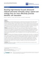
Figure 1 Microarray analysis of anti-inflammatory/type I IFN and pro-inflammatory gene expression. Microarray analysis revealed
enhanced anti-inflammatory/type I IFN and comparable pro-inflammatory gene expression profiles in the brain of LPS-preconditioned (0.2 mg/
kg, intraperitoneal injection) mice following 45 min MCAO. Heatmap representing level of gene expression immediately prior to (0 hr) MCAO
and 3 and 24 hr post MCAO; n = 4/treatment/timepoint. Lt. Select anti-inflammatory/type I IFN genes. Rt. Select pro-inflammatory genes. Color
scale from green to red represents relative decreased or increased gene expression levels, respectively.
Vartanian et al. Journal of Neuroinflammation 2011, 8:140
/>Page 3 of 12
preconditioning stimuli [14]. In brief, total RNA was iso-
lated from the ipsilateral cortex (n = 4 mice/treatment/
timepoint), using the Qiagen Rneasy Lipid Mini Kit
(Qiagen). Microarray assays were performed in the Affy-
metrix Microarray Core of the Oregon Health & Science
University Gene Microarray Shared Resource. RNA
samples were labeled using the NuGEN Ovation Biotin
RNA Amplification and Labeling System_V1. Hybridiza-
tion was performed as described in the Affymetrix tech-
nical manual (Affymetrix) with modification as
recommended for the Ovation labeling protocol
(NuGEN Technologies). Labeled cRNA target was qual -
ity-checked based on yield and size distribution. Qual-
ity-tested samples were hybridized to the MOE430 2.0
array. The array image was processed with Affymetrix
GeneChip Operating Software (GCOS). Affymetrix CEL
files were then uploaded into GeneSifter (http://www.
genesifter.net) and normalized using RMA.
RNA isolation, Reverse Transcription, and qtPCR
RNA was isolated from cortical tissue 72 hr post injec-
tion or from ipsilateral cortical tissue at 3 or 24 hr fol-
lowing MCAO (n ≥ 4 mice/treatment/timepoint) using
a Lipid Mini RNA isolation kit (Qiagen). Reverse tran-
scription was performed on 2 μgofRNAusingOmnis-
cript (Qiagen). Quantitative PCR was performed using
Taqman Gene Expression Assays (Applied Biosystems)
for each gene of interest on an ABI Prism 7700. Results
were normalized to b-Actin expression and analyzed
relative to their saline preconditioned counterparts. The
relative quantification of the gene of interest was deter-
mined using the comparative CT method (2
-DDCt
).
Western Blot
Protein extraction was performed as described pre-
viously [24] with some modifications. Briefly, tissue sam-
ples (n ≥ 4 mice/treatment/timepoint) were dissected
from the ipsilateral cortex and lysed in a buffer contain-
ing a protease inhibitor cocktail (Roche). Protein con-
centrations were determined using the BCA method
(Pierce-Endogen). Protein samples (50 μg) were dena-
tured in a gel-loading buffer (Bio-Rad Laboratories) at
100°C for 5 min and then loaded onto 12% Bis-Tris
polyacrylamide gels (Bio-R ad Laboratories). Following
electrophoresis, proteins were tra nsferred to polyvinylo-
dene difluoride membranes (Bio-Rad Laboratories) and
incubated with primary antibodies for Ship-1 (Santa
Cruz, sc8425), Tollip (AbCam, Ab37155), p105 (Santa
Cruz, sc7178), or b-Actin (Santa Cruz, sc161 6R) at 4°C
overnight. Membranes were then incubated with horse-
radish peroxidase conjugated anti-rabbit, anti-goat, or
anti-mouse antibody (Santa Cruz Biotechnology) and
detected by chemiluminescence (NEN Life Science Pro-
ducts) and exposed to Kodak film (Biomax). Images
were captured using an Epson scanner and the densito-
metry of the gel bands, including b-Actin loading con-
trol, was analyzed using ImageJ (NIH).
Electrophoretic Mobility Shift Assay (EMSA)
Nuclear protein extracts ( n = 4 mice/treatment/time-
point) were prepared from tissue dissected from the
ipsilateral cortex. Homogenized tissue was incubated in
Buffer A (10 mM Hepes-KOH pH7.9, 60 mM KCl, 1
mM EDTA, 1 mM DTT, 1 mM PMSF) for 5 min on ic e
and centrifuged at 3000 rpm for 5 min at 4°C. The pel-
lets were washed in Buffer B (10 mM Hepes-KOH
pH7.9, 60 mM KCl, 1 mM EDTA, 0.5% NP-40, 1 mM
DTT, 1 mM PMSF), resuspended in Buffer C (250 mM
Tris pH7.8, 60 mM KCl, 1 mM DTT, 1 mM PMSF),
and freeze-thawed 3 times in liquid nitrogen. All buffers
contained a protease inhibitor c ocktail (Roche). After
centrifug ing at 10,000 rpm for 10 min at 4°C, the super-
natant was collected and stored as nuclear extract at
-80°C. Nuclear protein concentrations were determined
using the BCA method (Pierce-Endogen). EMSAs were
performed using the Promega Gel Shift Assay System
according to the manufacturer ’s instructions. Briefly, 15
μg of nuclear protein was incubated with
32
P-labeled
NFB consensus oligonucleotide (Promega), either with
or without unlabeled competitor oligonucleotid e, unla-
beled noncompetitor oligonucleotide, or anti-p65 anti-
body (Santa Cruz). Samples were electrophoresed on a
4% acrylamide gel, dried and exposed to phosphorima-
ger overnight. The densitometry of the gel bands was
analyzed using scanning integrated optical density soft-
ware (ImageJ).
IRF3 Activity Assay
Nuclear protein (n ≥ 4 mice/trea tment/timepoint) was
isolated from fresh cortical tissue at 72 hr post injection
and from ipsilateral cortices at 3 or 24 hr following
MCAO using a Nuclear Extraction Kit (Active Motif,
Inc.). IRF3 activity was measured using 10 μgofnuclear
proteininanIRF3activityELISA(ActiveMotif,Inc),
that utilizes colorimetric detectio n of active IRF3 bound
to immobilized oligonucleotides.
Cytokine Analysis
Cytokine/chemokine analysis for IL1b,IL1a,MIP-1a,
MCP-1, RANTES, and IL10 was performed on plasma
samples (n ≥ 3 mice/treatment/timepoint) using a multi-
plex ELISA (Quansys). An IFNb ELISA (PBL Interferon
Source) was used to measure plasma levels of IFNb.
Statistical Analysis
Data is represented as mean ± SEM. The n for each
experiment is greater than or equal to 3, as specified in
each figure. Statistic al analysis was performed using
GraphPad Prism5 software. Two-way ANOVA with
Vartanian et al. Journal of Neuroinflammation 2011, 8:140
/>Page 4 of 12
Bonferroni Post Hoc test and Student’s t-test were uti-
lized as specified. Significance was determined as p <
0.05.
Results
LPS preconditioning does not affect inflammatory gene
expression in the brain following stroke
We used gene microarray analysis to elucidate the pat-
tern of inflammatory or anti-inflammatory/type I IFN
gene expression in the brain following stroke. In the set-
ting of stroke, LPS preconditioned animals exhibited
regulation of a number of genes typically found down-
stream of TLR signaling. The inflammatory profile
reveals that the gene regulation is similar at each time-
point following stroke in LPS or saline preconditioned
animals (Figure 1, Rt.). There is no evidence of inflam-
matory gene expression present immediately prior to
stroke (Figure 1, Rt. 0 hr). At 3 hr post MCAO, several
inflammatory genes are upregulated including IL6, IL1b,
Ptgs2/COX2, and CCL2/MCP-1 (Figure 1, R t.) and this
upregulation is sustained at the 24 hr timepoint follow-
ing MCAO (Figure 1, Rt.). TNFa, which is commonly
shown to be upregulated following MCAO [25,26], only
shows marginal levels of upregulation in LPS or saline
preconditioned mice (Figure 1, Rt.). To confirm the
microarray results, a subset of selected inflammatory
genes including IL6, IL1b, COX2, and TNFa, were ana-
lyzed using qtPCR. Each of these genes were upregu-
lated following MCAO in LPS and saline preconditioned
mice, but there were no significant differences based on
treatment at 3 hr (data not shown) and 24 hr (Figure 2)
following MCAO.
LPS preconditioning upregulates anti-inflammatory/type I
IFN gene expression in the brain following MCAO
Although pro-inflammatory gene expression was not dif-
ferentially modulated in preconditioned animals, micro-
array results revealed that the majority of the anti-
inflammatory/type I IFN genes, such as TGFb,IL1
receptor antagonist (IL1rn), RANTES, and IRF7, were
upregulated following stroke in the brains of LPS versus
saline preconditioned mice (Figure 1, Lt.). I L10 gene
expression was not detected at any timepoint (Figure 1,
Lt.). TGFb, IL10, RANTES, and IFIT1 were selected for
qtPCR analysis. TGFb, RANTES, and IFIT1 were signifi-
cantly upregulated in the LPS-preconditioned brain
compared to saline 24 hr following stroke (Figure 2).
RANTES was also significantly upregulated at 3 hr fol-
lowing stroke in LPS-preconditioned m ice compared to
saline (data not shown). IL10 expression remained unde-
tectable by qtPCR analysis (Figure 2), suggesting that
IL10 mRNA is not present at these timepoints in the
brain following stroke. These qtPCR results confirm the
gene expression profile observed on the microarray.
Taken together, these data indicate an enhanced anti-
inflammatory/type I IFN gene expression profile in the
brain of LPS-preconditioned animals following MCAO
while the inflammatory gene expression is unaffected.
NFB activity is suppressed in the brain of LPS-
preconditioned animals 24 hr post MCAO
NFB activity is associated with damage and inflamma-
tion in the brain that occurs in response to stroke. We
used EMSAs to evaluate the activity of the NFB subu-
nit p65 in the brain following stroke. The results indi-
cated that LPS and saline preconditioned mice have
comparable NFB activity at 3 hr post MCAO (Figure
3A). However, at 24 hr post MCAO, LPS-precondi-
tioned animals have significantly suppressed NFB activ-
ity compared to saline preconditioned mice (Figure 3A).
Ship1 and Tollip are cytosolic molecules that inhibit
TLR signaling, which leads to the suppression of NFB
activity.WefoundthatShip1andTollipmRNAare
upregulated in the brain 72 hr post injection versu s sal-
ine controls ( 2.06 ± 0.2 7 and 2.31 ± 0.35, respectively)
but not at 3 hr post stroke (1.09 ± 0.10 and 1.05 ± 0.09,
respectively). However, by 24 hr post MCAO, Ship1 and
Tollip mRNA are significantly enhanced in the brain of
LPS-preconditioned mice compared to saline controls
(2.62 ± 0.84 and 4.01 ± 1.06, respectively, Figure 3B).
Ship1 protein is not upregul ated at 72 hr post injection
(Fold change vs. saline: 1.01 ± 0.32), but becomes signif-
icantly enhanced in LPS-preconditioned mice at 3 hr
(Foldchangevssaline:1.83±0.13)andat24hr(Fold
Figure 2 Enhanced anti-inflammatory/type I IFN gene
expression but comparable pro-inflammatory gene expression
in LPS-preconditioned mice post MCAO. Gene regulation 24 hr
post MCAO measured by qtPCR reveals that anti-inflammatory/type
I IFN-associated genes TGFb, RANTES, and IFIT1 are significantly
upregulated in LPS preconditioned mice compared to saline. Pro-
inflammatory genes IL6, IL1b, COX2, and TNFa show similar
regulation in LPS and saline preconditioned mice. These results
confirm the gene microarray data. Samples from mice receiving a
45 (LPS: 0.2 mg/kg) or 60 min (LPS: 0.8 mg/kg) MCAO were
combined due to comparable gene regulation (see methods). ND =
not detected. Student’s t-test, LPS vs. saline 24 hr post MCAO, **p <
0.01, n ≥ 4 per treatment.
Vartanian et al. Journal of Neuroinflammation 2011, 8:140
/>Page 5 of 12
change vs. s aline: 8.81 ± 1.54, Figure 3C) post MCAO.
Tollip protein is not affected by LPS precondition ing at
72 hr post injection or 3 hr post MCAO (Fold change
vs. saline: 1.42 ± 0. 10 and 0.83 ± 0.10, respectively), but
it is significantly enhanced in LPS-preconditioned mice
compared to saline controls at 24 hr post MCAO (Fold
change vs. saline: 2.42 ± 0.20, Figure 3C). Additionally,
the p50 precursor protein p105, which inhibits NFB
activity by acting like an IB molecule by sequestering
NFB in the cytosol [27,28], was significantly upregu-
lated24hrpoststrokeinLPS-preconditionedmice
compared to saline (Figure 3D). Thus, despite the upre-
gulation of inflammatory genes, the activity of NFBis
suppressed in the late-phas e of the neuroprotective
response of LPS-preconditioned mice.
IRF3 activity in the brain is enhanced following MCAO in
LPS-preconditioned mice
IRF3 activation downstream of TLR4 is associated with
anti-inflammatory/type I IFN responses. Using an IRF3
activity ELISA, we determined that IRF3 activity is com-
parable immediately prior to stroke (data not shown)
and subsequently enhanced in the brains of LPS-precon-
ditioned mice following MCAO (Figure 4). The trend
for increased IRF3 activity is present at 3 hr post
MCAO and is significantl y increased at 24 hr in LPS-
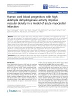
Figure 3 NFB is suppressed 24 hr post MCAO in LPS-preconditioned mice. (A) Nuclear protein obtained from ipsilateral cortices was used
to measure p65 activity by EMSA analysis. EMSA gel of pooled samples (n = 4) following 45 min MCAO for saline and LPS preconditioned (0.8
mg/kg) mice (Lt.). Quantification of band intensity of individual mice following MCAO (Rt.). NFB is significantly decreased in LPS-preconditioned
mice 24 hr post MCAO compared to saline. Supershift assay confirmed specificity for p65 oligos (data not shown). (B) Ship1 and Tollip mRNA are
significantly upregulated 24 hr post 60 minute MCAO in LPS-preconditioned (0.8 mg/kg) mice compared to saline, n ≥ 4 per treatment/
timepoint. (C) Western blot for Ship1 and Tollip and relative band quantification showing significant upregulation of Ship1 and Tollip protein 24
hr post 45 min MCAO in LPS-preconditioned mice (0.8 mg/kg), n ≥ 3 per treatment/timepoint. (D) Western blot and relative band quantification
for p105 at 24 hr post 45 minute MCAO showing significant upregulation in LPS-preconditioned (0.8 mg/kg) mice, n ≥ 3 per treatment/
timepoint. (A) Two-Way ANOVA, Bonferroni Post Hoc, *p < 0.05. (B-D) Student’s t-test, LPS vs. saline, **p < 0.01.
Vartanian et al. Journal of Neuroinflammation 2011, 8:140
/>Page 6 of 12
preconditio ned mice (Figure 4). Saline treated animals
showed no evidence of increased IRF3 activity follo wing
stroke (Figure 4). This indicates that LPS precondition-
ing alters the response to is chemic injury by activating
IRF3–a finding that is consistent with the enhanced
anti-inflammatory/type I IFN gene expression.
Blood cytokine/chemokine levels parallel the expression
in the brain
Evidence indicates that stroke alters the cytokine profile
in the plasma of circulating blood [5,29]. To determine
whether LPS preconditioning changes the balance of pro-
and anti-inflammatory cytokines and chemokines in the
plasma we examined the levels of seven molecules using
ELISAs. The results indicate that the level of pro-inflam-
matory cytokines, such as IL6, IL1b,andMCP-1,are
increased in both LPS and saline preconditioned mice
(Figure 5). The pro-inflammatory cytokines MIP-1a and
IL1a were not detected in the serum (data not shown).
The anti-inflammatory cytokine IL10 was significantly
increased only in the plasma of LPS-preconditioned mice
compa red to saline preconditioned mice following stroke
(Figure 5). RANTES, which is a chemokine associated
with IRF3 and IRF7 activity [30], was present in the
blood of LPS-preconditione d mice at significantly greater
levels than saline preconditioned mice (Figure 5). IFNb
was not detectable in the blood of LPS or saline precon-
ditioned animals following stroke (data not shown).
Overall, this suggests that the pro-inflammatory and
anti-inflammatory/type I IFN-associated response in the
blood parallels the response in the brain following stroke.
TRIF dependent LPS preconditioning induced
neuroprotection
Evidence presented here and previously suggests that sig-
naling following stroke is redirected towards IRF3
[13,14]. TLR4 signaling, which activates IRF3, is initiat ed
by the adaptor molecule TRIF, while TLR4 signaling that
activates NFB is initiated by the adaptor molecule
MyD88. The individual roles of these adaptor molecules
in neuroprotection induced by LPS preconditioning are
unknown. To test whether either of these key molecular
adaptors were important in mediating the neuroprotec-
tive effects of LPS, we exposed MyD88-/- and TRIF-/-
mice to LPS preconditioning (n = 4-10 mice/treatment ).
We found that MyD88-/- mice preconditioned with LPS
had significantly reduced infa rct sizes in response to
MCAO compared to saline controls (Figure 6), indicating
that LPS preconditioning is able to induce neuroprotec-
tion in mice lacking MyD88. In contrast, TRIF-/- mice
preconditioned with LPS or saline had comparable infarct
sizes (Figure 6), indicating that LPS preconditioning is
not able to induce neuroprotection in mice lacking TRIF.
Importantly, the TRIF adaptor is responsible for activa-
tion of IRF3, thus, our finding that TRIF is required for
LPS preconditioning provides further support for a pro-
tective role of IRF3 activity in neuroprotection.
Discussion
Here we sought to describe the LPS-induced repro-
grammed response to stroke and to determine the
important signaling events involved in neuroprotection
against ischemic injury. Our results demonstrated that
NFB activity was suppressed and that the cytosolic
inhibitors of NFB, Ship1, Tollip, and p105, were pre-
sent 24 hr post MCAO although pro-inflamma tory gene
expression was unaffected (diagrammed in Figure 7).
Interestingly, there is evidence that suppression of NFB
can promote protection against cerebral ischemia with-
out influencing pro-inflammatory cytokine production
[3,31]. In particular, administration of the NFB inhib i-
tor Tat-NEMO Binding Domain provided protection
against hypoxia-ischemia in neonatal rats without affect-
ing TNFa or IL1b production [3]. Furthermore, TLR4
deficient mice have smaller infarcts in response to
MCAO, yet the production of TNFa and IL1b was unaf-
fected [6]. This suggests that reduced ischemic injury
can be achieved by suppressing NFB activity without
suppressing pro-inflammatory cytokines and t hat TLR4
signaling and NFBactivationisnotthesolesourceof
these pro-inflammatory cytokines in response to
ischemic injury, implicating other signaling cascades and
transcription facto rs in the inflammatory response.
Thus, consistent with our result, reprogramming the
TLR4 response would not alter inflammatory gene
expression in the brain.
Figure 4 IRF3 activity is enhanced following MCAO in LPS-
preconditioned mice. Nuclear protein obtained from ipsilateral
cortex post 60 min MCAO analyzed using an IRF3 activity ELISA
(Active Motif, Inc.) revealed a significant increase in IRF3 activity in
LPS-preconditioned (0.8 mg/kg) mice. Two-way ANOVA, Bonferroni
Post Hoc, LPS vs. saline, *p < 0.05, n ≥ 4 per treatment.
Vartanian et al. Journal of Neuroinflammation 2011, 8:140
/>Page 7 of 12
NFB is known to be induced acutely in response to
ischemic injury; however, investigation into the role of
NFB activity has revealed conflicting results [2]. For
instance, NFB is constitutively active in neurons, a
requirement for their survival, while the surrounding
glial cells have inducible NFB activity [32]. In response
to ischemic challenge, NFB activity in astrocytes is
responsible for detrimental inflammation [33]. This
concept of pleotropic roles also applies to many of the
inflamma tory genes expressed in the brain in the setting
of stroke [34,35]. For example, intracerebroventricular
injection of recombinant IL6 significantly decreased the
infarct size in rats 24 hr post MCAO [36]. IL1b is a
potent inducer of IL1 receptor antagonist (IL-rn), which
significantly reduces damage in response to stroke [37]
and, notably, is upregulated in our microarray (Figure 1,
Lt.). TNFa is considered to play multiple roles in stroke
injury mediating many neuroprotective and injurious
effects [34]. Furthermo re, in response to viral challenge,
the simultaneous presence of inflammatory cytokines,
such as TNFa, and type I IFNs can alter their effects
and synergize to promote a more protective state [38].
Thus, alterations in the environment in which NFBis
activat ed and inflammatory genes are present may affect
the roles pro-inflam matory mediator s play in injury and
may even contribute to the protective phenotype.
IRF3 activity induces the expression of anti-inflamma-
tory and t ype I IFN-associated genes. Interestingly, mice
deficient in IRF3 are not protected against cerebral
ischemia by LPS preconditioning [13]. We have further
established the importance of IRF3 in neuroprotection
by identifying that multiple preconditioning paradigms
including LPS, CpG (TLR9 agonist) and brief ischemia
induce a common set of IRF-mediated genes in the
Figure 6 LPS preconditioning requires TLR si gnaling through
TRIF to promote neuroprotection. WT, MyD88-/-, and TRIF-/- mice
were preconditioned with LPS (0.8 mg/kg) 3 days prior to 40 min
MCAO. MyD88-/- mice were protected by LPS preconditioning
resulting in smaller infarct sizes. TRIF-/- mice did not have reduced
infarct sizes, demonstrating that TRIF deficient mice are not
protected by LPS preconditioning. Thus, TRIF is required for LPS
preconditioning induced neuroprotection. Student’s t-test, LPS vs.
saline, *p < 0.05, n = 4-10 per treatment.
Figure 5 Blood cytokine/chemokine levels show alterations in gene expression patterns comparable to the brain. Plasma collected from
saline or LPS-preconditioned (0.8 mg/kg) mice at the time of or following 60 min MCAO was examined using a multikine ELISA (Quansys).
Results indicated that pro-inflammatory cytokines IL1b, IL6 and MCP-1 are similar in saline and LPS-preconditioned mice. In contrast, LPS-
preconditioned mice have significantly enhanced levels of the anti-inflammatory/type I IFN-associated cytokine and chemokine IL10 and RANTES
compared to saline following MCAO. Two-way ANOVA, LPS vs. saline, *p < 0.05, n ≥ 3 per treatment.
Vartanian et al. Journal of Neuroinflammation 2011, 8:140
/>Page 8 of 12
neuroprotective environment following MCAO [14].
Here we demonstrate that IRF3 activity is upregulated
in the brain of LPS-preconditioned mice in response to
MCAO and that se veral IRF3-mediated genes are also
upregulated, including RANTES and IFIT1 (diagram med
in Figure 7), which may mitigate the damaging effects of
ischemia.
Many of the upregulated anti-inflammatory /type I IFN
genes in the brain following stroke have several identi-
fied neuroprotective functions. TGFb has been shown to
protect neurons from apoptosis, promote angiogenesis,
decrease microglial activation, and reduce edema
[34,39]. RANTES, which is induced by IRF3 and IRF7
[30], has been shown to protect neurons from cell death
in response to HIV-1 glycoprotein gp120 [40]. In the
setting of brain ischemia, mice deficient in the RANTES
receptor, CCR5, have larger infarcts, suggesting a neuro-
protective role for CCR5 activation [41]. Notably, the
expression of CCR5 is upregulated in our microarray
data (Figure 1, Lt). IFIT1 is commonly associated with
IRF3 signaling in response to IFN treatment and viral
infection [42]. Little is known about a role for IFIT1 in
ischemic injury; however, it is inducible in microglia and
neurons and has be en shown to af fect NFBandIRF3
activation [42-45]. Additional anti-inflammatory/type I
IFN genes shown to be upregulated in our microarray
studies have potential roles in neuroprotection including
IL-receptor antagonist (IL-rn), which is associated with
reduced infarct size in response to stroke [34,46]. A
recombinantformofIL-rnisbeingtestedinPhaseII
Figure 7 Schematic of TLR4 signaling and gene expression following stroke. (Top) TLR4 signaling cascades following stroke. In the
absence of LPS preconditioning, stroke leads to NFB activation without IRF3 activation. LPS preconditioning prior to stroke leads to robust
activation of IRF3 and suppressed NFB activity compared to stroke alone. (Bottom) Gene expression 24 hr post stroke. Stroke alone
dramatically upregulates pro-inflammatory genes. LPS preconditioning prior to stroke dramatically upregulates anti-inflammatory/Type I IFN
genes, many of which are associated with IRF3, while still maintaining a pro-inflammatory response.
Vartanian et al. Journal of Neuroinflammation 2011, 8:140
/>Page 9 of 12
clinical trials as an acute stroke therapy [47,48].
Although not detected in our gene microarray studies
here, perhaps due to assay sensitivity for IFNb transcript
on the microarray, we have previously published that
IFNb mRNA, a type I IFN known to have neuroprotec-
tive properties, is upregulated following stroke in the
brain of LPS-preconditioned mice using qtPCR [13].
Theprotectivefunctionsofthesegenesmaybeofcon-
siderable importance to the neuroprotective pheno type
following MCAO induced by LPS preconditioning.
Research strongly suggests that cerebral ischemia dra-
matically alters the protein and gene e xpression profile
in the per ipheral blood [5,29,49,50]. Our results demon-
stratethatthecytokineandchemokineresponseinthe
blood paralleled the pattern of gene expression in the
brain. Overall, inflammatory cytokine protein levels
were similarly induced in LPS and saline preconditioned
mice following stroke. However, we have previously
publishedthatTNFa is significantly reduced in the
plasma of LPS-preconditioned mice follo wing MCAO
[51]. The anti-inflammatory and type I IFN-induced
cytokines and chemokines measured in the blood were
enhanced in LPS-preconditioned mice compared to sal-
ine. In particular, IL10 was significantly upregulated in
the blood following MCAO of LPS-preconditioned mice.
Importantly, in humans, upregulation of IL10 in the
blood has been correlated with improved outcome in
stroke [52]. While IL10 mRNA was not detectable in
the brain, IL10 can be induced by IRF3 activity and
therefore is indicative of the same redirected response
seen in the brain. IFNb was not detected in the blood
24 hr post MCAO. This may be due to the kinetics of
IFNb expression. Further investigation into the time
course of IFNb induction in the blood is necessary to
fully understand the role of IFNb in this system. The
redirected signaling observed in the blood may stem
from the brain’s response to injury by leaking proteins
into the peripheral circulation; however, this is not con-
sidered a major source of plasma cytokines at these
early timepoints following stroke [29]. Alternately,
because LPS administration occurs by a systemic route,
target cells in the periphery may become tolerant to
activation by the secondary stimuli resulting from
ischemic injury. Although our data does not distinguish
between these possibilities, it is clear that LPS precondi-
tioning alters the response to injury in the brain and the
blood in a manner that promotes a protective
phenotype.
TLR4 signals through the adaptor molecules MyD88
and T RIF. MyD88 signaling culminates in NFBactiva-
tion. TRIF signaling can activate both IRF3 and NFB,
although IRF3 activation often i s more rapid and robust,
while activation of NFB is a secondary effect that
occurs as part of late-phase TLR signaling [53]. The
data presented in this paper and Marsh et al., 2009 [13]
suggests a dominant role for IRF3 signaling in LPS-
induced neuroprotection, which implicates the TRIF
adaptor as a key player in the r eprogrammed TLR4
response to stroke. Support for this lies in our finding
that mice deficient in TRIF are not protected by LPS
preconditioning. In contrast, MyD88 deficient mice pre-
conditioned with LPS are still protected against MCAO.
Taken together, these data strong ly support a protective
role for TRIF-me diated IRF3 activ ation in the neuropro-
tective phenotype induced by LPS preconditioning.
TLRs have the ability to self regulate in a manner that
redirects their signaling. The classic example is endo-
toxin tolerance, whereby a low dose of the TLR4 ligand
LPS reprograms TLR4 signaling in response to a subse-
quent toxic dose of LPS, leading to a protective pheno-
type [54]. This r eprogrammed response comes in two
major forms: (1.) suppressed pro-inflammatory signaling
and enhanced anti-inflammatory/type I IFN signaling, or
(2.) enhanced anti-inflammatory/type I IFN signaling in
the absence of suppressed pro-inflammatory signaling.
Thus, the suppressed NFB activity, the enhanced IRF3
activity, and the upregulated anti-inflammatory/type I
IFN associated genes seen in the LPS-preconditioned
brain following stroke is reminiscent of endotoxin toler-
ance–a phenomenon that has been best described in
macrophages in vitro, but more recently in animals.
Many other key features of endotoxin tolerance are seen
in the reprogrammed response to stroke produced by
LPS preconditioning. For example, Tollip and Ship1 are
known to be induced in endotoxin tolerance and lead to
suppressed NFB activity. TGFb has been shown to play
an important role in endotoxin tolerance, whereby
TGFb-mediated induction of SMAD4 is required to pro-
mote complete endotoxin tolerance and to induce the
NFB inhibitor, Ship1 [55]. Interestingly, in our system
the upregulation of TGFb corresponds to Ship1 upregu-
lation 24 hr post MCAO in LPS-preconditioned mice
compared to saline. Furthermore, cells deficient in TRIF
or IRF3 are unable to develop tolerance to endotoxin
[56]. This is similar to TRIF deficient or IRF3 deficient
mice not being protected by LPS preconditioning
against cerebral ischemia. Taken together, this suggests
that the cellular phenomenon of endotoxin tolerance is
potentially the same response observed in LPS precondi-
tioning w herein LPS exposure leads to a reprogrammed
TLR signaling response in the brain following stroke to
produce protection.
Conclusions
The findings reported here provide an important char-
acterization of the LPS-induced neuroprotective
response following stroke. We show that LPS precondi-
tioning induces a reprogrammed response to stroke,
Vartanian et al. Journal of Neuroinflammation 2011, 8:140
/>Page 10 of 12
whereby NFB activity is suppressed, IRF3 activity is
enhanced, and anti-inflammatory/type-I IFN genes are
upregulated (diagrammed in Figure 7). Interestingly, the
suppression of pro-inflammatory genes is not a neces-
sary part of the neuroprotective response induced by
LPS preconditioning. Further evaluatio n into the TLR4
signaling cascades revealed a seminal role for the TRIF
cascade in producing the neuroprotection initiated by
LPS preconditioning. As TRIF signaling culminates in
IRF3 activation, this finding provides further evidence
for the importance of IRF3 in the neuroprotective
response to stroke.
Acknowledgements
This work was supported by funding from the National Institutes of Health
NINDS RO1 NS050567. The authors would also like to thank Amy Packard
and Tao Yang for technical support.
Authors’ contributions
KBV performed experiments, collected data, conceived of the idea for the
paper, and wrote the manuscript. SLS worked on the microarray, provided
guidance in the production of data, and edited the paper. BJM performed
experiments and contributed to the writing of the Methods section. RWK
performed experiments. NL performed the MCAO surgeries. MSP provided
critical guidance and worked on the manuscript. All authors approved of the
final manuscript.
Competing interests
The authors declare that they have no competing interests.
Received: 16 June 2011 Accepted: 14 October 2011
Published: 14 October 2011
References
1. Rosamond W, Flegal K, Furie K, Go A, Greenlund K, Haase N, Hailpern SM,
Ho M, Howard V, Kissela B, et al: Heart disease and stroke statistics–2008
update: a report from the American Heart Association Statistics
Committee and Stroke Statistics Subcommittee. Circulation 2008, 117:
e25-146.
2. Harari OA, Liao JK: NF-kappaB and innate immunity in ischemic stroke.
Ann N Y Acad Sci 1207:32-40.
3. Nijboer CH, Heijnen CJ, Groenendaal F, May MJ, van Bel F, Kavelaars A:
Strong neuroprotection by inhibition of NF-kappaB after neonatal
hypoxia-ischemia involves apoptotic mechanisms but is independent of
cytokines. Stroke 2008, 39:2129-2137.
4. Gelderblom M, Leypoldt F, Steinbach K, Behrens D, Choe CU, Siler DA,
Arumugam TV, Orthey E, Gerloff C, Tolosa E, Magnus T: Temporal and
spatial dynamics of cerebral immune cell accumulation in stroke. Stroke
2009, 40:1849-1857.
5. Offner H, Subramanian S, Parker SM, Afentoulis ME, Vandenbark AA,
Hurn PD: Experimental stroke induces massive, rapid activation of the
peripheral immune system. J Cereb Blood Flow Metab 2006, 26:654-665.
6. Caso JR, Pradillo JM, Hurtado O, Lorenzo P, Moro MA, Lizasoain I: Toll-like
receptor 4 is involved in brain damage and inflammation after
experimental stroke. Circulation 2007, 115:1599-1608.
7. Caso JR, Pradillo JM, Hurtado O, Leza JC, Moro MA, Lizasoain I: Toll-like
receptor 4 is involved in subacute stress-induced neuroinflammation
and in the worsening of experimental stroke. Stroke 2008, 39:1314-1320.
8. Cao CX, Yang QW, Lv FL, Cui J, Fu HB, Wang JZ: Reduced cerebral
ischemia-reperfusion injury in Toll-like receptor 4 deficient mice. Biochem
Biophys Res Commun 2007, 353:509-514.
9. Hua F, Ma J, Ha T, Xia Y, Kelley J, Williams DL, Kao RL, Browder IW,
Schweitzer JB, Kalbfleisch JH, Li C: Activation of Toll-like receptor 4
signaling contributes to hippocampal neuronal death following global
cerebral ischemia/reperfusion. J Neuroimmunol 2007, 190:101-111.
10. Ziegler G, Harhausen D, Schepers C, Hoffmann O, Rohr C, Prinz V, Konig J,
Lehrach H, Nietfeld W, Trendelenburg G: TLR2 has a detrimental role in
mouse transient focal cerebral ischemia. Biochem Biophys Res Commun
2007, 359:574-579.
11. Lehnardt S, Lehmann S, Kaul D, Tschimmel K, Hoffmann O, Cho S,
Krueger C, Nitsch R, Meisel A, Weber JR: Toll-like receptor 2 mediates CNS
injury in focal cerebral ischemia. J Neuroimmunol 2007, 190:28-33.
12. Brea D, Blanco M, Ramos-Cabrer P, Moldes O, Arias S, Perez-Mato M, Leira R,
Sobrino T, Castillo J: Toll-like receptors 2 and 4 in ischemic stroke:
outcome and therapeutic values. J Cereb Blood Flow Metab .
13. Marsh B, Stevens SL, Packard AE, Gopalan B, Hunter B, Leung PY,
Harrington CA, Stenzel-Poore MP: Systemic lipopolysaccharide protects
the brain from ischemic injury by reprogramming the response of the
brain to stroke: a critical role for IRF3. J Neurosci 2009,
29:9839-9849.
14.
Stevens SL, Leung PY, Vartanian KB, Gopalan B, Yang T, Simon RP, Stenzel-
Poore MP: Multiple Preconditioning Paradigms Converge on Interferon
Regulatory Factor-Dependent Signaling to Promote Tolerance to
Ischemic Brain Injury. The Journal of Neuroscience 2011, 31:8456-8463.
15. McKhann GM, Grega MA, Borowicz LM Jr, Baumgartner WA, Selnes OA:
Stroke and encephalopathy after cardiac surgery: an update. Stroke 2006,
37:562-571.
16. Bond R, Rerkasem K, Shearman CP, Rothwell PM: Time trends in the
published risks of stroke and death due to endarterectomy for
symptomatic carotid stenosis. Cerebrovasc Dis 2004, 18:37-46.
17. Bucerius J, Gummert JF, Borger MA, Walther T, Doll N, Onnasch JF, Metz S,
Falk V, Mohr FW: Stroke after cardiac surgery: a risk factor analysis of
16,184 consecutive adult patients. Ann Thorac Surg 2003, 75:472-478.
18. Barber PA, Darby DG, Desmond PM, Gerraty RP, Yang Q, Li T, Jolley D,
Donnan GA, Tress BM, Davis SM: Identification of major ischemic change.
Diffusion-weighted imaging versus computed tomography. Stroke 1999,
30:2059-2065.
19. Tasaki K, Ruetzler CA, Ohtsuki T, Martin D, Nawashiro H, Hallenbeck JM:
Lipopolysaccharide pre-treatment induces resistance against subsequent
focal cerebral ischemic damage in spontaneously hypertensive rats.
Brain Res 1997, 748:267-270.
20. Hua F, Ma J, Ha T, Kelley J, Williams DL, Kao RL, Kalbfleisch JH, Browder IW,
Li C: Preconditioning with a TLR2 specific ligand increases resistance to
cerebral ischemia/reperfusion injury. J Neuroimmunol 2008, 199:75-82.
21. Stevens SL, Ciesielski TM, Marsh BJ, Yang T, Homen DS, Boule JL, Lessov NS,
Simon RP, Stenzel-Poore MP: Toll-like receptor 9: a new target of
ischemic preconditioning in the brain. J Cereb Blood Flow Metab 2008,
28:1040-1047.
22. Bahjat FR, Williams-Karnesky RL, Kohama SG, West GA, Doyle KP,
Spector MD, Hobbs TR, Stenzel-Poore MP: Proof of concept:
pharmacological preconditioning with a Toll-like receptor agonist
protects against cerebrovascular injury in a primate model of stroke. J
Cereb Blood Flow Metab 31:1229-1242.
23. Stevens SL, Bao J, Hollis J, Lessov NS, Clark WM, Stenzel-Poore MP: The use
of flow cytometry to evaluate temporal changes in inflammation. Brain
Research 2002, 932:110-119.
24. Meller R, Stevens SL, Minami M, Cameron JA, King S, Rosenzweig H,
Doyle K, Lessov NS, Simon RP, Stenzel-Poore MP: Neuroprotection by
Osteopontin in Stroke. J Cereb Blood Flow & Metab 2005, 25:217-225.
25. Gong C, Qin Z, Betz AL, Liu XH, Yang GY: Cellular localization of tumor
necrosis factor alpha following focal cerebral ischemia in mice. Brain
Research 1998, 801:1-8.
26. Uno H, Matsuyama T, Akita H, Nishimura H, Sugita M: Induction of tumor
necrosis factor-alpha in the mouse hippocampus following transient
forebrain ischemia. J Cereb Blood Flow Metab 1997, 17:491-499.
27. Hatada EN, Naumann M, Scheidereit C:
Common structural constituents
confer
I kappa B activity to NF-kappa B p105 and I kappa B/MAD-3.
Embo J 1993, 12:2781-2788.
28. Naumann M, Wulczyn FG, Scheidereit C: The NF-kappa B precursor p105
and the proto-oncogene product Bcl-3 are I kappa B molecules and
control nuclear translocation of NF-kappa B. Embo J 1993, 12:213-222.
29. Ferrarese C, Mascarucci P, Zoia C, Cavarretta R, Frigo M, Begni B, Sarinella F,
Frattola L, De Simoni MG: Increased cytokine release from peripheral
blood cells after acute stroke. J Cereb Blood Flow Metab 1999,
19:1004-1009.
30. Genin P, Algarte M, Roff P, Lin R, Hiscott J: Regulation of RANTES
chemokine gene expression requires cooperativity between NF-kappa B
Vartanian et al. Journal of Neuroinflammation 2011, 8:140
/>Page 11 of 12
and IFN-regulatory factor transcription factors. J Immunol 2000,
164:5352-5361.
31. Nijboer CH, Heijnen CJ, Groenendaal F, van Bel F, Kavelaars A: Alternate
pathways preserve tumor necrosis factor-alpha production after nuclear
factor-kappaB inhibition in neonatal cerebral hypoxia-ischemia. Stroke
2009, 40:3362-3368.
32. Bhakar AL, Tannis LL, Zeindler C, Russo MP, Jobin C, Park DS, MacPherson S,
Barker PA: Constitutive nuclear factor-kappa B activity is required for
central neuron survival. J Neurosci 2002, 22:8466-8475.
33. Dvoriantchikova G, Barakat D, Brambilla R, Agudelo C, Hernandez E,
Bethea JR, Shestopalov VI, Ivanov D: Inactivation of astroglial NF-kappa B
promotes survival of retinal neurons following ischemic injury. Eur J
Neurosci 2009, 30:175-185.
34. Ceulemans AG, Zgavc T, Kooijman R, Hachimi-Idrissi S, Sarre S, Michotte Y:
The dual role of the neuroinflammatory response after ischemic stroke:
modulatory effects of hypothermia. J Neuroinflammation 7:74.
35. Suzuki S, Tanaka K, Suzuki N: Ambivalent aspects of interleukin-6 in
cerebral ischemia: inflammatory versus neurotrophic aspects. J Cereb
Blood Flow Metab 2009, 29:464-479.
36. Loddick SA, Turnbull AV, Rothwell NJ: Cerebral interleukin-6 is
neuroprotective during permanent focal cerebral ischemia in the rat. J
Cereb Blood Flow Metab 1998, 18:176-179.
37. Rothwell N: Interleukin-1 and neuronal injury: mechanisms, modification,
and therapeutic potential. Brain Behav Immun 2003, 17:152-157.
38. Bartee E, Mohamed MR, McFadden G: Tumor necrosis factor and
interferon: cytokines in harmony. Curr Opin Microbiol 2008, 11:378-383.
39. Pang L, Ye W, Che XM, Roessler BJ, Betz AL, Yang GY: Reduction of
inflammatory response in the mouse brain with adenoviral-mediated
transforming growth factor-ss1 expression. Stroke 2001, 32:544-552.
40. Kaul M, Lipton SA: Chemokines and activated macrophages in HIV
gp120-induced neuronal apoptosis. Proc Natl Acad Sci USA 1999,
96:8212-8216.
41. Sorce S, Bonnefont J, Julien S, Marq-Lin N, Rodriguez I, Dubois-Dauphin M,
Krause KH: Increased brain damage after ischaemic stroke in mice
lacking the chemokine receptor CCR5. Br J Pharmacol 160:311-321.
42. Fensterl V, Sen GC: The ISG56/IFIT1 gene family. J Interferon Cytokine Res
31:71-78.
43. Wacher C, Muller M, Hofer MJ, Getts DR, Zabaras R, Ousman SS, Terenzi F,
Sen GC, King NJ, Campbell IL: Coordinated regulation and widespread
cellular expression of interferon-stimulated genes (ISG) ISG-49, ISG-54,
and ISG-56 in the central nervous system after infection with distinct
viruses. J Virol 2007, 81:860-871.
44. Lund S, Christensen KV, Hedtjarn M, Mortensen AL, Hagberg H, Falsig J,
Hasseldam H, Schrattenholz A, Porzgen P, Leist M: The dynamics of the
LPS triggered inflammatory response of murine microglia under
different culture and in vivo conditions. J Neuroimmunol 2006, 180:71-87.
45. Li Y, Li C, Xue P, Zhong B, Mao AP, Ran Y, Chen H, Wang YY, Yang F,
Shu HB: ISG56 is a negative-feedback regulator of virus-triggered
signaling and cellular antiviral response. Proc Natl Acad Sci USA 2009,
106:7945-7950.
46. Mulcahy NJ, Ross J, Rothwell NJ, Loddick SA: Delayed administration of
interleukin-1 receptor antagonist protects against transient cerebral
ischaemia in the rat. Br J Pharmacol 2003, 140:471-476.
47. Emsley HC, Smith CJ, Georgiou RF, Vail A, Hopkins SJ, Rothwell NJ,
Tyrrell PJ: A randomised phase II study of interleukin-1 receptor
antagonist in acute stroke patients. J Neurol Neurosurg Psychiatry 2005,
76:1366-1372.
48. Galea J, Ogungbenro K, Hulme S, Greenhalgh A, Aarons L, Scarth S,
Hutchinson P, Grainger S, King A, Hopkins SJ, et al: Intravenous anakinra
can achieve experimentally effective concentrations in the central
nervous system within a therapeutic time window: results of a dose-
ranging study. J Cereb Blood Flow Metab 31:439-447.
49. Smith CJ, Emsley HC, Gavin CM, Georgiou RF, Vail A, Barberan EM, del
Zoppo GJ, Hallenbeck JM, Rothwell NJ, Hopkins SJ, Tyrrell PJ: Peak plasma
interleukin-6 and other peripheral markers of inflammation in the first
week of ischaemic stroke correlate with brain infarct volume, stroke
severity and long-term outcome. BMC Neurol 2004, 4:2.
50. Tang Y, Xu H, Du X, Lit L, Walker W, Lu A, Ran R, Gregg JP, Reilly M,
Pancioli A, et al: Gene expression in blood changes rapidly in neutrophils
and monocytes after ischemic stroke in humans: a microarray study. J
Cereb Blood Flow Metab 2006, 26:1089-1102.
51. Rosenzweig HL, Minami M, Lessov NS, Coste SC, Stevens SL, Henshall DC,
Meller R, Simon RP, Stenzel-Poore MP: Endotoxin preconditioning protects
against the cytotoxic effects of TNFa after stroke: a novel role for TNFa
in LPS-ischemic tolerance. J Cereb Blood Flow & Metab 2007, 27:1663-1674.
52. Nayak AR, Kashyap RS, Purohit HJ, Kabra D, Taori GM, Daginawala HF:
Evaluation of the inflammatory response in sera from acute ischemic
stroke patients by measurement of IL-2 and IL-10. Inflamm Res 2009,
58:687-691.
53. Akira S, Takeda K: Toll-like receptor signalling. Nat Rev Immunol 2004,
4:499-511.
54. Virca GD, Kim SY, Glaser KB, Ulevitch RJ: Lipopolysaccharide induced
hyporesponsiveness to its own action in RAW 264.7 cells. J Biol Chem
1989, 264:21951-21956.
55. Pan H, Ding E, Hu M, Lagoo AS, Datto MB, Lagoo-Deenadayalan SA:
SMAD4 is required for development of maximal endotoxin tolerance. J
Immunol 184:5502-5509.
56. Biswas SK, Bist P, Dhillon MK, Kajiji T, Del Fresno C, Yamamoto M, Lopez-
Collazo E, Akira S, Tergaonkar V: Role for MyD88-independent, TRIF
pathway in lipid A/TLR4-induced endotoxin tolerance. J Immunol 2007,
179:4083-4092.
doi:10.1186/1742-2094-8-140
Cite this article as: Vartanian et al.: LPS preconditioning redirects TLR
signaling following stroke: TRIF-IRF3 plays a seminal role in mediating
tolerance to ischemic injury. Journal of Neuroinflammation 2011 8:140.
Submit your next manuscript to BioMed Central
and take full advantage of:
• Convenient online submission
• Thorough peer review
• No space constraints or color figure charges
• Immediate publication on acceptance
• Inclusion in PubMed, CAS, Scopus and Google Scholar
• Research which is freely available for redistribution
Submit your manuscript at
www.biomedcentral.com/submit
Vartanian et al. Journal of Neuroinflammation 2011, 8:140
/>Page 12 of 12