báo cáo hóa học: " Lateral fluid percussion injury of the brain induces CCL20 inflammatory chemokine expression in rats" pdf
Bạn đang xem bản rút gọn của tài liệu. Xem và tải ngay bản đầy đủ của tài liệu tại đây (1.13 MB, 16 trang )
RESEARC H Open Access
Lateral fluid percussion injury of the brain induces
CCL20 inflammatory chemokine expression in rats
Mahasweta Das
1
, Christopher C Leonardo
2
, Saniya Rangooni
1
, Shyam S Mohapatra
1,4*
, Subhra Mohapatra
3,4*
and
Keith R Pennypacker
2*
Abstract
Background: Traumatic brain injury (TBI) evokes a systemic immune response including leukocyte migration into
the brain and release of pro-inflammatory cytokines; however, the mechanisms underlying TBI pathogenesis and
protection are poorly understood. Due to the high incidence of head trauma in the sports field, battlefield and
automobile accidents identification of the molecular signals involved in TBI progression is critical for the
development of novel therapeutics.
Methods: In this report, we used a rat lateral fluid percussion impact (LFPI) model of TBI to characterize
neurodegeneration, apoptosis and alterations in pro-inflammatory mediators at two time points within the
secondary injury phase. Brain histopathology was evaluated by fluoro-jade (FJ) staining and terminal
deoxynucleotidyl transferase dUTP nick end labelling (TUNEL) assay, polymerase chain reaction (qRT PCR), enzyme
linked immunosorbent assay (ELISA) and immunohistochemistry were employed to evaluate the CCL20 gene
expression in different tissues.
Results: Histological analysis of neurodegeneration by FJ staining showed mild injury in the cerebral cortex,
hippocampus and thalamus. TUNEL staining confirmed the presence of apoptotic cells and CD11b
+
microglia
indicated initiation of an inflammatory reaction lead ing to secondary damage in these areas. Analysis of spleen
mRNA by PCR microarray of an inflammation panel led to the identification of CCL20 as an important pro-
inflammatory signal upregulated 24 h after TBI. Although, CCL20 expression was observed in spleen and thymus
after 24h of TBI, it was not expressed in degenerating cortex or hippocampal neurons until 48 h after insult.
Splenectomy partially but significantly decreased the CCL20 expression in brain tissues.
Conclusion: These results demonstrate that the systemic inflammatory reaction to TBI starts earlier than the local
brain response and suggest that spleen- and/ or thymus-derived CCL20 might play a role in promoting neuronal
injury and central nervous system inflammation in response to mild TBI.
Keywords: TBI, LFPI, CCL20, inflammation, neural damage, spleen, cortex, hippocampus
Background
Head wounds and brain in juries following blast explo-
sions a ffect more than 1.2 million Americans annually,
including U.S. soldiers involved in com bat operations
and public safety personnel surviving terrorist attacks. It
is estimated that 150-300,000 military personnel from
Operation Iraqi Freedom and Operation Enduring Free-
dom suffered from traumatic brain injury (TBI) [1-3]
Despite the increased recognition and prevalence of
TBI, the pathogenesis of TBI-induced brain injury is still
poorly understood and there are currently no effective
treatments. TBI is a complex process encompassing
three overlapping phases: primary injury t o brain tissue
and cerebral vasculature by virtue of the initial impact,
secondary injury including neuroinflammatory processes
triggered by the primary insult, and regenerative
responses including enhanced proliferation of neural
* Correspondence: ; ;
1
Department of Internal Medicine, University of South Florida College of
Medicine, 12901 Bruce B Downs Blvd, Tampa, FL 33612, USA
2
Department of Molecular Pharmacology and Physiology, University of South
Florida College of Medicine, 12901 Bruce B Downs Blvd, Tampa, FL 33612,
USA
Full list of author information is available at the end of the article
Das et al. Journal of Neuroinflammation 2011, 8:148
/>JOURNAL OF
NEUROINFLAMMATION
© 2011 Das et al; licensee BioMed Central Ltd. This is an Open Access article distributed under the terms of the Creative Commons
Attribution License ( which permits unres tricted use, distribution, and reproduction in
any medium, provided the original work is properly cited.
progenitor cells and endothelial cells. Therapies aimed
at reducin g TBI injury must be focu sed on blocki ng the
secondary in flammatory response or promoting regen-
eration and repair mechanisms.
The secondary damage is progressive, evolving from
hours to days after the initial trauma, and is largely due
to injury of the cerebral vasculature. Degradation of the
blood brain barrier (BBB) permits extravasation of circu-
lating neutrophils, mo nocytes and lymphocytes into the
brain parenchyma [4-6]. Inflammatory factors released
by these infiltrating immune cells as well as resident
microglia can cause cell death. Also, multi-organ
damage in trauma patients can lead to elevated circula-
tory levels of inflammatory cytokines that may contri-
bute to the post-TBI pathog enesis of the brain [7].
Spleen, a reservoir of immune cells, plays an important
role in initiating the systemic ischemic response to
stroke and neurodegeneration [8]. Reduction in splenic
mass with corresponding increase of immune cells in
circulation following TBI has been observed recently by
Walker et al. [9]. Various cyto kines and chemokines
have been reported to be involved in TBI, including IL-
1, IL-6, IL-8, IL-10, granulocyte colony-stimulating fac-
tor, tumour necrosis factor-a, FAS ligand and monocyte
chemo-attractant protein 1 [7,10] and are thought to
account for the progressive injury. But, there is a paucity
of mechanistic data implicating activated microglia,
reactive astrocytes, or peripheral leukocytes in the
release o f inflammatory molecules that exacerbate TBI
injury.
While profiling of inflammatory markers provides
some clues regarding the source and progression of TBI
pathology, it has not led to the development of a suc-
cessful therapy to combat TBI-induced brain damage
and its long term outcome. Therefore, identification of
oneormorespecificmoleculesasuniquebiomarkers
and therapeutic targets is o f critical importance in
extending experimental treatments to patients. The pre-
sent study was conducted to exa mine the relationship
between the brain response to TBI and the systemic
immune response in a rat model of TBI. The LFPI
model of TBI used in this study offers an excellent
model of clinical contusion without skull fracture
[11,12], expressing the features of the primary injury
including the disruption of the BBB, secondary injury
and diffuse axonal injury [13]. In this study, we charac-
terized the injury caused by LFPI in the rat and identi-
fied CCL20 as both a peripheral and local immune
signal in the pathogenesis of TBI.
Methods
Animals
All animal procedures were conducted in accordance
with the NIH Guide for the Care and Use of Laboratory
Animals following a protocol approved by the Institu-
tional Animal Care and Use Committee at the Univer-
sity of South Florida. Male Sprague-Dawley rats (Harlan,
Indianapolis, IN) weighi ng 250 to 300 g were housed in
a climate-controlled room with water and laboratory
chow available ad libitum. A total of 33 animals were
used in this study.
Induction of Lateral Fluid Percussion Injury (LFPI)
Animals were anesthetized using a mixture of ketamine
(90 mg/kg)/xylazine (10 mg/kg) (IP). To deliver LFPI, a
1 mm diameter craniotomy was performed centered at
2 mm lateral and 2.3 mm caudal to the bregma on the
right side of the midline. A female luer-lock hub was
implanted at the cranio tomy site and secured with den-
tal cement. The FPI device was then fastened to the
luer-lock. All tubing was checked to ensure that no air
bubbles had been introduced, after which a mild impact
ranging from 2.0 -2.2 atm. was administered [14]. Impact
pressures were measured using a transd ucer attached to
the point of impact on the fluid percussive device. The
luer-lock was then detached, the craniotomy hole was
sealed with bone wax and the scalp was sutured. Keto-
profen (5 mg/kg) was administered to minimize postsur-
gical pain and discomfort. Rats were then replaced in
their home cages and allowed to recover for 24-48 h
prior to subsequent experiments. Animals were excluded
from further tests if the impact did not register between
2.0 and 2.2 atm. or if the dura was disturbed during the
craniotomy prior to impact. In sham (control) animals,
craniotomy was performed at the same coordinates as
the TBI animals but no impact was delivered.
Splenectomy
To remove the spleen from the anesthetized rat a cra-
nial-caudal incision was made lateral to the spine with
the c ranial terminus of the incision just behind the left
rib cage. A small incision was made on the exposed
muscle layer to access the spleen. The spleen was then
pulled out through the incision, the splenic blood ves-
sels were tied with 4.0 silk sutures and the spleen was
removed by transecting the blood vessels distal to the
ligature. The attached pa ncreatic tissues were detached
from the spleen by blunt dissection and returned to the
abdominal cavity before removal of the spleen. The
muscle and skin incisions were sutured and the animals
were allowed to survive for 24 or 48 hours.
Tissue collection
Animals were deeply anesthetized with ketamine (75
mg/kg) and xylazine (7.5 mg/kg) 24 or 48 hours after
TBI. Thymuses and spleens were removed and immedi-
ately snap frozen on dry ice. Animals were then per-
fused with 0.9% saline followed by 4% paraformaldehyde
in phosphate buffer (pH 7.4). The brains were harvested,
Das et al. Journal of Neuroinflammation 2011, 8:148
/>Page 2 of 16
post-fixed in 2% paraformaldehyde and saturated with
increasing sucrose concentrations (20% to 30%) in phos-
phate-buffered saline (PBS, pH 7.4). Brains were then
frozen, sectioned coronally at 30 μmthicknessusinga
cryostat, thaw-mounted onto glass slides and stored at
-20°C prior to staining. In the initial studies 80% of the
injured neurons were fo und in the brain region between
3.5 and 5.5 mm caudal to the bregma. Therefore, for all
subsequent staining experiments, three sections from
each brain corresponding to 3.5, 4.5, and 5.5 mm caudal
to the bregma were selected for analysis.
RNA extraction, purification and cDNA synthesis
Total RNA w as extracted from 50 mg of frozen spleen
tissue using TRIZOL reagent (Invitrogen, Carlsbad, CA).
Briefly, the samples were homogenized with 1 ml of
TRIZOL, inc ubated at room temperature for 5 minutes
and phase-separated by chloroform. Total RNA was pre-
cipitated b y isopropyl alcohol, collected by centrifuga-
tion and purified using an RNeasy mini kit (Qiagen,
Valencia, CA). The RNA concentration and purity was
determined by spectrophotomet ry at 260/280 nm and
260/230 nm. First strand cDNA wa s synthesized from
the isolated RNA using the Superscript III system
(Invitrogen).
mRNA SuperArray analysis
A p anel of proinflammatory cytokines and chemokines
and their receptors was analyzed using a SYBR green-
optimized primer assay (RT
2
Prolifer PCR Array) from
SA bioscience (Frederick, MD). Briefly, cDNA was
synthesized from fresh frozen spleens as stated above.
cDNA was mixed with the RT2 qPCR master mix and
the mixture was aliquoted across the PCR array. The
PCR was done in a CFX96 Real-Time C1000 thermcy-
cler (BioRad) for 5 min at 65C, 50 min at 50C and 5
min at 85C. Control gene expression was normalized
and target gene expression was expressed as fold
increase or decrease compared to control. PCR data
were analyzed using the SA Bioscience Excel program.
Enzyme-linked immunosorbent assay (ELISA) for CCL20
Spleen tissue lysates were prepared from 5 mg o f fresh
frozen tissue using protein lysis buffer containing NP-
40. CCL20 was estimated by ELISA using t he DuoSet
ELISA Development kit for CCL20 from R & D systems
(Minneapolis, MN). Briefly, 96 well sterile ELISA micro-
plates were coated with anti-rat CCL20a antibody over-
night at room temperature. Next day, the plates were
washed and blocked with bovine serum albumin (BSA).
Plates were incubated sequentially with standards or
samples for 2 h, detection antibody (biotinylated goat
anti-rat CCL20a antibody) for 2 h, streptavidin-HRP for
20 minutes and substrate solution (1:1 mixture of H
2
O
2
and tetramethylbenzidine) for 20 minutes. Reactions
were stopped with 2N H
2
SO
4
. All incubations were per-
formed at room temperature and the microplate was
thoroughly washed after each incubation. The absor-
bance of each well was determined at 450 nm using a
Synergy H4 Hybrid reader (BioTek). Total protein con-
centrations from the same samples were determined by
BCA protein assay (Pierce). CCL20 was expressed as pg
per μg of total protein in the tissue.
Fluoro-Jade histochemistry
Fluoro-Jade (Histochem, Jefferson, AR) staining was per-
formed to label degenerating neurons. This method was
adapted from that originally developed by Schmued et
at [15] and subsequently detailed by Duckworth [16].
Thaw-mounted sections were placed in 100% ethanol
for 3 minutes followed by 70% ethanol and deionized
water for 1 minute each. Sections were then oxidized
using a 0.06% KMnO
4
solution for 15 minutes followed
by thee rinses in ddH2O for 1 minute each. Sections
were then st ained in a 0.001% solution of Fluoro-Jade in
0.1% acetic acid for 30 min. Slides were rinsed, dried at
45°C for 20 min, cleared with xylene, and cover-slipped
using DPX mounting medium (Electron Microscopy
Sciences, Ft. Washington, PA).
TUNEL staining
Nuclear DNA fragmentation, a marker of apoptotic cells
was measured using the DeadEnd Fluorimetric TUNEL
system (Promega, Madison, WI). Fixed cryosections
(30μ thick) were permeabilized with 20 μg/ml proteinase
K at r oom temperature for 8 minutes followed by 4%
PFA in PBS for 5 minutes. The sections were washed in
PBS and equilibrated with 200 mM potassium cacody-
late,pH6.6;25mMTris-HCl,pH6.6;0.2mMDTT;
0.25 mg/ml BSA and 2.5 cobalt chloride (equilibration
buffer) for 10 minutes a t room temperature. The sec-
tions were then incubated at 37°C for 1 hour wit h incu-
bation buffer containing equilibration buffer, nucleot ide
mix and rTdT enzyme mix, covered with plastic cover
slip an d placed away from exposure t o light. The cover
slips were removed and the reactions were stopped with
2X SSC. The sections were then washed with PBS and
mounted with VectaShield mounting medium contain-
ing DAPI. The green fluorescence of fluorescein-12-
dUTP was detected in the blue background of DAPI
under the fluorescence microscope. Images were taken
and apoptotic nuclei were quantified using the Image J
quantitation program.
Immunohistochemistry
Spleen, thymus or brain tissue sections were washed
with PBS for 5 min, incubated in 3% hydrogen peroxide
for 20 min and washed 3 tim es in P BS. They were then
Das et al. Journal of Neuroinflammation 2011, 8:148
/>Page 3 of 16
heated in antigen unmasking solution (1:100; Vector
Laboratories Inc., Burlingame, CA) for 20 min at 90°C,
incubated for 1 h in permeabilization buffer (10% goat
serum, 0.1% Triton X-100 in PBS) and incubated over-
night at 4°C with either rabbit anti-CCL20 primary anti-
body (1:1000) or mouse mono clonal anti-CD11b
antibody (1:400) (Abcam, Cambridge, MA) in antibody
solution (5% goat serum, 0.05% Triton X-100 in PBS).
The following day, sections were washed with PBS and
incubated 1 h at room te mperature with secondary anti-
body (biotinylated goat anti-rabbit, 1:400, V ector
Laboratories Inc., Burlingame, Ca or Alexafluor 594
conjugated antimouse antibody, 1:50 or DyLight 594
conjugated antirabbit antibody, 1:50) in antibody solu-
tion. Sections incubated with biotinylated antirabbit
antibody were then washed in PBS, incubated in avidin-
biotin complex mixture (ABC,1:100; Vector Laboratories
Inc, Burlingame, Ca) for 1 h, washed again and visua-
lized using DAB/peroxide solution (Vector Laboratories
Inc). After three washes, sections were dried, dehydrated
with increasing con centrations of ethanol (70%, 95%,
100%), cleared with xylene and cover-slipped with Vec-
tamount mounting medium. Sections incubated with
mouse anti-C D11b antibody followed by alexafluor 594-
conjugated antimouse antibody were washed three times
with PBS and used for double staining with IB4. Some
of the anti-CCL20 antibodies followed by DyLight 594-
conjugated antirabbit antibody treated sections were
incubated with Alexa fluor 488-conjugated mouse anti-
neuronal nuclei (NeuN) monoclonal antibody (1:100;
Millipore, Temecula, CA) 3 hours at room temperature,
washed with PBS, dried and cover slipped with vecta-
mount mounting medium with DAPI.
CCL20 - Fluoro-Jade double staining
Slide mounted sections were washed in PBS and CCl20
immunostaining was performed as described above and
developed with DyLight 594 conjugated anti rabbit anti-
body. Sections were then incubated in acidic 0.0001% FJ
solution for 20 min on shaker. Slides were washed,
dried and cover slipped with Vecta Shield mounting
medium.
Isolectin IB4 histochemistry
Brain sections were washed with modified PBS (PBS
with 0. 5mM CaCl
2
, pH 7.2) and permeabilized with buf-
fer containing 10% goat serum, 3% lysine, 0.3% triton X-
100 in modified PBS for 1 hour at room temperature.
Brain sections already immunostained were transferred
to modified PBS. Sections were then incubated over-
nightat4°Cwith5μg/ml Alexa 488-conjuga ted isolec-
tin IB4 (Molecular Probes) dissolved in m odified PBS
with 0.3% triton X-100 and 2% goat serum. Staine d sec-
tions were washed w ith modified PBS, mounted with
Vecta-Shield mounting medium with DAPI and viewed
with an Olympus IX71 fluorescent microscope using the
FITC filter. Images were taken using the Olympus DP70
imaging system and IB4-positive cells were quantified
using the Image J quantitation program.
Image analysis and quantitation
All quantitation was performed using the NIH Image J
software. For immunohistochemical analysis, images
were acquired using an Olympus IX71 microscope con-
trolled by DP70 manager software (Olympus America
Inc., Melville, NY). Photomicrographs captured at 200x
magnification with an Olympus DP70 camera were used
for quantification. Images were taken at the same expo-
sure and digital gain settings for a given magnification to
minimize differential background intensity or false-posi-
tive immunoreactivity across sections. The channels of
the RGB images were split and the green channel was
used for quantitation of the FJ, IB4 and TUNEL staining
images. The CCL20 images were converted to gray-scale
before quantitation. The single channel or gray-scale
images were then adjusted for brightness and contrast to
exclude noise pixels. The images were al so adjusted for
the threshold to highlight all the positive cells to be
counted and a binary version of the image was created
with pixel intensities 0 and 255. Particle size was adjusted
to exclude the small noise pixels from the count. Circu-
larity was adjusted to between 0 and 1 to discard any cell
fragments, processes or tissue aggregates resulting in
false labelling from the quantitation. The same specifica -
tions were used for all sections. Cell counts of sections
from 3.5, 4.5 and 5.5 mm caudal to the bregma were
summed to represent the number of positive cells from
each brain. The results for the FJ, TUNEL, IB4 and
CCL20 immunoreactivity were expressed as mean num-
ber of po sitive cells ± S.E. M. CCL20 immunoreactivity of
the thymus or the spleen was expressed as mean area of
immunoreactivity ± S.E.M.
Statistical analysis
All data are presented as mean ± S.E.M. Statistical sig-
nificance was evaluated by one-way ANOVA with Bon-
ferroni’s post-hoc test. A p value of less than 0.05 was
considered statistically significant for all comparisons.
Results
Regional distribution of neurodegeneration after TBI
Inconsistencies in injury assessment across laboratories
and lack of a reliable, quantitative approach to assessing
neural injury ha ve impeded efforts to develop novel
treatments for TBI pathology. Therefore, a detailed
investigation throughout the brain was sought to deter-
mine which regions show consistent, prominent neuro-
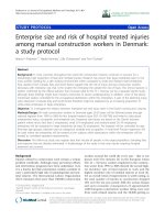
degeneration in rats subjected to mild LFPI (Figure 1).
Das et al. Journal of Neuroinflammation 2011, 8:148
/>Page 4 of 16
A consistent profile emergedinwhichthemajorityof
Fluoro-Jade (FJ)-positive cells were found within the cer-
ebral cortex (Figure 1), hippocampus (Figure 1), and
thalamus (Figure 1). Cortical Fluoro-Jade was ubiquitous
and was present at various levels throughout the brain.
Hippocampal FJ staining w as localized to the pyramidal
cell layers (Figure 1), while some diffuse labelling
throughout the general structure was also evident. The
thalamic staining was diffuse and sparsely distributed.
Quantitation revealed that the neurodegeneration in
these regions significantly increased at both 24 and 48 h
post-impact relative to sham-operated controls.
CA3
CA3
CA3
Sham
24 H
48 H
A
B
Cortex
Hippocampus
Thalamus
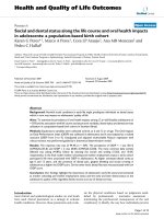
Figure 1 TBI induces neurodegeneration in different areas of the rat brain. Fluoro Jade (FJ) staining was performed on cryosections from
rat brains to identify the damaged neurons 24 hours or 48 hours after the induction of mild lateral fluid percussion impact (LFPI). A.
Representative low magnification (40X) photomicrographs showing FJ-positive neurons indicating neurodegeneration in cortex (left column),
hippocampus (middle column) and thalamus (right column) 24 hours or 48 hours after LFPI. No degenerating neurons were observed in the
corresponding brain regions in the sham animals. Scale bar = 500 μ. High magnification (400X) images from selected areas of respective sections
are shown in the inset. Scale bar = 50 μ. B. The FJ-positive neurons were quantitated using the Image J program. The histograms show the
estimation of FJ-positive neurons in cortex, hippocampus and thalamus. Cortex showed the highest number of injured neurons compared to
other regions. Most FJ-positive neurons were observed after 24 hours of injury in all three regions. The numbers of degenerating neurons went
down 48 hours after TBI but were significantly higher compared to sham animals. *** p < 0.001 compared to sham animals.
Das et al. Journal of Neuroinflammation 2011, 8:148
/>Page 5 of 16
Additionally, data showed that FJ-stained degenerating
hippocampal neurons were restricted to the ipsilateral
hemisphere, whereas few cortical and thalamic FJ-posi-
tive neurons were also detected in the contralateral
hemisphere in some animals.
Mild TBI-induced internucleosomal DNA fragmentation in
the cortex and hippocampus
Internucleosomal DNA fragmentation, an important
marker for apoptotic cells, w as assessed by terminal
deoxynucleotidyl transferase biotin-dUTP nick end
labelling (TUNEL) histochemistry. Few TUNEL-positive
cells were detected in the contralateral hemisphere, and,
while the ipsilateral thalamus showed sparse TUNEL
staining in some sections, this was not a consistent find-
ing throughout the experiment (data not shown). The
majority of TUNEL-stained nuclei were detected at 24 h
post-TBI in the ipsilateral cortex (Figure 2A) and hippo-
campus (Figure 2B), while sections from sham-operated
controls were predominantly devoid of TUNEL staining
in these regions (Figure 2A, Figure 2B) and showed only
background levels of fluorescence. By 48 h after TBI,
Sham TBI
TUNEL
DAPI
Merge
Sham
TBI
A
B
Cortex
Hippocampus
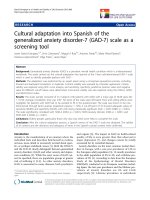
Figure 2 TBI causes DNA damage 24 hours after impact . A. Photomic rographs of representative sections from rat cortex or hippocampus
showing TUNEL histochemistry 24 hours after mild LFPI. TUNEL-positive nuclei (green fluorescence) were distributed throughout the ipsilateral
cortex or hippocampus 24 h after TBI. Intense signals are observed as rims on the nuclear boundaries with a diffuse homogeneous signal on the
interior of the nucleus. Arrows indicate the TUNEL positive nuclei. (Scale bar 500 μ). B. Histograms show the number of TUNEL-positive nuclei in
the cortex or hippocampus 24 or 48 hours after TBI. Significant increase in the TUNEL-positive nuclei at the 24 h time point indicates the DNA
damage occurs in these brain regions as early as 24 hours post-TBI although at 48 hours after TBI the damage was not significantly different in
TBI animals compared to sham-treated animals. (** p < 0.001 compared to sham animals)
Das et al. Journal of Neuroinflammation 2011, 8:148
/>Page 6 of 16
sections showed very few TUNEL-positive cells in the
cortex and hippocampus and resembled sham-operated
controls. Quan titation revealed a significant increase in
TUNEL-positive cells in both cortex and hippocampus
24 h post TBI as compared to sham-operated control
groups (Figure 2C).
Microglia are activated in the brain following mild TBI
Isolectin-IB4, a 114 kD protein isolated from the seeds
of the African legume, Griffonia simplicifolia has been
shown to have a stron g affinity for res ident microglia in
the central nervous system and peripheral macrophages
that are activated in response to neural injury. To assess
the local inflammatory response following mild TBI,
Alexa-Fluor 488-conjugated IB4 was used to label
microglia/macrophages in the brain tissue. While IB4
labelling was primarily restricted to the ipsilateral hemi-
sphere, sparse labelling was d etected within the contral-
ateral hippocampus (data not shown). IB4-positive cells
were abundant in the hippocampus, especially in the
dentate gyrus (Figure 3A). Microglia were also found in
the cortex and thalamus (data not shown) following
TBI. CD11b, an activated microglial marker, was also
found in the cells of the cortex and hippocampus (den-
tate gyrus, Figure 3A) of the ipsilateral side. Confocal
microscopy revealed that most but not all IB4
+
cells in
the cortex or hippocampus were also CD11b
+
(Figure
3A). Quantitation showed that the n umber of IB4-posi-
tive cells was significantly inc reased in each of these
brain regions 24 h after TBI, while number of IB4
+
cells
in these regions 48 h post-TBI did not significantly dif-
fer from sham-operated controls (Figure 3B). These
observations indicate that an inflammatory response was
mounted within the brain parenchyma as early as 24 h
after the injury involving microglial activation/ migra-
tion to the site of injury.
CCL20 is identified as a major inflammatory gene
expressed in the spleen and thymus following TBI
Several s tudies have suggested that in addition to the
local response, activation of the systemic inflammatory
response is criti cal in inducing TBI-associated neuropa-
thies. Although a number of cytokines and chemokines
have been studied, the key systemic inflammatory mole-
cules have not yet been identified. Because the spleen
has been shown to be involved in the systemic inflam-
matory response in various injury models, SuperArray
analysis was performed on spleen RNA from three sepa-
rate experiments to identify alterations in the expression
of genes associated with pro-inflammatory signalling
after LFPI (Figure 4). SuperArray data indicates that
more genes were down-regulated (Figure 4B) than were
up-regulated (Figure 4A). Among the genes that were
up-regulated, CCL20 was uniquely up-regulated by five-
fold compared to controls (Figure 4A) 24 h after TBI.
These studies led to the identification of CCL20 as a
potentially important pro-inflammatory, systemic marker
of TBI. To confirm this observation as well as to deter-
mine whether alterations in CCL20 mRN A paralleled
protein e xpression, ELISAs and immunohisto chemistry
were performed on spleen tissues. Immunohistochemis-
try on spleen tissues indic ated significant up-regulation
of CCL20 expr ession at 24 h after TBI as in dicated by
the increase in mean area of CCL20 intensity. Signi fi-
cant expression of the protein was also observed 48 h
aft er impact (Figures 5A, B). The immunohist ochemical
observation was further supported by the data obtained
from ELISA of spleen tissues showing at least two-fold
up-regulation of CCL20 protein expression 24 h after
TBI (Figure 5C). In addition to spleen, the thymus also
expressed CCL20 at 24 h after TBI as evident from the
immunohistochemical labelling of thymus (Figure 5A
and 5B) an d ELISA for CCL20 of thymic tissues (Figure
5C). These observations support the notion that CCL20
chemokine signalling contri butes to the systemic inflam-
matory response, and that the spleen and thymus
respond as early as 24 h after TBI.
CCL20 is expressed in the brain following TBI-induced
neurodegeneration
Data from the regional injury distribution experiments
showed that mild TBI resulted in highly reproducible
cellular injury within th e cortex as well as the hippo-
campus. Because splenic CC L20 expression was
increased in the acute phase of TBI injury (24 h post-
insult) and the splenic inflammatory response is known
to exacerbate neural injury [10,17,18] experiments were
performed to determine whether CCL20 expression is
associated with neural injury. Brain sections from ani-
mals subjected to mild TBI or sham-TBI were immu-
nostained for CCL20 expression using an antibody
generated against the same CCL20 antigen that was
used to immunostain the spleen and thymus sections
(Figure 6).
CCL20 immunoreactivity was observed in the cortex
and hippocampus 48 h after TBI. In the cortex CCL20
was expressed in the ipsilateral as well as contralateral
sides. The immunoreactivity was observed in the CA1
and CA3 hippocampal pyramidal cell layers and was
restricted to ipsilateral side of the brain. CCL20 immu-
noreactivity was absent in the 24 h group. Additionally,
CCL20-positive neuronal cell bodies displayed pyknotic
morphology and were surrounded by areas devoid of tis-
sue (Figure 6A; Figure 7A). The immunohistochemical
observation was further supported by the quantitation of
the CCL20-positive cell bodies which showed a signifi-
cant increase in CCL20-positive neurons in the cortex
and hippocampus of rats euthanized 48 h post-TBI
Das et al. Journal of Neuroinflammation 2011, 8:148
/>Page 7 of 16
compared to 24 h or sham control rats (Figure 6B). It is
noteworthy that although CCL20 immunoreactivity was
not seen in the damaged neurons at 24 h, it was
expressed by the neurons of cortex and hippocampus
(Figure 7A), including the degenerating ones in these
regions at 48 h after impact as evident by the co-local i-
zation of FJ and CCL20 stainings (Figure 7B). Impor-
tantly, CCL20 expressing cells in the cortex (Figure 8)
and hippocampus (data not shown) were mostly neu-
rons as they were also NeuN positive. Taken together,
Sham
IB4
Merge
TBI
DG
DG
DG
CD11b
A
B
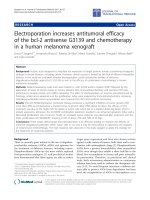
Figure 3 Mild TBI activates microglia 24 hours after impact. IB4-positive cells were observed in different areas of brain 24 hours after TBI.
Some of these cells were CD11b-positive. This labelling was absent in the sham animals and significantly less on the contralateral side or 48h
after TBI. A. Confocal microscopic images showing IB4-positive (Alexafluor 488-conjugated, green fluorescence), CD11b-positive (red fluorescence)
or IB4/CD11b-positive (red-green overlap) microglia in representative sections of ipsilateral dentate gyrus 24 hours after moderate TBI. The left
column shows CD11b immunostaining, the middle column IB4 labelling and the right column is an overlay of CD11b and IB4 double labelling.
Arrows indicate the CD11b or IB4 or CD11b-IB4 positive cells. Scale bar 30μ.B. Histograms show the quantitation of IB4-positive microglia in the
ipsilateral cortex, hippocampus and thalamus 24 or 48 hours after TBI. In all three regions, the number of IB4-positive cells was significantly
increased 24 h after TBI compared to sham animals. ** p < 0.001; * p < 0.05; compared to sham; # p < 0.05, ## p < 0.001 compared to 24H TBI.
Das et al. Journal of Neuroinflammation 2011, 8:148
/>Page 8 of 16
these observations demonstrate that CCL20 expression
is increased in the brain due to TBI-induced neuronal
injury at a later time point than the systemic increase of
the same che mokine in response to mild TBI and may
play a role in the neural injury and inflammatory reac-
tion in the brain.
Splenectomy attenuates TBI-induced neurodegeneration
and CCL20 expression in the cortex
To evaluate the significance of the spleen in LFPI-
induced neurodegeneration, splenectomy was perfo rmed
immediately after the induction of TBI. FJ histochemis-
try and CCL20 immunostaining were performed to eval-
uate the extent of damage in splene ctomised animals. It
was observed that in splenectomised rats the number of
FJ-positive cells was significantly reduced compared to
non-splenectomised animals a t the same time points,
while within the splenectomy group the number of FJ-
positive cells was significantly increased after TBI
compared to splenectomised shams (Figure 9A). Sple-
nectomy also reduced CCL20 expression in the cortex
48 h after TBI. In splenectomised rats, CCL20 expres-
sion increa sed significantly when compared to splenec-
tomised sham animals; but the CCL20 expression was
reduced signif icantly when the spenectomised TBI rats
were compared to the non-splenecto mised TBI group.
These observations indicate that the spleen plays a role
in TBI induced ne urodegeneration and CCL20 expres-
sion in the rat brain after mild TBI.
Discussion
Mild TBI comprises almost 80% of clinical TBI. Despite
continuing research and accumulated knowledge, an
effective treatment for mild TBI is still not available. In
the present study, we have adopted the LFPI model of
TBI originally characterized by McIntosh et. al. [19] to
develop a methodology that results in quantifiable
reproducible injury. Because pressure pulses within the
Fold increase
Mean ± SEM
6
-4
-2
0
Ccl12
Ccl19
Ccl22
Ccl7
Ccr8
Crp
Cxcl2
Cxcl9
Ifng
Il3
Il4
Il8ra
Fold decrease
Mean S.E.M.
Fold decrease
Mean ± SEM
0
-2
-4
-6
A
B
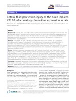
Figure 4 CCL20 is up-regulated in spleen 24 hours after mild TBI. PCR super array analysis was performed to analyze the gene expression in
spleen tissues following TBI. The histograms show the mRNA expressional changes of different cytokines, chemokines and their receptors 24
hours after TBI. A: The up-regulated genes: CCL20 mRNA increased 5-fold in TBI animals compared to the sham animals. B: The down-regulated
genes with 2-fold or more down-regulation.
Das et al. Journal of Neuroinflammation 2011, 8:148
/>Page 9 of 16
48H TBI
24H TBI
Sham
Spleen
Thymus
A
B
C
Figure 5 CCL20 expression is up-regulated in spleen and thymus after mild TBI. A: Low magnification (scale bar 500 μ) photomicrographs
showing the immunohistochemical labelling of CCL20 in spleen and thymus tissues in sham, and 24 h or 48 h after TBI. High magnification
(scale bar 20 μ) images of the selected areas from each section are shown in the inset of the corresponding image. B. CCL20 immunoreactivity
in spleen or thymus in sham or TBI animals was quantitated using the Image J program and expressed as mean area ± S.E.M. CCL20
immunoreactivity increased significantly 24 h and 48 h after TBI compared to sham animals. *p < 0.05, **p < 0.001 compared to sham. C. The
histograms show the changes of CCL20 expression in spleen and thymus 24 or 48 hours post TBI. ELISA was performed with rat anti-CCL20
antibody using a Duo set ELISA kit from R&D systems. In both tissues CCL20 expression increased significantly 24 h after TBI. *p < 0.05, ** p <
0.001 compared to sham animals.
Das et al. Journal of Neuroinflammation 2011, 8:148
/>Page 10 of 16
range used here (2.0-2.2 atm.) are generally c onsider ed
to reflect mild injury in the rat model [14], this para-
digm is particularly attractive in that it lends relevance
to the clinical population suffering from mild injury.
However, conflicting data in the literature regarding the
regional and temporal injury distribution prompted us
to conduct a comprehensive investigation throughout
the brain to determine where approximately 80% of the
CA3
CA1
LV
CA3
CA3
CA1
CA1
LV
LV
Sham
24H TBI
48H TBI
A
B
Cortex
Hippocampus
Figure 6 CCL20 is expressed in rat brain cortex and hippocampus 48 h after TBI. A. Immunostaining with anti CCL20 antibody shows CCL20-
expressing cells in cortex and hippocampus 48 h after TBI. Low magnification (scale bar 500μ) photomicrographs with high magnification (scale bar
50μ) images from selected areas are shown in the inset. The immunostaining was localized in the pyknotic cell bodies (arrows) devoid of
surrounding tissues indicating tissue damage. This immunostaining was not evident 24 h after TBI. Arrows indicate the CCL20-expressing cells. B.
CCL20-positive neurons in ipsilateral cortex and hippocampus were counted using the NIH Image J program and compared with corresponding
areas from sham animals. CCL20 expression significantly increased in TBI animals 48 hours after impact. **p < 0.001 compared to sham.
Das et al. Journal of Neuroinflammation 2011, 8:148
/>Page 11 of 16
injury is found at 24 and 48 h post-impact. These end-
points were se lected because they represe nt a de layed
window within the secondary injury phase that can be
targeted by novel therapeutics.
Consistent with previous studies using the LFPI
[20,18,21] FJ and TUNEL staining in this study showed
that the predominant areas of neurodegeneration and
apoptosis includ e the cerebral cortex, hippocampus, and
FJ
CCL20
24 h
FJ
CCL20
FJ
CCL20
FJ
CCL20
48
h
24 h
48h
FJ
CCL20
Merge
A
B
Cortex
Hippocampus
Figure 7 CCL20 expression is observed in t he areas of neurodegeneration of cortex and hippocamp us 48 hours after TBI. A.High
magnification photomicrographs of brain sections from animals subjected to TBI and sacrificed 24 or 48 h post-impact were stained with
Fluoro-Jade or anti-CCL20 antibody. Fluoro-Jade staining was observed in the cortex and in the hippocampal CA1 and CA3 pyramidal cell layers
24 and 48 hours after TBI. While no CCL20 immunoreactivity was observed in the same regions of adjacent sections 24 h after TBI, CCL20
immunoreactivity was observed in the cortical neurons as well as within the hippocampal CA1 and CA3 pyramidal cell layers at 48 h. FJ, Fluoro
Jade. Scale bar 50μ. B. Representative photomicrographs showing the FJ - CCL20 double staining in the cortex. CCL20 immunoreactivity was
observed in most of the degenerating neurons (FJ positive) as indicated by arrows. CCL20 immunoreactivity was also observed in other cells
those were not FJ positive. Scale bar 100μ.
Sham
TBI
CCL20
NeuN
Merge
Figure 8 CCL20 is expressed in rat brain cortical neurons 48 h after TBI. Fluorescence microscopic images double immunostained with anti
CCL20 antibody and the neuronal marker NeuN antibody showed most of the CCL20-expressing cells in the cortex were also NeuN positive.
White arrows indicate CCL20 positive neurons, blue arrows indicate CCL20 positive non neuronal cells. Scale bar 100 μ.
Das et al. Journal of Neuroinflammation 2011, 8:148
/>Page 12 of 16
A
B
Figure 9 Immediate splenect omy reduces TBI-induced neurodegeneration and CCL20 expression in the corte x. Degenerating neurons
(FJ positive) were observed 24 hours or 48 hours after the induction of LFPI in animals with (splenectomy group) or without (no splenectomy
group) immediate splenectomy. A. The histograms show the estimation of FJ-positive neurons as quantitated by the Image J program in the
cortex. B. CCL20 expression was observed in the cortex 48 hours after LFPI in animals with (splenectomy) or without (no splenectomy)
immediate splenectomy. The histograms show the estimation of CCL20-positive cells in the cortex. ** p < 0.0001 and * p < 0.001 compared to
sham animals within the group. # p < 0.001 compared to 24h or 48h TBI between the groups.
Das et al. Journal of Neuroinflammation 2011, 8:148
/>Page 13 of 16
thalamus. In a previous study using similar TBI metho-
dology, Sato et al. [21] showed Fluoro-Jade and TUNEL
staining that persisted from 3 hours to 7 days and
included cerebellar damage in addition to damage in
those regions identified here. Furthermore, we have
demonstrated that LFPI-induced neurodegeneration par-
alleled with increase in activated microglia in the injured
brain. Also, in accordance with the observation of Stahel
et al., the dying neurons showed characteristics of both
necrosis and apoptosis [22]. These observations indicate
that substantial inflammation takes place i n the brai n
parenchyma in response to mild LFPI in this study.
The nature and progression of TBI-induced brain
pathology limits the goals of treatment to either blocking
the secondary injury phase or facilitating plasticity and
repair at some point after the initial impact. The second-
ary phase is largely a result of the migration of activated
microglia towards the site of injury, secreting toxic cyto-
kines and oxygen radicals and thereby causing further
neuronal damage [13,23,24]. Lunemann et. al. [25] have
shown that following the formation of a brain lesion,
microglia invade the damaged brain tissue after maturing
and becoming activated by producing macrophage acti-
vating factor (MAF) in a CD11b-positive pathway. In
agreement with this finding, we have also observed that
within 24 h of initial damage the brain parenchyma is
invaded by activated microglial cells. This indicates that
an active inflammatory reaction is generated locally in
the brain as early as 24 hours after injury.
The spleen is a reservoir of peripheral macrophages
and other immune cells in the body, and it is now well
known that splenic signalling contributes to injury of
various tissues after ischemic insult. For example, sple-
nectomy prior to insult protects both the liver [26] and
brain [8] from ischemic damage. In a recent study, L i et
al. hav e shown that splenectomy immediately after TBI
in rats decreased[18] proinflammatory cytokine produc-
tion and mortality rate and improved cognitive function.
In our study, we observed that splenectomy immediately
after induction of TBI attenuated TBI-induce d neurode-
generation and CCL20 expression in the brain. Although
it is not clear how this spleen-brain interaction takes
place, Lee et al. [27] suggested that vagal nerve stimula-
tion may reduce immune cell infiltration and conse-
quent decrease in brain inflammation and edema while
Stewart and McKenzie [28] suggested a role of sympa-
thetic stimulation in causing the release of immune cells
from spleen and subsequent i nfiltration into the brain
tis sues. Regardless of the neural mechanism, removal of
the spleen immediately after the insult would r emove
the largest pool of immune cells, resulting in decreased
infiltration and consequent neuroinflammation. Our
study clearly shows that reduction in the splenic
immune cell population reduced neuronal damage and
CCL20 production.
Interestingly, CCL20 is a unique chemokine known to
interact specifically with CC chemokine receptor 6
(CCR6) and induce chemo taxis of dendritic cells, T cells
and B cells [29], all of which reside in the spleen and
have the potential to promote neuroinflammation. Sev-
eral lines of evidence support this hypothesis. Ohta et
al. [30] have shown that CCL20 was up-regu lated under
normothermic conditions in a rat middle cerebral ar tery
occlusion (MCAO) model. CCL20 is also expressed in
inflamed epithelial cells [31] and in the synovial tissues
of rheumatoid arthritis patients [32,33], while up-regula-
tion of CCL20 along with other cytokines has been
observed in human subjects one day after severe trau-
matic brain injury [34]. Furthermore, a recent study
identified CCL20 as a dual-acting chemokine with the
potential for inhibiting immune r eactions and more
importantly in attracting inflammatory effectors and
activators [35]. Although a great deal of investigation
has recently been done to elucidate the relationship
between brai n trauma and the immune system, very lit-
tle is known about the function of the thymus after
brain trauma. Since the thymus is the major source of
mature circulating T cells, CCL2 0 expression in the thy-
mus as observed in this study seems significant,
although further investigation is needed to identify the
specific function of thymus after TBI in adult rats.
Because CCL20-CCR6 signalling is now known to
facilitate the immune response in pathological cir cum-
stances, data from the present study demonstrating up-
regulated CCL20 in spleen and thymus 24 h post-LFPI
likely reflects the initiation or persistence of a systemic
signal that drives neural inflammation and cell death.
Mouse models of autoimmune encephalomyelitis (EAE)
have provided some evidence that T cells may be tar-
geted by the splenic signal. A recent knockout study
demon strated that CCR6 modulates the infiltration of T
cells into the brains of EAE-infected mice, although
reduced infiltration of Treg in CCR6-/- mice was a sso-
ciated with increased neurological damage [36]. Despit e
evidence of a protective role for CCR6 activation,
CCL20 signaling through CCR6 on Th1 or Th17 cells,
rather than Treg cells, would be expected to promote
inflammation. CCR6 is constitutively expressed in the
choroid plexus of mo use and human and there are data
showing that the binding of CCL20 to CCR6 on Th17
cells is critical for T cell infiltration into the CNS
through the choroid plexus [37]. Indeed, T cells are well
known for infiltrating the brain in neural injury models
characterized by a compromised BBB. Beca use BBB
degradation is also a critical component of TBI [19,23],
peripheral CCL20 signalling may be an impor tant
Das et al. Journal of Neuroinflammation 2011, 8:148
/>Page 14 of 16
initiator of T cell chemotaxis and extravasation into the
brain parenchyma.
Data presented in this report also show that CCL20
was not expressed in degenerating cortical or hippocam-
pal cell layers until 48 h after the impact. This raises the
question of why cortical and hippocampal neurons
expressed CCL20 at 48 h, which i s 24 h after the sys-
temic expression of the same chemokine and the neuro-
degenerationinthesameareasoftheinjuredbrain.
Although CCL20 is produced by astrocytes in response
to bacterial infections [38] and EAE [39], to the best of
our knowledge, ours is the first report to demonstrate
neuronal expression of CCL20. One possibility is that
cellular injury induces exp ression of CCL20 as a signal
for peripheral or local immune cell recruitment to the
injured site. If so, it is also likely that the neuronal cells
tha t expresse d CCL20 were in the immediate vicinity of
those cells undergoing neurodegeneration. However,
another possibility is that neuronal CCL20 expression is
a ‘tombstone’ marker in cells that are beyond repair and
need to be removed from the surrounding viable tissues.
This latter explanation is supported by the pyknotic
morphology that was observed in CCL20-expressing
neurons, as well as the fact that the areas surrounding
the cell bodies appeared to be devoid of tissue. The
morphological analysis, anatomical localization and colo-
calization with FJ and NeuN protein of CCL20-positive
cells strongly suggest that neurons represent the predo -
minant cell type expressing this chemokine following
TBI. Preli minary observations from this laboratory indi-
cate downregulation of peroxysome proliferator-acti-
vated receptor g (PPARg) in neuronal cells (data not
shown) after TBI; however, a causal role of PPARg in
regulating CCL20 signalling and/or expression in these
cells remains to be established.
While results here demonstrate a link between CCL20
expression a nd LFPI-induced injury and indicate invol-
vement of peripheral immune organs like the spleen in
this resp onse, further experiments are required to define
the precise mechanisms by which CCL20 signalling con-
tributes to cell death and the exact role played by spleen
and thymus in inducing neuronal death. Furthermore, if
CCL20 exerts direct actions on neurons, the 11 kDa
protein could easily enter the CNS from the systemic
circulation and promote injury even in the absence of
peripheral leukocyte recruitment. If this latter scenario
is the case, plasma CCL20 levels could be utilized as an
important bioma rker indicating the pres ence and sever-
ity of TBI.
Conclusion
This study identified CCL20 as a potential novel target
for anti-inflammatory therapeutic intervention. Data
from this study clearly showed that LFPI-induced brain
injury evoked an inflammatory reaction in the injured
brain and attracted a population of activated microglia
resulting in further damage of the brain. The fact that
CCL20 expression is elevated in the spleen and thymus
prior to it s appearance in the br ain, and that brai n
CCL20 expression is decreased in splenectomised rats
provide evidence that a peripheral CCL20 signal med-
iates the neuropathological response to TBI. These
results suggest that CCL20 plays an important role in
neuroinflammation in the brain after TBI, and that per-
ipheral CCL20 signalling promotes the secondary phase
of neural injury. Future studies investigating an
extended time course encompassing hours to weeks
after LFPI will be critical in determining the tissue- and
cell-specific origins, mechanisms, and overall effects of
CCL20 signalling on TBI.
Acknowledgements
This work is supported by a VA career scientist award to Shyam Mohapatra,
a USF Health development grant to Subhra Mohapatra and 5R21NS060907
to Keith Pennypacker. We would like to acknowledge Xiaoyuan Kong , Jordan
Heft and Sowndharya Rajavel for technical assistance and James Musso (III)
for assistance in initial experiments.
Author details
1
Department of Internal Medicine, University of South Florida College of
Medicine, 12901 Bruce B Downs Blvd, Tampa, FL 33612, USA.
2
Department
of Molecular Pharmacology and Physiology, University of South Florida
College of Medicine, 12901 Bruce B Downs Blvd, Tampa, FL 33612, USA.
3
Department of Molecular Medicine, University of South Florida College of
Medicine, 12901 Bruce B Downs Blvd, Tampa, FL 33612, USA.
4
JAH-VA
Hospital, Tampa, FL, 13000 Bruce B. Downs Blvd. Tampa, FL 33612, USA.
Authors’ contributions
SM and SSM have contributed to the conception and experimental design
of the study. MD carried out most of the experimental work, analysed the
data and prepared the figures of the manuscript. SR contributed to the
immunostaining. SM, MD and CCL wrote the manuscript. SSM, SM and KRP
reviewed and rated the manuscript. All authors have read and approved the
final manuscript.
Competing interests
The authors declare that they have no competing interests.
Received: 12 April 2011 Accepted: 31 October 2011
Published: 31 October 2011
References
1. Fabrizio KS, Keltner NL: Traumatic brain injury in operation enduring
freedom/operation iraqi freedom: a primer. Nurs Clin North Am 2010,
45:569-580, vi.
2. Galarneau MR, Woodruff SI, Dye JL, Mohrle CR, Wade AL: Traumatic brain
injury during Operation Iraqi Freedom: findings from the United States
Navy-Marine Corps Combat Trauma Registry. J Neurosurg 2008,
108:950-957.
3. Macgregor AJ, Dougherty AL, Galarneau MR: Injury-Specific Correlates of
Combat-Related Traumatic Brain Injury in Operation Iraqi Freedom. J
Head Trauma Rehabil 2010.
4. Fernaud-Espinosa I, Nieto-Sampedro M, Bovolenta P: Differential activation
of microglia and astrocytes in aniso- and isomorphic gliotic tissue. Glia
1993, 8:277-291.
5. Reid DM, Perry VH, Andersson PB, Gordon S: Mitosis and apoptosis of
microglia in vivo induced by an anti-CR3 antibody which crosses the
blood-brain barrier. Neuroscience 1993, 56:529-533.
Das et al. Journal of Neuroinflammation 2011, 8:148
/>Page 15 of 16
6. Ghirnikar RS, Lee YL, Eng LF: Inflammation in traumatic brain injury: role
of cytokines and chemokines. Neurochem Res 1998, 23:329-340.
7. Utagawa A, Truettner JS, Dietrich WD, Bramlett HM: Systemic inflammation
exacerbates behavioral and histopathological consequences of isolated
traumatic brain injury in rats. Exp Neurol 2008, 211:283-291.
8. Ajmo CT, Vernon DO, Collier L, Hall AA, Garbuzova-Davis S, Willing A,
Pennypacker KR: The spleen contributes to stroke-induced
neurodegeneration. J Neurosci Res 2008, 86:2227-2234.
9. Walker PA, Jimenez F, Cox CS Jr: Progenitor cell therapy for traumatic
brain injury: effect of serum osmolarity on cell viability and cytokine
production. Regen Med 2010, 5:65-71.
10. Morganti-Kossmann MC, Satgunaseelan L, Bye N, Kossmann T: Modulation
of immune response by head injury. Injury 2007, 38:1392-1400.
11. Thompson HJ, Lifshitz J, Marklund N, Grady MS, Graham DI, Hovda DA,
McIntosh TK: Lateral fluid percussion brain injury: a 15-year review and
evaluation. J Neurotrauma 2005, 22:42-75.
12. Cernak I: Animal models of head trauma. NeuroRx 2005, 2:410-422.
13. Cernak I, Vink R, Zapple DN, Cruz MI, Ahmed F, Chang T, Fricke ST,
Faden AI: The pathobiology of moderate diffuse traumatic brain injury as
identified using a new experimental model of injury in rats. Neurobiol Dis
2004, 17:29-43.
14. Kabadi SV, Hilton GD, Stoica BA, Zapple DN, Faden AI: Fluid-percussion-
induced traumatic brain injury model in rats. Nat Protoc 2010,
5:1552-1563.
15. Schmued LC, Albertson C, Slikker W Jr: Fluoro-Jade: a novel fluorochrome
for the sensitive and reliable histochemical localization of neuronal
degeneration. Brain Res 1997, 751:37-46.
16. Duckworth EA, Butler TL, De Mesquita D, Collier SN, Collier L,
Pennypacker KR: Temporary focal ischemia in the mouse: technical
aspects and patterns of Fluoro-Jade evident neurodegeneration. Brain
Res 2005, 1042:29-36.
17. Lucas SM, Rothwell NJ, Gibson RM: The role of inflammation in CNS injury
and disease. Br J Pharmacol 2006, 147(Suppl 1):S232-240.
18. Li M, Li F, Luo C, Shan Y, Zhang L, Qian Z, Zhu G, Lin J, Feng H: Immediate
Splenectomy Decreases Mortality and Improves Cognitive Function of
Rats After Severe Traumatic Brain Injury. J Trauma 2011.
19. McIntosh TK, Vink R, Noble L, Yamakami I, Fernyak S, Soares H, Faden AL:
Traumatic brain injury in the rat: characterization of a lateral fluid-
percussion model. Neuroscience 1989, 28:233-244.
20. Hellmich HL, Eidson KA, Capra BA, Garcia JM, Boone DR, Hawkins BE,
Uchida T, Dewitt DS, Prough DS: Injured Fluoro-Jade-positive
hippocampal neurons contain high levels of zinc after traumatic brain
injury. Brain Res 2007, 1127:119-126.
21. Sato M, Chang E, Igarashi T, Noble LJ: Neuronal injury and loss after
traumatic brain injury: time course and regional variability. Brain Res
2001, 917:45-54.
22. Stahel PF, Shohami E, Younis FM, Kariya K, Otto VI, Lenzlinger PM,
Grosjean MB, Eugster HP, Trentz O, Kossmann T, Morganti-Kossmann MC:
Experimental closed head injury: analysis of neurological outcome,
blood-brain barrier dysfunction, intracranial neutrophil infiltration, and
neuronal cell death in mice deficient in genes for pro-inflammatory
cytokines. J Cereb Blood Flow Metab 2000, 20:369-380.
23. Cernak I, Chang T, Ahmed FA, Cruz MI, Vink R, Stoica B, Faden AI:
Pathophysiological response to experimental diffuse brain trauma differs
as a function of developmental age. Dev Neurosci 2010, 32:442-453.
24. Cernak I, Noble-Haeusslein LJ: Traumatic brain injury: an overview of
pathobiology with emphasis on military populations. J Cereb Blood Flow
Metab 2010, 30:255-266.
25. Lunemann A, Ullrich O, Diestel A, Jons T, Ninnemann O, Kovac A, Pohl EE,
Hass R, Nitsch R, Hendrix S: Macrophage/microglia activation factor
expression is restricted to lesion-associated microglial cells after brain
trauma. Glia 2006, 53:412-419.
26. Okuaki Y, Miyazaki H, Zeniya M, Ishikawa T, Ohkawa Y, Tsuno S,
Sakaguchi M, Hara M, Takahashi H, Toda G: Splenectomy-reduced hepatic
injury induced by ischemia/reperfusion in the rat. Liver 1996, 16:188-194.
27. Lee ST, Chu K, Jung KH, Kim SJ, Kim DH, Kang KM, Hong NH, Kim JH,
Ban JJ, Park HK, et al: Anti-inflammatory mechanism of intravascular
neural stem cell transplantation in haemorrhagic stroke. Brain 2008,
131:616-629.
28. Stewart IB, McKenzie DC: The human spleen during physiological stress.
Sports Med 2002, 32:361-369.
29. Schutyser E, Struyf S, Van Damme J: The CC chemokine CCL20 and its
receptor CCR6. Cytokine Growth Factor Rev 2003, 14:409-426.
30. Ohta H, Terao Y, Shintani Y, Kiyota Y: Therapeutic time window of post-
ischemic mild hypothermia and the gene expression associated with
the neuroprotection in rat focal cerebral ischemia. Neurosci Res 2007,
57:424-433.
31. Dieu-Nosjean MC, Massacrier C, Homey B, Vanbervliet B, Pin JJ, Vicari A,
Lebecque S, Dezutter-Dambuyant C, Schmitt D, Zlotnik A, Caux C:
Macrophage inflammatory protein 3alpha is expressed at inflamed
epithelial surfaces and is the most potent chemokine known in
attracting Langerhans cell precursors. J Exp Med 2000, 192:705-718.
32. Chabaud M, Page G, Miossec P: Enhancing effect of IL-1, IL-17, and TNF-
alpha on macrophage inflammatory protein-3alpha production in
rheumatoid arthritis: regulation by soluble receptors and Th2 cytokines.
J Immunol 2001, 167:6015-6020.
33. Matsui T, Akahoshi T, Namai R, Hashimoto A, Kurihara Y, Rana M,
Nishimura A, Endo H, Kitasato H, Kawai S, et al: Selective recruitment of
CCR6-expressing cells by increased production of MIP-3 alpha in
rheumatoid arthritis.
Clin Exp Immunol 2001, 125:155-161.
34. Helmy A, Carpenter KL, Menon DK, Pickard JD, Hutchinson PJ: The cytokine
response to human traumatic brain injury: temporal profiles and
evidence for cerebral parenchymal production. J Cereb Blood Flow Metab
2010.
35. Comerford I, Bunting M, Fenix K, Haylock-Jacobs S, Litchfield W, Harata-
Lee Y, Turvey M, Brazzatti J, Gregor C, Nguyen P, et al: An immune
paradox: how can the same chemokine axis regulate both immune
tolerance and activation?: CCR6/CCL20: a chemokine axis balancing
immunological tolerance and inflammation in autoimmune disease.
Bioessays 2010, 32:1067-1076.
36. Villares R, Cadenas V, Lozano M, Almonacid L, Zaballos A, Martinez AC,
Varona R: CCR6 regulates EAE pathogenesis by controlling regulatory
CD4+ T-cell recruitment to target tissues. Eur J Immunol 2009,
39:1671-1681.
37. Reboldi A, Coisne C, Baumjohann D, Benvenuto F, Bottinelli D, Lira S,
Uccelli A, Lanzavecchia A, Engelhardt B, Sallusto F: C-C chemokine
receptor 6-regulated entry of TH-17 cells into the CNS through the
choroid plexus is required for the initiation of EAE. Nat Immunol 2009,
10:514-523.
38. McKimmie CS, Graham GJ: Astrocytes modulate the chemokine network
in a pathogen-specific manner. Biochem Biophys Res Commun 2010,
394:1006-1011.
39. Ambrosini E, Columba-Cabezas S, Serafini B, Muscella A, Aloisi F: Astrocytes
are the major intracerebral source of macrophage inflammatory protein-
3alpha/CCL20 in relapsing experimental autoimmune encephalomyelitis
and in vitro. Glia 2003, 41:290-300.
doi:10.1186/1742-2094-8-148
Cite this article as: Das et al.: Lateral fluid percussion injury of the brain
induces CCL20 inflammatory chemokine expression in rats. Journal of
Neuroinflammation 2011 8:148.
Submit your next manuscript to BioMed Central
and take full advantage of:
• Convenient online submission
• Thorough peer review
• No space constraints or color figure charges
• Immediate publication on acceptance
• Inclusion in PubMed, CAS, Scopus and Google Scholar
• Research which is freely available for redistribution
Submit your manuscript at
www.biomedcentral.com/submit
Das et al. Journal of Neuroinflammation 2011, 8:148
/>Page 16 of 16