báo cáo hóa học: " Low ficolin-3 levels in early follow-up serum samples are associated with the severity and unfavorable outcome of acute ischemic stroke" pptx
Bạn đang xem bản rút gọn của tài liệu. Xem và tải ngay bản đầy đủ của tài liệu tại đây (1.39 MB, 27 trang )
This Provisional PDF corresponds to the article as it appeared upon acceptance. Fully formatted
PDF and full text (HTML) versions will be made available soon.
Low ficolin-3 levels in early follow-up serum samples are associated with the
severity and unfavorable outcome of acute ischemic stroke
Journal of Neuroinflammation 2011, 8:185 doi:10.1186/1742-2094-8-185
George Fust ()
Lea Munthe-Fog ()
Zsolt Illes ()
Gabor Szeplaki ()
Tihamer Molnar ()
Gabriella Pusch ()
Kristof Hirschberg ()
Robert Szegedi ()
Zoltan Szeplaki ()
Zoltan Prohaszka ()
Mikkel-Ole Skjoedt ()
Peter Garred ()
ISSN 1742-2094
Article type Research
Submission date 7 September 2011
Acceptance date 29 December 2011
Publication date 29 December 2011
Article URL />This peer-reviewed article was published immediately upon acceptance. It can be downloaded,
printed and distributed freely for any purposes (see copyright notice below).
Articles in JNI are listed in PubMed and archived at PubMed Central.
For information about publishing your research in JNI or any BioMed Central journal, go to
/>For information about other BioMed Central publications go to
Journal of Neuroinflammation
© 2011 Fust et al. ; licensee BioMed Central Ltd.
This is an open access article distributed under the terms of the Creative Commons Attribution License ( />which permits unrestricted use, distribution, and reproduction in any medium, provided the original work is properly cited.
/>Journal of Neuroinflammation
© 2011 Fust et al. ; licensee BioMed Central Ltd.
This is an open access article distributed under the terms of the Creative Commons Attribution License ( />which permits unrestricted use, distribution, and reproduction in any medium, provided the original work is properly cited.
1
Low ficolin-3 levels in early follow-up serum samples are associated with the severity
and unfavorable outcome of acute ischemic stroke
George Füst
1
, Lea Munthe-Fog
2
, Zsolt Illes
3
, Gábor Széplaki
1
, Tihamér Molnar
4
, Gabriella
Pusch
3
, Kristóf Hirschberg
5,7
, Robert Szegedi
6
, Zoltán Széplaki
6
, Zoltán Prohászka
1
, Mikkel-
Ole Skjoedt
2
, Peter Garred
2
1
3rd Department of Internal Medicine, Semmelwies University, Budapest, Hungary,
2
Laboratory of Molecular Medicine, Department of Clinical Immunology-7631,
Rigshospitalet, University of Copenhagen, Copenhagen, Denmark,
3
Division of Clinical and
Experimental Neuroimmunology, Department of Neurology, University of Pecs, Pecs,
Hungary,
4
Institute of Anaesthesia and Intensive Therapy, Faculty of Medicine, University of
Pecs, Pecs, Hungary,
5
Heart Center, Semmelweis University, Budapest, Hungary,
6
Department of Neurology, Kútvölgyi Clinical Centre, Semmelweis University, Budapest,
Hungary,
7
Experimental Laboratory of Cardiac Surgery, University of Heidelberg, Germany
Address correspondence to Dr George Füst
3rd Dept Internal Medicine
Semmelweis University
Budapest
Kútvölgyi út 4
Phone: 361-212-9351, fax: 361-225-3899
e-mail:
2
Abstract
Background. A number of data indicate that the lectin pathway of complement activation
contributes to the pathophysiology of ischemic stroke. The lectin pathway may be triggered
by the binding of mannose-binding lectin (MBL), ficolin-2 or ficolin-3 to different ligands.
Although several papers demonstrated the significance of MBL in ischemic stroke, the role of
ficolins has not been examined.
Methods. Sera were obtained within 12 hours after the onset of ischemic stroke (admission
samples) and 3-4 days later (follow-up samples) from 65 patients. The control group
comprised 100 healthy individuals and 135 patients with significant carotid stenosis (patient
controls). The concentrations of ficolin-2 and ficolin-3, initiator molecules of the lectin
complement pathway, were measured by ELISA methods. Concentration of C-reactive
protein (CRP) was also determined by a particle-enhanced immunturbidimetric assay.
Results. Concentrations of both ficolin-2 and ficolin-3 were significantly (p<0.001) decreased
in both the admission and in the follow-up samples of patients with definite ischemic stroke as
compared to healthy subjects. Concentrations of ficolin-2 and ficolin-3 were even higher in
patient controls than in healthy subjects, indicating that the decreased levels in sera during the
acute phase of stroke are related to the acute ischemic event. Ficolin-3 levels in the follow-up
samples inversely correlated with the severity of stroke indicated by NIH scale on admission.
In follow-up samples an inverse correlation was observed between ficolin-3 levels and
concentration of S100β, an indicator of the size of cerebral infarct. Patients with low ficolin-3
levels and high CRP levels in the follow up samples had a significantly worse outcome
(adjusted ORs 5.6 and 3.9, respectively) as measured by the modified Rankin scale compared
to patients with higher ficolin-3 and lower CRP concentrations. High CRP concentrations
were similarly predictive for worse outcome, and the effects of low ficolin-3 and high CRP
were independent.
Conclusions: Our findings indicate that ficolin-mediated lectin pathways of complement
activation contribute to the pathogenesis of ischemic stroke and may be additive to
complement-independent inflammatory processes.
Keywords: stroke, ischemic stroke, outcome, complement, lectin pathway, ficolins, ficolin-2,
ficolin-3, CRP
3
Background
Neuroinflammation is a key element in the ischemic cascade after cerebral ischemia that
results in cell damage and death in the subacute phase.[1]
Complement activation is one of the pathological mechanisms that contribute to the
ischemic/reperfusion injury in ischemic stroke [2-4]. Among other neuroinflammatory
processes, the complement system is also activated during tissue injury and has recently been
considered as a new potential therapeutic target in ischemic stroke [5] and in intracerebral
haemorrhage [6]. Both animal experiments and observations made in stroke patients indicate
that activation of the complement system is one of the mechanisms contributing to the
extension of the cerebral infarct after ischemic stroke [7]. Several studies have demonstrated
the essential role of complement activation in brain damage following cerebral ischemia. Such
evidence includes (i) an increased expression of complement proteins and complement
receptors after permanent middle cerebral artery occlusion (MCAO) [8-11] (ii) different
pathological events in complement-deficient/-sufficient animals after the onset of cerebral
ischemia compared to wild-type littermates: complement deficient animals are at least
partially protected after transient MCAO [12-15] . (iii) In rodent experimental models,
complement depletion induced using the cobra venom factor (CVF) [16, 17], as well as
complement inhibition by a plasma-derived C1-inhibitor [18, 19], a recombinant C1 inhibitor
[20] , CR2-Crry [13] and intravenous immunoglobulin administration [14] were proven to
exert beneficial, neuroprotective effects, indicating the protective role of complement
antagonism and inhibition.
Only a few studies have explored complement activation in patients with ischemic stroke [21,
22]. Recently, we found that sC5b-9 levels determined at admission exhibited a significant
positive correlation with the clinical severity of stroke, as well as with the extent of the
neurological deficit as determined by different scales [3]. Our findings suggested that the
lectin pathway is primarily responsible for the activation of complement in ischemic stroke. In
agreement with these findings, Cervera et al. [4] demonstrated both in mice and stroke
patients that genetically determined MBL-deficiency is associated with a better outcome after
acute ischemic stroke. In a high number of patients with ischemic stroke, Osthoff et al. [23]
found that a deficiency of the mannose-binding lectin is associated with smaller infarction
size and a more favorable outcome. More recently, the group of De Simoni [24] reported on
the formation of functional MBL/MASP-2 complexes in plasma in mice after MCAO, and
4
demonstrated that molecules, which strongly bound to MBL, induced significant reduction in
neurological deficits and infarct volume, when administered 6 h after transient MCAO. These
data support the notion that the lectin pathway plays a crucial role in the development of
ischemic stroke.
Apart from MBL, the ficolins also serve as recognition molecules in the lectin complement
pathway. Three different ficolins have been described in humans. Ficolin-1, -2, and -3 are
derived from the genes FCN1, FCN2, and FCN3, respectively. In healthy individuals, ficolin-
2 and -3 are present in the serum and plasma in relatively high concentrations, while the
concentration of ficolin-1 is much lower [25]. Similar to MBL, the ficolins are associated
with a set of three serine proteases, termed MBL-associated serine proteases (MASPs),
enabling activation of the complement system. The primary activator of the lectin pathway
appears to be MASP-2.
As described above, there are abundant data about the significance of MBL in ischemic
stroke. The role of the ficolins, initiator molecules of the lectin complement pathway,
however, has never been studied in this disease. Therefore, we measured the levels of ficolin-
2 and ficolin-3 in sera from 65 patients with ischemic stroke and from controls. In order to
assess the clinical significance of the results, serum concentrations of these proteins were
correlated to an indirect measure of the stroke severity (NIHss), S100β concentration on day
3, which is an indicator of the size of cerebral infarct, [26, 27] as well as the outcome of the
disease expressed by the modified Rankin scale.
Besides complement activation, other inflammatory processes are also known to contribute to
the pathogenesis of the ischemic stroke [1]. Among them, CRP-associated processes were
mostly studied. In 2005, Di Napoli et al [28] summarized evidence for CRP as an independent
predictor of cerebrovascular events in at-risk individuals and its usefulness in evaluating
prognosis after stroke. It was also demonstrated that C-reactive protein predicts the prognosis
of patients with functional disability after the first occurrence of ischemic stroke [29] and
correlates to the infarct volume [30]. Recently, Ormstad et al. [31] provided evidence that
CRP plays an important role in the progression of cerebral tissue injury. In addition, in our
previous study [3] we found that complement activation and elevated CRP levels were
independently associated with the clinical severity and different outcome measures of
ischemic stroke, indicating their additive effect. Therefore, serum concentrations of CRP and
its relationship to the ficolin levels were also examined here.
5
Methods
Patients and control subjects
Patients with ischemic stroke included in the present work were admitted to two centers: the
Department of Neurology, University of Pecs, Hungary (39 patients:20 men and 19 women,
aged 49-84 years) and the Department of Neurology, Kútvölgyi Clinical Centre, Semmelweis
University, Budapest, Hungary (26 patients:10 men and 16 women, aged 58–87 years)
(Table 1). The management of ischemic stroke was in accordance with the guidelines of the
Stroke Council of the American Heart Association/ American Stroke Association [32] None
of the patients were treated by intravenous thrombolysis. Patients with stroke were enrolled
upon the first occurrence of acute ischemic stroke only; all patients had neuroimaging (most
of them brain MRI, but at least cranial CT). No patients had hemorrhagic infarction. All
patients with definite acute clinical symptoms were enrolled regardless of etiology i.e. lacunar
or territorial infarct caused by thrombosis or emboli. Exclusion criteria were infectious
diseases, fever <4 weeks before stroke, an elevated WBC, erythrocyte sedimentation rate
(ESR), high-sensitivity CRP (hsCRP, cut-off value <10 mg/L), procalcitonin on admission
(cut-off value <0.05 ng/mL), positive chest X-ray, hemorrhagic stroke defined by an acute
cranial CT scan, and those who declined to participate in the study. Almost all patients had
hypertension and elevated cholesterol/triglyceride levels. All patients were therefore treated
for such risk factors; nevertheless the effect of such treatments on the ficolin pathway is
unlikely. An evidence-based guideline [33] was followed to detect post-stroke infectious
complications (in short, physical and laboratory measures including WBC, ESR, hsCRP,
PCT, fever, abnormal urine, chest X-ray or positive cultures). Such complications occurred on
the 4
th
day as an average, and were located to the respiratory system and urinary tract even in
the absence of catheterization; in addition, thrombophlebitis occurred in a single case.
At the time of admission, severity of stroke was assessed using the National Institutes of
Health Stroke Scale (NIHSS). [34]. Blood samples were obtained at the time of admission
(admission samples: the median time from the onset of symptoms was 7 hours in the
Budapest cohort and 8.5 hours in the Pecs cohort), and 72 to 96 hours later (follow-up
samples). Five and four patients in the Pecs and Budapest cohorts, respectively, developed
infections. The outcome of disease was assessed with the modified Rankin scale [35].
Serum samples were also taken from 100 healthy volunteers as controls (Table 1).
Additionally, 134 patients with significant carotid atherosclerosis served as controls (Table
1). In agreement with international guidelines, significant carotid atherosclerosis was defined
as 70–100% stenosis of the carotid artery determined by Duplex scan sonography. The
6
examination was indicated in the case of other vascular disorders or risk factors of vascular
disorders. None of the patients had definite residual signs and no symptoms suggesting acute
ischemia. Some of these patients had either peripheral arterial disease or coronary disease, and
in these patients carotid Duplex scans were performed to detect asymptomatic severe carotid
stenosis (as a common comorbidity). Some of the patients had non-specific symptoms (i.e.
dizziness) or transient ischemic attack previously; in these patients diagnostic carotid Duplex
scans were performed. Lacunar strokes defined by neuroimaging were no exclusion criteria.
Serum samples of the patients and of the controls were stored at -80
o
C in the Hungarian
laboratories until transported on dry ice to Copenhagen.
The study was approved by the local ethics committees, and all patients and control subjects
gave informed consent.
Laboratory methods
The serum concentrations of the proteins ficolin-2 [36] and ficolin-3 [37] were determined by
ELISA-based methods at the Laboratory of Molecular Medicine, Department of Clinical
Immunology, Rigshospitalet, Copenhagen, Denmark. Briefly, microtiter plates were coated
with either monoclonal anti-ficolin-2 antibody (FCN216) or monoclonal anti-ficolin-3
antibody (FCN334) in phosphate buffered saline (PBS) overnight at 4°C. Samples diluted
1:50 or 1:640 in sample buffer (PBS-T with 1% mouse serum and bovine serum) were added
in triplets to washed wells and incubated for 3 hours at 37°C. Ficolin-2 was detected with
biotinylated monoclonal anti-ficolin-2 antibody (FCN219) and ficolin-3 was detected with
biotinylated monoclonal anti-ficolin-3 antibody (FCN334) by incubation overnight at 4°C.
Washed wells were incubated for 1 hour at 37°C with HRP-conjugated streptavidin. Plates
were developed for 15 min with OPD (o-phenylenediamine) substrate solution and stopped by
adding 1M H
2
SO
4
. The optical density was measured at 490 nm. A standard dilution series of
pooled human serum were added to each assay as were a sample control. The lower limit of
detection in these assays is 5 ng/ml of ficolin-2 and 1 ng/ml of ficolin-3. The inter-assay
coefficient of variation (CV) is 7.1% and 4.7% and the intra-assay CV 4.3% and 3.9% for the
ficolin-2 and ficolin-3 assay, respectively.
Human S100β concentrations were measured by an ELISA method (BioVendor, Modrice,
Czech Republic). In our previous study, we found that the concentration of S100β was the
7
highest 72 hours after the onset of stroke, therefore concentration was determined at this
timepoint [27].
Serum CRP concentrations were measured by particle-enhanced immunturbidimetric assay,
using an automated laboratory analyzer (Roche Cobas Integra 400, Basel, Switzerland).
Statistical evaluation of the results
Statistical analysis was performed using the GraphPad Prism 3.0 (GraphPad Software Inc,
San Diego, CA, www.graphpad.com) and SPSS 13.0 (SPSS Inc., Chicago, IL) software.
Between-group differences were evaluated by the Mann-Whitney test. Correlations between
the variables were expressed using non-parametric Spearman’s correlation coefficients. The
categorical variables were compared with the χ2 test for trend. The association between the
serum concentration of selected proteins and the outcome of stroke was calculated by multiple
logistic regression, adjusted for the sex and the age of the patients. All tests were two-tailed.
All data are presented as median values with the 25
th
to 75
th
percentiles in parentheses unless
stated otherwise.
Results
The concentrations of the proteins of the lectin pathway and CRP in the sera of patients with
ischemic stroke, as compared to healthy controls and patient controls
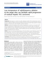
The serum levels of ficolin-2, ficolin-3 and CRP were measured in the samples obtained from
65 stroke patients on admission and 3-4 days later (follow-up samples), as well as in the sera
of 100 healthy volunteers and 134 patient controls (patients with severe carotid
atherosclerosis without acute stroke) (Figure 1). Compared to both healthy controls and
patient controls, both ficolin-2 ficolin-3 levels were significantly lower both in the admission
and follow-up sera of stroke patients. When all patients were considered, CRP levels were
significantly higher in the admission samples than in the sera of healthy controls but were
nearly equal to that measured in the sera of patient controls. By contrast in the follow up
samples, CRP levels were significantly higher as compared to both control groups. When
patients who developed infections were not considered, the difference between stroke patients
and controls became non-significant (data not shown). As for the two controls groups, all the
three variables had significantly higher concentration in the sera of patient controls than in the
healthy controls, although the difference in the ficolin-3 levels was small.
8
Follow-up ficolin-3 levels correlated with the indirect measures of stroke severity and infarct
size
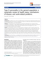
Ficolin-3 concentrations measured in the follow-up samples but not in the admission samples
exhibited a significant, negative correlation with indirect measures of stroke severity i.e. the
NIH score determined on admission (Figure 2, panel A). Patients were divided into two
groups in a similar manner to Foerch 2005 [26]; those with a NIH scale of <16 with relatively
good expected outcome and those with NIH scale of >16 with poor expected outcome, and the
ficolin-3 levels were compared accordingly. There were significantly (p=0.017) lower ficolin-
3 levels in the former than in the latter group. By contrast, no significant differences in the
ficolin-2 levels (p=0.309) were found between the two groups (data not shown).
In addition, we found significant negative correlation between ficolin-3 concentrations and
the S100β level measured in the follow-up samples but not in the admission samples (Figure
2, panel B). The levels of ficolin-2 did not correlate with the S10B concentrations (data not
shown).
CRP concentrations in follow-up samples were significantly higher in patients with high
(>16) NIH score (Figure 2, Panel C), but did not significantly correlate with the S100β levels
(Figure 2, panel D).
Ficolin-3 and CRP levels in follow-up samples correlate with the outcome of acute ischemic
stroke
The levels of the ficolins and CRP were related to the outcome of the disease, as assessed by
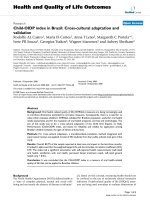
the modified Rankin scale (Figure 3). When patients were divided according to unfavorable
(3 to 6) and favorable (0 to 2) modified Rankin scores, ficolin-3 levels were lower in the
former group, supporting the association with an unfavorable outcome. The difference was
significant only in the follow-up samples, while almost significant in the admission samples
(Figure 3, panels A and B). When the 9 patients, who developed infectious complications
were excluded, CRP levels both in admission and follow up samples were significantly higher
in the patient group with unfavorable compared to favorable outcome (Figure 3, panels C
and D).
We confirmed these data by performing a multiple logistic regression analysis. Unfavorable
(modified Rankin scale 3 to 6) vs. favorable (modified Rankin scale 0 to 2) outcome was
regarded as a dependent variable, whereas ficolin-3 levels, CRP levels, age and sex were
considered as independent variables (Table 2). Since both ficolin-3 and CRP levels were
9
included in the analysis, those 9 patients who developed infectious complications were
excluded.
Both ficolin-3 and CRP levels measured in the follow up samples were significantly
associated with the outcome of the disease: lower ficolin-3 and higher CRP values were found
in the unfavorable compared to the favorable outcome group. Similar but only, marginally
significant (ficolin-3) or weakly significant (CRP) associations were found when the
admission samples were analyzed.
Next, in order to assess the strength of association between the low ficolin-3 and high CRP
levels on the one hand and the unfavorable outcome of the disease on the other hand, we
repeated the analysis as above in the follow-up samples by including ficolin-3 and CRP levels
as low/high values. Ficolin-3 levels below or equal to the median (16 µg/ml for both the
admission and follow up samples) were considered low, while those above the median value
were considered high. CRP levels above median (7.7 mg/L) were considered high (Table 3).
In the analysis, adjusted for sex and age of the patients, both the low ficolin-3 and the high
CRP levels significantly predicted an unfavorable outcome, with odds ratios of 5.6 and 3.9,
respectively.
Correlation between the baseline NIH score, serum S100β concentration in the follow up
samples as well as the outcome of the disease
Finally, we assessed the relationship between the baseline NIH scale as an indirect measure of
the severity of the stroke, the concentration of the S100β in the follow up samples as an
indicator of the infarct size, and the outcome of the disease assessed by the modified Rankin
scale (Figure 4). Both measures exhibited highly significant correlation to the outcome:
patients with high baseline NIHSS scale had much worse outcome than those with low NIH
scale, and patients with unfavorable outcome had higher serum S100β concentrations at 72
hours than those with a favorable outcome.
10
Discussion
We report here on three novel observations: (i) the decrease of serum concentrations of two
proteins of the lectin pathway during the acute phase of ischemic stroke; (ii) an inverse
correlation of ficolin-3 levels obtained 3-4 days post-admission with the severity and outcome
of acute ischemic stroke; (iii) the independent effect of low ficolin-3 and high CRP levels on
the outcome of the disease.
As compared to healthy subjects, the serum concentrations of both ficolin-2 and ficolin-3,
initiator proteins of the lectin complement pathway, were significantly lower in the samples
taken from patients with ischemic stroke immediately after admission (i.e. within hours after
the onset of the symptoms). The levels of these proteins did not further change during the
initial 3-4 days of stroke. The differences observed between stroke patients and healthy
individuals seem to be valid, since the ficolin-2 and ficolin-3 concentrations measured in the
sera of healthy subjects are similar to previously reported data [38]. In addition, ficolin-2 and
ficolin-3 levels were significantly lower in sera of patients with definite stroke compared to
patients with severe carotid atherosclerosis without clinical event as well. The main age of
this group was equal to that of stroke patients. This control group of patients exhibited even
higher ficolin levels than healthy subjects. These data may suggest that the decreased levels of
ficolins in the acute phase of stroke were not related to the chronic and severe atherosclerosis,
but rather a decrease in ficolin-2 and ficolin-3 concentrations may happen in the very early
phase of the acute ischemic event. In addition, these data indicated that the difference in
ficolin concentrations comparing healthy controls and stroke patients were not related to the
difference between their ages.,.
The decreased concentration of ficolins could be observed in the very early phase of ischemic
stroke and remained unchanged during the next 3-4 days. It seems reasonable to surmise that
this decrease was due to consumption through the binding of the molecules to the apoptotic
and necrotic cells in the penumbra of the cerebral infarct [2]. Moreover, ficolin-2 and ficolin-3
have also been shown to be involved in the sequestration of dying host cells [39]. The
observations made by Wang et al. [40] are of particular interest, since these authors reported
that maternal plasma concentrations of ficolin-3 and ficolin-2 were significantly (p<0.001)
lower in preeclamptic pregnancies than in uncomplicated pregnancies, due to the
sequestration of the proteins in placenta. Additionally, they found that both ficolins but
particularly ficolin-3 were associated with ischemic placenta tissue.
11
According to our second observation, lower level of ficolin-3 in the follow-up samples were
associated with greater size of the cerebral infarct indicated by higher S100β levels in the
sera. Astrocyte-derived S100β concentration is a marker of the degree and the severity of
cellular injury in acute ischemic stroke [41]. The examination of S100β protein has been
accepted as a good biomarker of the infarct size [26, 42-44]. The concentration of S100β is
known to be the highest 72 hours after the onset of stroke [27].
In addition, ficolin-3 levels inversely correlated with the indirect measure of the severity of
ischemic stroke, i.e. with the NIHSS neurological deficit score. Higher NIHSS scores define
more severe deficits [34].
Additionally, a strong negative correlation was found between ficolin-3 concentration and the
outcome of the disease measured with modified Rankin scale. This negative correlation
indicates that low ficolin-3 levels are associated with an unfavorable prognosis. This
association is most probably secondary to the negative correlation between ficolin-3 on the
one hand and the severity of ischemic stroke and the infarct size on the other hand, as
discussed above. It is well known that both the high baseline NIHSS score and the high serum
S100β levels predict poor prognosis of ischemic stroke, which was also found in the present
study (Figure 4). The lack of clinical correlates of ficolin-2 could be explained by the
observation that ficolin-3 has the highest concentration and the greatest complement-
activating capacity among the lectin pathway initiators [45].
Complement activation is one of the pathological mechanisms contributing to
ischemic/reperfusion injury in ischemic stroke [2-4]. The selective ability for complement
activation after the binding of ficolin-3 to dying cells may be responsible for the selective
clinical correlation with the levels of this protein. Further studies, including simultaneous
measurement of ficolin-3 levels and of the generation of complement activation products, are
necessary to confirm this assumption.
Our present findings also support previous data [3, 4, 23], which showed that the lectin
complement pathway indeed plays an important role in the pathogenesis of acute ischemic
stroke; here we show that a ficolin-3-dependent activation of the lectin pathway also
contributes to the pathological processes besides the previously suggested MBL-dependent
activation.
Third, in accordance with the previous data [46-48] and earlier work from our groups [3, 27],
we measured higher CRP levels in the sera of patients obtained at admission as compared to
healthy controls, and high CRP levels measured on day 3 were strongly associated with an
12
unfavorable outcome of ischemic stroke. This latter observation is in accordance with the
recent findings of Song et al. [29]. According to our present findings, the clinical associations
with the low ficolin-3 and high CRP levels measured in the follow up samples are
independent, indicating that they reflect two different pathways of inflammation contributing
to the pathogenesis of the disease. These findings may have important therapeutic
implications. Anti-inflammatory drugs have already been used for the treatment of ischemic
stroke with limited success. Since many pharmacological agents, which are able to inhibit
pathological complement activation are either approved for therapeutic purposes (such as C1-
inhibitor [49] or eculizimab [50]) or are under clinical trials [51], these may be more
efficiently used for treatment of ischemic stroke either alone or in combination with anti-
inflammatory drugs.
The paper has some limitations. First of all, the number of patients tested is rather low and no
late follow-up samples were collected for ficolin measurements. Nevertheless our
observations are novel and may initiate a number of studies.
Conclusions
Our findings indicate that two seemingly different but only partially identified pathways of
neuroinflammation, the ficolin-3-dependent activation of lectin pathway of complement and
CRP-dependent processes independently contribute to the pathogenesis and poor outcome of
acute ischemic stroke. These findings may lead to the introduction of novel treatment
approaches for a disease with a rather limited therapeutic arsenal at present.
13
List of abbreviations
CR1: complement receptor type 1; CR2: complement receptor type 2; CRP: C-reactive
protein, CVF: cobra venom factor; MCAO: middle cerebral artery occlusion, MASP: MBL-
associated serine protease, MBL: mannose-binding lectin, NIHSS: National Institutes of
Health Stroke Scale OR: odds ratio
Competing interests
The author(s) declare that they have no competing interests
Authors' contributions
GF, ZsI and PG conceived of the study, and participated in its design and coordination, and
helped to draft the manuscript; L-MT and M-OS carried out the immunoassays; GSZ, T, GP,
KH, RSZ and ZSz participated in the collection and analysis of clinical data; ZP participated
at the design of the study and drafting the manuscript. All authors read and approved the final
manuscript.
Acknowledgement
The authors wish to thank Ms Sandra Færch and Ms Vibeke Witved for skilful technical
assistance. This study was supported by grants from the Novo Nordisk Research Foundation,
Svend Andersens Foundation, Rigshospitalet and Research Foundation of the Capital Region
of Denmark, as well as by grants from the Hungarian National Research Fund (OTKA 77892
to ZI), the Hungarian Ministry of Health (ETT 036/2009 to GF) and the Hungarian
Neuroimaging Foundation (to ZI).
14
References
1. Ceulemans AG, Zgavc T, Kooijman R, Hachimi-Idrissi S, Sarre S, Michotte Y: The
dual role of the neuroinflammatory response after ischemic stroke: modulatory
effects of hypothermia. J Neuroinflammation 2010, 7:74.
2. Yanamadala V, Friedlander RM: Complement in neuroprotection and
neurodegeneration. Trends Mol Med 2010, 16:69-76.
3. Szeplaki G, Szegedi R, Hirschberg K, et al.: Strong complement activation after
acute ischemic stroke is associated with unfavorable outcomes. Atherosclerosis
2009, 204:315-320.
4. Cervera A, Planas AM, Justicia C, et al.: Genetically-defined deficiency of
mannose-binding lectin is associated with protection after experimental stroke in
mice and outcome in human stroke. PLoS One 2010, 5:e8433.
5. Mocco J, Sughrue ME, Ducruet AF, Komotar RJ, Sosunov SA, Connolly ES, Jr.: The
complement system: a potential target for stroke therapy. Adv Exp Med Biol 2006,
586:189-201.
6. Ducruet AF, Zacharia BE, Hickman ZL, et al.: The complement cascade as a
therapeutic target in intracerebral hemorrhage. Exp Neurol 2009, 219:398-403.
7. D'Ambrosio AL, Pinsky DJ, Connolly ES: The role of the complement cascade in
ischemia/reperfusion injury: implications for neuroprotection. Mol Med 2001,
7:367-382.
8. Nishino H, Czurko A, Fukuda A, et al.: Pathophysiological process after transient
ischemia of the middle cerebral artery in the rat. Brain Res Bull 1994, 35:51-56.
9. Van Beek J, Bernaudin M, Petit E, et al.: Expression of receptors for complement
anaphylatoxins C3a and C5a following permanent focal cerebral ischemia in the
mouse. Exp Neurol 2000, 161:373-382.
10. Huang J, Kim LJ, Mealey R, et al.: Neuronal protection in stroke by an sLex-
glycosylated complement inhibitory protein. Science 1999, 285:595-599.
11. Pedersen ED, Froyland E, Kvissel AK, et al.: Expression of complement regulators
and receptors on human NT2-N neurons effect of hypoxia and reoxygenation.
Mol Immunol 2007, 44:2459-2468.
12. Mocco J, Mack WJ, Ducruet AF, et al.: Complement component C3 mediates
inflammatory injury following focal cerebral ischemia. Circ Res 2006, 99:209-217.
13. Atkinson C, Zhu H, Qiao F, et al.: Complement-dependent P-selectin expression
and injury following ischemic stroke. J Immunol 2006, 177:7266-7274.
14. Arumugam TV, Tang SC, Lathia JD, et al.: Intravenous immunoglobulin (IVIG)
protects the brain against experimental stroke by preventing complement-
mediated neuronal cell death. Proc Natl Acad Sci U S A 2007, 104:14104-14109.
15. Harhausen D, Khojasteh U, Stahel PF, et al.: Membrane attack complex inhibitor
CD59a protects against focal cerebral ischemia in mice. J Neuroinflammation
2010, 7:15.
16. Vasthare US, Barone FC, Sarau HM, et al.: Complement depletion improves
neurological function in cerebral ischemia. Brain Res Bull 1998, 45:413-419.
17. Figueroa E, Gordon LE, Feldhoff PW, Lassiter HA: The administration of cobra
venom factor reduces post-ischemic cerebral injury in adult and neonatal rats.
Neurosci Lett 2005, 380:48-53.
18. Akita N, Nakase H, Kaido T, Kanemoto Y, Sakaki T: Protective effect of C1 esterase
inhibitor on reperfusion injury in the rat middle cerebral artery occlusion model.
Neurosurgery 2003, 52:395-400.
15
19. De Simoni MG, Storini C, Barba M, et al.: Neuroprotection by complement (C1)
inhibitor in mouse transient brain ischemia. J Cereb Blood Flow Metab 2003,
23:232-239.
20. Gesuete R, Storini C, Fantin A, et al.: Recombinant C1 inhibitor in brain ischemic
injury. Ann Neurol 2009, 66:332-342.
21. Pedersen ED, Waje-Andreassen U, Vedeler CA, Aamodt G, Mollnes TE: Systemic
complement activation following human acute ischaemic stroke. Clin Exp
Immunol 2004, 137:117-122.
22. Mocco J, Wilson DA, Komotar RJ, et al.: Alterations in plasma complement levels
after human ischemic stroke. Neurosurgery 2006, 59:28-33.
23. Osthoff M, Katan M, Fluri F, et al.: Mannose-binding lectin deficiency is associated
with smaller infarction size and favorable outcome in ischemic stroke patients.
PLoS One 2011, 6:e21338.
24. Orsini F, Parrella S, Villa P, et al.: Mannose binding lectin as a target for cerebral
ischemic injury. Molecular Immunology 2011, 48:1677.
25. Garred P, Honore C, Ma YJ, et al.: The genetics of ficolins. J Innate Immun 2009,
2:3-16.
26. Foerch C, Singer OC, Neumann-Haefelin T, du Mesnil de Rochemont R, Steinmetz H,
Sitzer M: Evaluation of serum S100B as a surrogate marker for long-term
outcome and infarct volume in acute middle cerebral artery infarction. Arch
Neurol 2005, 62:1130-1134.
27. Molnar T, Papp V, Banati M, et al.: Relationship between C-reactive protein and
early activation of leukocytes indicated by leukocyte antisedimentation rate
(LAR) in patients with acute cerebrovascular events. Clin Hemorheol Microcirc
2010, 44:183-192.
28. Di Napoli M, Schwaninger M, Cappelli R, et al.: Evaluation of C-reactive protein
measurement for assessing the risk and prognosis in ischemic stroke: a statement
for health care professionals from the CRP Pooling Project members. Stroke
2005, 36:1316-1329.
29. Song IU, Kim YD, Kim JS, Lee KS, Chung SW: Can high-sensitivity C-reactive
protein and plasma homocysteine levels independently predict the prognosis of
patients with functional disability after first-ever ischemic stroke? Eur Neurol
2010, 64:304-310.
30. Youn CS, Choi SP, Kim SH, et al.: Serum highly selective C-reactive protein
concentration is associated with the volume of ischemic tissue in acute ischemic
stroke. The American journal of emergency medicine 2010, 30(1):124-8.
31. Ormstad H, Aass HC, Lund-Sorensen N, Amthor KF, Sandvik L: Serum levels of
cytokines and C-reactive protein in acute ischemic stroke patients, and their
relationship to stroke lateralization, type, and infarct volume. Journal of
neurology 2011, 258:677-685.
32. Adams RJ, Albers G, Alberts MJ, et al.: Update to the AHA/ASA recommendations
for the prevention of stroke in patients with stroke and transient ischemic attack.
Stroke 2008, 39:1647-1652.
33. Cohen J, Brun-Buisson C, Torres A, Jorgensen J: Diagnosis of infection in sepsis: an
evidence-based review. Critical care medicine 2004, 32:S466-494.
34. Brott T, Adams HP, Jr., Olinger CP, et al.: Measurements of acute cerebral
infarction: a clinical examination scale. Stroke 1989, 20:864-870.
35. Bonita R, Beaglehole R: Recovery of motor function after stroke. Stroke 1988,
19:1497-1500.
16
36. Munthe-Fog L, Hummelshoj T, Hansen BE, et al.: The impact of FCN2
polymorphisms and haplotypes on the Ficolin-2 serum levels. Scand J Immunol
2007, 65:383-392.
37. Munthe-Fog L, Hummelshoj T, Ma YJ, et al.: Characterization of a polymorphism
in the coding sequence of FCN3 resulting in a Ficolin-3 (Hakata antigen)
deficiency state. Mol Immunol 2008, 45:2660-2666.
38. Sallenbach S, Thiel S, Aebi C, et al.: Serum concentrations of lectin-pathway
components in healthy neonates, children and adults: mannan-binding lectin
(MBL), M-, L-, and H-ficolin, and MBL-associated serine protease-2 (MASP-2).
Pediatr Allergy Immunol 2011, 22(4):424-30.
39. Jensen ML, Honore C, Hummelshoj T, Hansen BE, Madsen HO, Garred P: Ficolin-2
recognizes DNA and participates in the clearance of dying host cells. Mol Immunol
2007, 44:856-865.
40. Wang CC, Yim KW, Poon TC, et al.: Innate immune response by ficolin binding in
apoptotic placenta is associated with the clinical syndrome of preeclampsia. Clin
Chem 2007, 53:42-52.
41. Beer C, Blacker D, Bynevelt M, Hankey GJ, Puddey IB: Systemic markers of
inflammation are independently associated with S100B concentration: results of
an observational study in subjects with acute ischaemic stroke. J
Neuroinflammation 2010, 7:71.
42. Herrmann M, Vos P, Wunderlich MT, de Bruijn CH, Lamers KJ: Release of glial
tissue-specific proteins after acute stroke: A comparative analysis of serum
concentrations of protein S-100B and glial fibrillary acidic protein. Stroke 2000,
31:2670-2677.
43. Jauch EC, Lindsell C, Broderick J, Fagan SC, Tilley BC, Levine SR: Association of
serial biochemical markers with acute ischemic stroke: the National Institute of
Neurological Disorders and Stroke recombinant tissue plasminogen activator
Stroke Study. Stroke 2006, 37:2508-2513.
44. Laskowitz DT, Kasner SE, Saver J, Remmel KS, Jauch EC: Clinical usefulness of a
biomarker-based diagnostic test for acute stroke: the Biomarker Rapid
Assessment in Ischemic Injury (BRAIN) study. Stroke 2009, 40:77-85.
45. Hummelshoj T, Fog LM, Madsen HO, Sim RB, Garred P: Comparative study of the
human ficolins reveals unique features of Ficolin-3 (Hakata antigen). Mol
Immunol 2008, 45:1623-1632.
46. Di Napoli M: Systemic complement activation in ischemic stroke. Stroke 2001,
32:1443-1448.
47. Elkind MS, Tai W, Coates K, Paik MC, Sacco RL: High-sensitivity C-reactive
protein, lipoprotein-associated phospholipase A2, and outcome after ischemic
stroke. Arch Intern Med 2006, 166:2073-2080.
48. Youssef MY, Mojiminiyi OA, Abdella NA: Plasma concentrations of C-reactive
protein and total homocysteine in relation to the severity and risk factors for
cerebrovascular disease. Transl Res 2007, 150:158-163.
49. Cicardi M, Zanichelli A: Replacement therapy with C1 esterase inhibitors for
hereditary angioedema. Drugs Today (Barc) 2010, 46:867-874.
50. Schrezenmeier H, Hochsmann B: Eculizumab opens a new era of treatment for
paroxysmal nocturnal hemoglobinuria. Expert Rev Hematol 2009, 2:7-16.
51. Emlen W, Li W, Kirschfink M: Therapeutic complement inhibition: new
developments. Semin Thromb Hemost 2010, 36:660-668.
17
Figures Legends
Figure 1
Concentrations of ficolin-2, ficolin-3 and C-reactive protein in the sera of patients with
acute ischemic stroke
Concentrations at the time of hospital admission and on day 3, as compared to healthy
controls (HC) and patient controls (PC, patients with >70% stenosis of the carotid artery
without acute stroke) are shown. P values (*<0.05, ***<0.01) for the non-parametric Kruskal-
Wallis test followed by the Dunn post hoc test are indicated.
Figure 2
Correlation between ficolin-3 levels with severity and outcome of stroke and size of
infarct
Panel A: Negative correlation between serum ficolin-3 levels in follow-up (FU) samples and
the severity of stroke as assessed by the NIH stroke scale at admission in 65 patients with
ischemic stroke. Patients with unfavorable (>16) vs. favorable (<16) NIH scale were
compared. P value of Mann-Whitney test is indicated. Panel B: Negative correlation between
serum ficolin-3 levels in follow-up samples and the size of cerebral infarct as assessed by the
S100β level in follow-up samples. Spearman’s correlation coefficient and its significance is
indicated. Panel C: Positive correlation between serum CRP levels in follow-up samples and
the severity of stroke as assessed by the NIH scale at admission in 65 patients with ischemic
stroke. Patients with unfavorable (>16) vs. favorable (<16) NIH scale were compared. P value
of Mann-Whitney test is indicated. Panel D: No significant correlation between serum CRP
levels in follow-up samples and the size of cerebral infarct as assessed by the S100β level in
follow-up samples. Spearman’s correlation coefficient and its significance is indicated.
Figure 3
Relationship of serum ficolin-3 and CRP levels with outcome
Differences in ficolin 3 (left panels, A, C) and CRP (right panels, B, D) levels measured in
admission samples (upper panels, A, B) and in follow-up samples (lower panel, C, D)
comparing patients with a favorable (modified Rankin scale: 1 or 2) and an unfavorable
(modified Rankin scale 3 to 6) outcome. In the case of the CRP calculations, nine patients
with infectious complications were excluded from the analysis. The significance of the Mann-
Whitney test is indicated.
18
Figure 4
Relationship between the baseline NIH score scale values and the 3-day serum S100β
concentration with the outcome of the disease in 65 patients with ischemic stroke
Panel A: Distribution of the patients with different outcome of the disease among patients
with low (<6), medium (6-10) and high (>10) baseline NIHSS scale. P value for χ2 test is
indicated. Panel B: Differences in the S100β concentration between patients with favorable
(modified Rankin score: 0-2) and unfavorable modified Rankin score: 3-6) outcome.
19
Table 1
Main characteristics of the cohorts tested
Cohort Patients with
ischemic stroke
Healthy controls Patients with
severe athero-
sclerosis
(patient
controls)
Number of subjects 65 100 134
Sex, males/females 20/19 47/53 88/46
Age, years, mean + S.D. 69.8 ± 9.8 35.5 ± 9 69.8 ± 9.9
Median time between the onset of
symptoms and blood sampling, in
hours
7.0 or 8.5*
Infection, yes/no 9/56
Lethal outcome yes/no 7/58
NIH scale at admission, >16 vs
<16
58/7
Serum S100β levels, pg/ml,
median (IQ range)
0,27 (0.12-0.93)
Outcome: modified Rankin scale
at discharge: 0/1/2/3/4/5/6
4/13/14/7/9/7/1
*in the Pécs and Budapest groups, respectively.
20
Table 2
Relationship between the ficolin-3 and CRP levels and unfavorable (modified Rankin scale 3
to 6) vs. favorable (modified Rankin scale: 1 to 2) outcome of ischemic stroke as calculated
by multiple logistic regression analysis
OR (95% CI) (p value)**
Admission samples Follow-up samples
ficolin 3, µg/ml
0.989 (0.776-1.020)
(p=0.093)
0.736 (0.603-0.899)
(p=0.003)
CRP, mg/L 1.226 (1.040-1.446)
(p=0.015)
1.375 (1.095-1.727)
(p=0.006)
Sex (females/males) 0.951 (0.238-3.794)
(p=0.943)
0.969 (0.207-4.535)
P=0.968)
Age, years 1.005 (0.925-1.092)
(p=0.904)
0.981 (0.902-1.066)
(p=0.652)
Multiple logistic regression analysis adjusted for sex and age was used. Nine patients with
infectious complications were excluded from the analysis.
21
Table 3
Relationship between the low ficolin-3 and high CRP levels and outcome of ischemic stroke
OR*** (95% CI) P value
Low vs. high ficolin 3* 5.628 (1.497-21.153) 0.044
High vs. low CRP** 3.949 (1.036-15.055) 0.011
Sex (females/males) 1.171 (0.306-4.491) 0.818
Age, years 1.041 (0.966-1.122) 0.294
*low ficolin 3 defined as <16 µg/ml (median), **high CRP levels defined as >7,7 mg/L
(median), ***unfavorable (modified Rankin scale: 3 to 6) vs. favorable (modified Rankin
scale: 0 to 2) outcome. Multiple logistic regression analysis adjusted to sex and age was used.
Nine patients with infectious complications were excluded from the analysis.
Figure 1
Figure 2