Báo cáo hóa học: " Do bullae and emphysema increase risk of pneumothorax in silicosis?" pot
Bạn đang xem bản rút gọn của tài liệu. Xem và tải ngay bản đầy đủ của tài liệu tại đây (392.97 KB, 6 trang )
BioMed Central
Page 1 of 6
(page number not for citation purposes)
Journal of Occupational Medicine
and Toxicology
Open Access
Research
Do bullae and emphysema increase risk of pneumothorax in
silicosis?
Iraj Mohebbi*
1
, Ebrahim Hassani
2
, Shaker Salarilak
3
and
Abdul Rahman Bahrami
4
Address:
1
Department of Occupational Medicine, Urmia University of Medical Sciences, Urmia, Iran,
2
Department of Anesthesiology, Urmia
University of Medical Sciences, Urmia, Iran,
3
Department of Community Medicine, Urmia University of Medical Sciences, Urmia, Iran and
4
Department of Occupational Health, Hamadan University of Medical Sciences, Hamadan, Iran
Email: Iraj Mohebbi* - ; Ebrahim Hassani - ; Shaker Salarilak - ;
Abdul Rahman Bahrami -
* Corresponding author
Abstract
Background: The occurrence of occupational lung diseases is decreasing due to improvements in
occupational health in recent years; however, silicosis and its complications remain important
occupational health problems. We have studied the role of emphysema and bullae as predictive
factors of secondary spontaneous pneumothorax in acute and accelerated silicosis.
Methods: This study was carried out using questionnaire items on occupational history and
conventional computed tomography of lungs. Differences between two groups (silicosis with and
without secondary spontaneous pneumothorax) in terms of age, interval of exposure-diagnosis and
therefore silica exposure duration were assessed by independent t-test. Fisher's exact test was
used to determine the association between secondary spontaneous pneumothorax and both
emphysema and bullae.
Results: We found a significant association between secondary spontaneous pneumothorax and
bullae in acute and accelerated silicosis.
Conclusion: Pneumothorax in silicosis could be attributed to previous bullae.
Background
Pneumothorax is the presence of air in the pleural cavity
[1]. The spontaneous form is generally due to the rupture
of subpleural blebs. Clinical diagnosis of pneumothorax
is established by history, physical examination and where
possible, by radiological investigations [2-4]. Secondary
spontaneous pneumothorax (SSP) occurs as a complica-
tion of an underlying lung disease, which can be identi-
fied. Chronic obstructive pulmonary disease (COPD) and
Pneumocystis carinii pneumonia are the most common
conditions associated with SSP [1,5]. Pleural involvement
in silicosis is rare and SSP is the only recognised pleural
complication of silicosis [6-11]. SSP is usually unilateral,
when it occurs in the course of silicosis. There are only a
few reports in which patients with silicosis had bilateral
SSP [9,11]. Some investigators have reported that emphy-
sema and bullae formation may lead to the occurrence of
SSP [3,12]. Emphysema is defined as enlargement of air-
spaces distal to the terminal bronchiole, accompanied by
destructive changes of alveolar walls [13]. It is usually clas-
Published: 15 September 2007
Journal of Occupational Medicine and Toxicology 2007, 2:8 doi:10.1186/1745-6673-2-8
Received: 22 April 2007
Accepted: 15 September 2007
This article is available from: />© 2007 Mohebbi et al; licensee BioMed Central Ltd.
This is an Open Access article distributed under the terms of the Creative Commons Attribution License ( />),
which permits unrestricted use, distribution, and reproduction in any medium, provided the original work is properly cited.
Journal of Occupational Medicine and Toxicology 2007, 2:8 />Page 2 of 6
(page number not for citation purposes)
sified into the following three main subtypes: centrilobu-
lar emphysema; panlobular emphysema; and paraseptal
emphysema [14]. Bullae can develop in association with
any type of emphysema. A bulla is a sharply demarcated
area with a diameter greater than 1 cm and possessing a
wall less than 1 mm in thickness [15].
This case-control study was performed to evaluate the
association between SSP and both emphysema and bullae
on the basis of lung conventional computerized tomogra-
phy (CCT) scan in the non-smoker silicotic patients.
Methods
We studied all of registered silicotic patients who had
immigrated as seasonal temporary workers from West
Azarbaijan province to the stone grinding factories of
Azandarian area. Pneumothorax was defined by detection
of the thin, visceral line in the expiratory postero-anterior
chest radiographs that was displaced from the chest wall.
Those who suffered pneumothorax selected as case group
and others as control.
Characteristics of the factories and study population
From September 2000 to March 2006, 21 subjects with
silicosis had been registered in the Urmia Occupational
Medical Center of West Azarbaijan province in Iran. They
had immigrated as seasonal workers from West Azarbai-
jan province (in the west of Iran) to the Azandarian area,
a suburb of Hamadan province in the center of Iran. This
area located 750 Km far away from West Azarbaijan prov-
ince, and also is a base for stone-cutting and stone-grind-
ing factories in Hamadan province. Each factory usually
employs 5 to 10 seasonal temporary workers from various
districts. We studied only workers who were resident in
the West Azarbaijan province with history of employment
in aforementioned factories. We did not have accesses to
other workers, because they were scattered in other prov-
inces. All of the workers who are included in this study
had previously worked in the same stone-grinding facto-
ries in the Azandarian area. They did not have any
reported silica exposure before working at those work-
places. They described the working environment as being
very dusty, with no provision of engineering control or
exhaust ventilation.
Diagnostic methods
Diagnosis of silicosis was made on the basis of clinical
findings, chest radiological criteria in accordance with the
ILO International Classification of Radiographs of Pneu-
moconiosis, an unequivocal history of substantial silica
dust exposure and an appropriate interval of time after
exposure. We defined acute and accelerated silicosis
according to NIOSH definition [16]. Patients with silico-
sis were entered into the study if the high voltage poste-
rior-anterior radiograph of the chest with acceptable
quality showed reticular-nodular shadowing at least 1/1
profusion grade according to the ILO system. Expiratory
postero-anterior chest radiography was reviewed for
detection of pneumothorax and also SSP was defined by
identification of the thin, visceral line that was displaced
from the chest wall. In each subject, CCT of lung was
reviewed for recognition of emphysema and bullae. For
each subject, a detailed history of work occupations was
recorded as follows: (a) the age of onset of exposure to sil-
ica dust; (b) the age when the exposure to silica dust
ended; (c) the age when the initial diagnosis was made.
CCT procedures
The scanners of GE 4000 SYTEC (GE Medical Systems,
Milwaukee, WI, USA) and X VISION-EX CT (Toshiba;
Tokyo, Japan) were used for recognition of emphysema
and bullae. For each subject, CCT scans had been
obtained with 10 mm collimation at 1 cm intervals from
the upper to the lower thorax in the supine position.
Emphysema was defined as well-delineated focal periph-
eral areas surrounded by a thin wall and less than 10 mm
in diameter. If an area of emphysema was greater than 1
cm in diameter, this was defined as a bulla.
Stone grinding process
There was the same process in the 20 stone grinding facto-
ries as follows: The quartz stone is first put into a jaw
crusher where large stone is broken into smaller pieces,
which are then taken through a conveyor belt to disinte-
grator, which makes powder out of these small pieces. It is
then separated according to its fineness through a vibrat-
ing screen. The quartz powder is also passed through a
magnetic separator in order to remove the extraneous fer-
rous material from the product raw material.
Characteristics of the occupational exposure
In the Azandarian area where the patients had been
employed, all of the stone-grinding factories were selected
for the assessment of environmental exposure of silica
particles. The determination of quartz was carried out
according to the NIOSH method number 7500. A rotam-
eter was used to adjust the flow. The respirable dust sam-
ples were collected on 25 mm cellulose acetate filters
(pore size 0.8 μm) placed in a 25 mm conductive plastic
cyclone. The cyclone was attached to the worker's overalls
as closely as possible to the face in order to determine
respirable dust in the breathing zone. The filters were con-
ditioned in desiccator environmental chamber for 24
hours at 25°C and weighed before and after testing to
determine total penetrating weights. The analysis was
done by X-ray diffraction (XRD) using a Siemens Model
D5000 diffractometer equipped with variable slit in
research laboratory of X-ray at the Faculty of Science,
Tehran University.
Journal of Occupational Medicine and Toxicology 2007, 2:8 />Page 3 of 6
(page number not for citation purposes)
Statistical analysis
Statistical analysis was performed using commercially
available software (SPSS 10.0 for Windows; SPSS; Chi-
cago, IL, USA). Differences between the two groups (sili-
cosis with SSP and silicosis without SSP) in age, interval
between the onset of exposure to silica dust and clinical
diagnosis of silicosis, and silica exposure duration were
assessed by independent t-test. We used Fisher's exact test
to determine the association between SSP and predictive
factors that included emphysema and bullae.
Results
The total working period was 12 to 14 h a day for 1 to 5
consecutive years. All of the subjects were male and non-
smokers with the youngest being 20 and the oldest 79, of
whom 18 (86%) were younger than 40. The acute and
accelerated silicosis were found in 43% and 57%, respec-
tively. Latency periods of the acute and accelerated forms
were 3.2 ± 0.83 (mean ± SD) and 6.4 ± 1.6 (mean ± SD)
years, respectively. In the chest radiographs, 3(14.3%)
had radiological profusion category 1, and8(38.1%) cate-
gory 2. 10 (47.7%) showed category3. The most common
subcategories of small opacities profusion was 3/3
(42.9%).
In patients who experienced SSP, the interval between the
onset of exposure to silica dust and clinical diagnosis of
silicosis was 3.2 ± 1.1 (mean ± SD) years and in those
without SSP, it was 5.42 ± 1.60 years. The mean of expo-
sure duration in patients with SSP was 2.14 ± 1 years and
in those without SSP, it was 2.86 ± 1.2 years. The differ-
ence in age between the two groups at the end of exposure
was not significant. There was a significant difference
between the two groups in the interval from first silica
exposure to diagnosis of SSP and no significant differ-
ences in exposure duration (Table 1).
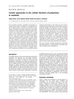
In our study, SSP was identified in 34% of subjects (Figure
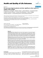
1) of which 19% was bilateral. Emphysema and bullae
were found in 49% and 52%, respectively (Figure 2).
There was a high odds ratio between SSP and emphysema
(odds ratio = 10.8 CI; 0.997–117). We found that the
probabilities of SSP were higher in those with bullae
(odds ratio = 15 CI; 1.3–168), as shown in Table 2.
Table 1: Comparison of age, interval between onset of exposure
and clinical diagnosis, and exposure duration between the two
groups
Variable Silicosis with
SSP(mean ±
SD)
Silicosis
without SSP
(mean ± SD)
T p value
Age at the end
of exposure
(years)
26.43 ± 5.85 34.50 ± 17.7 1.22 0.073
Interval between
the onset of
exposure and
clinical diagnosis
(years)
3.2 ± 1.1 5.42 ± 1.6 3.5 0.002
Exposure
duration (years)
2.14 ± 1 2.86 ± 1.2 1.3 0.19
CT. 1: Large localized pneumothorax in right lungFigure 1
CT. 1: Large localized pneumothorax in right lung. 2: Mixed
alveolar and interstitial fibrosis. 3: Pleural thickening in right
lung. 4: Several bullae in right lung. 5: Alveolar and interstitial
shadowing. 6: Paraseptal emphysema in anterior segment of
left upper lobe.
CT. 1: Hydropneumothorax in right lungFigure 2
CT. 1: Hydropneumothorax in right lung. 2: Multiple bullae in
both lungs. 3: Paraseptal emphysema in left lung. 4: Mixed
alveolar and interstitial pattern as silicoproteinosis and fibro-
sis. 5: Air bronchogram
Journal of Occupational Medicine and Toxicology 2007, 2:8 />Page 4 of 6
(page number not for citation purposes)
The silica particles in the ambient air at all of the factories
were higher than OSHA level, as shown in Table 3.
Discussion
The results of the present study demonstrate that in indi-
viduals with advanced silicosis, SSP is significantly associ-
ated with the presence of bullae. The results also indicate
that advanced silicosis causes distal airspace enlargement
independent of smoking.
To the best of our knowledge, predictive factors of SSP as
a complication of silicosis have not been extensively
described in the literature [1,5]. Choi et al. studied 458
patients who underwent transthoracic needle biopsy
(TTNB). They found a significantly higher (p < 0.001) risk
of pneumothorax in patients with emphysema and con-
cluded that development of pneumothorax may be pre-
vented by the elastic recoil of the normal lung
parenchyma and pleura [17]. Mitlehner et al. suggested
that the presence of bullae in patients with primary spon-
taneous pneumothorax has no predictive value for the
future development of recurrences [18]. Conversely, bul-
lae rupture in SSP appears to be due to local airway
obstruction, emphysema susceptibility, and the presence
of bronchial abnormalities [3]. Functionally, massive
fibrosis results in stiff nondistensible lungs with increased
elastic recoil [19]. In advanced silicosis, coalescence of
perinodular emphysematous regions may lead to forma-
tion of macroscopic blebs, which can rupture causing a
pneumothorax [12]. Our findings were consistent with
these findings and indicate that the occurrence of SSP
could be attributed to the presence of bullae.
In the absence of smoking, coal pneumoconiosis and con-
fluent silicosis are associated with emphysematous
changes in the lungs [20]. Some investigators have
reported that emphysema with silicosis has also been
observed to occur independently of smoking [21,22].
Emphysema is common in silicosis and has been attrib-
uted as the major cause of corpulmonale and disability
rather than fibrosis by some investigators [23].
In this study, all of the subjects were non-smokers and the
results confirmed that emphysematous changes are com-
mon in non-smoker silicotic patients.
In a case series study, Kawano et al. suggested that there
was no correlation between the onset of SSP and duration
of occupational exposure to silica [24]. Our findings sup-
port this hypothesis as shown in Table 1. According to an
investigation by Bahrami and Mahjub in the stone-grind-
ing factories where the study subjects had been employed,
the concentration of silica compounds in the ambient air
had been 25–50 times higher than Occupational Safety
and Health Administration (OSHA) levels [25]. Our find-
ings were consistent with these findings. Our study also
showed that the interval between the onset of exposure
and clinical diagnosis of silicosis was statistically different
for both groups, but both of them were exposed to
extremely high levels of respirable silica. In summary,
development of SSP may be enhanced by increased elastic
recoil of the lung parenchyma, and bullae rupture. There
is a high probability of SSP occurring in acute and acceler-
ated silicosis.
Historically, people employed in stone-grinding factories
are from lower income backgrounds and have not had the
advantage of regular medical surveillance or specialized
care until their conditions become very advanced. A sig-
nificant difference between the two groups in the interval
from first silica dust exposure to clinical diagnosis of sili-
cosis might possibly be a confounding issue that silicosis
may be misdiagnosed due to lack of appropriate health
Table 3: Exposure assessment in each industrial unit (mg/m 3)
Industrial unit Total dust Total Respirable
dust
Respirable
quartz
1 1355.37 66.70 17.35
2 1438.21 78.34 19.60
3 1442.21 79.45 17.89
4 1472.73 83.87 20.34
5 1542.32 88.45 22.87
6 1618.71 94.45 26.00
7 1624.45 83.45 23.90
8 1641.34 105.54 31.89
9 1645.13 104.79 32.90
10 1725.75 109.86 31.00
11 1747.00 111.27 39.90
12 1747.00 121.32 42.27
13 1747.45 121.30 42.80
14 1836.73 126.97 44.90
15 1845.37 127.12 44.89
16 1924.14 134.12 45.89
17 1995.34 134.78 50.90
18 2032.80 159.45 56.90
19 2058.81 141.00 48.50
20 2067.83 134.60 44.80
Table 2: Odds ratio and comparison of risk factors between the
two groups
Risk Factor Pneumoth
orax
Odds ratio 95%
Confidence
Interval
No Yes
Bullae No 10 1 15 1.3–168
Yes 4 6
Emphysema No 9 1 10.8 .997–117
Yes 5 6
Journal of Occupational Medicine and Toxicology 2007, 2:8 />Page 5 of 6
(page number not for citation purposes)
surveillance. We concluded that a brief, but intensive
exposure to silica dust could cause SSP after a short
latency period. It is recommended that an effective
hygiene program be implemented to monitor the health
of these workers.
Our methodology for assessing odds ratios and the asso-
ciation between pneumothorax and both bullae and
emphysema in silicosis has both strengths and weak-
nesses. Its strengths are as follows. All subjects had worked
in the same unregulated stone-grinding workplaces and
had no history of smoking, underlying disease of COPD
and/or other mineral dust exposure. Information was
available on the specific occupational exposure to silica
powder in the stone-grinding factories. Although the sam-
ple size in our study was only 21, to the best of our knowl-
edge, it is larger than any previous study of pneumothorax
in silicosis worldwide, so its major strength is due to the
number of cases. A final strength is that information was
available on possible confounding factors such as tobacco
use.
The main limitation of our study also concerns the
number of subjects; however, we attempted to find all of
scattered workers. We used the opportunity and discussed
the implications for enforcement of regulations on a
national level to prevent the occurrence of silicosis (espe-
cially in Hamadan province where this outbreak had
occurred). Due to the rarity of acute and accelerated silico-
sis and also SSP in silicosis, statistical analysis of binary
outcomes is almost always based on odds ratios and it is
the same as the risk ratio [26]. Therefore, we believe this
study might be a good estimation of odds ratio or relative
risk of SSP in acute and accelerated silicosis.
Conclusion
Our study findings reemphasize the clinical importance of
SSP and its association with bullae. The results also indi-
cate that advanced silicosis causes distal airspace enlarge-
ment independently of smoking.
Abbreviations
CCT: Conventional computerized tomography
COPD: Chronic obstructive pulmonary disease
ILO: International Labour Office
NIOSH: National Institute for Occupational Safety and
Health
OSHA: Occupational Safety and Health Administration
SSP: Secondary spontaneous pneumothorax
TTNB: Transthoracic needle biopsy
Competing interests
We have not received any financial support or grant from
any organization for carrying out this research, and all of
expenditure has been met by the researchers with the aim
of benefiting humanity. The authors declare that they
have no completing interests.
Authors' contributions
Iraj Mohebbi carried out the clinical and imaging studies,
participated in the study design, sequence alignment and
drafted the manuscript. Ebrahim Hassani carried out the
scientific editing of the manuscript. Shaker Salarilak per-
formed the statistical data analysis/interpretation. Abdul
Rahman Bahrami helped in the assessment of environ-
mental monitoring. All authors read and approved the
final manuscript.
Acknowledgements
This work was performed at the Urmia Medical Sciences University, Urmia,
Iran. None of the authors has any financial interest in any of the products
mentioned in this article. We would like to thank Dr Mehrdad Arjomandi
for his distinguished recommendations and review of the manuscript. The
authors are grateful to Dr Zubeyri, Dr Mohammadi, and Dr Jalili in the CT
imaging suite of Urmia Medical Sciences University for their assistance.
Thanks also to Alpha Science Editors for English editing services and to all
West Azerbaijan Healthcare staff for their help.
References
1. Sahn SA, Heffner JE: Spontaneous pneumothorax. N Engl J Med
2000, 342:868-874.
2. Strobel SL: Pathologic quiz case: Recurrent spontaneous
pneumothorax in an industrial worker. Arch Pathol Lab Med
2002, 126:749-750.
3. Al-Qudah A: Treatment options of spontaneous pneumotho-
rax. Indian J Chest Dis Allied Sci 2006, 48:191-200.
4. Weill H, Jones RN, Parkes WR: Silicosis and related diseases. In
Occupational Lung Disorders 3rd edition. Edited by: Parkes WR.
Oxford, UK: Butterworth-Heinemann; 1994:285-339.
5. Gupta D, Hansell A, Nichols T, Duong T, Ayres JG, Strachan D: Epi-
demiology of pneumothorax in England. Thorax 2000,
55:666-671.
6. Light RW: Disease of the pleura, mediastinum, chest wall, and
diaphragm. In Chest Medicine, Essentials of Pulmonary and Critical Care
Medicine 4th edition. Edited by: George RB, Light RW, Matthay MA,
Matthay RA. Philadelphia, PA: Lippincott, Williams & Wilkins;
2000:441-477.
7. Kobashi Y, Manabe T, Hara H, Nakashima T, Matsushima T: A case
of silicoproteinosis with pneumothorax. Nihon Kokyuki Gakkai
Zasshi 2003, 41:117-122.
8. Suratt PM, Winn WC, Brody AR, Bolton WK, Giles RD: Acute sili-
cosis in tombstone sandblasters. Am Rev Respir Dis 1977,
115:521-529.
9. Rao S, Rau PV: Bilateral spontaneous pneumothorax in silico-
sis. Indian J Chest Dis Allied Sci 1993, 35:47-49.
10. Gupta KB, Manchanda M, Kaur P: Bilateral spontaneous pneu-
mothorax in silicosis. Indian J Chest Dis Allied Sci 2006, 48:201-203.
11. Arora VK, Seetharaman ML, Veliath AJ: Silicotic alveolar protei-
nosis with bilateral spontaneous pneumothorax. J Assoc Physi-
cians India 1992, 40:760-762.
12. Davis SG: Silicosis. In
Occupational Disorders of the Lung 1st edition.
Edited by: Hendrick DJ, Burge PS, Becket WS, Churg A. WB Saun-
ders; 2002:105-127.
13. American Thoracic Society: Chronic bronchitis, asthma, and
pulmonary emphysema: statement by the Committee on
Publish with BioMed Central and every
scientist can read your work free of charge
"BioMed Central will be the most significant development for
disseminating the results of biomedical research in our lifetime."
Sir Paul Nurse, Cancer Research UK
Your research papers will be:
available free of charge to the entire biomedical community
peer reviewed and published immediately upon acceptance
cited in PubMed and archived on PubMed Central
yours — you keep the copyright
Submit your manuscript here:
/>BioMedcentral
Journal of Occupational Medicine and Toxicology 2007, 2:8 />Page 6 of 6
(page number not for citation purposes)
Diagnostic Standards for Nontuberculous Respiratory Dis-
ease. Am Rev Respir Dis 1962, 85:762-768. [ISI]
14. Thurlbeck WM, Müller NL: Emphysema: definition, imaging,
and quantification. AJR Am J Roentgenol 1995, 163:1017-1025.
15. Tuddenham WJ: Glossary of terms for thoracic radiology: rec-
ommendations of the Nomenclature Committee of the
Fleishner Society. AJR Am J Roentgenol 1984, 143:509-517.
16. NIOSH: NIOSH Case Study in Occupational Epidemiology.
SILICOSIS IN SANDBLASTERS. DHHS (NIOSH) Publication;
2002:1-21.
17. Choi CM, Um SW, Yoo CG, Kim YW, Han SK, Shim YS, Lee CT:
Incidence and risk factors of delayed pneumothorax after
transthoracic needle biopsy of the lung. Chest 2004,
126:1516-1521.
18. Mitlehner W, Friedrich M, Dissmann W: Value of computer tom-
ography in the detection of bullae and blebs in patients with
primary spontaneous pneumothorax. Respiration 1992,
59:221-227.
19. Redlich CA: Pulmonary fibrosis and interstitial lung diseases.
In Occupational and Environmental Respiratory Disease 1st edition.
Edited by: Harber P, Schenker MB, Balmes JR. Mosby; 1996:216-227.
20. Begin R, Filion R, Ostiguy G: Emphysema in silica- and asbestos-
exposed workers seeking compensation. A CT scan study.
Chest 1995, 108:647-655.
21. American Thoracic Society Committee of the Scientific Assembly of
Environmental and Occupational Health: Adverse effects of crys-
talline silica exposure. Am J Respir Crit Care Med 1997,
155:761-765.
22. Oxman AD, Muir DCF, Shannon HS, Stock SR, Hnizdo E, Lange HJ:
Occupational dust exposure and chronic obstructive pulmo-
nary disease. Am Rev Respir Dis 1993, 148:38-48.
23. Murray J, Reid G, Kielkowski D, de Beer M: Cor pulmonale and
silicosis: a necropsy based case control study. Br J Ind Med
1993, 50:544-548.
24. Kawano M, Miura H, Anan H, Shimizu M: Treatment of secondary
spontaneous pneumothorax complicating silicosis and pro-
gressive massive fibrosis. Kurume Med J 2002, 49:35-40.
25. Bahrami AR, Mahjub H: Comparative study of lung function in
Iranian factory workers exposed to silica dust. East Mediterr
Health J 2003, 9:390-398.
26. Kirkwood BR, Stern JAC: Comparing two proportions. In Essen-
tial Medical Statistics 2nd edition. Edited by: Kirkwood BR, Stern JAC.
Blackwell Science; 2003:148-164.