báo cáo hóa học: " Immune sensitization to methylene diphenyl diisocyanate (MDI) resulting from skin exposure: albumin as a carrier protein connecting skin exposure to subsequent respiratory responses" ppt
Bạn đang xem bản rút gọn của tài liệu. Xem và tải ngay bản đầy đủ của tài liệu tại đây (1.66 MB, 12 trang )
RESEARC H Open Access
Immune sensitization to methylene diphenyl
diisocyanate (MDI) resulting from skin exposure:
albumin as a carrier protein connecting skin
exposure to subsequent respiratory responses
Adam V Wisnewski
1*
, Lan Xu
2
, Eve Robinson
2
, Jian Liu
1
, Carrie A Redlich
1
, Christina A Herrick
2
Abstract
Background: Methylene diphenyl diisocyanate (MDI), a reactive chemical used for commercial polyurethane
production, is a well-recognized cause of occupational asthma. The major focus of disease prevention efforts to
date has been respiratory tract exposure; however, skin exposure may also be an important route for inducing
immune sensitization, which may promote subsequent airway inflammatory responses. We developed a murine
model to investigate pathogenic mechanisms by which MDI skin exposure might promote subsequent immune
responses, including respiratory tract inflammation.
Methods: Mice exposed via the skin to varying doses (0.1-10% w/v) of MDI diluted in acetone/olive oil were
subsequently evaluated for MDI immune sensitization. Serum levels of MDI-specific IgG and IgE were measured by
enzyme-linked immunosorbant assay (ELISA), while respiratory tract inflammation, induced by intranasal deliver y of
MDI-mouse albumin conjugates, was evaluated based on bronchoalveolar lavage (BAL). Autologous serum IgG
from “skin only” exposed mice was used to detect and guide the purification/identification of skin proteins
antigenically modified by MDI exposure in vivo.
Results: Skin exposure to MDI resulte d in specific antibody production and promoted subsequent respiratory tract
inflammation in animals challenged intranasally with MDI-mouse albumin conjugates. The degree of (secondary)
respiratory tract inflammation and eosinophilia depended upon the (primary) skin exposure dose, and was maximal
in mice exposed to 1% MDI, but paradoxically limited in mice receiving 10-fold higher doses (e.g. 10% MDI). The
major antigenically-modified protein at the local MDI skin exposure site was identified as albumin, and
demonstrated biophysical changes consistent with MDI conjugation.
Conclusions: MDI skin exposure can induce MDI-specific immune sensitivity and promote sub sequent respiratory
tract inflammatory responses and thus, may play an important role in MDI asthma pathogenesis. MDI conjugation
and antigenic modification of albumin at local (skin/respiratory tract) exposure sites may represent the common
antigenic link connecting skin exposure to subsequent respiratory tract inflammation.
* Correspondence:
1
Department of Internal Medicine; Yale University School of Medicine; 300
Cedar Street; New Haven, CT; 06510, USA
Full list of author information is available at the end of the article
Wisnewski et al. Journal of Occupational Medicine and Toxicology 2011, 6:6
/>© 2011 Wisnewski et al; licensee BioMed Central Ltd. This is an Open Access article distributed under the term s of the Creative
Commons Attribution License (http://cr eativecommons.org/l icenses/by/2 .0), which permits unrestricted use, distribut ion, and
reproduction in any medium, provided the original work is properly cited.
Background
Isocyanates, the reactive chemicals used in the produc-
tion of polyurethane foams, coatings, and adhesives
remain a leading cause of occupational asthma world-
wide, despite substantial efforts at disease prevention
[1]. MDI has become the most commonly used isocya-
nate for multiple reaso ns, including its relatively low
volatility at room temperature, which has been pre-
sumed to make it “safer” than other major isocyanates,
e.g. hexamethylene and toluene diisoc yanate (HDI and
TDI respectively) [2,3]. However, respirable forms of
MDI are inherent to its common applications, which
often involve heating and/or spraying the chemical, thus
creating vapor and aerosols. The number of people at
risk from MDI exposure continues to increase with
increasing demand for polyurethane containing pro-
ducts; for example, “ environmentally-friendly” or “green”
construction using MDI-based spray-foam insulation
made with soybean (vs. petroleum)-derived polyols
[2,4,5]. A better understanding of MDI asthma patho-
genesis is central to multiple approaches toward protect-
ing the health of occupationally expose d individuals,
including hygiene, engineering controls, personal protec-
tive equipment, exposure/disease surveillance and treat-
ment [6-9].
Despite decades of research, the pathogenesis of MDI,
and other isocyanate (TDI, HDI)-induced asthma
remains unclear; however, contemporary theories suggest
one important step involves the chemical’s reactivity with
“self” proteins in the respiratory tract, causing antigenic
changes in their structure/conformation, which trigger
an immune response [10,11]. The self-proteins crucial to
this process remain incompletely defined, however in ani-
mal models, the major target for isocyanate in the air-
ways has been identified as albumin, by multiple
investigators using several distinct approaches (immuno-
chemical, radiotracing) [12-15]. Albumin has also been
foundconjugatedwithisocyanate in vivo in occupation-
ally exposed humans, and is the only known “ carrier”
protein for human antibody recognition and binding (e.g.
IgE/IgG from exposed individuals specifically bind to iso-
cyanate conjugates with human albumin, but not other
proteins) [16]. Furthermore, in animal models of TDI
and HDI asthma, albumin conjugates have been shown
to induce asthma-like airway inflammation and/or phy-
siologic responses in previously (isocyanate) sensitized
animals [17-2 2]. Thus, w hile the pathogenesis of MDI
(and other isocyanate-induced) asthma remains unclear,
previous studies support an important role for chemical
conjugation with albumin present in the airways.
Given the airway localization of inflammation in iso-
cyanate asthma patients, inhalation was originally
assumed to be the primary exposure route responsible
for the immune activation associated with exposure.
However, evidence continues to increase in support of
an a lternative hypothesis; that skin exposure is equally
(if not more) effective for isocyanate immune sensitiza-
tion. Skin exposure to isocyanates is relatively common
during polyurethane pro duction (likely mo re common
than airway exposure for “low volatility” isocyanates
such as MDI) and thus could play a major role in sensi-
tizing workers, despite appropriate respiratory tract pro-
tection, and without “warning” (methods for monitoring
skin exposure remain poorly developed, and skin reac-
tions are rare). Once immune sensitization to isocyanate
occurs, extremely low air borne levels (below OS HA
established permissible exp osure levels) can trigger asth-
matic reactions [23,24]. Thus, while research, practice
and regulation have focused almost exclusively on
understanding and preventing inhalation exposures
[6,25-27], skin exposure may be an equally critical, yet,
under-recognized target for isocyanate asthma preven-
tion [6,8,28,29].
In this study, we developed a murine model to investi-
gate the capacity of MDI skin exposure to induce sys-
temic immune sensitization, and to identify key “ MDI
antigens” in this process. The investigation builds upon
previous studies in guinea pi gs and rats, which pio-
neered the hypothesis that isocyanate skin exposure
might promote airway inflammation/asthma [30-33].
The investigation also builds upon more recent mouse
mod els of HDI and TDI asthma, which developed tech-
niques for effectively delivering isocyanates (as mouse
albumin conjugates) to the lower airways; thus overcom-
ing t echnical challenges imposed by species difference
between humans and mice ("scrubbing” action of nasal
cavities and obligatory nasal breathing of mice), as well
as respiratory tract irritation/toxicity by organic solvents
(acetone, toluene) typically used for diluting isocyanate
[15,22,31,34-37]. The findings of the p resent study are
discussed in the context of disease (MDI asthma) patho-
genesis and prevention.
Materials and methods
Reagents
Mouse and bovine albumin, triton X-100, sodium chlor-
ide, dithiothreitol (DTT), MDI, protease inhibitor cock-
tail and Tween 20 were from Sigma (St. Louis, MO).
Urea and Tris-HCl were from American Bioanalytical
(Natick, MA) . Nonidet P40 substitute (Igepal CA-360)
was from USB Corporation (Cleveland, OH). Acetone
was from J.T. Baker (Phillipsburg, NJ). Ethyl enediamine-
tetraacetic acid (EDTA) and phosphate buffered saline
(PBS) were from Gibco (Grand Island, NY). Nunc Maxi-
sorp™ microtiter plates were obtained through VWR
International (Bridgeport, NJ). SuperSignal West Femto
Wisnewski et al. Journal of Occupational Medicine and Toxicology 2011, 6:6
/>Page 2 of 12
Maximum Sensitivity enhanced chemiluminescence sub-
strate was obtained through Thermo Fisher Scientific
(Rochester, NY). Tetramethylbenzidine (TMB) substrate
was from BD Bios cience (San Jose, CA). Streptavidin
conjugated alkaline phosphatase and p-nitrophenyl
phosphate (pNPP) substrate were from Kirkegaard &
Perry Laboratories (Gaithersburg, MD). Pero xidase con-
jugated rat anti-mouse anti-IgG
1
,andanti-IgG
2a
were
from Pharmingen (San Diego, CA). Protein G Sepharose
4 Fast Flow was from GE Healthcare (Piscataway, NJ).
Biotin-labeled rat anti-mouse IgE w as from BioSource
International, Inc. (Camarillo, CA). Imperial protein
stain and rabbit anti-mouse IgG were from Pierce
(Rockford, IL). Nitrocellulose and reducing gel electro-
phoresis buffer were from Bio-Rad (Hurcules, CA). Rab-
bit anti-tropomyosin, rabbit anti-collagen type 1/a2, and
mouse anti-cytokeratin 14 were from Santa Cruz Bio-
technology, Inc (Santa Cruz, CA).
Animals and skin sensitization
Female BALB/c mice, 9 to 12 weeks, from the National
Cancer Institute (Frederick, MD), were used in all experi-
men ts. The backs of mice were sha ved with electric clip-
pers 1 day before exposure to 50 μlofMDIrangingin
dose from 0.1%-10% weight/volume (w/v), delivered in a
4:1 acetone/olive oil “vehicle” (approximate surface area
0.5 - 1 cm
2
on right side). Control mice were identically
exposed to 50 μL of an acetone/olive oil mixture wi thout
MDI. Mice were anesthetized during the skin ex posure,
and 20 minutes after application, the exposed area was
cleansed with 70% ethanol. Mice were re-exposed a sec-
ond time 7 days later on the opposite (left) side of their
back. Serum of exposed mice was obtained on day 21
andanalyzedbyELISAforMDI-specificantibodies,and
used as a probe to detect MDI (exposure)-induced anti-
genic-modification of “self” mouse skin proteins. In some
studies MDI skin exposed mice were subsequently
exposed to MDI-albumin conjugates via the respiratory
tract (see below). A time line of skin/airway exposures
and sample acquisition is shown in Figure 1.
Measurement of serum antibodies
Mouse sera s amples were analyzed for M DI-spec ific antibo-
dies using an enz yme-linked immun osorbant a ssay (ELISA),
similar to that our laboratory has recently developed for
measuring MDI-specific human antibodies [38]. Microtiter
plates were coated with 1 μg/well of mouse a lbumin conju-
gated with MDI (see below), or control “mock exposed”
mouse al bumin, by overnight incubation at 4°C, in 0.1 M
carbonate buffer (pH 9.5). Plates were “blocked” with 3%
(w/v) bovine serum albumin before murine serum samples
were titrated in blocking buffer. Sera were incubated for 1
hour at 25°C, followed by a 1:2000 dilution of peroxidase
conjugated rat anti-mouse anti-IgG
1
or anti-IgG
2a
. MDI -
specific IgE was detected with biotin-labeled secondary rat
anti-mouse IgE, followed by streptavidin-conjugated alka-
line phosphatase. ELISAs were developed with TMB or p-
NPP substrate and optical density (OD) measurements
were obta ined o n a Benchmark microtiter plate reader from
Bio-Rad. A ll samples were te sted in triplicate to obtain aver-
age values expressed i n figures.
MDI-specific IgG data are reported as end-titers; the
reciprocal of the highest dilution that yields a positive
OD reading, > 3 S.D. units above control serum from
unexposed mice. Isocyanate-specific IgE data are repre-
sented as a binding ratio, as recommended in previous
clinical studies, which is calculated as the (OD of well s
coated with MDI-albumin) ÷ (OD of wells coated with
control albumin) [39]. Total serum IgE levels were me a-
sured as previously described [40].
MDI-albumin
MDI-mouse albumin conjugates used for ELISA and
respiratory tract challenge were prepared under the
Figure 1 Experimental time line. The major time points of dermal and/or subsequent airway exposure as well as sample acquisition are
depicted.
Wisnewski et al. Journal of Occupational Medicine and Toxicology 2011, 6:6
/>Page 3 of 12
reaction conditions recently defined to yield optimally
antigenic MDI-conjugates with human albumin [ 38].
Mouse albumin in phosphates buffered saline (pH 7.2)
at 5 mg/ml was mixed with a freshly prepared solution
of 10% (w/v) MDI dissolved in acetone, to achieve a
final MDI concentration of 0.1% (w/v). The reaction
mixture was rotated end-over-end for 2 hours at room
temperature, dialyz ed against PBS and (0.2 μM) filtered.
“Mock exposed” albumin was identically prepared, using
only acetone (1% v/v final concentration) for the 2-hr
exposure period. MDI conjugation to mouse albumin
was verified based on characteristic shift in electro-
phoretic mobility, and absorbance at 250 nm, due to
MDI’ s double ring structure [41]. In later experiments,
for comparative purposes (with albumin purified from
skin exposed to MDI in vivo, s ee below), we generated
MDI-mouse albumin conjuga tes in vitro with varying
levels of MDI/protein molecule, b y varying t he MDI
concentration during conjugation reactions.
Respiratory Tract Challenge with MDI-mouse albumin
conjugates
Mice were lightly anesthetiz ed with methoxyfluran e and
exposed to 50 μL of a 2 mg/ml solution of MDI -albu-
min or control “ mock exposed” albumin in PBS by
means of an intranasal droplet on day s 14, 15, 18, and
19; and sacrificed by means of CO
2
asphyxiation on day
21. Bronchoal veolar lavage (BAL) cell counts and differ-
entials were performed as previously described [40].
Processing of skin proteins
Mice were skin exposed to MDI or vehicle for 20 min-
utes, as described above; following which, the exposed
area was wiped clean with 70% ethanol, surgically
excised, a nd snap fr ozen in liquid nitrogen. Skin samples
were then homogenized in a glass tissue grinder in an
isotonic, pH buffered, detergent solution (20 mM Tris-
HCl, 0.15 M NaCl, 1 mM EDTA, 1% Triton X-100, 0.5%
Nonidet P40 and a cocktail of protease inhibitors). The
homogenized samples were then microfuged at 16,000 x
g for 5 minutes to obtain a “ detergent soluble” fraction
(supernatant) of skin proteins. Before Western blot analy-
sis, detergent extracted skin samples were depleted of
endogenous murine immunoglobulins by incubation with
Protein G-coated sepharose beads, and clearance by cen-
trifugation. The detergent insoluble fraction of skin sam-
ples was further homogenized in a strong denaturing
buffer containing 9M urea and 50 mM DTT, to obtain a
urea soluble fraction of skin proteins.
Detection of antigenically modified skin proteins (MDI
antigens)
Skin samples from MDI exposed mice were Western
blotted with serum IgG from autologous mice that had
been “skin-only” exposed to MDI, to detect “self” pro-
teins antigenically modified by MDI exposure. Specificity
controls included parallel blots with sera from mice
exposed to vehicle only, and irrelevant (anti- ovalbumin)
hyperimmune sera. Electrophoresis and Western blot
were performed as previously described using pre-cast
sodium dodecyl sulfate (SDS) acrylamide gels (4-15%
gradient) from BioRad, and nitrocellulose membrane
[42,43]. Nitrocellulose strips were incubated for 2 hrs
with a 1:100 dilution of sera, washed extensively wit h
PBS containing 0.05% Tween 20, incubated with a
1:2000 dilution of peroxidase conjugated anti-mouse
IgG, and developed with enhanced chemiluminescence
substrate.
Purification of “MDI antigens” from exposed skin
Proteins from MDI exposed mouse skin were p urified
bya2-step(isoelectricfocusing/electroelution) process,
guided by serum IgG from “skin only ” exposed autolo-
gous mice, to detect antigenic modification. Preparative
isoelectric focusing was performed using a Rotofor
®
sys-
tem from Bio-Rad, according to the manufacturers
recommendations, to initially se parate skin proteins into
20 fractions between pH 3 and 10, with subsequent re-
focusing between pH 3 to 6, to increase resolution.
Rotofor fractions containing proteins antigenically modi-
fied by MDI exposure were further fractionated and
analyzed by parallel Western blot/SDS-PAGE, from
whichtheywereexcisedusingaBio-RadModel422
Electro-Eluter run at constant current (8-10 mA/glass
tube) f or 3-5 hrs. Purified proteins were aliquoted and
further analyzed for protein sequence (see below) and
confirmation of MDI-ant igenicity via immunoblot with
serum IgG from exposed mice.
Protein identification
Liquid c hromatography (LC) followed by tandem mass
spectrometry (MS/MS) was performed by the Yale Keck
Center o n a Thermo Scientific LTQ-Orbitrap XL mass
spectrometer, as previously described [44]. Briefly, puri-
fied proteins were reduced and carboxamidomethylated,
trypsin digest ed and desalted with a C18 zip-tip column
before MS/MS analysis. From uninterrupted MS/MS
spectra, MASCOT compatible files (http://www.
matrixscience.com/home.html) were generated, and
searched against the NCBI non-redundant database
[45,46]. For true positive protein identification, the 95%
confidence level was set as a threshold within the MAS-
COT search engine (for protein hits based on random-
ness search). In addition, the following criteria must also
have been met (1) two or more MS/MS spectra match
the same protein entry in the database searched, (2)
matched peptides were derived from trypsin digestion of
the protein, (3) the peptides be murine in origin, and (4)
Wisnewski et al. Journal of Occupational Medicine and Toxicology 2011, 6:6
/>Page 4 of 12
the electrophoretic mobility must agree with the mole-
cular w eight. The identity of the purified proteins was
further confirmed by Western blots with commercially
available polyclonal or monoclonal antibodies (type I
collagen, keratin-14, and tropomyosin), using hyperim-
mune anti-ovalbumin rabbit or mouse serum as a (nega-
tive) specificity control.
Statistical analyses
Statistical significance was determined using ANOVA
with a block design for pooled data from more than one
experiment. Antibody data, calculated through 2-fold
dilutions, were log(2) transformed for analysis.
Results
Skin exposure induces an MDI-specific antibody (i.e.
systemic) response
The capacity of MDI skin exposure to induce an MDI-
specific antibody response was evaluated through ELISA
analysis of sera from mice expo sed to MDI diluted in
acetone, at varying concentrations ranging from 0.1-10%
weight/v olum e (w/v). We found that skin exposure to ≥
1% MDI resulted in the development of high serum
levels of MDI-specific antibodies. As shown in Figure 2,
the end titers for MDI-specific antibod y reached
>1:100,000 and >1:30,000 for IgG
1
and IgG
2a
subclasses
respectively. MDI -specific IgE and total IgE serum levels
were also elevated, up to 6-fold above control l evels.
The IgG and IgE induced by MDI skin exposure did not
bind to unexposed proteins, or other reactive chemical
“ haptens” such as DNCB or adipoyl chloride (not
shown).
Influence of skin exposure on (secondary) respiratory
tract exposure
Mice initially exposed to MDI via the skin, were subse-
quently exposed via the respiratory, to a w ater soluble
derivative of MDI (mouse albumin conjugates), in an
adaptation of our murine HDI asthma model [22]. In
the present experiments, mice that received only vehicle
(acetone /olive oil) skin exposure, exhibited no change in
bronchoalveolar lavage (BAL) cell numbers or differen-
tials, when (airway) challenged with MDI-albumin con-
jugates. However, mice with previous (≥1%) MDI skin
exposure developed significant airway inflammatory
responses to respiratory challenge. The observed
increase in t otal cell numbers of BAL samples (obtained
48 hours post exposure) was primarily due to increases
in eosinophils and lymphocytes (Figure 3). Thus,
respiratory tract exposure, to concentrations of MDI
(albumin conjugates) that normally do not evoke cellular
inflammation, causes pathologic changes (incre ased
number of airway cells with Th2-profile) in mice pre-
viously exposed to MDI via the skin.
The initial MDI (skin) exposure dose was found to
have a strong affect on the level of air way inflammation
subsequently induced by respiratory tract challenge. The
largest degree of airway inflammat ion was observed in
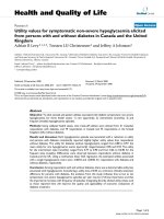
Figure 2 Serum antibody responses to MDI skin exposure. BALB/c mice were skin (on ly) exposed to vehicle (acetone/olive oil) or varying
concentrations of MDI (0.1 - 10% w/v) as shown on X-axis. On day 21, serum levels of MDI-specific IgG
1
/IgG
2a
(inverse end-titer), IgE binding
(ratio) and total IgE (ng/ml) were measured. Data shown are the mean ± SEM of 12 mice per group.
Wisnewski et al. Journal of Occupational Medicine and Toxicology 2011, 6:6
/>Page 5 of 12
mice initially (skin) exposed to MDI at a 1% (w/v) con-
centration, with more limited, albeit sig nificant, inflam-
mation in mice that had been skin exposed to 10% (w/
v). T he reason for the paradoxically limited airway
inflammation in mice (skin) exposed to the highest test
dose of MDI (10% w/v) remains u nclear; however, ana-
logous findings have been reported in HDI exposed
mice [22]. A similar ( non-linear dose-response) phe-
nomenon is well-described for contact sensitization to
many other reactive chemica ls, e.g. form aldehyde, picryl
chloride, DNCB [47].
Respiratory tract exposure boosts serum levels of MDI-
specific antibodies elicited by primary skin exposure
In mice with prior MDI skin exposure, subsequent
respiratory tract exposure to MDI-albumin conjugates
was found to boost MDI-immune sensitization, based
on levels of MDI-specific serum IgG and IgE. As s hown
in Figure 4, statistically significant increases were detect-
able among Th2-associated subclasses/isotypes, IgG
1
and IgE, but not in the Th1-associated subclass, IgG
2a
.
Thus,inmicepreviouslyexposedtoMDIviatheskin,
subsequent respiratory tract exposure to MDI ( albumin
conjugates) further boosts MDI immune sensitivity.
Identification of MDI antigens in exposed skin
As shown in Figure 5A, detergent extracts from 1% MDI
exposed skin contained a single antigenically-modified
protein, specifica lly recogn ized by antibodies from auto-
logous MDI skin (only) exposed mice, but not control
mouse sera. The “ MDI antigen” was purified from
exposed skin by a 2-step process (Figure 5B, and 6A),
and identified as albumin through LC-MS/MS a nalysis
(see Additional file 1). The antigenically modified albu-
min from exposed skin exhibited biophysical properties
consistent with MDI conjugation, w hen compared with
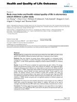
Figure 3 Airway inflammatory responses to MDI in mice sensitized via skin exposure.BALB/cmicethatwereinitiallyskinexposedto
vehicle or varying doses of MDI were subsequently exposed via the respiratory tract as described. On day 21, the number of cells recovered (by
BAL) was determined. The data shown, are the mean ±SEM of 12 mice per group; *(p < .005) and
#
(p < .05) compared to all other groups.
Figure 4 Respiratory trac t exposure boosts serum levels of MDI-s pecific antibodies elicited by primary skin exposure. Serum levels of
MDI-specific antibodies from mice (with (+) or without (-) prior skin exposure) following respiratory tract exposure to MDI albumin conjugates
(+) or mock exposed albumin (-). Each bar represents the mean ± SEM for 12 mice; * p < .001 comparing skin exposed vs. skin + airway
exposed.
Wisnewski et al. Journal of Occupational Medicine and Toxicology 2011, 6:6
/>Page 6 of 12
albumin purified from vehicle-only exposed skin, or
MDI-mouse albumin conjugates prepared in vitro; speci-
fically, alterations in electrophoretic migration and
change in absorbance at 250 nm (Figure 6A&6B).
Additional “MD I antigens”, specifically recognized by
antibodies from MDI skin (only) exposed autologous
mice, but not control mouse sera, were detectable in
urea extracts from skin exposed to th e highest test dose
of MDI (10%), as shown (Figure 7A). Among these ant i-
genically-modified proteins, the most prominent, based
on recognition by serum IgG from skin exposed autolo-
gous mice, were purified through elecrophoretic fractio-
nation methods, and identified by LC-MS/MS as pro-
collagen type 1/a2, keratin 14, and tropomyosin (see
Additional file 1). Their (MDI) antigenicity and identity
were further confirmed by Western blot with autologous
serum IgG from skin exposed mice (Figure 7B) and
commercially available protein-specific (collagen, kera-
tin, tropomysosin) antibodies (not shown).
Discussion
In the present study, we utilized a murine MDI expo-
sure model to demonstrate the capacity of skin exposure
to induce immune sensitization to MDI, and promote
airway inflammation upon subsequent respiratory tract
exposure. The degree of secondary (respiratory tract)
inflammation was found to depend upon the primary
(skin) exposure dose, and exhibited a non-linear
Figure 5 Detection and fractionation of the major MDI antigen in detergent extracts of exposed skin. (A) Proteins from (-) control or (+)
1% MDI exposed mouse skin, were separated by SDS-PAGE and stained with commassie blue or Western blotted with autologous sera from
MDI skin exposed mice (lanes 3 and 4) or control mice (lanes 5 and 6). Arrow highlights major antigenic protein from exposed skin, with
apparent shift in migration, indicating change in conformation/charge. (B) The MDI antigen, highlighted by arrows, was separated from other
skin proteins by isoelectric focusing. Shown is Ponceau S protein staining of Rotofor
®
fractions 2-16 after SDS-PAGE and transfer to nitrocellulose
membrane. Lanes 1 and 17 contain prestained molecular weight markers.
Wisnewski et al. Journal of Occupational Medicine and Toxicology 2011, 6:6
/>Page 7 of 12
relationship tha t peaked when mice were skin exposed
to 1 % (w/v) MDI, and was paradoxically limited at 10-
fold higher (skin) exposure doses; a phenomenon similar
to that reported for HDI. Albumin in exposed skin was
found to undergo antigenic as well as structural/ confor-
mational changes, consistent with MDI conjugation.
Furthermore, MDI-mouse albumin conjugates were spe-
cifical ly recognized by serum IgE and IgG, and triggered
heightened respiratory tract responses, in previously
skin exposed mice. The data highlight mechanisms by
which MDI skin exposure might contribute to the
development of systemic immune sensitization and pos-
sibly MDI asthma.
The present findings are consistent with limited
reports on MDI skin exposure in mice, despite differ-
ences in exposure protocols, and methods of assessing
immunologic responses [48-51]. The findings are also
consistent with data on the smaller, more volatile 6-car-
bon isocyanates, HDI and TDI, including, the non-linear
“ (skin) dose/(respiratory tract) response” and mixed
Th1/Th2-like response to skin exposure [22,31,34,36,52].
Importantly, in all of these studies, the isocyanate
Figure 6 Purification of antigenically modified albumin from in vivo exposed mouse skin. (A) SDS-PAGE analysis (top) and Western blot
with serum IgG from skin exposed mice (bottom) of the major MDI antigen (highlighted with *), purified from skin exposed in vivo to (+) 1%
MDI and its corresponding protein purified from (-) control skin (highlighted with #). For comparison, MDI-albumin conjugates prepared in vitro
using varying doses of MDI (0.001%, 0.01% and 0.1%, lanes 4 to 6 respectively) are shown to the right of the molecular weght markers. The MDI
antigen was not recognized using control sera from vehicle expose mice or irrelevant hyperimmune mouse serum (not shown). (B) Ultraviolet
light absorbance spectra of albumin purified from control or 1% MDI exposed skin. (C) For comparison, commercially purified mouse albumin
and MDI-mouse serum albumin conjugates prepared in vitro were similarly analyzed. *Note increase in absorbance in the 250 nm range due to
MDI’s aromatic rings.
Wisnewski et al. Journal of Occupational Medicine and Toxicology 2011, 6:6
/>Page 8 of 12
concentrations found to induce immune responses via
skin exposure (≤%1 w/v) were within the range com-
monly used in polyurethane production, and are likely
experienced by workers in multiple occupational settings
[8,28,53].
The presently described mouse model possesses dis-
tinct strengths as well as limitations compared with pre-
viously published animal stu dies of MD I and/or other
isocyanate-induced asthma. One major strength is the
use of skin as the primary exposure route for inducing a
state of MDI-specific immune sensitization in which
subsequent respiratory tract exposure leads to asthma-
like inflammation. In this regard, the present investiga-
tion differs from prior studies attempting to model iso-
cyanate-induced airway inflammation through
“respiratory tract only” exposure, whic h have met lim-
ited success [15,31,49,54-60]. Another strength of the
present study is the use o f autologous serum IgG from
skin exposed mice to identify immunologically-relevant
protein targets for MDI conjugation and (antigenic)
modification. The major weakness of the study, as
viewed a priori, was the use of MDI-albumin conjugates,
rather than MDI itself, for respiratory tract exposure
(see Introduction for rationale), thus bypassing a major
step between inhalation and inflammation. Retrospec-
tively, however, the data suggest that albumin conjugates
maybeuniquelysuitedasantigensinmodeling
isocyanate asthma, e specially secondary to initial skin
exposure.
The data provide new insight into the reactivity of
MDI with proteins present in the skin, which likely con-
tributes to the development of MDI immune sensitiza-
tion. At the 1% MDI exposure dose (which promoted
the strongest secondary respiratory tract responses),
only 1 skin protein, albumin, exhibited changes consis-
tent with MDI conjugation (charge/conformation, ultra-
violet light absorbance, antigenicity). Albumin is a major
protein of the extracellular compartment of the skin,
but has not been previously recognized as a target for
isocyanate at that anatomical location [61]. However,
albumin in airway fluid has been described as a major
target for isocyanate conjugation in vivo following
respiratory tract exposure [12-14,16,43,62]. Furthermore,
albumin is the only known human protein whose conju-
gation with isocyanate confers specific recognition by
human antibodies from expo sed individuals [43,63].
Thus, the pr esent data suggest that MDI conjugatio n to
albumin in exposed skin creates an antigenic trigger
that promotes subsequent airway inflammatory
responses to respiratory tract exposure [22,35].
While albumin was the only MDI antigen detectable
in skin exposed to 1% MDI, additional proteins were
found to be antigenically-modified in skin samples
exposedtothehighesttestdose(10%)ofMDI.The
Figure 7 Identification of MDI antigens in urea extracts of exposed skin. (A) The detergent insoluble fraction of (-) control or (+) 10% MDI
exposed skin tissue were further homogenized in 9 M urea, separated by SDS-PAGE, and stained for total proteins (lanes 1 and 2). Parallel
Western blot with sera from autologous MDI skin exposed mice (lanes 3 and 4) vs. control mouse sera (lanes 5 and 6) identified at least three
antigenically modified proteins (MDI antigens) in these samples; see arrows. (B) The MDI antigens from 10% MDI exposed mouse skin were
purified and reanalyzed by protein stain following SDS-PAGE, and parallel Western blot with autologous sera from MDI skin exposed mice.
Arrows highlight antigenically modified collagen (*1), keratin (*2) and tropomyosin (*3) from MDI exposed skin. Actin from unexposed mouse
skin, which was not recognized by autologous sera, was run as a negative control (lane 3). MDI antigens were not detectable using control sera
from vehicle expose mice or irrelevant hyperimmune mouse serum (not shown).
Wisnewski et al. Journal of Occupational Medicine and Toxicology 2011, 6:6
/>Page 9 of 12
significance of these proteins in response to MDI skin
exposure wi ll require further investigation. However, it
is interesting to speculate the possibility that reactivity
with MDI may alter their normal conformation in a
manner that breaks “ immune tolerance” given the
reported association of anti-keratin antibodies with iso-
cyanate asthma, and the pan-al lergenic ity of non-mam-
malian tropomyosin [64-66].
If the present data translate across species, they will
provide important insight into pathogenic mechanisms
of MDI asthma as well as practical guidance for d isease
prevention, among occupationally exposed individuals.
The murine model will facilitate investigation of the role
of specific genes, through transgenic technology, and
provide a system for evaluating the effectiveness of dif-
ferent exposure interventions. The ELISA assay for
MDI-specific IgG, described herein, may be helpful in
assessing workplace skin exposure, which currently goes
largely undetected, due to the lack of practical metho-
dology for measuring. Furthermore, recognition of the
ability to generate systemicimmunesensitizationto
MDI v ia skin exposure, may promote increased aware-
ness and use of personal (skin) protection, including
gloves, overalls and head coverings.
Conclusions
In summary, we developed a murine model to investi-
gate the potential consequences of MDI skin exposure,
which is relatively common in the numerous industries
that utilize MDI to make polyurethane products. The
present data demonstrate that MDI ski n exposure can
induce systemic immune sensitization and asthmatic-
like inflammatory responses to subsequent respiratory
tract exposure. Albumin was found to be a major target
for MDI conjugation in exposed skin, and MDI-albumin
conjugates were also shown to trigger heightened
respiratory tract inflammation in pr eviously skin
exposed mice (vs. unexposed controls). The data may
help explain the devel opment of new MDI asthma cases
despite extremely low workplace airborne MDI levels
and provide practical guidance for exposure and disease
prevention.
Additional material
Additional file 1: Antigenically modified proteins from exposed
mouse skin identified by LC-MS/MS. A table listing the positively
identified peptides from the purified protein bands specifically
recognized by serum IgG from MDI skin exposed mice.
Acknowledgements
The authors would like to Acknowledge Dr. Kathy Stone and Tom Abbot for
their expert help with the LC/MS-MS studies. Funding was provided by
grant support from the National Institutes of Health (NIH), the National
Institute of Environmental Health Safety (NIEHS), and the National Institute
for Occupational Safety and Health (NIOSH).
Author details
1
Department of Internal Medicine; Yale University School of Medicine; 300
Cedar Street; New Haven, CT; 06510, USA.
2
Department of Dermatology; Yale
University School of Medicine; 300 Cedar Street; New Haven, CT; 06510, USA.
Authors’ contributions
AVW drafted the manuscript and supervised the in vitro immunology/
biochemistry experiments. LX and ER performed in vivo skin and respiratory
tract exposure studies, as well as BAL, and cell counts/differentials. JL
performed the in vitro immunology/biochemistry experiments; ELISAs for
MDI-specific IgG/IgE and total IgE, SDS-PAGE, Western blot, protein
purification, and MDI-mouse albumin conjugate preparation. CAR organized
the project and edited the manuscript. CAH conceived the original
hypotheses underlying the overall project and supervised all aspects of the
in vivo mouse studies. AVW, CAR, and CAH were together responsible for
experiment design and data interpretation. All authors reviewed and
approved the final manuscript.
Competing interests
The authors declare that they have no competing interests.
Received: 23 November 2010 Accepted: 17 March 2011
Published: 17 March 2011
References
1. Dykewicz MS: Occupational asthma: current concepts in pathogenesis,
diagnosis, and management. J Allergy Clin Immunol 2009, 123:519-28, quiz
529-30.
2. Redlich CA, Wisnewski AV, Bello D: In Environmental and Occupational
Medicine. Edited by: Rom, W. N. Lippincott, Williams and Wilkins,
Philadelphia, PA; 2007:.
3. Allport DC, Gilbert DS, Outterside SM, (Eds): MDI and TDI: Safety, Health
and the Environment: A Source Book and Practical Guide. Wiley, Wiley,
Chichester Wiley; 2003.
4. ACC Center for the Polyurethane Industry: End Use Market Survey on the
Polyurethanes Industry. 2008, October 2009.
5. Spray Foam Insulation Saving Lives & Billions of Dollars in Iraq &
Afghanistan: Increased energy efficiency at US military structures
reduces fuel requirements. SprayFoam.com [ />npps/story.cfm?nppage=418].
6. Petsonk EL, Wang ML, Lewis DM, Siegel PD, Husberg BJ: Asthma-like
symptoms in wood product plant workers exposed to methylene
diphenyl diisocyanate. Chest 2000, 118:1183-93.
7. Sabbioni G, Wesp H, Lewalter J, Rumler R: Determination of isocyanate
biomarkers in construction site workers. Biomarkers 2007, 12:468-83.
8. Liljelind I, Norberg C, Egelrud L, Westberg H, Eriksson K, Nylander-
French LA: Dermal and inhalation exposure to methylene bisphenyl
isocyanate (MDI) in iron foundry workers. Ann Occup Hyg 2010, 54:31-40.
9. Chester DA, Hanna EA, Pickelman BG, Rosenman KD: Asthma death after
spraying polyurethane truck bedliner. Am J Ind Med 2005, 48:78-84.
10. Bernstein JA: Overview of diisocyanate occupational asthma. Toxicology
1996, 111:181-9.
11. Chen SE, Bernstein IL: The guinea pig model of diisocyanate sensitization.
I. Immunologic studies. J Allergy Clin Immunol 1982, 70 :383-92.
12. Kennedy AL, Stock MF, Alarie Y, Brown WE: Uptake and distribution of 14C
during and following inhalation exposure to radioactive toluene
diisocyanate. Toxicol Appl Pharmacol 1989, 100:280-92.
13. Jin R, Day BW, Karol MH: Toluene diisocyanate protein adducts in the
bronchoalveolar lavage of guinea pigs exposed to vapors of the
chemical. Chem Res Toxicol 1993, 6:906-12.
14. Kennedy AL, Wilson TR, Stock MF, Alarie Y, Brown WE: Distribution and
reactivity of inhaled 14C-labeled toluene diisocyanate (TDI) in rats. Arch
Toxicol 1994, 68:434-43.
15. Kennedy AL, Singh G, Alarie Y, Brown WE: Autoradiographic analyses of
guinea pig airway tissues following inhalation exposure to 14C-labeled
methyl isocyanate. Fundam Appl Toxicol 1993, 20:57-67.
16. Liu Q, Wisnewski AV:
Recent developments in diisocyanate asthma. Ann
Allergy
Asthma Immunol 2003, 90:35-41.
Wisnewski et al. Journal of Occupational Medicine and Toxicology 2011, 6:6
/>Page 10 of 12
17. Bernstein IL, Splansky GL, Chen SE, Vinegar A: The guinea pig model of
diisocyanate sensitization. II. Physiologic studies. J Allergy Clin Immunol
1982, 70:393-8.
18. Patterson R, Zeiss CR, Harris KE: Immunologic and respiratory responses to
airway challenges of dogs with toluene diisocyanate. J Allergy Clin
Immunol 1983, 71:604-11.
19. Pauluhn J: Assessment of respiratory hypersensitivity in guinea pigs
sensitized to toluene diisocyanate: improvements on analysis of
respiratory response. Fundam Appl Toxicol 1997, 40:211-9.
20. Sugawara Y, Okamoto Y, Sawahata T, Tanaka K: An asthma model
developed in the guinea pig by intranasal application of 2,4-toluene
diisocyanate. Int Arch Allergy Immunol 1993, 101:95-101.
21. Huang J, Millecchia LL, Frazer DG, Fedan JS: Airway hyperreactivity elicited
by toluene diisocyanate (TDI)-albumin conjugate is not accompanied by
airway eosinophilic infiltration in guinea pigs. Arch Toxicol 1998, 72:141-6.
22. Herrick CA, Xu L, Wisnewski AV, Das J, Redlich CA, Bottomly K: A novel
mouse model of diisocyanate-induced asthma showing allergic-type
inflammation in the lung after inhaled antigen challenge. J Allergy Clin
Immunol 2002, 109:873-8.
23. Bello D, Herrick CA, Smith TJ, Woskie SR, Streicher RP, Cullen MR, Liu Y,
Redlich CA: Skin exposure to isocyanates: reasons for concern. Environ
Health Perspect 2007, 115:328-35.
24. Redlich CA, Herrick CA: Lung/skin connections in occupational lung
disease. Curr Opin Allergy Clin Immunol 2008, 8:115-9.
25. Tinnerberg H, Mattsson C: Usage of air monitoring and biomarkers of
isocyanate exposure to assess the effect of a control intervention. Ann
Occup Hyg 2008, 52:187-94.
26. Liu Y, Stowe MH, Bello D, Woskie SR, Sparer J, Gore R, Youngs F, Cullen MR,
Redlich CA: Respiratory protection from isocyanate exposure in the
autobody repair and refinishing industry. J Occup Environ Hyg 2006, 3:234-49.
27. Pauluhn J, Woolhiser MR, Bloemen L: Repeated inhalation challenge with
diphenylmethane-4,4’-diisocyanate in brown Norway rats leads to a
time-related increase of neutrophils in bronchoalveolar lavage after
topical induction. Inhal Toxicol 2005, 17:67-78.
28. Liu Y, Stowe MH, Bello D, Sparer J, Gore RJ, Cullen MR, Redlich CA,
Woskie SR: Skin exposure to aliphatic polyisocyanates in the auto body
repair and refinishing industry: III. A personal exposure algorithm. Ann
Occup Hyg 2009, 53:33-40.
29. Bello D, Redlich CA, Stowe MH, Sparer J, Woskie SR, Streicher RP,
Hosgood HD, Liu Y: Skin exposure to aliphatic polyisocyanates in the
auto body repair and refinishing industry: II. A quantitative assessment.
Ann Occup Hyg 2008,
52:117-24.
30.
Karol MH, Hauth BA, Riley EJ, Magreni CM: Dermal contact with toluene
diisocyanate (TDI) produces respiratory tract hypersensitivity in guinea
pigs. Toxicol Appl Pharmacol 1981, 58:221-30.
31. Ban M, Morel G, Langonne I, Huguet N, Pepin E, Binet S: TDI can induce
respiratory allergy with Th2-dominated response in mice. Toxicology
2006, 218:39-47.
32. Pauluhn J: Brown Norway rat asthma model of diphenylmethane-4,4’-
diisocyanate (MDI): impact of vehicle for topical induction. Regul Toxicol
Pharmacol 2008, 50:144-54.
33. Pauluhn J: Brown Norway rat asthma model of diphenylmethane-4,4’-
diisocyanate (MDI): analysis of the elicitation dose-response relationship.
Toxicol Sci 2008, 104:320-31.
34. Tarkowski M, Vanoirbeek JA, Vanhooren HM, De Vooght V, Mercier CM,
Ceuppens J, Nemery B, Hoet PH: Immunological determinants of
ventilatory changes induced in mice by dermal sensitization and
respiratory challenge with toluene diisocyanate. Am J Physiol Lung Cell
Mol Physiol 2007, 292:L207-14.
35. Herrick CA, Das J, Xu L, Wisnewski AV, Redlich CA, Bottomly K: Differential
roles for CD4 and CD8 T cells after diisocyanate sensitization: genetic
control of TH2-induced lung inflammation. J Allergy Clin Immunol 2003,
111:1087-94.
36. Vanoirbeek JA, Tarkowski M, Ceuppens JL, Verbeken EK, Nemery B, Hoet PH:
Respiratory response to toluene diisocyanate depends on prior
frequency and concentration of dermal sensitization in mice. Toxicol Sci
2004, 80:310-21.
37. Dearman RJ, Moussavi A, Kemeny DM, Kimber I: Contribution of CD4+ and
CD8+ T lymphocyte subsets to the cytokine secretion patterns induced
in mice during sensitization to contact and respiratory chemical
allergens. Immunology 1996, 89:502-10.
38. Wisnewski AV, Liu J, Redlich CA: Antigenic changes in human albumin
caused by reactivity with the occupational allergen diphenylmethane
diisocyanate. Anal Biochem 2010, 400:251-8.
39. Karol MH, Kramarik JA, Ferguson J: Methods to assess RAST results in
patients exposed to chemical allergens. Allergy 1995, 50:48-54.
40. Herrick CA, MacLeod H, Glusac E, Tigelaar RE, Bottomly K: Th2 responses
induced by epicutaneous or inhalational protein exposure are
differentially dependent on IL-4. J Clin Invest 2000, 105 :765-75.
41. Jin RZ, Karol MH: Intra- and intermolecular reactions of 4,4’-
diisocyanatodiphenylmethane with human serum albumin. Chem Res
Toxicol 1988, 1:281-7.
42. Coligan J, Kruisbeck A, Marguiles D, Sevacch E, Stober W, editors: Current
Protocols in Immunology.
Wiley and Sons Inc, West Sussex; 1998.
43.
Wisnewski AV, Srivastava R, Herick C, Xu L, Lemus R, Cain H, Magoski NM,
Karol MH, Bottomly K, Redlich CA: Identification of human lung and skin
proteins conjugated with hexamethylene diisocyanate in vitro and in
vivo. Am J Respir Crit Care Med 2000, 162:2330-6.
44. Stone KL, DeAngelis R, LoPresti M, Jones J, Papov VV, Williams KR: Use of
liquid chromatography-electrospray ionization-tandem mass
spectrometry (LC-ESI-MS/MS) for routine identification of enzymatically
digested proteins separated by sodium dodecyl sulfate-polyacrylamide
gel electrophoresis. Electrophoresis 1998, 19:1046-52.
45. Perkins DN, Pappin DJ, Creasy DM, Cottrell JS: Probability-based protein
identification by searching sequence databases using mass
spectrometry data. Electrophoresis 1999, 20:3551-67.
46. Hirosawa M, Hoshida M, Ishikawa M, Toya T: MASCOT: multiple alignment
system for protein sequences based on three-way dynamic
programming. Comput Appl Biosci 1993, 9:161-7.
47. Andersen KE: Testing for contact allergy in experimental animals.
Pharmacol Toxicol 1987, 61:1-8.
48. Dearman RJ, Basketter DA, Kimber I: Characterization of chemical allergens
as a function of divergent cytokine secretion profiles induced in mice.
Toxicol Appl Pharmacol 1996, 138:308-16.
49. Farraj AK, Boykin E, Haykal-Coates N, Gavett SH, Doerfler D, Selgrade M: Th2
Cytokines in Skin Draining Lymph Nodes and Serum IgE Do Not Predict
Airway Hypersensitivity to Intranasal Isocyanate Exposure in Mice. Toxicol
Sci 2007, 100:99-108.
50. Selgrade M, Boykin EH, Haykal-Coates N, Woolhiser MR, Wiescinski C,
Andrews DL, Farraj AK, Doerfler DL, Gavett SH: Inconsistencies between
cytokine profiles, antibody responses, and respiratory
hyperresponsiveness following dermal exposure to isocyanates. Toxicol
Sci 2006, 94:108-17.
51. Potter DW, Wederbrand KS: Total IgE antibody production in BALB/c mice
after dermal exposure to chemicals. Fundam Appl Toxicol 1995, 26:127-35.
52. Vanoirbeek JA, De Vooght V, Vanhooren HM, Nawrot TS, Nemery B, Hoet PH:
How long do the systemic and ventilatory responses to toluene diisocyanate
persist in dermally sensitized mice? J Allergy Clin Immunol 2008, 121:456-463e5.
53. Lesage J, Stanley J, Karoly WJ, Lichtenberg FW: Airborne methylene
diphenyl diisocyanate (MDI) concentrations associated with the
application of polyurethane spray foam in residential construction.
J Occup Environ Hyg 2007, 4:145-55.
54. Vanoirbeek JA, De Vooght V, Nemery B, Hoet PH: Multiple challenges in a
mouse model of chemical-induced asthma lead to tolerance: ventilatory
and inflammatory responses are blunted, immunologic humoral
responses are not. Toxicology 2009, 257:144-52.
55. Satoh T, Kramarik JA, Tollerud DJ, Karol MH: A murine model for assessing
the respiratory hypersensitivity potential of chemical allergens. Toxicol
Lett 1995, 78:57-66.
56. Pauluhn J, Dearman R, Doe J, Hext P, Landry TD: Respiratory
hypersensitivity to diphenylmethane-4,4’-diisocyanate in guinea pigs:
comparison with trimellitic anhydride.
Inhal Toxicol 1999, 11:187-214.
57.
Nabe T, Yamauchi K, Shinjo Y, Niwa T, Imoto K, Koda A, Kohno S: Delayed-
type asthmatic response induced by repeated intratracheal exposure to
toluene-2,4-diisocyanate in guinea pigs. Int Arch Allergy Immunol 2005,
137:115-24.
58. Karol MH: Concentration-dependent immunologic response to toluene
diisocyanate (TDI) following inhalation exposure. Toxicol Appl Pharmacol
1983, 68:229-41.
59. Johnson VJ, Yucesoy B, Reynolds JS, Fluharty K, Wang W, Richardson D,
Luster MI: Inhalation of toluene diisocyanate vapor induces allergic
rhinitis in mice. J Immunol 2007, 179:1864-71.
Wisnewski et al. Journal of Occupational Medicine and Toxicology 2011, 6:6
/>Page 11 of 12
60. Blaikie L, Morrow T, Wilson AP, Hext P, Hartop PJ, Rattray NJ, Woodcock D,
Botham PA: A two-centre study for the evaluation and validation of an
animal model for the assessment of the potential of small molecular
weight chemicals to cause respiratory allergy. Toxicology 1995, 96:37-50.
61. Quinlan GJ, Martin GS, Evans TW: Albumin: biochemical properties and
therapeutic potential. Hepatology 2005, 41:1211-9.
62. Wass U, Belin L: Immunologic specificity of isocyanate-induced IgE
antibodies in serum from 10 sensitized workers. J Allergy Clin Immunol
1989, 83:126-35.
63. Wisnewski AV, Stowe MH, Cartier A, Liu Q, Liu J, Chen L, Redlich CA:
Isocyanate vapor-induced antigenicity of human albumin. J Allergy Clin
Immunol 2004, 113:1178-84.
64. Ye YM, Nahm DH, Kim CW, Kim HR, Hong CS, Park CS, Suh CH, Park HS:
Cytokeratin autoantibodies: useful serologic markers for toluene
diisocyanate-induced asthma. Yonsei Med J 2006, 47:773-81.
65. Arlian LG, Morgan MS, Vyszenski-Moher DL, Sharra D: Cross-reactivity
between storage and dust mites and between mites and shrimp. Exp
Appl Acarol 2009, 47:159-72.
66. Reese G, Ayuso R, Lehrer SB: Tropomyosin: an invertebrate pan-allergen.
Int Arch Allergy Immunol 1999, 119:247-58.
doi:10.1186/1745-6673-6-6
Cite this article as: Wisnewski et al.: Immune sensitization to methylene
diphenyl diisocyanate (MDI) resulting from skin exposure: albumin as a
carrier protein connecting skin exposure to subsequent respiratory
responses. Journal of Occupational Medicine and Toxicology 2011 6:6.
Submit your next manuscript to BioMed Central
and take full advantage of:
• Convenient online submission
• Thorough peer review
• No space constraints or color figure charges
• Immediate publication on acceptance
• Inclusion in PubMed, CAS, Scopus and Google Scholar
• Research which is freely available for redistribution
Submit your manuscript at
www.biomedcentral.com/submit
Wisnewski et al. Journal of Occupational Medicine and Toxicology 2011, 6:6
/>Page 12 of 12