báo cáo hóa học: " Thoracic aorta pseudoaneurysm with hemopericardium: unusual presentation of warfarin overdose" potx
Bạn đang xem bản rút gọn của tài liệu. Xem và tải ngay bản đầy đủ của tài liệu tại đây (1.07 MB, 3 trang )
CAS E REP O R T Open Access
Thoracic aorta pseudoaneurysm with
hemopericardium: unusual presentation of
warfarin overdose
Ya-Chih Tien
1
, Ying-Cheng Chen
2
, Chiung-Ying Liao
3
and Chia-Chu Chang
1*
Abstract
There have been few case reports which discuss a relationship between warfarin overdose and aortic
pseudoaneurysm leakage. We report the case of a female receiving warfarin who presented with dsypnea. Her
international normalized ratio was > 10. Chest radiograph revealed cardiomegaly, and chest computed
tomography (CT) showed a bulging pouch-like lesion below the aortic arch greater than 6x6 cm in size and a fluid
collection suggesting blood in the pericardium. Thoracic endovascular aneurysm repair (TEVAR) was successfully
performed by a cardiovascular surgeon. Aortic pseudoaneurysm formation and leakage may be considered as a
rare complication in patients receiving warfarin therapy. Further study regarding warfarin use and the incidence of
pseudoaneurysm leakage is needed.
Keywords: Warfarin pseudoaneurysm, hemopericardium, TEVAR
Background
A patient with a pseudoaneurysm will typically have had
a traumatic event such as a recent b lunt or penetrating
trauma, or an endovascular procedure [1,2]. Heart fail-
ure and chest pain are the most common manifestations
of a pseudoaneurysm of the ascending aorta [3]. Herein
we report the case of a female receiving warfarin whose
international normalized ratio (INR) was >10, who pre-
sented with dyspnea. Chest computed tomography (CT)
revealed an aortic arch pseudoa neurysm and a fluid col-
lection suggesting blood in the pericardium. We discuss
the risk o f bleeding as it is related to warfarin overdose
and pseudoaneurysm leakage.
Case presentation
A 78-year-old female, presenting with progressive short-
ness of breath and general weakness was admitted to
our hospital on March 15, 2010. She experienced palpi-
tations and tachycardia, and mild chest tightness when
palpitations occurred. Her history was significant for
primary cancer of the appendix with ovarian metastases,
and was status post a debunking operation in Decem ber
of 2006, complicated by chronic right leg lymphedema.
She had been taking warfarin as prescribed by the cardi-
ovascular surgery department for deep vein thrombosis
of the right leg.
On admission, her blood pressure was 148/96 mmHg,
heart rate 114 beats/min, respiratoryrate26breaths/
min, and temperature 37.8°C. Laboratory studies
revealed: white blood cell (WBC) count, 17200/uL (neu-
trophil-segment 89.1%); hemoglobin, 7.6 gm/dL; platelet
count, 455000/uL; NT-proBNP, 6776 pg/mL; PT, 143s
(INR >10); blood urea nitrogen (BUN), 33 mg/dL; crea-
tinine, 0.77 mg/dL; Na 131 mmol/L; K 2.5, mmol/L; Ca
8.4 mg/dL; Mg, 2.4 mg/dL; and albumin 1.7 g/dL. The
thyroid function tests were normal. Artery gas analysis
showed hypoxia (pH, 7.4; PCO
2
, 36.9 mm Hg; PO
2
,75.7
mm Hg; HCO
3
, 23.4 mmol/L; SaO
2
, 95%). The elevated
PT and INR suggested warfarin overdose. We prescribed
VitK
1
1 ample per-12h and transfused frozen fresh
plasma 12 units per-day. Three days later, the PT was
normalized, 21s (INR2.0).
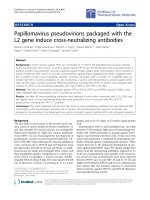
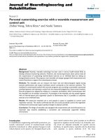
As admitted, her chest radiograph revealed cardiome-
galy with pulmonary edema and blunting of the left cost-
ophrenic angle (Figure 1). Echocardiography revealed
* Correspondence:
1
Department of Medicine, Changhua Christian Hospital, 135 Nan-Siau Street,
Changhua city, 500 Taiwan
Full list of author information is available at the end of the article
Tien et al. Journal of Occupational Medicine and Toxicology 2011, 6:12
/>© 2011 Tien et al; licensee BioMed Central Ltd. This is an Open Access article distributed under the terms of the Creative Commons
Attribution License (http://creativecommons.o rg/licenses/by/2.0), which permits unrestricted use, distribution, and reproduction in
any medium, provided the original work is properly cited.
normal left ventricular systolic function with an ejection
fraction of 70%, dilatation of the left atrium, right ventri-
cle, and ascend ing aorta, moderate tricuspid valve regur-
gitation, mild pulmonary, mitral, and aortic valve
regurgitation, and pericardial effusion; no valvular steno-
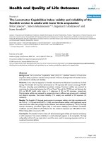
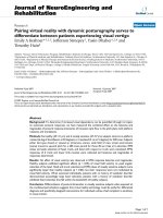
sis problem was identified. Chest CT was performed in
consideration of an organic lesion, such as a pulmonary
embolism or malignancy. A l arge bulging pouch-like
lesion below the aortic arch greater than 6x6 cm i n size
and a fluid collection in the pericardium (relative high
density) was found (Figure 2, 3). Results were consistent
with a pseudoaneurysm in the aortic arch and hemor-
rhage into the pericardium.
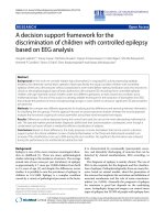
Thoracic endovascular aneurysm repair (TEVAR) was
successfully performed by a cardiovascular surgeon one
day later. Clinical presentation including serial CXR
(Figure 4) and patient status s howed dramatic improve-
ment. The procedure was successful, and the p atient
was discharged 2 weeks later in good condition. At fol-
low-up in the cardiovascular surgery department she
remained in stable condition.
Discussion
Etiologies of ascending aortic pseudoaneurysms include
trauma, connective tissue disease, vasculitis, and prior
aortic surgery [1,2]. Doppler ultrasound can detect pseu-
doaneurysm, and is inexpensive and widely available;
however, CT, arteriography, and CT angiography are
superior at showing the anatomy of the arterial system
[4]. Once a pseudoaneurysm is diagnosed, endovascular
management is the best treatment option [5].
Figure 1 Chest AP film on admission revealed cardiomegaly
with widening of the mediastinum, as well as blunting of left
costo-pleural angle suggesting pleural effusion.
Figure 2 Chest computed tomography (CT) in sagital oblique
reformation: a pseudoaneurysm size over 6*6 cm arises from aortic
arch (black arrow) and suspicious hemorrhage into pericardium.
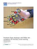
Figure 3 Cross section of chest CT: ar row (white) points the
pseuoaneurysm, compression of pulmonary artery by
pseudoaneuysm was noted. Pericardium effusion is identified in
hyper-density (white arrow head) suggesting bloody component
that may resulted from the pseudoaneurysm hemorrhage into
pericardium space.
Tien et al. Journal of Occupational Medicine and Toxicology 2011, 6:12
/>Page 2 of 3
Major bleeding has been reported in 1.1%-8.1% of
patients during ea ch year of long term warfarin therapy,
and risk factors include o ld age, hypoalbuminemia, ser-
ious illness (cardiac, kidney, or liver disease), cerebrovas-
cular or peripheral vascular disease, and an unstable
anticoagulant effect [6]. This effect is related to warfarin
being absorbed after oral administration, and then being
highly bound to albumin in plasma [7]. Thus, hypoalbu-
minemia is associated with an increased risk of over-
anticoagulation. One study showed that in patients on
long term warfarin therapy, there was a 32% increase in
all forms of bleeding, and a 46% increase in major bleeds
for every 10 years of age over 40 years [8].
Blunt et al. reported a wa rfarin-associated thoracic
aortic dissection in an elderly woman, and concluded
that the mechanism of aortic dissection was a ble ed into
an atheromatous plaque in the thoracic aorta, which
was related to warfarin therapy [7].
Conclusion
Aortic aneurysm formation and leakage may be a rare
complication in patients receiving warfarin therapy
that has not been previously reported. Further study
regarding warfarin use and the incidence of aneurysm
leakage may be an interesting topic worthy of addi-
tional examination.
Consent
Written informed consent was obtained from the patient
for publication of this case report and accompanying
images
Author details
1
Department of Medicine, Changhua Christian Hospital, 135 Nan-Siau Street,
Changhua city, 500 Taiwan.
2
Department of Cardiovascular Surgery,
Changhua Christian Hospital, 135 Nan-Siau Street, Changhua city, 500
Taiwan.
3
Department of Radiology, Changhua Christian Hospital, 135 Nan-
Siau Street, Changhua city, 500 Taiwan.
Authors’ contributions
YCT contributed in visiting the case, all authors contributed in editing the
manuscript, all authors contributed in drafting the manuscript, all authors
read and approved the final manuscript.
Competing interests
The authors declare that they have no competing interests.
Received: 5 November 2010 Accepted: 26 April 2011
Published: 26 April 2011
References
1. Dumont E, Carrier M, Cartier R, Pellerin M, Poirier N, Bouchard D,
Perrault LP: Repair of aortic false aneurysm using deep hypothermia and
circulatory arrest. Ann Thorac Surg 2004, 78:117-120.
2. Tammelin A, Hambraeus A, Stahle E: Mediastinitis after cardiac surgery:
improvement of bacteriological diagnosis by use of multiple tissue
samples and strain typing. J Clin Micorbiol 2002, 40:2936-2941.
3. Atik FA, Navia JL, Svensson LG, Vega P R, Feng J, Brizzio ME, Gillinov AM,
Pettersson BG, Blackstone EH, Lytle BW: Surgical treatment of
pseudoaneurysm of the thoracic aorta. The Journal of Thoracic and
Cardiovascular Surgery 2006, 132:379-385.
4. Davidm M, Tthomaps P, Sinda B, Robert L: Diagnosis of Aortic
Pseudoaneurysm by Echocardiography. Clin Cardiol 1992, 15:773-776.
5. Sozen D, Ahmet M, Arzum K, Suat B: Endovascular Stent Graft Placement
in the Treatment of Ruptured Tuberculous Pseudoaneurysm of the
Descending Thoracic Aorta: Case Report and Review of the Literature.
Cardiovasc Intervent Radiol 2009, 32:572-576.
6. Enrico T, Fausto M, Lorenzo M: Hypoalbuminemia as a risk factor for over-
anticoagulation. The American Journal of Medicine 2002, 112:247-248.
7. Blunt DM, Implloment MG: Warfarin-associated thoracic aortic dissection
in an elderly woman. Age and Ageing 2004, 33:201-203.
8. Van der Meer FJM, Rosendaal FR, Vanderbouke BE, Briët E: Bleeding
complications in oral anti-coagulant therapy. An analysis of risk factor.
Arch Intern Med 1993, 153:1557-1562.
doi:10.1186/1745-6673-6-12
Cite this article as: Tien et al.: Thoracic aorta pseudoaneurysm with
hemopericardium: unusual presentation of warfarin overdose. Journal of
Occupational Medicine and Toxicology 2011 6:12.
Submit your next manuscript to BioMed Central
and take full advantage of:
• Convenient online submission
• Thorough peer review
• No space constraints or color figure charges
• Immediate publication on acceptance
• Inclusion in PubMed, CAS, Scopus and Google Scholar
• Research which is freely available for redistribution
Submit your manuscript at
www.biomedcentral.com/submit
Figure 4 Chest X ray: after thoracic endovascular aneurysm
repair (black arrow point stent in aortic arch).
Tien et al. Journal of Occupational Medicine and Toxicology 2011, 6:12
/>Page 3 of 3