báo cáo hóa học:" Case report: intra-tendinous ganglion of the anterior cruciate ligament in a young footballer" docx
Bạn đang xem bản rút gọn của tài liệu. Xem và tải ngay bản đầy đủ của tài liệu tại đây (1.29 MB, 6 trang )
BioMed Central
Page 1 of 6
(page number not for citation purposes)
Journal of Orthopaedic Surgery and
Research
Open Access
Case report
Case report: intra-tendinous ganglion of the anterior cruciate
ligament in a young footballer
Christer Rolf and Thomas P Watson*
Address: From the Sheffield Centre of Sports Medicine, Division of Clinical Sciences South, University Of Sheffield, UK
Email: Christer Rolf - ; Thomas P Watson* -
* Corresponding author
Abstract
A 20-year-old male medical student and keen rugby player presented with a 12-month history of
progressively worsening right knee pain and stiffness with no history of trauma. Clinical
examination revealed effusion and posterior knee pain exacerbated by end range movement and
an extension lag of 15 degrees. Physiotherapy to improve the range of motion proved unsuccessful.
Magnetic resonance imaging showed that the ACL was grossly thickened and displaced by material
reported as mucoid in nature. There were also areas of focally high signal in relation to its tibial
attachment and intra osseous small cysts. Arthroscopic examination revealed a ganglion related to
the tibial attachment of the ACL and gross thickening and discoloration of the ACL. Biopsies were
taken showing foci of mucoid degeneration in the ACL. A large intra-ACL mass of brownish
coloured tissue was excised arthroscopically. Already at 2 weeks follow up the patient had greatly
improved range of movement and was pain free. However, upon returning to rugby, joint instability
was noticed and a tear of the ACL was confirmed.
This rare clinical condition can be diagnosed with MRI and arthroscopic debridement effectively
relieves symptoms. This case report illustrates that augmentation or reconstruction may end up
being the definitive treatment for athletes. It may also offer some support to the argument that
mucoid degeneration and ganglion cyst formation share a similar pathogenesis to intra-osseous cyst
formation.
Background
A ganglion is a cystic lesion containing mucin-rich fluid
associated with a joint or tendon sheath [1]. Ganglia of
the anterior cruciate ligament (ACL) are uncommon. The
prevalence of ganglia associated with the ACL is reported
to be 0.12–0.44% on MRI [2,3]. Mucoid degeneration is
characterized by an increase in the mucoid ground sub-
stance in the connective tissue containing glycoprotein
and mucoprotein [4]. The prevalence of mucoid degener-
ation of the ACL has been reported to be 0.43% [5].
Bergin et al. reported on the co-existence of mucoid
degeneration and ganglia of the ACL. From 4221 knee
MRI examinations they found 26 patients (0.62%) who
had both mucoid degeneration and ganglia of the ACL.
Although they can coexist, the existence of a relationship
between the two is an area of debate [5-8]. The aetiology
of both ganglion cysts and mucoid degeneration is
unknown [9-11]. Further relationships with intra-osseous
bone formation are not explained in the literature. ACL
ganglia and mucoid degeneration can cause pain and
decreased range of movement in the knee [12,3,13-15]. A
Published: 02 November 2006
Journal of Orthopaedic Surgery and Research 2006, 1:11 doi:10.1186/1749-799X-1-11
Received: 21 August 2006
Accepted: 02 November 2006
This article is available from: />© 2006 Rolf and Watson; licensee BioMed Central Ltd.
This is an Open Access article distributed under the terms of the Creative Commons Attribution License ( />),
which permits unrestricted use, distribution, and reproduction in any medium, provided the original work is properly cited.
Journal of Orthopaedic Surgery and Research 2006, 1:11 />Page 2 of 6
(page number not for citation purposes)
limited number of cases have been described in the litera-
ture.
In this report we describe a case of intra ACL mucoid
degeneration and ganglion cyst combined with intra-
osseous cysts in a 20-year-old medical student and keen
rugby player who had to stop playing because of gradually
increasing dysfunction of his knee. The clinical findings,
MRI, histology and arthroscopic findings and outcome are
described.
Case presentation
A 20-year-old male medical student presented with a 12-
month history of right knee pain of insidious onset. He
had been a regular amateur rugby union player for 12
years. No episodes of trauma and no previous knee prob-
lems were reported. The problem began as stiffness and
pain and had worsened in the last 3 months to the extent
that he had to stop playing. The patient described the pain
as an intermittent deep ache. The pain was exacerbated by
end of range flexion and extension.
Physical examination revealed right-sided posterior knee
pain exacerbated by end range flexion and extension and
mild effusion. Active range of movement was -15°–95°
and passive range of movement -10°–100° (Fig 5 and 7).
Atrophy of the right quadriceps was present. There was no
joint-line tenderness on palpation. Anterior and posterior
drawer and Lachmann tests were negative whilst compres-
sion rotation test was positive.
Physiotherapy was initially undertaken in order to
improve range of movement. At review no improvement
at all was seen and compression rotation test was still pos-
itive. MRI scans showed an abnormal anterior cruciate lig-
ament (ACL). Its fibres were intact but displaced by
material containing high signal on the fat sat sequence,
reported as appearing mucoid in nature. There were also
areas of focally high signal in relation to its tibial attach-
ment. These areas had the appearance of tiny ganglia. The
report presumed that these ganglia extended into and
thickened the ACL. The findings were in keeping with
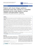
mucinous degeneration. There was evidence of intra-
osseous cyst formation as shown by high signal at the
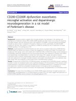
attachment sites of the ligament (Fig. 1 – arrows). X-ray
was unremarkable. Based on these findings an arthros-
copy with biopsies was undertaken.
Arthroscopy revealed several small ganglia in the ACL
around the tibial insertion. Both menisci were intact and
cartilage was normal. The posterior cruciate ligament
(PCL) was normal. The histology report from biopsies
described ligament with multilocular foci of mucoid
degeneration. There was no evidence of neoplasia.
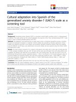
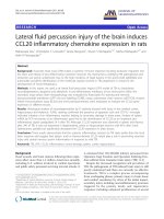
As much as possible of the macroscopically abnormal tis-
sue was excised arthroscopically (Fig 2, 3, 4). Examination
under anaesthesia showed a range of movement of -15°–
110° pre-operatively and -5 – 140 degrees post surgery.
Lachmann's test and anterior drawer tests were negative
after extensive debridement. Post-operatively the range of
movement was improved to 5°–140+° (Fig 5, 6, 7, 8).
Following surgery the patient was referred for early mobi-
lisation and physiotherapy. At 6 week follow up range of
movement was increased to full passive flexion and full
passive extension.
The patient remained pain free and returned to rugby 14
weeks after operation. No major trauma occurred during
this time. However, instability was described following
several games and positive anterior drawer and Lach-
mann's tests were present on examination. Arthroscopy
confirmed the presence of an ACL tear (Fig 9) and also
showed cartilage and meniscal damage. The patient later
had a patellar tendon graft performed.
Discussion
For intra-tendinous ganglia of the ACL, MRI identified the
lesion site, although it was not entirely diagnostic.
Arthroscopy and biopsy was necessary to rule out an early
neoplastic process. Debridement of the abnormal mucoid
tissue relieved symptoms effectively, which has been
described previously in the literature
[16,3,7,17,13,18,19]. However, we have not found as
thorough documentation of clinical findings, MRI, histol-
ogy, arthroscopy findings and outcomes as in the present
case. Gradually decreased range of movement and stiff-
ness of the knee joint in a young athlete without preced-
ing trauma should therefore lead to this suspicion and an
MRI and arthroscopy should be undertaken [3,11-13,20].
In these cases there is usually no preceding major trauma
[8,7,10,12] or instability of the joint [5,16,17,12,13].
Common MRI findings are high signal on T2-weighted
MRI images thickening the ACL with a 'celery-stalk'
appearance [16,11,6,17,12,21], erosion of cortical bone
[22,11,10] and intraosseous cyst formation [5,10].
Arthroscopically ligament fibres are interspersed with a
yellow-brown substance and the ACL displaces anteriorly
and posteriorly [3,11,12,21]. All of these features were
seen in this case. Mucoid degeneration and ganglia of the
anterior cruciate ligament are uncommon [7,9,13,3]. Fur-
ther more so is their coexistence. Bergin et al. reported the
prevalence of this to be 0.62% on MRI [5].
The aetiology of ganglion cysts and mucoid degeneration
is unclear [9,11,10]. One theory is that mucoid degenera-
tion leads to ganglia formation [7]. This relationship is
commonly theorised in the literature but its existence is
Journal of Orthopaedic Surgery and Research 2006, 1:11 />Page 3 of 6
(page number not for citation purposes)
A sagittal T2-weighted MRI showing thickening and pathological appearance of the ACLFigure 1
A sagittal T2-weighted MRI showing thickening and pathological appearance of the ACL.
Abnormal tissue displacing the ACL anteriorly out of the notchFigure 2
Abnormal tissue displacing the ACL anteriorly out of the
notch.
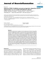
Posterior abnormal tissue close to the posterior lateral meniscus horn displacing the ACLFigure 3
Posterior abnormal tissue close to the posterior lateral
meniscus horn displacing the ACL.
Journal of Orthopaedic Surgery and Research 2006, 1:11 />Page 4 of 6
(page number not for citation purposes)
unproven. Bergin et al. reported that ACL ganglia and
mucoid degeneration commonly coexist and gave some
evidence to suggest these two entities may share a similar
pathogenesis [5]. Another theory suggests that herniation
of synovial tissue through a defect in the tendon sheath
causes ganglia formation [15]. A third describes displace-
ment of synovial tissue during embryogenesis [3]. The
relationship to trauma is unknown. One theory involves
the cellular response to trauma that liberates a mucin sub-
stance, hyaluronic acid. This is interspersed with the fibres
of the ligament, causing its fusiform dilatation. With joint
and tissue motion, the mucin substance dissects the liga-
ment fibers and may be found at the ligament attach-
ments or in the intercondylar notch of the knee [9]. Many
cases in the literature describe ganglia formation in the
absence of trauma. However, excessive training or repeti-
tive minor trauma such as rugby tackles could well be a
triggering factor [8,7,10,12]. Although repetitive trauma
from rugby may be a contributing factor, the aetiology of
the current case is not known and there are no known
hereditary factors in the history.
There are no reported cases of ACL rupture following
pathogenesis of this type. The literature shows that arthro-
scopic debridement of the abnormal tissue effectively
relieves symptoms [16,3,6,17,13,18,19]. However, this
inevitably results in a thinned ACL, which could compro-
mise joint stability. Cases in the literature report no insta-
bility in day-to-day activities following debridement
Reduced flexion of the right leg prior to surgeryFigure 7
Reduced flexion of the right leg prior to surgery.
Reduced extension of the right leg prior to surgeryFigure 5
Reduced extension of the right leg prior to surgery.
Improved position of ACL following excision of the diseased tissueFigure 4
Improved position of ACL following excision of the diseased
tissue.
Improved extension 2 weeks post-operativelyFigure 6
Improved extension 2 weeks post-operatively.
Journal of Orthopaedic Surgery and Research 2006, 1:11 />Page 5 of 6
(page number not for citation purposes)
[16,11,17,12]. However, none of these patients played
sport. Reporting on five cases, Narvekar et al concluded
that because none of the patients participated in any type
of sporting activity, the thinned ACL mass probably suf-
ficed to provide the requisite stability for day-to-day activ-
ities [17]. Nishimori et al concluded that if their patients
had participated in any type of sport, they might have had
to consider augmentation or reconstruction of the ACL
after resection of the lesion [12].
Only one previous case of an athlete is reported; Fealy et
al describe a successful return to sport following arthro-
scopic debridement of the ACL of a volleyball player [16].
This rare diagnosis and treatment option should be con-
sidered when a young athlete presents with reduced ROM
of the knee without preceding trauma.
Arthroscopic debridement of the abnormal tissue effec-
tively relieves symptoms.
Augmentation or reconstruction of the ACL may end up
being the definitive treatment if the patient returns to a
sport demanding high levels of stability.
This report may also offer some support to the argument
that mucoid degeneration and ganglion in the ACL and
intraosseous cyst formation share a similar pathogenesis.
Abbreviations
ACL – Anterior cruciate ligament
PCL – Posterior cruciate ligament
ROM – Range of movement
Competing interests
The author(s) declare that they have no competing inter-
ests.
Authors' contributions
CR conceived of the study, participated in its design and
coordination and helped to draft the manuscript. CR
revised the article for intellectual content details. TW con-
ducted the literature review and carried out the review of
the patient's medical record in order to collect all the
available information. TW helped draft the manuscript.
Both authors read and approved the final manuscript.
Acknowledgements
Written consent was obtained from the patient for publication of study.
References
1. Underwood JCE, Hunter J: Underwood JCE General and systemic pathol-
ogy Edinburgh: Churchill Livingstone; 2004.
2. Do-Dai DD, Youngberg RA, Lanchbury FD, Pitcher JD: Intraligmen-
tous ganglion cysts of the anterior cruciate ligament: MR
findings with clinical and arthroscopic correlations. J Comput
Assist Tomogr 1996, 20:80-84.
3. Huang GS, Lee CH, Chan WP, Taylor JA, Hsueh CJ, Juan Cj, Chen CY,
Yu JS: Ganglion cysts of the cruciate ligaments. Acta Radiol
2002, 43:419-424.
4. Boya H, Pinar H, Gulay Z, Oktay G, Ozer E: Clinical and arthro-
scopic features of meniscal tears and a search for the role of
infection in histologically confirmed meniscal mucoid degen-
eration. Knee Surg Sports Traumatol Arthrosc 2003, 12:294-299.
5. Bergin D, Morrison WB, Carrino JA, Nallamshetty SN, Bartolozzi AR:
Anterior cruciate ligament ganglia and mucoid degenera-
tion: coexistence and clinical correlation. AJR Am J Roentgenol
2004, 182(5):1283-1287.
6. McIntyre J, Moelleken S, Tirman P: Mucoid degeneration of the
anterior cruciate ligament mistaken for ligamentous tears.
Skeletal Radiol 2001, 30:312-315.
7. Krudwig WK, Schulte K-K, Heinemann C: Intra-articular ganglion
cysts of the knee joint: a report of 85 cases and review of the
literature. Knee Surg Sports Traumatol Arthrosc 2004, 12:123-129.
Torn ACL prior to reconstructive surgeryFigure 9
Torn ACL prior to reconstructive surgery.
Improved flexion 2 weeks post-operativelyFigure 8
Improved flexion 2 weeks post-operatively.
Publish with BioMed Central and every
scientist can read your work free of charge
"BioMed Central will be the most significant development for
disseminating the results of biomedical researc h in our lifetime."
Sir Paul Nurse, Cancer Research UK
Your research papers will be:
available free of charge to the entire biomedical community
peer reviewed and published immediately upon acceptance
cited in PubMed and archived on PubMed Central
yours — you keep the copyright
Submit your manuscript here:
/>BioMedcentral
Journal of Orthopaedic Surgery and Research 2006, 1:11 />Page 6 of 6
(page number not for citation purposes)
8. García-Alvarez F, García-Pequerul JM, Avila Jl, Sainz JM, Castiliella T:
Ganglion cysts associated with cruciate ligaments of the
knee: a possible cause of recurrent knee pain. Acta Orthop Belg
2000, 66:490-494.
9. Bui-Mansfield LT, Youngberg RA: Intra-articular ganglia of the
knee: prevalence, presentation, etiology, and management.
AJR Am J Roentgenol 1997, 168:123-127.
10. Melloni P, Valss R, Yuguero M, Sáez A: Mucoid degeneration of
the anterior cruciate ligament with erosion of the lateral
femoral condyle. Skeletal Radiol 2004, 33:359-362.
11. Kumar A, Bickerstaff DR, Grimwood JS, Suvarna SK: Mucoid cystic
degeneration of the cruciate ligament. J Bone Joint Surg Br 1999,
81:304-305.
12. Nishimori M, Sumen Y, Sakaridani K: Mucoid degeneration of the
anterior cruciate ligament – a report of two cases. Magn
Reson Imaging 2004, 22:1325-1328.
13. Parish EN, Dixon P, Cross MJ: Ganglion cysts of the anterior cru-
ciate ligament: a series of 15 cases. Arthroscopy 2005,
21:445-447.
14. Tyrrell Pn, Cassar-Pullicino VN, McCall IW: Intra-articular gan-
glion cysts of the cruciate ligaments. Eur Radiol 2000,
10:1233-1238.
15. Zantop T, Rusch A, Hassenpflug J, Petersen W: Intra-articular gan-
glion cysts of the cruciate ligaments: case report and review
of the literature. Arch Orthop Trauma Surg 2003, 123:195-198.
16. Fealy S, Kenter K, Dines JS, Warren RF: Mucoid degeneration of
the anterior cruciate ligament. Arthroscopy 2001, 17:E37.
17. Narvekar A, Gajjar S: Mucoid degeneration of the anterior cru-
ciate ligament. Arthroscopy 2004, 20:141-146.
18. Sarimo J, Rantanen J, Helttula I, Orava S: Intra-articular cysts and
ganglia of the knee: a report of nine patients. Knee Surg Sports
Traumatol Arthrosc 2005, 13:44-47.
19. Sumen Y, Ochi M, Deie M, Adachi N, Ikuta Y: Ganglion cysts of the
cruciate ligaments detected by MRI. Int Orthop
1999, 23:58-60.
20. Pedrinelli A, Castellana FB, Fontes RB, Coelho RF, Menezes FLIL:
Anterior cruciate ligament ganglion: case report. Sao Paulo
Med J 2002, 120:195-197.
21. Proscan Imaging [ />May_2004_to_print-304.html]
22. Kaatee R, Kjartansson Ó, Brekkan Á: Intra-articular ganglion
between the cruciate ligaments of the knee a case report.
Acta Radiol 1994, 35:434-436.