báo cáo hóa học:" Isokinetic eccentric exercise can induce skeletal muscle injury within the physiologic excursion of muscle-tendon unit: a rabbit model" doc
Bạn đang xem bản rút gọn của tài liệu. Xem và tải ngay bản đầy đủ của tài liệu tại đây (334.83 KB, 7 trang )
BioMed Central
Page 1 of 7
(page number not for citation purposes)
Journal of Orthopaedic Surgery and
Research
Open Access
Research article
Isokinetic eccentric exercise can induce skeletal muscle injury
within the physiologic excursion of muscle-tendon unit: a rabbit
model
Yang-Hwei Tsuang
1
, Shui-Ling Lam
2
, Lien-Chen Wu
1
, Chang-Jung Chiang
1
,
Li-Ting Chen
3
, Pei-Yu Chen
1
, Jui-Sheng Sun*
4,5
and Chien-Che Wang
6
Address:
1
Department of Orthopedic Surgery, Taipei City Hospital, Taipei, Taiwan,
2
Department of Physical Medicine & Rehabilitation, Cardinal
Tien Hospital, Taipei, Taiwan,
3
Department of Research and Development, Healthbanks Biotechnology Corporation Ltd, Taipei, Taiwan,
4
Department of Orthopedic Surgery, National Taiwan University Hospital, Taipei, Taiwan,
5
Institute of Biomedical Engineering, National Yang-
Ming University, Taipei, Taiwan and
6
Department of Orthopedic Surgery, PoJen General Hospital, Taipei, Taiwan
Email: Yang-Hwei Tsuang - ; Shui-Ling Lam - ; Lien-Chen Wu - ; Chang-
Jung Chiang - ; Li-Ting Chen - ; Pei-Yu Chen - ; Jui-
Sheng Sun* - ; Chien-Che Wang -
* Corresponding author
Abstract
Background and Purpose: Intensive eccentric exercise can cause muscle damage. We simulated
an animal model of isokinetic eccentric exercise by repetitively stretching stimulated triceps surae
muscle-tendon units to determine if such exercise affects the mechanical properties of the unit
within its physiologic excursion.
Methods: Biomechanical parameters of the muscle-tendon unit were monitored during isokinetic
eccentric loading in 12 rabbits. In each animal, one limb (control group) was stretched until failure.
The other limb (study group) was first subjected to isokinetic and eccentric cyclic loading at the
rate of 10.0 cm/min to 112% (group I) or 120% (group II) of its initial length for 1 hour and then
stretched to failure. Load-deformation curves and biomechanical parameters were compared
between the study and control groups.
Results: When the muscle-tendon unit received eccentric cyclic loading to 112%, changes in all
biomechanical parameters – except for the slope of the load-deformation curve – were not
significant. In contrast, most parameters, including the slope of the load-deformation curve, peak
load, deformation at peak load, total energy absorption, and energy absorption before peak load,
significantly decreased after isokinetic eccentric cyclic loading to 120%.
Conclusion: We found a threshold for eccentrically induced injury of the rabbit triceps surae
muscle at between 12% and 20% strain, which is within the physiologic excursion of the muscle-
tendon units. Our study provided evidence that eccentric exercise may induce changes in the
biomechanical properties of skeletal muscles, even within the physiologic range of the excursion of
the muscle-tendon unit.
Published: 21 August 2007
Journal of Orthopaedic Surgery and Research 2007, 2:13 doi:10.1186/1749-799X-2-13
Received: 18 February 2007
Accepted: 21 August 2007
This article is available from: />© 2007 Tsuang et al; licensee BioMed Central Ltd.
This is an Open Access article distributed under the terms of the Creative Commons Attribution License ( />),
which permits unrestricted use, distribution, and reproduction in any medium, provided the original work is properly cited.
Journal of Orthopaedic Surgery and Research 2007, 2:13 />Page 2 of 7
(page number not for citation purposes)
Background
In the musculoskeletal system, muscle is the only tissue
that can actively develop tension. When skeletal muscle is
stimulated, it rapidly changes from passive tissue to active
tissue. This change can cause muscular injuries, primarily
strains or tears, which are extremely common in profes-
sional and amateur athletes [1,2]. In sports medicine,
stretching exercises are often recommended to prevent
injury and to improve performance [3,4]. However, inten-
sive exercise training can result in muscular damage and
soreness, especially when the exercise involves eccentric
contraction [5,6].
Researchers have demonstrated that eccentric contrac-
tions create more force than either isometric or concentric
contractions [7,8]. McCully and Faulkner reported that
the extent of injury was related to the peak force devel-
oped during a lengthening contraction [8]. The increased
development of force may be responsible for muscular
injury in eccentric contraction [7]. Later, Jones et al stud-
ied the influence of mechanical factors (ie, force on long-
lasting changes in voluntary force occurrence) and found
that the generation of low-frequency fatigue and muscular
injury is length dependent rather than force dependent
[9].
To investigate the deleterious effects of eccentric exercise
on humans, scientists usually use biochemical and elec-
trophysiologic parameters to indirectly monitor the
degrees of muscular injury [10-14]. An evaluation of the
biomechanical properties of skeletal muscle includes an
assessment for macroscopic tears. However, this method
does not apply to evaluate the potential deleterious effect
of eccentric exercise on the biomechanical properties of
human skeletal muscles.
In a rabbit model, Lieber and Frieden demonstrated that
high force per se does not cause muscular damage after
eccentric contraction, but rather the magnitude of the
active strain does [15]. It has been demonstrated that the
triceps surae muscle-tendon unit behaved viscoelastically
and the extent of muscle injuries was closely related with
the stretch rate. The muscle-tendon unit tolerated great
tensile force and endured high energy at fast stretch status
[16]. The extent of muscular injuries were closely related
to the stretch rate; with fast stretch rates, an increased peak
tensile force was required, and energy absorption
increased [16]. In later studies of eccentric contraction, we
found that when the stimulated muscle failed, the passive
muscle force was dominant and closely related to the
extent of stretch [17]. In these studies, a single stretch to
failure produced injury.
In previous reports on injuries induced by eccentric con-
traction [18,19] activation of muscle tissue was usually
induced by tetanic stimulation, and this kind of distur-
bances could result in structural changes in the muscle-
tendon unit [20]. In the present study, we used low-fre-
quency nerve stimulation (10 Hz) to prevent the possible
confounding effect of tetanic-nerve stimulation on the
muscle during the experiment.
Cyclic stretching of the triceps surae muscle-tendon unit
can substantially affect its tensile properties [21]. How-
ever, the effect of cyclic loading on the skeletal muscle-
tendon unit during an eccentric model is still unclear. A
threshold for stretch-induced injury can be reproduced at
25% strain of the triceps surae muscle-tendon unit [18].
In this study, the muscle eccentric contraction was simu-
lated by repetitively stretching stimulated muscle-tendon
units. We hypothesized that eccentric cyclic loading could
produce a deleterious effect on the unit at relatively low
strain level and that isokinetic eccentric exercise affected
the mechanical properties of the unit, even within its
physiologic excursion.
Methods
Animal preparation
This study was approved by the National Taiwan Univer-
sity Medical College's Animal Research Committee.
Twelve New Zealand White rabbits (4 months old, mean
weight 2.5 kg, SD 0.2 kg) were equally divided into two
groups. In group I, the triceps muscle-tendon unit was
passively stretched to 112% of its resting length, and in
group II, it was stretched to 120%. The leg in each tested
rabbit chosen to be the study or control leg was randomly
assigned.
Preparation of the animals was the same as previously
reported [16]. After the animals were anesthetized with
ketamine 50 mg/kg given subcutaneously, an incision was
made from the midcalf to the plantar surface of the foot
on the lateral aspect of each hind limb. The Achilles ten-
don was isolated with special care to maintain the integ-
rity of the neurovascular bundle and tendon insertion.
Biomechanical test
During the test procedure, the sedated rabbits were put on
supine position with the hip fixed in 90 degrees of flexion.
To determine the in situ length of the muscle-tendon unit,
a dial calipers accurate to 0.05 mm was used to measure
the distance between the origin of the triceps surae at the
distal femur and the insertion site at calcaneus with the
knee while the ankle was flexed 90° [16]. The anesthe-
tized rabbit was then placed in a frame attached to a test-
ing machine (MTS Bionix 858, Minneapolis, MN, USA).
The hind limbs were immobilized with K-wire transfixa-
tion through the proximal tibia. The distal tendinous
insertion was freed by means of osteotomy at the calca-
neal tuberosity and then clamped to the load cell of the
Journal of Orthopaedic Surgery and Research 2007, 2:13 />Page 3 of 7
(page number not for citation purposes)
test system. The muscle was passively extended to its orig-
inal length before osteotomy. A 3-N preload was applied
on the muscle, and its length was measured again [16].
Before the experiment, a skin incision was made over
bilateral buttock region to expose the sciatic nerve. The
nerve was isolated and clamped with a nerve stimulator
(TENS SkylarkTM transcutaneous electrical nerve stimula-
tor; Skylark Device Co., Ltd., ROC).
For the study group, the muscle-tendon unit of one hind
limb was cyclically loaded for 1 hour at a rate of 10.0 cm/
min to a strain amplitude of 12% or 20%. After the peak
stretch amplitude was reached, stretching was discontin-
ued, and the muscle-tendon unit returned to its initial
resting length. To avoid the confounding effect of tetanic
stimulation, low-frequency nerve stimulation was simul-
taneously applied to the sciatic nerve (pulse width 120
µsec, frequency 10 Hz, amplitude 12 mA) during cyclic
loading. The magnitude of supramaximal nerve stimula-
tion was based on our previous finding that muscle con-
traction was maximal under this condition [20]. After
eccentric-cyclic passive stretching, the muscle was
stretched without further electric stimulation at a constant
rate of 10.0 cm/min until a macroscopic tear or a full divi-
sion of ruptured muscle fragments occurred. For the con-
trol group, the muscle-tendon unit in the other hind limb
was stretched at the same rate of 10.0 cm/min until fail-
ure.
The load and deformation required to deform the muscles
were simultaneously recorded by using a personal compu-
ter and software (Testlink PCLAB Data Translation; Data
Translation Inc., Marlboro, USA). All muscles were kept
moist and at physiologic temperature (37°C) by irrigating
them with warm normal saline. Additional anesthesia was
given when needed. The rabbits were sacrificed at the
completion of the study.
For each triceps surae muscle, the load and deformation
of the muscle-tendon unit were recorded and plotted by
using the computer. Deformation of the unit was meas-
ured when peak load was evident. Deformation was calcu-
lated as the length of the muscle at peak load minus its
length before stretching. Load-deformation curves were
generated, and slopes were measured at every linear por-
tion. Energy absorption was calculated by measuring the
area beneath the load-deformation curve; the area before
the failure point represented the relative energy the mus-
cle-tendon unit absorbed before it failed. A ratio of the
energy absorption before peak load was measured by
dividing the energy absorption before peak load with the
total energy absorption during each test.
Statistical analysis
Differences in the energy that the muscle-tendon unit
absorbed before peak load and at full separation of the
ruptured fragments were analyzed by using the paired t
test. Because of the great individual variation in the
strength of the triceps surae muscle, the paired t test was
also used to evaluate differences between limbs of the rab-
bits in each group. The level of statistical significance was
set at P < 0.05.
Results
After isokinetic eccentric loading, all muscle-tendon units
under stretch had similar curve patterns. The load-defor-
mation curve began with an initially increasing slope and
ultimately reached the peak load. After this point, a steep
decline was observed, followed by a curve with gradual
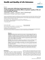
increasing and decreasing of the load. After 12% strain for
1 hour, the curve shows a slope of 54.9 N/mm for the
study group, compared with 36.5 N/mm for the control
sample. The slope of the curve was steeper in the study
group than in the control group (Fig. 1). When the mus-
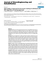
cle-tendon unit was loaded to 20% strain for 1 hour, we
observed a significant change on the load-deformation
curve between the control and study groups. All biome-
chanical parameters were substantially decreased in the
study group. For the control and study groups, respec-
tively, peak load was 850.5 and 305.4 N, deformation at
peak load was 35.93 and 20.9 mm, the slope of the curve
was 31.1 and 20.5 N/mm, total energy absorption was
23764.6 and 3989.5 N-mm, and energy absorption before
peak load was 11564.5 and 2194.0 N-mm. The peak load
was lower in the study group than in the control group
(Fig. 2).
Representative load-deformation curve of a triceps surae muscle-tendon unit after isokinetic eccentric cyclic loading for 1 hour at 12% strainFigure 1
Representative load-deformation curve of a triceps surae
muscle-tendon unit after isokinetic eccentric cyclic loading
for 1 hour at 12% strain. The curve shows a slope of 54.9 N/
mm for the study group, compared with 36.5 N/mm for the
control sample.
Load-Deformation Curve (12% of Cyclic Loading)
0.00
200.00
400.00
600.00
800.00
1000.00
0.00 20.00 40.00 60.00 80.00
Deformation (mm)
Load (Newton)
Study
Control
Journal of Orthopaedic Surgery and Research 2007, 2:13 />Page 4 of 7
(page number not for citation purposes)
In group I (isokinetic eccentric cyclic loading to 112% of
resting length), all biomechanical parameters were similar
between the control and experimental limbs, except for
the slope of the load-deformation curve (Fig. 3, Tables 1
&2). In group II (loading to 120% loading of resting
length), all biomechanical parameters significant differed
between the control and study groups, except for the ratio
of energy absorption before peak load (Fig. 3, Tables 1
&2). After 1 hour of 120% loading, the slope of the load-
deformation curve decreased 33.9%, the peak load
decreased 57.2%, and the deformation at peak load
decreased 44.0%, (Fig. 3, Table 1).
Figure 3 and Table 2 show the average total energy absorp-
tion, the energy absorption before peak load, and the ratio
of energy absorption before peak load. In group I, the
average total energy absorption and energy absorption
before peak load remained constant. In group II, the aver-
age total energy absorption and energy absorption before
peak load decreased significantly. Average total energy
absorption decreased 73.3%, and energy absorption
before peak load decreased 72.0% (Fig. 3, Table 2); the
differences were statistically significant (both P < 0.001).
No significant difference was found between the ratios of
energy absorption before peak load in either groups (P >
0.05).
The sites of failure were within 0.1 to 1.0 mm from the
distal musculotendinous junction for soleus muscle and
within 5 to 10 mm from the distal musculotendinous
junction in the lateral head of the gastrocnemius muscle.
In the medial head of the gastrocnemius muscle, failure
occurred within 15 to 30 mm from the distal musculo-
tendinous junction, as previous reported [16].
Discussion
Musculotendinous strain injuries are reportedly the most
common injury in competitive athletics [1,3,22]. Their
frequency and disabling effects have been documented in
many epidemiologic studies [23,24]. For example, strains
can cause athletes to lose time from their sport, impair
their performance, and produce pain.
Eccentric contractions have been shown to produce mus-
cle damage [25-27]. Patel et al. reported that increasing
the oxidative capacity of muscle with isometric training
did not protect it against eccentric contraction-induced
injury [28]. The magnitude of this damage may strongly
depend on the number of stretches performed, the ampli-
tude of each stretch, and the maximum tension reached
[29]. In a preliminary study, we measured excursion of the
Representative load-deformation curve of the triceps surae muscle-tendon unit after isokinetic eccentric cyclic loading for 1 hour at 20% strainFigure 2
Representative load-deformation curve of the triceps surae
muscle-tendon unit after isokinetic eccentric cyclic loading
for 1 hour at 20% strain. All biomechanical parameters were
substantially decreased in the study group. For the control
and study groups, respectively, peak load was 850.5 and
305.4 N, deformation at peak load was 35.93 and 20.9 mm,
the slope of the curve was 31.1 and 20.5 N/mm, total energy
absorption was 23764.6 and 3989.5 N-mm, and energy
absorption before peak load was 11564.5 and 2194.0 N-mm.
Load Deformation Curve (20% of Cyclic Loading)
0.00
200.00
400.00
600.00
800.00
1000.00
0.00 20.00 40.00 60.00 80.00
Deformation (mm)
Load (Newton)
Study
Control
Table 1: Biomechanical data for triceps surae muscle-tendon units subjected to eccentric cyclic loading (n = 6)
Parameter Group I, 112% Load Group II, 120% Load
Slope (N/mm)
Study 51.1 ± 5.7 22.5 ± 10.8
Control 36.5 ± 7.5 34.0 ± 2.8
P value 0.004 0.025
Peak load (N)
Study 970.2 ± 42.1 368.6 ± 238.6
Control 934.6 ± 165. 5 840.8 ± 111.3
P value 0.327 0.002
Deformation at peak load (mm)
Study 34.8 ± 9.9 19.9 ± 3.9
Control 33.2 ± 5.2 35.5 ± 4.9
P value 0.381 < 0.001
Note: Data other than P values are the mean (standard deviation). For all groups, the stretch rate was 10 cm/min. In the study group, stimulation
was applied with 12 mA at a frequency of 10 Hz.
Journal of Orthopaedic Surgery and Research 2007, 2:13 />Page 5 of 7
(page number not for citation purposes)
Table 2: Energy absorption of triceps surae muscle-tendon units during eccentric cyclic loading (n = 6)
Group I, 112% Load Group II, 120% Load
Total energy absorbed (N-mm)
Study 18,950.0 ± 3083.8 6869.0 ± 6598.1
Control 20,740.0 ± 5380.5 25,746.0 ± 3275.0
P value 0.268 < 0.001
Energy absorbed before peak load (N-mm)
Study 9515.4 ± 607.3 3117.7 ± 2819.3
Control 12,298.0 ± 3601.3 11,117.0 ± 2065.4
P value 0.063 < 0.001
Ratio of Energy Absorption Before Peak Load (%)
Study 51.4 ± 10.1 48.7 ± 9.2
Control 59.3 8.46 43.3 ± 6.7
P value 0.108 0.161
Note: Data other than P values are the mean (standard deviation). For all groups, the stretch rate was 10 cm/min. In the study group, stimulation
was applied with 12 mA at a frequency of 10 Hz.
Achilles tendon between 17.8% and 22.6% strain [17]. In
the present study, we investigated eccentric loading of
muscle-tendon units using 12% and 20% strain under 10-
Hz and 12-mA nerve stimulation to determine whether
such a specific eccentric cyclic load within the physiologic
range can induce muscular injury.
We previously elucidated that the loss of nerve function
significantly reduced the peak force and the energy
absorption before peak force [30]. The aforementioned
studies were based on the tests in which specimens were
loaded to rupture during a single loading test. No unload-
ing phase was performed before rupture.
In most activities of daily living, the repetitive contrac-
tion-relaxation cycles of muscle-tendon unit are similar to
dynamic cyclic loading. In this study, after isokinetic
eccentric loading with 12% strain for 1 hour, the slope of
the load-deformation curve was steeper in the study group
than in the control group (Fig. 1). Nerve function was well
preserved, and the anesthetic we used did not inhibit
reflex activity [30]. We suggest that isokinetic eccentric
loading with 12% strain for 1 hour can increase muscle
tone of the muscle-tendon unit and thus increase the
slope of load-deformation curve.
When the muscle-tendon unit was eccentrically loaded to
20% strain, we observed significant changes in the biome-
chanical parameters of the study group (Fig. 2). After iso-
kinetic eccentric loading to 120% of the resting length for
1 hour, the slope of the load-deformation decreased
33.9%, the peak load decreased 57.2%, and the deforma-
tion at peak load decreased 44.0% (Fig. 3, Table 1). The
average total energy absorption before the unit failed
decreased 73.3%; the energy absorption before peak load
decreased 72.0% (Fig. 3, Table 2). These findings suggest
that eccentric contractions cause profound changes in the
muscular parenchyma and that they may be the result of
mechanical trauma caused by the high tension generated
in relatively few active fibers during eccentric contractions
[31]. Eccentric loading within the physiologic range of
muscular excursion for 1 hour can induce injury of the
muscle-tendon unit under this experimental condition.
This observation can partially explain the mechanism of
muscular injury induced by eccentric contraction during
daily activities.
At a given angular velocity, the eccentric moment is
greater than the corresponding concentric moment. The
mode specificity of both concentric and eccentric exercises
has been investigated, but the results are conflicting [32].
Eccentric activation has been well associated with delayed
muscle soreness and muscle damage [31,33]. A limited
number of studies have shown that isokinetic eccentric
efforts may produce less muscle soreness than other exer-
cise modalities do [31]. As a consequence, the use of this
exercise modality to prevent and assess musculoskeletal
injuries should be investigated further.
In 1995, Hasselman et al. studied muscular injury by
using active cyclic stretching or stretching of the muscle to
the point of complete muscle-tendon dissociation. They
found a threshold and a continuum for active stretch-
induced injury. Disruption of the muscle fibers occurred
initially, and disruption of the connective tissue occurred
only with large displacements of the muscle [34]. Our
results are consistent with those of Kellis and Baltzopou-
los. That is, eccentric activation is associated with muscu-
lar damage, even it is performed in the physiologic range
[31].
Muscle strain is one of the most common injuries practic-
ing physicians see. Until recently, little data were available
on the basic science and the clinical application for the
treatment and prevention of muscle strains. Certain mus-
cles (muscles that cross several joints or those with com-
Journal of Orthopaedic Surgery and Research 2007, 2:13 />Page 6 of 7
(page number not for citation purposes)
Changes in biomechanical parameters of the triceps surae muscle-tendon unit after isokinetic eccentric cyclic loading to 112% of its resting length for 1 hourFigure 3
Changes in biomechanical parameters of the triceps surae muscle-tendon unit after isokinetic eccentric cyclic loading to 112%
of its resting length for 1 hour. Only the slope of the load-deformation curve significantly changed. In contrast, after isokinetic
eccentric cyclic loading to 120% for 1 hour, all biomechanical parameters except for the ratio of energy absorption before peak
load significantly changed (*P < 0.05, **P < 0.005).
Slope
0
10
20
30
40
50
60
Group I - 112% Group II - 120%
Newton/mm.
Control
Study
**
*
Deformation at Peak Load
0
10
20
30
40
50
Group I - 112% Group II - 120%
mm.
Control
Study
**
Energy Absorption Before Peak Load
0
5000
10000
15000
20000
Group I - 112% Group II - 120%
Newton-mm.
Contr ol
Study
ʽʽ
Peak Load
0
200
400
600
800
1000
1200
Group I - 112% Group II - 120%
Newton
Control
Study
**
Ratio of Energy Absorption
0
20
40
60
80
Group I - 112% Group II - 120%
%
Contr ol
Study
Deformation at Peak Load
0
10
20
30
40
50
Group I - 112% Group II - 120%
mm.
Control
Study
ʽʽ
Journal of Orthopaedic Surgery and Research 2007, 2:13 />Page 7 of 7
(page number not for citation purposes)
plex architecture) are susceptible to strain injury.
Commonly injured muscles include the hamstring, rectus
femoris, gastrocnemius, and adductor longus muscles. All
of these muscles have a strain threshold for both passive
and active injury [35]. Eccentric muscle activation pro-
duces more tension in the muscle than concentric activa-
tion does, increasing susceptibility of the muscle to
tearing [36]. We previously demonstrated that cyclic
stretching of muscle-tendon units above a threshold dras-
tically altered both load-deformation and failure proper-
ties [21]. Using a rabbit model in vivo, we have further
demonstrated that the biomechanical parameters signifi-
cantly changed after eccentric cyclic loading for 1 hour,
even within physiologic range of muscular excursion
(20% strain).
In summary, we demonstrated a threshold for eccentri-
cally induced injury of the rabbit triceps surae muscle at
between 12% and 20% strain, which is within the physi-
ologic excursion of the muscle-tendon units. Our study
provided evidence that eccentric exercise may induce
changes in the biomechanical properties of skeletal mus-
cles, even within the physiologic range of the excursion of
the muscle-tendon unit.
Acknowledgements
The authors sincerely thank the National Science Council, Republic of
China, for their financial support of this research and John DeRisco for his
assistance in the editorial preparation of this manuscript.
References
1. Friden J, Sjostrom M, Ekblom B: Myofibrillar damage following
intense eccentric exercise in man. Int J Sports Med 1983,
4:170-6.
2. Wiktorsson-Moller M, Oberg B, Ekstrand J, Gillquist J: Effects of
warming up, massage, and stretching on range of motion
and muscle strength in the lower extremity. Am J Sports Med
1983, 11:249-52.
3. Garrett WE Jr: Muscle strain injuries: clinical and basic aspects.
Med Sci Sports Exerc 1990, 22:436-43.
4. Noonan TJ, Best TM, Seaber AV, Garrett WE Jr: Thermal effects
on skeletal muscle tensile behavior. Am J Sports Med 1993,
21:517-22.
5. Jones DA, Newham DJ, Clarkson PM: Skeletal muscle stiffness
and pain following eccentric exercise of the elbow flexors.
Pain 1987, 30:233-42.
6. Evans WJ, Meredith CN, Cannon JG, Dinarello CA, Frontera WR,
Hughes VA, Jones BH, Knuttgen HG: Metabolic changes following
eccentric exercise in trained and untrained men. J Appl Physiol
1986, 61:1864-8.
7. Flitney FW, Hirst DG: Cross-bridge detachment and sarcom-
ere "give" during stretch of active frog's muscle. J Physiol Lond
1978, 276:449-65.
8. MuCully KK, Faulkner JA: Characteristics of lengthening con-
tractions associated with injury to skeletal muscle fibers. J
Appl Physiol 1986, 61:293-9.
9. Jones DA, Newham DJ, Torgan C: Mechanical influences on long-
lasting human muscle fatigue and delayed-onset pain. J Physiol
1989, 412:415-27.
10. MacIntyre DL, Reid WD, Lyster DM, Szasz IJ, McKenzie DC: Pres-
ence of WBC, decreased strength, and delayed soreness in
muscle after eccentric exercise. J Appl Physiol 1996, 80:1006-13.
11. Warren GL, Hermann KM, Ingalls CP, Masselli MR, Armstrong RB:
Decreased EMG median frequency during a second bout of
eccentric contractions. Med Sci Sports Exer 2000, 32:820-9.
12. Schwane JA, Buckley RT, Dipaolo DP, Atkinson MA, Shepherd JR:
Plasma creatine kinase responses of 18- to 30-yr-old African-
American men to eccentric exercise. Med Sci Sports Exer 2000,
32:370-8.
13. Teague BN, Schwane JA: Effect of intermittent eccentric con-
tractions on symptoms of muscle microinjury. Med Sci Sports
Exer 1995, 27(10):1378-1384.
14. De Ruiter CJ, Jones DA, Sargeant AJ, De Haan A: The measure-
ment of force/velocity relationships of fresh and fatigued
human adductor pollicis muscle. European J App Physiol Occup
Physiol 1999, 80(4):386-93.
15. Lieber RL, Friden J: Muscle damage is not a function of muscle
force but active muscle strain. J Appl Physiol 1993, 74:520-6.
16. Sun JS, Tsuang YH, Liu TK, Hang YS, Cheng CK: Failure Sites and
Peak Tensile Forces of the Composite Triceps Surae Muscle
by Passive Extension in Rabbit. Clin Biomech 1994, 9:310-4.
17. Hang YS, Tsuang YH, Sun JS, Cheng CK, Liu TK: Failure of Stimu-
lated Skeletal Muscle Mainly Contributed by Passive Force:
An In-Vivo Rabbit's Model. Clin Biomech 1996, 11:343-7.
18. Woittiez RD, Huijing PA, Boom HB, Rozendal RH: A three-dimen-
sional muscle model: a quantified relation between form and
function of skeletal muscles. J Morphol 1984, 182:95-113.
19. Kaufman KR, An KN, Chao EYS: Incorporation of muscle archi-
tecture into the muscle length-tension relationahip. J Biomech
1989, 22:943-8.
20. Sun JS, Tsuang YH, Hang YS, Liu TK, Lee WL, Cheng CK: The Del-
eterious Effect of Tetanic Contraction on Rabbit's Triceps
Surae Muscle During Cyclic Loading. Clin Biomech 1996,
11:46-50.
21. Tsuang YH, Sun JS, Chen IH, Hsu SH, Tsao KY, Wei KY, Hang YS:
The Effects of Cyclic Stretching on Tensile Properties of the
Rabbit's Skeletal Muscle. Clin Biomech 1998, 13:48-53.
22. Safran MR, Garrett WE Jr, Seaber AV, Glisson RR, Ribbeck BM: The
role of warmup in muscular injury prevention. Am J Sports Med
1988, 16:123-9.
23. Apple DV, O'Toole J, Annis C: Professional basketball injuries.
Physician Sports Med 1982, 10:81-6.
24. Berson BL, Rolnick AM, Ramos CG, Thornton J: An epidemiologic
study of squash injuries. Am J Sports Med 1982, 9:103-6.
25. Best TM: Soft-tissue injuries and muscle tears. Clin Sports Med
1997, 16:419-34.
26. Clarkson PM: Eccentric exercise and muscle damage. Int J
Sports Med 1997, 18:S314-7.
27. McHugh MP, Connolly DA, Eston RG, Gleim GW: Exercise-
induced muscle damage and potential mechanisms for the
repeated bout effect. Sports Med 1999, 27:157-70.
28. Patel TJ, Cuizon D, Mathieu-Costello O, Fridén J, Lieber RL:
Increased oxidative capacity does not protect skeletal mus-
cle fibers from eccentric contraction-induced injury. Am J
Physiol 1998, 274:R1300-8.
29. Talbot JA, Morgan DL: The effects of stretch parameters on
eccentric exercise-induced damage to toad skeletal muscle.
J Muscle Res Cell Motl 1998, 19:237-45.
30. Sun JS, Tsuang YH, Cheng CC, Hang YS, Liu TK: The effect of nerve
function on the failure mechanism of the triceps surae mus-
cle by passive extension in the rabbit. J Formosan Med Assoc
1994, 93:51-5.
31. Newham DJ, Mills KR, Quigley BM, Edwards RH: Pain and fatigue
after concentric and eccentric muscle contractions. Clin Sci
(Colch) 1983, 64:55-62.
32. Kellis E, Baltzopoulos V: Isokinetic eccentric exercise. Sports Med
1995, 19:202-22.
33. Newham DJ, Jones DA, Clarkson PM: Repeated high-force eccen-
tric exercise: effects on muscle pain and damage. J Appl Physiol
1987, 63:1381-6.
34. Hasselman CT, Best TM, Seaber AV, Garrett WE Jr: A threshold
and continuum of injury during active stretch of rabbit skel-
etal muscle. Am J Sports Med 1995, 23:65-73.
35. Garrett WE Jr: Muscle strain injuries. Am J Sports Med 1996,
24:S2-8.
36. El-Khoury GY, Brandser EA, Kathol MH, Tearse DS, Callaghan JJ:
Imaging of muscle injuries. Skeletal Radiol 1996, 25:3-11.