báo cáo hóa học:" Tissue specific characteristics of cells isolated from human and rat tendons and ligaments" pptx
Bạn đang xem bản rút gọn của tài liệu. Xem và tải ngay bản đầy đủ của tài liệu tại đây (481.78 KB, 11 trang )
BioMed Central
Page 1 of 11
(page number not for citation purposes)
Journal of Orthopaedic Surgery and
Research
Open Access
Research article
Tissue specific characteristics of cells isolated from human and rat
tendons and ligaments
NScutt
1
, CG Rolf
2
and A Scutt*
3
Address:
1
Dept. of Engineering Materials, Sir Robert Hadfield Building, Mappin Street, Sheffield, S1 3JD, UK,
2
Sheffield Centre of Sports Medicine,
5 Broomfield Road, Sheffield, S10 2SE, UK and
3
Section of Musculoskeletal Science, School of Medicine and Biomedical Sciences, University of
Sheffield, Beech Hill Road, Sheffield, S10 2RX, UK
Email: N Scutt - ; CG Rolf - ; A Scutt* -
* Corresponding author
Abstract
Background: Tendon and ligament injuries are common and costly in terms of surgery and
rehabilitation. This might be improved by using tissue engineered constructs to accelerate the
repair process; a method used successfully for skin wound healing and cartilage repair. Progress in
this field has however been limited; possibly due to an over-simplistic choice of donor cell. For
tissue engineering purposes it is often assumed that all tendon and ligament cells are similar despite
their differing roles and biomechanics. To clarify this, we have characterised cells from various
tendons and ligaments of human and rat origin in terms of proliferation, response to
dexamethasone and cell surface marker expression.
Methods: Cells isolated from tendons by collagenase digestion were plated out in DMEM
containing 10% fetal calf serum, penicillin/streptomycin and ultraglutamine. Cell number and
collagen accumulation were by determined methylene blue and Sirius red staining respectively.
Expression of cell surface markers was established by flow cytometry.
Results: In the CFU-f assay, human PT-derived cells produced more and bigger colonies suggesting
the presence of more progenitor cells with a higher proliferative capacity. Dexamethasone had no
effect on colony number in ACL or PT cells but 10 nM dexamethasone increased colony size in
ACL cultures whereas higher concentrations decreased colony size in both ACL and PT cultures.
In secondary subcultures, dexamethasone had no significant effect on PT cultures whereas a
stimulation was seen at low concentrations in the ACL cultures and an inhibition at higher
concentrations. Collagen accumulation was inhibited with increasing doses in both ACL and PT
cultures. This differential response was also seen in rat-derived cells with similar differences being
seen between Achilles, Patellar and tail tendon cells. Cell surface marker expression was also
source dependent; CD90 was expressed at higher levels by PT cells and in both humans and rats
whereas D7fib was expressed at lower levels by PT cells in humans.
Conclusion: These data show that tendon & ligament cells from different sources possess intrinsic
differences in terms of their growth, dexamethasone responsiveness and cell surface marker
expression. This suggests that for tissue engineering purposes the cell source must be carefully
considered to maximise their efficacy.
Published: 24 July 2008
Journal of Orthopaedic Surgery and Research 2008, 3:32 doi:10.1186/1749-799X-3-32
Received: 31 December 2007
Accepted: 24 July 2008
This article is available from: />© 2008 Scutt et al; licensee BioMed Central Ltd.
This is an Open Access article distributed under the terms of the Creative Commons Attribution License ( />),
which permits unrestricted use, distribution, and reproduction in any medium, provided the original work is properly cited.
Journal of Orthopaedic Surgery and Research 2008, 3:32 />Page 2 of 11
(page number not for citation purposes)
Background
Tendon and ligament injuries are very common in sports
as well as in sedentary population, comprising chronic
pain conditions, and acute complete or partial ruptures
[1]. Treatment of tendon and ligament injuries is costly in
terms of both the time taken for the repair process and the
cost of surgery and rehabilitation. It has been estimated
that about 30,000 tendon repair processes take place
annually in the USA costing billions of dollars in their
evaluation and management [2]. One possible way to
improve this situation is to generate cell-seeded tissue
engineered constructs to speed the repair process [3]. This
line of research has proved successful in the fields of skin
wound healing and cartilage repair [4,5]. However,
although the relatively simple structure of tendons and
ligaments and their minimal vascularisation would sug-
gest that tendons and ligaments are ideal candidates for
this type of treatment, progress in this field has been lim-
ited and has not progressed to the clinic. One reason for
this might be an over-simplistic choice of cell type for
incorporation into the tissue engineered constructs. To
date a variety of cell types have been used including MSCs,
dermal fibroblasts and cells extracted from tendons and
ligaments themselves, with varying amounts of success [6-
8]. To date the vast majority of tissue engineered tendons
have been populated using MSCs and although a small
number of studies have been published using tenocytes
populated constructs, to our knowledge no attention has
been paid to the exact source of the tenocytes.
Although it is often assumed that tendon and ligament-
derived cells are similar, regardless of source, this is by no
means the case and it may well be that the choice of cell
type is an important factor in the successful generation of
different tendon/ligament tissue engineered constructs.
Differing matrix properties between tendons have been
well described. For example Rumian et al [9] showed dif-
ferences in collagen fibril diameter distribution, water
content, GAG content and collagen dry weight between
ligaments and tendons of the ovine hind limb. It is well
documented that there are functional differences between
tendons depending on their role in the body. Tendons can
be classified according to their function into positional,
which are relatively fixed, and energy storing which are
involved in activities such as running, and are therefore
subjected to much higher strains during activity, and these
differences are reflected in differences in the matrix of the
tendons [10]. Matrix turnover (degradation of damaged
collagen by MMPs, and its replacement by newly synthe-
sised collagen) in turn is a function of the cells present in
the tendon. Whether phenotypic variation between ten-
don cells leading to alterations in matrix production
occurs as a result of stimuli in the individual tendon
microenvironment or is intrinsic to the individual tendon
is unclear.
Although the literature is limited, tendon cells have been
shown to express a number of markers including the
human fibroblastic markers D7-FIB [11,12] and CD90
(data not shown) as well as other markers characteristic of
mesenchymal cells (CD13 & CD44) (data not shown).
The degree of expression of these markers by tendon and
ligament cells has not been thoroughly investigated. How-
ever, differences in the intensity of expression of CD90
have been seen in other mesenchymal tissues; e.g.
between human bone marrow or synovial-derived MSCs
with the expression being higher in cells derived from the
synovium [13]. The expression of this marker has also
been shown to lose intensity in cultures of human MSC
subjected to mechanical strain [14]. As MSC and tendon/
ligament cells are phenotypically closely related, it would
seem likely that a similar tissue/context specific expres-
sion will occur in tendons.
The response to treatment with glucocorticoids is another
area where tendon/ligament cells appear to show some
degree of site specificity. Glucocorticoids are used to treat
a number of overuse-induced tendinoses however, the
response to this form of treatment is variable and may
result in recovery or induce side effects leading to rupture
[15-18]. Clinical trials investigating the effectiveness of
glucocorticoid therapy show varying results depending on
the tendon treated. [19-22]. This diverse range of
responses to glucocorticoid therapy may be due to a
number of factors including type/preparation of drug
used, its dosage and also the sensitivity of the various ten-
dons and resident cell populations to glucocorticoids.
In vitro studies on the effects of corticosteroids on cul-
tured tendon/ligament cells are also bedevilled by varying
conditions of dosage, time of culture and usage of differ-
ent tendons and ligaments from various species making
direct comparisons difficult. Wong et al found a dexame-
thasone-induced reduction in collagen synthesis, cell pro-
liferation and proteoglycan synthesis in passaged human
patellar tenocytes [23,24]. In contrast Fermor et al found
a dexamethasone-induced increase in cell proliferation
and collagen synthesis in ACL-derived cells [25]. In vitro
studies on rat tendons have all shown reduction in cell
proliferation in the presence of dexamethasone but again
direct comparisons between studies are difficult due to
varying doses and culture conditions [26-28]. We have
previously shown that in long term culture of rat tail ten-
don cells in the presence of dexamethasone, there is a con-
centration dependant decrease in cell number and
collagen accumulation as compared to control cultures.
We also showed that increasing doses of dexamethasone
lead to decreasing colony size and above dexamethasone
concentrations of 10 nM there was also a decrease in col-
ony number in the fibroblastic -colony forming unit assay
indicating reduction in progenitor cell recruitment in the
Journal of Orthopaedic Surgery and Research 2008, 3:32 />Page 3 of 11
(page number not for citation purposes)
presence of increasing dexamethasone concentrations
[29]. However, the relevance of these data to human ten-
dons remains unclear.
As the majority of in vitro studies have used a single ten-
don or ligament, tested in isolation, it is not possible to
draw conclusions regarding the relative behaviour of dif-
ferent tendons and ligaments. This is particularly critical
in relation to the use of tenocytes in a cell therapy or tissue
engineering context. It is likely that not all tendons or lig-
aments will be prove to be optimal cell sources for thera-
peutic purposes. We therefore decided to compare cells
derived from a range of human and rat tendons and liga-
ments in terms of their progenitor cell population,
responsiveness to dexamethasone, and their expression of
cell surface markers.
Methods
Reagents and consumables
Unless stated otherwise, all chemicals were purchased
from Sigma-Aldrich (Poole, Dorset U.K.), tissue culture
media from Lonza (Wokingham, U.K.) and plasticware
from Nunc (Nottingham, U.K.), or Greiner Bio One
(Gloucester, U.K) and used as supplied.
Tissue samples and preparation
Human patellar tendon (PT) and anterior cruciate liga-
ment (ACL) samples were collected from The Sheffield
Centre for Sports Medicine during operations for the
repair of ACL rupture. Samples were donated following
informed patient consent and with local Ethical Commit-
tee approval. The samples were all from young male
donors with an age range of 18–23 (mean age 20). The
choice of males with as tight an age range as possible was
to avoid possible age or gender related effects, both of
which are known to affect tendon pathophysiology [30-
33]. Samples were collected into tissue culture medium
(DMEM) without additives and then transported directly
to the laboratory.
Rat tendon samples were collected from male Wistar rats,
(200–250 g) that were killed by a schedule 1 method. The
PT from each knee was dissected free from the surround-
ing tissues with sharp scissors and both tendons com-
bined for digestion. The Achilles tendons were cut free at
the Calcanius and the adjoining muscles with scissors and
combined for digestion. The tails were removed and the
tendon fascicles were dissected from the surrounding tis-
sues and combined. Under sterile conditions tissue sam-
ples were rinsed in DMEM, and then diced into small
pieces and digested in sterile crude collagenase solution
(Sigma crude collagenase from Clostridium histolyticum
1 mg/ml). The samples were incubated for 18 h at 37°C
on a rotary blood mixer, after which time the majority of
the collagen in the sample was digested and the cells freed
into the medium. Following digestion, the sample was fil-
tered through a 70 micron sieve, washed and assessed for
cell number and vitality using the Guava Viacount system
(Guava Technologies, Stamford, UK). After digestion, the
tendon-derived cells (TDC) typically had a viability of
greater than 90%.
Fibroblastic-colony forming unit cultures
Fibroblastic-colony forming unit cultures (CFU-f) were
performed as described previously for the investigation of
CFU-f in bone marrow cells but with modifications [29].
Briefly, 1 × 10
3
primary TDC were plated out in 55 cm
2
petri dishes in Dulbecco's modified Eagle's medium con-
taining 10% fetal calf serum pen/strep, ultraglutamine
and 50 μg/ml ascorbate-2-phosphate and an appropriate
concentration of dexamethasone. The medium was
replaced after 5 days and thereafter twice weekly. Fresh
supplements were added each time the culture medium
was replenished. The cultures were maintained for 11 days
after which time the cells were washed with PBS and fixed
by the addition of cold ethanol. After fixation, the cultures
were stained for total colonies with 0.1% methylene blue
in 10 mM borate buffer for 30 min, excess stain was then
removed by washing under running tap water. The cul-
tures were dried and photographed, then analyzed using
"Gene Tools" image analysis software (Syngene, Cam-
bridge, UK) and the number and size of colonies calcu-
lated.
High density cultures
Tendon or ligament-derived cells were expanded in tissue
culture flasks in DMEM plus 10% fetal calf serum pen/
strep, and ultraglutamine for up to 2 passages. They were
then plated into 24 well plates at a density of 5 × 10
3
cells
per well in 0.5 ml medium containing ascorbate-2-phos-
phate and varying doses of dexamethasone. The medium
was changed twice weekly. Fresh dexamethasone was
added at each media changed. After one or two weeks, the
cultures were washed with PBS and fixed using ethanol
and analysed as described below.
Collagen accumulation
Total collagen was assessed using a modification of the
method of Lopez-de Leon and Rojkind [34]. After fixa-
tion, the cell layers were stained with 0.1% Sirius red F3BA
in saturated picric acid for 18 h, after which excess Sirius
red was removed by washing under running tap water. In
high-density cultures, the dye was then eluted with 0.1 N
NaOH/methanol (50:50), and the collagen quantitated
by measuring spectrophotometrically at 490 nm.
Cell number
Cell number was assessed by the method of Currie [35].
After fixation, the cells were washed with borate buffer (10
mM, pH 8.8), stained with 0.1% methylene blue in borate
Journal of Orthopaedic Surgery and Research 2008, 3:32 />Page 4 of 11
(page number not for citation purposes)
buffer for 30 min, and then rewashed three times with
borate buffer. Bound methylene blue was eluted with 1%
HCl in ethanol, and the absorbance measured at 650 nm.
Flow cytometric analysis of cell surface markers
Flow cytometry was used to assess the expression of cell
surface markers on tendon-derived cells. Antibodies were
used at the manufacturers recommended dilutions and
were purchased from Serotec (Oxford). Cells were used
after one passage and were resuspended in microcentri-
fuge tubes at a density of 1 × 10
4
cells in 20 μl of medium.
20 μl of the appropriate primary antibody was added to
the vial and the cells were then incubated at 4°C for 30
minutes. At the end of this time the cells were washed and
incubated with a PE conjugated secondary antibody at
4°C for a further 20 minutes. The cells were again washed
then analysed in the Guava Personal Cytometry system
using the 'Protein Express' software package, analysing at
least 5000 cells per sample.
Data handling and statistical analyses
Data are presented as group means +/- standard devia-
tions. At least three replicates of each experiment were
performed, and the results presented in the figures are rep-
resentative of these. For each variable, effects across treat-
ment groups were compared by one-way analysis of
variance (ANOVA) using SPSS14.0 software. If the overall
difference was significant, multiple comparisons were per-
formed between groups using Tukey's test. Differences are
considered significant at a probability of <0.05 on a two
tailed test.
Results
In order to compare human tendon/ligaments from dif-
ferent sources, cells from pairs of ACL and PTs isolated
from the same subjects and during the same procedure
were isolated. They were then compared in terms of their
proliferation, collagen accumulation, levels of progenitor
cells and response to dexamethasone.
Cells from 3 pairs of PT and ACLs were examined for their
levels of progenitor cells and their response to a range of
concentrations of dexamethasone using the CFU-f assay.
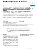
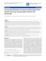
A typical example is shown in fig 1. The data shows that
dexamethasone had no significant effect on the number
of colonies formed over a range of concentrations in
either the ACL samples or the PT samples. There were
however, significantly more colonies per 1000 cells
seeded in the patellar sample than in the ACL sample,
indicating a higher proportion of proliferative cells in this
tissue (Fig. 1A) and in addition, the patellar samples pro-
duced bigger colonies in the control and 1 nM cultures
indicating a higher proliferative capacity (Fig. 1B).
Although these results indicate that dexamethasone did
not affect progenitor cell recruitment in terms of colony
number, there was a differential effect on subsequent col-
ony expansion. When the mean colony size was calcu-
lated, it was found that in the patellar samples there was a
Effect of dexamethasone on colony formation by primary human patellar or anterior cruciate ligament derived cellFigure 1
Effect of dexamethasone on colony formation by primary human patellar or anterior cruciate ligament
derived cell. 1 × 10
3
cells were cultured in 56 cm
2
Petri dishes for 11 days as described in the text. The cultures were then
stopped, stained with methylene blue and colony number (A) and colony size (B) determined by image analysis. * denotes sta-
tistical significance compared to appropriate control cultures, p < 0.05.
A. B.
Journal of Orthopaedic Surgery and Research 2008, 3:32 />Page 5 of 11
(page number not for citation purposes)
significant reduction in mean colony size at 100 and 1000
nM dexamethasone indicating a reduction in cell prolifer-
ation at these concentrations. Similarly, dexamethasone
caused a reduction in colony size in the ACL samples at
100 and 1000 nM as compared with the controls, how-
ever, at 10 nM dexamethasone lead to a significant
increase in mean colony size (Fig. 1B). Although this con-
centration response relationship was somewhat unex-
pected, it was reproducible and in 5 ACL samples, the
mean colony size at 10 nM dexamethasone ranged from
between 111% and 209% (mean 155%) of the mean con-
trol colony size (data not shown). Together these data sug-
gest that dexamethasone at 10 nM (i.e. physiological
levels) stimulated ACL cell proliferation but not that of
the PT cells whereas at higher concentrations it repressed
cell proliferation in both ACL and PT cells.
In order to see if this response persisted in culture, we
looked at the response of secondary human tendon/liga-
ment cells to dexamethasone using high density cultures
in a series of 3 paired samples. 'The cells were used after 2
passages and cell number and collagen accumulation
were measured after 1 and 2 weeks in culture; the results
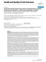
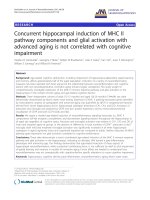
of a typical experiment are shown in fig 2. In the sub-cul-
tured PT cells, cell proliferation, as measured by methyl-
ene blue staining, after one week in culture showed no
significant changes although there was a slight downward
trend at the higher doses tested (Fig. 2A). However after 2
weeks of culture, cells in the presence of dexamethasone
at 1 nM showed a slight increase in proliferation as com-
pared to the control cultures but this increase was not sig-
nificant at the higher doses of dexamethasone (Fig. 2A).
These results differ from those obtained in the primary
CFU-f cultures where the higher concentrations of dexam-
ethasone lead to a reduction in cell proliferation as meas-
ured by mean colony size (Fig. 1B). This may be a result
of different culture conditions or a difference between pri-
mary and cultured PT cells.
In the passaged ACL cultures, again there was a significant
increase in cell number in the presence of low doses dex-
amethasone as compared to control cultures. After 1 week
of culture this was greatest at 1 nM dexamethasone but
after 2 weeks of culture the peak was at 10 nM dexameth-
asone (Fig. 2B). However at both time points, the increase
in cell number was not as large as that found in the ex vivo
CFU-f cultures, again suggesting different sensitivities to
dexamethasone using the different culture methods.
Collagen accumulation in the PT cell cultures followed
the same trend as the cell proliferation in that at both time
points there was a slight but insignificant increase at low
dexamethasone concentrations and a decrease at higher
dexamethasone concentrations, probably reflecting the
numbers of cells per culture. However the rate of increase
between the first and second week of culture was far
greater for collagen accumulation than for cell number
with a 100% increase in the amount of collagen compared
Effect of dexamethasone on secondary human patellar (A) or anterior cruciate ligament (B) derived cell proliferationFigure 2
Effect of dexamethasone on secondary human patellar (A) or anterior cruciate ligament (B) derived cell prolif-
eration. Cells were plated out in 24 well plates at a density of 5 × 10
3
cells per well as described above. The cultures were
stopped after 1 or 2 weeks and cell number determined by methylene blue staining. * denotes statistical significance compared
to appropriate control cultures, p < 0.05.
A. B.
Journal of Orthopaedic Surgery and Research 2008, 3:32 />Page 6 of 11
(page number not for citation purposes)
with about 25% increase in cell number in the control cul-
tures suggesting a process of differentiation and specific
increase in collagen accumulation (Fig. 3A).
In the ACL cultures, once again at week one the pattern of
collagen accumulation paralleled changes in cell number
indicating that the rate of collagen accumulation per cell
number was constant over the range of dexamethasone
doses. However, after 2 weeks in culture the pattern
changed in that the control cultures showed a proportion-
ally greater increase in collagen accumulation than the PT
cultures. Furthermore, no stimulation was seen at low
dexamethasone concentrations although, as in the 1 week
cultures, an inhibition was seen at higher concentrations
(Fig. 3B).
It was evident that cells derived from ACLs and PTs dis-
played a differential response to dexamethasone. How-
ever, as the PT samples used in these experiments were
healthy (trimmings taken from tissue used for ACL
autografts) whereas the ACL samples were derived from
ruptured tissue, it was thought that these differences
might reflect pathological changes rather than physiolog-
ical differences between tendon/ligament types. To exam-
ine this possibility we then compared the responses to
dexamethasone in 3 types of rat tendons derived from
healthy rats.
When cells derived from patellar, Achilles and tail ten-
dons from 3 male Wistar rats were examined for their
response to dexamethasone in the CFU-f assay, it was evi-
dent that the cells behaved differently in their growth and
response to dexamethasone. Examples of the cultures are
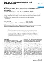
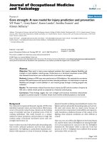
shown in Figure 4. On analysis, it was found that in terms
of mean colony number, only the tail tendon cells showed
any significant differences over the dexamethasone con-
centrations tested, with a significant reduction in colony
number with doses of 100 nM or higher (Fig. 5A). The tail
tendon results are in agreement with our previous find-
ings [29] however, the PT cells showed no reduction in
colony number over the range of doses tested, a similar
result to the human patellar samples; and the Achilles ten-
don cells also showed no reduction in colony number
(Fig. 5A). With regards to mean colony size it was evident
that in the absence of dexamethasone the largest colonies
(and therefore having the greatest rate of growth) was seen
in the PT cells (Fig. 5B). In the tail cells, as before, there
was a reduction in mean size at doses of 10 nM and above.
In the PT cultures there was also a reduction in mean col-
ony size, occurring at doses of 100 nM and above, again
similar to the results found in the human samples. Inter-
estingly, in the Achilles cells, although there was a reduc-
tion in the intensity of staining indicating a reduction in
cell number, on analysis there was no significant reduc-
tion in colony size at any of the doses tested. These results,
along with the human results suggest that the differences
in sensitivity to dexamethasone are not due to the disease
status of the tendons but rather due to specific differences
in sensitivity in the various tendon tissues.
Effect of dexamethasone on secondary human patellar (A) or anterior cruciate ligament (B) derived collagen accumaulationFigure 3
Effect of dexamethasone on secondary human patellar (A) or anterior cruciate ligament (B) derived collagen
accumaulation. Cells were cultured as in Figure 2 and stopped after 1 or 2 weeks at which point collagen accumulation was
determined by Sirius red staining. * denotes statistical significance compared to appropriate control cultures, p < 0.05.
A.
B.
Journal of Orthopaedic Surgery and Research 2008, 3:32 />Page 7 of 11
(page number not for citation purposes)
To see whether there were any intrinsic differences
between cells derived from the different tendons, we
determined the intensity of expression of cell surface
markers typical of mesenchymal cells on cells cultured
from all tendons used in the above experiments using
flow cytometry. Rat cells were analysed for CD44 and
CD90 and human cells for CD44, CD90 and D7fib. These
markers were chosen as they are expressed by between 90
Effect of dexamethasone on colony formation by primary rat Achilles, patellar or tail tendon derived cellsFigure 4
Effect of dexamethasone on colony formation by primary rat Achilles, patellar or tail tendon derived cells. 1 ×
10
3
cells were cultured in 56 cm
2
Petri dishes for 11 days as described above and then stopped, stained with methylene blue
and a digital image acquired.
Achilles Patellar Tail
Control
Dex 1nM
Dex 10nM
Dex 100M
Dex 1μM
Journal of Orthopaedic Surgery and Research 2008, 3:32 />Page 8 of 11
(page number not for citation purposes)
and 100% of cells from all sources facilitating accurate
comparison of intensity data between cell types (Tables 1
and 2).
Interestingly in both the rat and the human patellar sam-
ples, the marker CD90 showed a higher intensity of
expression than in the ACL (human) or Achilles and tail
(rat). In the rat there were no other obvious differences in
the markers tested. In the human samples, the levels of
expression varied greatly between patients, however, the
general pattern of expression remained the same through-
out. It was found that in all cases, ACL samples had a
higher intensity of expression of the fibroblastic marker
D7fib and a reduced CD90 expression compared to the
patellar samples in all four pairs tested, however there
were no consistent differences in CD13 or CD44.
Discussion
In this manuscript we have demonstrated differences
between tenocytes derived from tendons and ligaments
from different sources in terms of their proliferation, col-
lagen deposition, levels of progenitor cells, responses to
dexamethasone and expression of cell surface markers. It
is a sad fact that, despite considerable evidence to the con-
trary, tendons and ligaments tend to be considered to
metabolically and cell biologically similar if not identical.
In the clinic, treatment regimes often aim to treat only
parts of the pathology, such as neovascularisation, that is
similar for different structures without taking into account
possible tissue specific differences [36]. Although his-
topathologic features are similar between different condi-
tions, such as rotator cuff tears, patella tendinosis, Achilles
tendinosis and "lateral epicondylitis" of the elbow, this
does not necessarily mean that the response to treatment
will be similar. Differences between tendons & ligaments
and between flexor and extensor tendons have been refer-
enced. However, this approach is probably too simplistic
and given the different biomechanical stresses inflicted on
tendons combined with the differences in vasculature and
environment, it is likely that all tendons will be to some
degree unique in terms of their cell biology and metabo-
Effect of dexamethasone on colony formation by primary rat Achilles, patellar or tail tendon-derived cellFigure 5
Effect of dexamethasone on colony formation by primary rat Achilles, patellar or tail tendon-derived cell. Cells
were cultured as Figure 1 and then colony number (A) and colony size (B) determined by image analysis. * denotes statistical
significance compared to appropriate control cultures, p < 0.05.
A. B.
Table 1: Intensity of Expression of cell surface markers on rat
patellar, Achilles or tail tendon-derived cells.
Marker PT Achilles Tail
CD44 39.4 ± 4.3 42.8 ± 3.5 35.3 ± 3.9
CD90 105.9 ± 9.6 81.4 ± 5.1 83.2 ± 6.3
Table 2: Expression of cell surface markers on Human patellar
tendon or anterior cruciate ligament cells.
Patient 1 Patient 2 Patient 3 Patient 4
PT ACL PT ACL PT ACL PT ACL
CD44 25.3 37.3 297.5 203.7 329.4 316.1 92.8 49.3
CD90 62.2 37.8 274.7 59.5 176.6 100.9 76.2 42
D7fib 42.2 74.1 42.6 105.9 50.5 72.2 43.1 49.8
Journal of Orthopaedic Surgery and Research 2008, 3:32 />Page 9 of 11
(page number not for citation purposes)
lism. Such differences are likely to have considerable rele-
vance to therapies used to treat various tendinopathies, in
particular in terms of the response to glucocorticoids
treatment and to our knowledge; this is the first study
investigating such tendon specific effects in human or rat
tissues.
There is conflicting data regarding the effects of dexameth-
asone on tendon-derived cells in vitro. For example, Fer-
mor et al showed that dexamethasone at 10 nM
stimulated the proliferation of cells extracted from ACL's
[25] whereas Wong et al showed that at concentrations
between 1 nM and 100 uM dexamethasone inhibited the
proliferation of cells from healthy PTs [23]. This is consist-
ent with the in vitro data presented here in that the
responses to this drug differed between the PTs and ACLs
from the same individual, cultured under identical condi-
tions. In the CFU-f assay there were more colonies per
1000 cells seeded in the patellar as compared to the ACL
control samples although this difference was not signifi-
cant, indicating the presence of a greater percentage of
proliferative progenitor cells. The PT colonies were also
larger in the control cultures indicating a greater basal pro-
liferative rate in this tissue. In the PTs, a decrease in cell
proliferation, as indicated by colony size was seen in ex
vivo cultures in the CFU-f assay at doses greater than 10
nM although there was no effect on colony number. Sim-
ilarly, in high density cultures of passaged cells at one
week there was a small but non-significant decrease in cell
number at higher doses, but after two weeks of culture
there was an increase in cell number at doses of dexame-
thasone below 10 nM. This difference may well be a prod-
uct of the differing length of exposure to the drug – 2
weeks as compared to 4 days, indeed at one week our cul-
tures did not show increases in cell number at lower doses
of dexamethasone.
PT and ACL cultures derived from the same individuals
showed a different pattern of response to dexamethasone.
In the ACL cultures, colony size (and hence cell prolifera-
tion) showed a marked increase at 10 nM – the same dose
used by Fermor et al [25]. However this stimulation of cell
proliferation was only seen at this dose and an inhibition
was seen at doses of 100 nM and more. A similar pattern
of response was seen in the high density cultures although
here the differences were smaller. In contrast, no such
stimulation was seen on the PT cultures and these data
show that these two cell populations do indeed respond
in a different manner despite being cultured under identi-
cal conditions and derived from the same individual. This
could indicate an intrinsic difference in dexamethasone
sensitivity between the two tissues. However, an alterna-
tive explanation could be that the ACL tissue was from a
rupture and the PT was from clinically healthy tissue and
a repair response may be the cause of these differing
results. We therefore decided to compare the responses of
healthy rat tendons to dexamethasone.
In the rat we compared the responses of the patellar,
Achilles and tail tendons to dexamethasone. Ideally, we
would have used ACLs as well; however, due to their small
size we were unable to extract sufficient cells from this tis-
sue for accurate experiments. In the CFU-f assay, again in
the control cultures the PTs produced the largest colonies,
followed by the Achilles with the tail cells producing the
smallest colonies, indicating that the PT cells proliferate at
the fastest rate of the tissues tested, a result similar to that
found in the human tissue. These PT cells also responded
to dexamethasone in a similar manner to the human PT
cells suggesting that there is no difference in the response
to this drug in the PTs in the two species tested. When the
response to dexamethasone was tested in the three differ-
ent rat tendons, there was a difference in response
between the tendon cells as only the tail tendon cells
showed a decrease in progenitor cell recruitment (i.e. a
decrease in colony number) with increasing concentra-
tions on dexamethasone. However there was a dose
dependant decrease in colony size in the PT and tail cells
but not the Achilles cells at the doses tested. These results
therefore show that in the rat as in the human, there is a
difference in sensitivity to dexamethasone between ten-
don cells isolated from different tendons but from the
same subject.
To further characterise any intrinsic differences in cells
from different tendons we looked at the intensity of
expression on cell surface markers. Tendon cells have
been shown to express the human fibroblastic markers
D7-FIB [11,12] and CD90 as well as other markers
expressed by MSCs (eg. CD13 & CD44) (data not shown).
Here we found that in a group of four PT/ACL pairs the
intensity of expression of CD90 was consistently lower in
the ACL as compared to the patellar and the expression of
D7 FIB was consistently higher, whereas the pattern of
response was inconsistent in the other markers. In the rat,
the intensity of expression of CD90 was also higher in the
PT cells as compared to the Achilles and tail cells. Differ-
ences in intensity of expression of CD90 have been seen
between MSC's derived from synovial tissue and bone
marrow MSCs obtained from humans with the expression
being higher in cells derived from the synovium [13]. This
marker has also been shown to lose intensity of expres-
sion in cultures of human MSC subjected to mechanical
strain as compared to unstrained cultures [14]. Therefore
a possible explanation for this lower level of expression in
the ruptured ACL tissue is that these cells are exposed to
higher levels of mechanical strain in vivo prior to rupture.
The results presented here clearly show that cells taken
from different tendon/ligaments have different respon-
Journal of Orthopaedic Surgery and Research 2008, 3:32 />Page 10 of 11
(page number not for citation purposes)
siveness to dexamethasone, and degree of expression of
some cell surface markers. Such source specific differences
have been shown for other connective tissues including
osteoblasts [37,38], chondrocytes [39-41] and mesenchy-
mal stem cells [38] and the data presented above is con-
sistent with this. In the rat, tail tendon cells appear to be
most sensitive to dexamethasone followed by cells from
the PT and the Achilles being least affected. The responses
in the PT appear to be similar for both rat and human,
both in expression of CD90 and reactivity to dexametha-
sone, indicating therefore that this pattern of response is
not to be species specific. In the human ACL the effects of
dexamethasone concentrations produce a different pat-
tern of response to that of the PT in that there is a prolif-
erative response at 10
-8
M but an inhibition of cell number
at higher doses. At present we can only speculate as to the
mechanism underlying these effects. This might lie at the
level of the glucocorticoid receptor; although this is
derived from a single gene, alternative splicing and post-
translational modification give rise to multiple receptor
isoforms which together with site specific co-activators is
thought to be responsible for glucocorticoid tissue specif-
icity [42,43]. The non-linear dose-response to dexameth-
asone seen in the human ACL samples is quite different to
the responses seen in all other tendons or ligaments tests,
human or rodent. It is however, similar to the bell shaped
response of MSCs to dexamethasone, peaking at 10 nM
followed by an inhibitory effect [44]. As all of the ACL
samples were from ACL ruptures undergoing surgery it is
likely that this response was due to extrinsic cells invading
during the healing process. In support of this, it is now
widely accepted that MSC are associated with microvascu-
lature and are probably pericytic in nature [45]. The struc-
ture and biomechanical environment of a tendon varies
greatly both within and between tendons [46] and is also
likely that the cellular make-up of a tendon or ligament
reflects this. Both the patellar tendon and ACL are weight
bearing tissues; the ACL is under a chronic stress that is
dependent on the posture whereas the patellar tendon
experiences a more pulsatile loading related to the activity
of the Quadriceps femorus. This environment will play a
role in selecting the population of cells present in the ten-
don although exactly how and how long this phenotype
persists in culture is unknown. It might also be expected
that cells from ACL and Patellar tendons might respond
differently to mechanical loading in vitro and this is cur-
rently a focus of investigation in our laboratory. One
study comparing human finger flexor and extensor ten-
dons suggested that there is no difference [47] whereas a
similar investigation in horse flexor and extensor tendons
suggested an increased responsiveness in extensor ten-
dons [30] although these studies might not necessarily be
relevant to the human ACL/Patellar situation.
Conclusion
In conclusion, these data show that tendon & ligament-
derived cells from different sources possess intrinsic dif-
ferences in terms of their growth, dexamethasone respon-
siveness and cell surface marker expression. This is not
unexpected as different tendons perform radically differ-
ent functions and their cell populations must adapt their
behaviour to reflect this. The current rapid developments
in tissue engineering, in particular of mesenchymal tis-
sues, holds great promise for the treatment of chronic and
debilitating diseases. However, the source of cells for the
generation of tissue engineered constructs must be care-
fully considered to maximise their engraftment and effi-
cacy.
Competing interests
The authors declare that they have no competing interests.
Authors' contributions
NS: planning of experiments, majority of experimental
work, preparation of manuscript; CGR: sourcing of
human tissue, preparation of manuscript; AS: planning of
experiments, some experimental work, preparation of
manuscript.
Acknowledgements
We wish to thank the theatre staff at the Thornbury Hospital for assistance
with collecting clinical samples.
References
1. Chan KM, Fu F, Maffulli N, Rolf CG, Kurosaka M, Liu S: Controver-
sies in Orthopedic Sports Medicine. Hong Kong , Williams and
Wilkins Asia-Pacific ltd; 1998.
2. DeFranco MJ, Derwin K, Iannotti JP: New therapies in tendon
reconstruction. J Am Acad Orthop Surg 2004, 12(5):298-304.
3. Butler DL, Juncosa-Melvin N, Boivin GP, Galloway MT, Shearn JT,
Gooch C, Awad H: Functional tissue engineering for tendon
repair: A multidisciplinary strategy using mesenchymal stem
cells, bioscaffolds, and mechanical stimulation. J Orthop Res
2008, 26:1-9.
4. MacNeil S: Progress and opportunities for tissue-engineered
skin. Nature 2007, 445(7130):874-880.
5. Hollander AP, Dickinson SC, Sims TJ, Brun P, Cortivo R, Kon E, Mar-
cacci M, Zanasi S, Borrione A, De Luca C, Pavesio A, Soranzo C, Aba-
tangelo G: Maturation of tissue engineered cartilage
implanted in injured and osteoarthritic human knees. Tissue
Eng 2006, 12(7):1787-1798.
6. Awad HA, Boivin GP, Dressler MR, Smith FN, Young RG, Butler DL:
Repair of patellar tendon injuries using a cell-collagen com-
posite. J Orthop Res 2003, 21(3):420-431.
7. Liu W, Chen B, Deng D, Xu F, Cui L, Cao Y: Repair of tendon
defect with dermal fibroblast engineered tendon in a porcine
model. Tissue Eng 2006, 12(4):775-788.
8. Cao Y, Liu Y, Liu W, Shan Q, Buonocore SD, Cui L: Bridging ten-
don defects using autologous tenocyte engineered tendon in
a hen model. Plast Reconstr Surg 2002, 110(5):1280-1289.
9. Rumian AP, Wallace AL, Birch HL: Tendons and ligaments are
anatomically distinct but overlap in molecular and morpho-
logical features a comparative study in an ovine model. J
Orthop Res 2007, 25(4):458-464.
10. Birch HL: Tendon matrix composition and turnover in rela-
tion to functional requirements. Int J Exp Pathol 2007,
88(4):241-248.
11. de Mos M, Koevoet WJ, Jahr H, Verstegen MM, Heijboer MP, Kops
N, van Leeuwen JP, Weinans H, Verhaar JA, van Osch GJ: Intrinsic
Publish with BioMed Central and every
scientist can read your work free of charge
"BioMed Central will be the most significant development for
disseminating the results of biomedical research in our lifetime."
Sir Paul Nurse, Cancer Research UK
Your research papers will be:
available free of charge to the entire biomedical community
peer reviewed and published immediately upon acceptance
cited in PubMed and archived on PubMed Central
yours — you keep the copyright
Submit your manuscript here:
/>BioMedcentral
Journal of Orthopaedic Surgery and Research 2008, 3:32 />Page 11 of 11
(page number not for citation purposes)
differentiation potential of adolescent human tendon tissue:
an in-vitro cell differentiation study. BMC Musculoskelet Disord
2007, 8:16.
12. Machner A, Baier A, Wille A, Drynda S, Pap G, Drynda A, Mawrin C,
Buhling F, Gay S, Neumann W, Pap T: Higher susceptibility to Fas
ligand induced apoptosis and altered modulation of cell
death by tumor necrosis factor-alpha in periarticular teno-
cytes from patients with knee joint osteoarthritis. Arthritis Res
Ther 2003, 5(5):R253-61.
13. Djouad F, Bony C, Haupl T, Uze G, Lahlou N, Louis-Plence P, Appa-
railly F, Canovas F, Reme T, Sany J, Jorgensen C, Noel D: Transcrip-
tional profiles discriminate bone marrow-derived and
synovium-derived mesenchymal stem cells. Arthritis Res Ther
2005, 7(6):R1304-15.
14. Wiesmann A, Buhring HJ, Mentrup C, Wiesmann HP: Decreased
CD90 expression in human mesenchymal stem cells by
applying mechanical stimulation. Head Face Med 2006, 2:8.
15. Wiggins ME, Fadale PD, Ehrlich MG, Walsh WR: Effects of local
injection of corticosteroids on the healing of ligaments. A fol-
low-up report. J Bone Joint Surg Am 1995, 77(11):1682-1691.
16. Gottlieb NL, Riskin WG: Complications of local corticosteroid
injections. Jama 1980, 243(15):1547-1548.
17. Ismail AM, Balakrishnan R, Rajakumar MK, Lumpur K: Rupture of
patellar ligament after steroid infiltration. Report of a case.
J Bone Joint Surg Br 1969, 51(3):503-505.
18. McQuillan R, Gregan P: Tendon rupture as a complication of
corticosteroid therapy. Palliat Med 2005, 19(4):352-353.
19. Arroll B, Goodyear-Smith F: Corticosteroid injections for painful
shoulder: a meta-analysis. Br J Gen Pract 2005, 55(512):224-228.
20. Speed CA: Fortnightly review: Corticosteroid injections in
tendon lesions. Bmj 2001, 323(7309):382-386.
21. Paavola M, Kannus P, Jarvinen TA, Jarvinen TL, Jozsa L, Jarvinen M:
Treatment of tendon disorders. Is there a role for corticos-
teroid injection? Foot Ankle Clin
2002, 7(3):501-513.
22. Buchbinder R, Green S, Youd JM: Corticosteroid injections for
shoulder pain. Cochrane Database Syst Rev 2003:CD004016.
23. Wong MW, Tang YY, Lee SK, Fu BS, Chan BP, Chan CK: Effect of
dexamethasone on cultured human tenocytes and its revers-
ibility by platelet-derived growth factor. J Bone Joint Surg Am
2003, 85-A(10):1914-1920.
24. Wong MW, Tang YY, Lee SK, Fu BS: Glucocorticoids suppress
proteoglycan production by human tenocytes. Acta Orthop
2005, 76(6):927-931.
25. Fermor B, Urban J, Murray D, Pocock A, Lim E, Francis M, Gage J:
Proliferation and collagen synthesis of human anterior cruci-
ate ligament cells in vitro: effects of ascorbate-2-phosphate,
dexamethasone and oxygen tension. Cell Biol Int 1998, 22(9-
10):635-640.
26. Almekinders LC, Deol G: The effects of aging, antiinflammatory
drugs, and ultrasound on the in vitro response of tendon tis-
sue. Am J Sports Med 1999, 27(4):417-421.
27. Torricelli P, Fini M, Giavaresi G, Carpi A, Nicolini A, Giardino R:
Effects of systemic glucocorticoid administration on teno-
cytes. Biomed Pharmacother 2006, 60(8):380-385.
28. Tsai WC, Tang FT, Wong MK, Yen HC, Pang JH: Decreased
expression of proliferating cell nuclear antigen is associated
with dexamethasone inhibition of the proliferation of rat
tendon fibroblasts. J Rheumatol 2002, 29(11):2397-2402.
29. Scutt N, Rolf CG, Scutt A: Glucocorticoids inhibit tenocyte pro-
liferation and Tendon progenitor cell recruitment. J Orthop
Res 2006, 24(2):173-182.
30. Goodman SA, May SA, Heinegard D, Smith RK: Tenocyte response
to cyclical strain and transforming growth factor beta is
dependent upon age and site of origin. Biorheology 2004,
41(5):613-628.
31. Stanley RL, Fleck RA, Becker DL, Goodship AE, Ralphs JR, Patterson-
Kane JC: Gap junction protein expression and cellularity:
comparison of immature and adult equine digital tendons.
J
Anat 2007, 211(3):325-334.
32. Magnusson SP, Hansen M, Langberg H, Miller B, Haraldsson B, Westh
EK, Koskinen S, Aagaard P, Kjaer M: The adaptability of tendon
to loading differs in men and women. Int J Exp Pathol 2007,
88(4):237-240.
33. Miller BF, Hansen M, Olesen JL, Schwarz P, Babraj JA, Smith K, Rennie
MJ, Kjaer M: Tendon collagen synthesis at rest and after exer-
cise in women. J Appl Physiol 2007, 102(2):541-546.
34. Lopez-De Leon A, Rojkind M: A simple micromethod for colla-
gen and total protein determination in formalin-fixed paraf-
fin-embedded sections. J Histochem Cytochem 1985,
33(8):737-743.
35. Currie GA: Platelet-derived growth-factor requirements for
in vitro proliferation of normal and malignant mesenchymal
cells. Br J Cancer 1981, 43(3):335-343.
36. Alfredson H, Lorentzon R: Sclerosing polidocanol injections of
small vessels to treat the chronic painful tendon. Cardiovasc
Hematol Agents Med Chem 2007, 5(2):97-100.
37. Declercq H, Van den Vreken N, De Maeyer E, Verbeeck R, Schacht E,
De Ridder L, Cornelissen M: Isolation, proliferation and differ-
entiation of osteoblastic cells to study cell/biomaterial inter-
actions: comparison of different isolation techniques and
source. Biomaterials 2004, 25(5):757-768.
38. Yoshimura H, Muneta T, Nimura A, Yokoyama A, Koga H, Sekiya I:
Comparison of rat mesenchymal stem cells derived from
bone marrow, synovium, periosteum, adipose tissue, and
muscle. Cell Tissue Res 2007, 327(3):449-462.
39. Kuettner KE, Cole AA: Cartilage degeneration in different
human joints. Osteoarthritis Cartilage 2005, 13(2):93-103.
40. Billinghurst RC, Wu W, Ionescu M, Reiner A, Dahlberg L, Chen J, van
Wart H, Poole AR: Comparison of the degradation of type II
collagen and proteoglycan in nasal and articular cartilages
induced by interleukin-1 and the selective inhibition of type
II collagen cleavage by collagenase. Arthritis Rheum 2000,
43(3):664-672.
41. Xu C, Oyajobi BO, Frazer A, Kozaci LD, Russell RG, Hollander AP:
Effects of growth factors and interleukin-1 alpha on prote-
oglycan and type II collagen turnover in bovine nasal and
articular chondrocyte pellet cultures. Endocrinology 1996,
137(8):3557-3565.
42. Zhou J, Cidlowski JA: The human glucocorticoid receptor: one
gene, multiple proteins and diverse responses. Steroids 2005,
70(5-7):407-417.
43. Heitzer MD, Wolf IM, Sanchez ER, Witchel SF, DeFranco DB: Glu-
cocorticoid receptor physiology. Rev Endocr Metab Disord 2007,
8(4):321-330.
44. Scutt A, Bertram P, Brautigam M: The role of glucocorticoids and
prostaglandin E2 in the recruitment of bone marrow mesen-
chymal cells to the osteoblastic lineage: positive and nega-
tive effects. Calcif Tissue Int 1996, 59(3):154-162.
45. Shi S, Gronthos S: Perivascular niche of postnatal mesenchy-
mal stem cells in human bone marrow and dental pulp. J Bone
Miner Res 2003, 18(4):696-704.
46. Benjamin M, Kaiser E, Milz S: Structure-function relationships in
tendons: a review. J Anat 2008, 212(3):211-228.
47. Evans CE, Trail IA: An in vitro comparison of human flexor and
extensor tendon cells. J Hand Surg [Br] 2001, 26(4):307-313.