báo cáo hóa học:" New fluoroscopic imaging technique for investigation of 6DOF knee kinematics during treadmill gait" pot
Bạn đang xem bản rút gọn của tài liệu. Xem và tải ngay bản đầy đủ của tài liệu tại đây (296.85 KB, 5 trang )
BioMed Central
Page 1 of 5
(page number not for citation purposes)
Journal of Orthopaedic Surgery and
Research
Open Access
Technical Note
New fluoroscopic imaging technique for investigation of 6DOF knee
kinematics during treadmill gait
Guoan Li*, Michal Kozanek, Ali Hosseini, Fang Liu, Samuel K Van de Velde
and Harry E Rubash
Address: Bioengineering Laboratory, GRJ 1215, Massachusetts General Hospital, 55 Fruit Street, Boston, MA 02114, USA
Email: Guoan Li* - ; Michal Kozanek - ; Ali Hosseini - ;
Fang Liu - ; Samuel K Van de Velde - ; Harry E Rubash -
* Corresponding author
Abstract
Introduction: This report presents a new imaging technique for non-invasive study of six degrees
of freedom (DOF) knee kinematics during treadmill gait.
Materials and methods: A treadmill was integrated into a dual fluoroscopic imaging system
(DFIS) to formulate a gait analysis system. To demonstrate the application of the system, a healthy
subject walked on the treadmill at four different speeds (1.5, 2.0, 2.5 and 3.0 MPH) while the DFIS
captured the knee motion during three strides under each speed. Characters of knee joint motion
were analyzed in 6DOF during the treadmill walking.
Results: The speed of the knee motion was lower than that of the treadmill. Flexion amplitudes
increased with increasing walking speed. Motion patterns in other DOF were not affected by
increase in walking speed. The motion character was repeatable under each treadmill speed.
Conclusion: The presented technique can be used to accurately measure the 6DOF knee
kinematics at normal walking speeds.
Introduction
Accurate data of six degrees-of-freedom (6DOF) knee kin-
ematics is instrumental for investigation of biomechani-
cal mechanisms of knee pathology such as osteoarthritis,
ligamentous injuries and total knee arthroplasty. Tradi-
tional gait analysis used multiple video cameras to track
the three-dimensional (3D) motions of reflective markers
fixed to the skin[1], which was limited to reveal relative
motion of the femoral and tibial bones. Invasive meth-
ods, such as using reflective markers directly fixed to bone
using a thin rod[2] or opaque markers embedded within
the bones, [3-7] were applied to detect bony motion in
order to eliminate the effect of skin motion and enhance
the accuracy of motion data. In another way, a point-clus-
ter technique, which is noninvasive, has also been pro-
posed to improve the traditional gait analysis method in
order to reduce the effect of relative motion of the skin
and bones[8].
Recently, fluoroscopic imaging technique, due to its rela-
tive accessibility, easiness to operate, and low radiation
dosage compared to traditional X-rays, has been used for
the analysis of knee joint motion during gait [9-11]. How-
ever, the use of just a single image might not detect knee
joint motion in the out-of-plane degrees-of-freedom in
the same accuracy as compared to the accuracy in in-plane
Published: 13 March 2009
Journal of Orthopaedic Surgery and Research 2009, 4:6 doi:10.1186/1749-799X-4-6
Received: 14 November 2008
Accepted: 13 March 2009
This article is available from: />© 2009 Li et al; licensee BioMed Central Ltd.
This is an Open Access article distributed under the terms of the Creative Commons Attribution License ( />),
which permits unrestricted use, distribution, and reproduction in any medium, provided the original work is properly cited.
Journal of Orthopaedic Surgery and Research 2009, 4:6 />Page 2 of 5
(page number not for citation purposes)
motion[12,13]. In our laboratory, we validated the
method using the cine function of two fluoroscopes to
simultaneously capture dynamic knee joint motion[14].
This study presents how to use this technique to deter-
mine 6DOF knee joint motion during treadmill gait with
different speeds.
Methods
DFIS Setup
The dual fluoroscopic imaging system (DFIS) setup that
was validated previously is used for the treadmill gait
analysis (Fig. 1) [14,15]. The DFIS consists of two pulse
fluoroscopes (BV Pulsera, Philips) that are set to generate
8 ms width X-ray pulses with an effective dose of 13 mrem
per scanning. In this study, the fluoroscopes took 30
evenly distributed snapshot images per second during
dynamic knee joint motion.
The diameter of the image intensifier of the fluoroscopes
is ~310 mm. In general, given the size of the image inten-
sifier of the fluoroscopes might be difficult to capture the
entire range of knee motion during the treadmill gait.
Therefore, the two fluoroscopes are positioned so that
their intensifiers form an angle between 120 and 130°
(Fig. 1). In this setup, the dual fluoroscopic system has a
common field of view with a length of ~450 mm. There-
fore, the entire knee motion could be captured by both
fluoroscopes during the gait cycle.
Treadmill gait
To demonstrate the methodology of treadmill gait analy-
sis, one healthy subject (male, 45 years old) performed
gait on the treadmill at different speeds: 1.5, 2.0, 2.5 and
3.0 mile/hour MPH (or 0.67, 0.89, 1.12 and 1.34 m/s,
respectively). Two laser-positioning devices were attached
to the two fluoroscopes to help the subject align the target
knee (left) within the field of view of the fluoroscopes
during gait with the assistance of a technician. The knee
was then imaged from heel strike to toe-off during three
consecutive strides after about 30 seconds of practice. The
subject took 5 minute rest after testing for each speed.
Reproduction of in-vivo knee kinematics
The anatomic model of the target knee, including the
bony geometry of the tibia and femur, was reconstructed
by tracing the bony contours on sagittal plane magnetic
resonance (MR) images of the knee in solid modeling
software (Rhinoceros
®
, Robert McNeal & Associates, Seat-
tle, WA). The MR images were obtained using a 3.0 Tesla
MR scanner (MAGNETOM
®
Trio, Siemens, Erlangen, Ger-
many) while the subject was lying supine with the knee in
a relaxed, extended position. The MR scanner employed a
3D double echo water excitation sequence and the follow-
ing parameters: field-of-view = 160 × 160 × 120 mm,
voxel resolution = 0.31 × 0.31 × 1.00 mm, time of repeti-
tion (TR) = 24 ms, time of echo (TE) = 6.5 ms, and flip
angle = 25°. A joint coordinate system described previ-
ously (Fig. 1B) was adopted to determine the 6DOF knee
joint kinematics [16].
The model and the dual fluoroscopic images were placed
into a virtual DFIS environment where the in-vivo posi-
tions of knee were reproduced by matching projections of
the models to their outlines on the fluoroscopic images
[12]. The knee positions during three strides at each tread-
mill speed were reproduced. For each stride, the knee
position was analyzed at each 10% of the stance phase
from heel strike to toe-off.
The average speed of the knee during stance phase was cal-
culated by dividing the maximal traveling distance by the
corresponding traveling time. The data on 6DOF knee
kinematics, including knee flexion, internal-external tibial
rotation, as well as medial-lateral translation and varus-
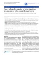
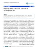
(A) Setup of the DFIS system and a treadmillFigure 1
(A) Setup of the DFIS system and a treadmill. (B) Knee model and virtual DFIS environment for reproduction of in-vivo
knee kinematics. A typical knee model is shown after reproducing its position in the virtual environment of the modeling soft-
ware.
Treadmill
F1
F2
(A) (B)
130°
Journal of Orthopaedic Surgery and Research 2009, 4:6 />Page 3 of 5
(page number not for citation purposes)
valgus rotation, were analyzed. The repeatability of the
treadmill gait was determined by the standard deviation
of the three strides of each treadmill speed.
Results
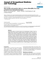
The duration of the stance phase decreased with the tread-
mill speed (Fig. 2). At 1.5 MPH, the stance phase time was
0.99 second, while at treadmill speed of 3.0 MPH, the
stance phase time decreased to 0.49 second. The average
speed of the knee during stance phase was lower than the
treadmill speed (Fig. 2). At 1.5 MPH treadmill speed, the
knee speed was 0.28 ± 0.02 m/second, only 41% of the
treadmill speed. At 2.5 MPH treadmill speed, the knee
speed was 0.39 ± 0.05 m/second that was 35% of the
treadmill speed. At 3.0 MPH treadmill speed, the knee
speed was 0.81 ± 0.02 m/second that was 60% of the
treadmill speed.
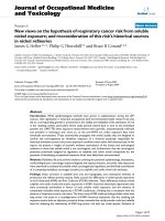
Knee kinematics under different treadmill speeds showed
similar patterns in both, the rotations and the translations
(Fig. 3). After heel strike, the tibia showed an increase in
flexion angle to 6.71 ± 0.86° and 13.81 ± 2.73° and an
increase in internal tibial rotation to 4.56 ± 0.29° and
5.45 ± 0.76° for the treadmill speeds of 1.5 MPH and 3.0
MPH, respectively (Fig. 2B). During mid-stance, the knee
showed maximal hyperextension of about -2.5° and
external tibial rotation of about -1.5° at all speeds. At toe-
off, the knee had flexion angles of 43.05 ± 2.18° and
52.35 ± 5.09° and internal tibial rotation of 4.73 ± 0.35°
and 7.56 ± 1.50° for treadmill speeds of 1.5 and 3.0 MPH,
respectively. The knee also showed an increase in valgus
rotation after heel strike, a decrease in valgus rotation dur-
ing stance phase and sharper increase in valgus rotation at
toe-off (Fig. 2B and Fig. 3C).
Femoral translations during gait at different speeds
showed similar patterns (Figs. 3D and 3E). The femur
translated anteriorly during loading response and early
midstance and moved posteriorly thereafter until termi-
nal stance when it shifted anteriorly again. In medial-lat-
eral direction, the femur moved laterally during early
stance and medially towards toe-off.
Discussion
This paper introduced the technique of using the DFIS for
measurement of 6DOF in-vivo knee kinematics during
treadmill gait. The data showed that this technique is fea-
sible to analyze the dynamic knee motion during a wide
Top: Average speed of the knee and duration of the stance phase of gait on treadmill at four different walking speedsFigure 2
Top: Average speed of the knee and duration of the stance phase of gait on treadmill at four different walking
speeds. Bottom: Peak kinematic values of the knee during different intervals of the stance phase. F/E: flexion (+)/extension(-
); IR/ER: internal(-)/external(+) femoral rotation; A/P: anterior(+)/posterior(-) femoral translation; ML: medial(-)/lateral(+) fem-
oral translation; SD: standard deviation.
Treadmill
Speed
1.5 8.23 0.20 5.65 0.28 4.15 0.17 0.45 0.16 5.58 0.22
2.
0
8.91 0.35 5.84 0.48 4.1
0
0.23 0.5
3
0.20 5.71 0.12
2.5 9.44 0.60 5.7
8
0.69 4.2
8
0.14 0.5
6
0.33 5.71 0.16
3.
0
10.8
2
1.75 5.91 0.95 4.14 0.35 0.55 0.48 5.8
0
0.21
1.5 -2.48 0.23 -1.40 0.25 3.64 0.12 -4.6
7
0.16 2.6
0
0.17
2.
0
-1.99 0.47 -1.3
7
0.21 3.6
0
0.17 -4.73 0.21 2.65 0.13
2.5 -1.83 0.16 -1.4
7
0.38 3.6
0
0.22 -4.89 0.12 2.64 0.19
3.
0
-1.79 1.08 -1.39 0.40 3.5
0
0.21 -4.76 0.3
7
2.6
2
0.2
2
1.5 40.9
0
1.15
7.3
7
1.03
5.51
0.15
2.51
0.39
2.65
0.32
2.
0
42.6
8
1.65 7.5
7
1.65 5.5
0
0.27 2.41 0.44 2.7
2
0.52
2.5 43.75 2.85 7.4
9
1.94 5.4
8
0.46 2.4
9
0.53 2.7
7
0.63
3.
0
46.4
0
1.98 7.8
3
2.3
7
5.4
3
0.65 2.35 0.66 2.65 0.5
7
SD
0-30
30-60
60-100
SD
A/P
SD
M/L
SD
IR/ER
SD
VR/VL% stance F/E
0
0.2
0.4
0.6
0.8
1
0.5 0.75 1 1.25 1.5
Treadmill speed (m/sec)
Knee speed (m/sec)
0.4
0.6
0.8
1
1.2
Stance phase duration (sec)
Knee speed
Stance phase
duration
Journal of Orthopaedic Surgery and Research 2009, 4:6 />Page 4 of 5
(page number not for citation purposes)
range of treadmill speeds (up to 3 MPH). Since this tech-
nique reproduced the knee positions using 3D anatomic
models of the knee, 6DOF tibiofemoral joint kinematics
during gait can be obtained.
Few studies have utilized fluoroscopes to investigate
human knee motion during gait [17]. For example, Zihl-
mann et al. [17] moved a fluoroscope to follow the knee
motion to overcome the limited field of view of the image
intensifier. They estimated an accuracy of 0.2 mm for in-
plane translation and of 3.25 mm for out-plane transla-
tion and an accuracy of 1.57° for rotation in a knee after
total joint arthroplasty during level walking. To overcome
the limitation of the image intensifier size in the DFIS set
up, the two fluoroscopes are positioned so that their com-
mon image zone covers the knee motion during the com-
plete gait cycle on a treadmill [14].
The DFIS has been recently validated to measure dynamic
knee motion [14]. Using standard geometry, the sphere
positions could be determined with a SD below 0.2 mm
when sphere moved at a speed up to 0.5 m/second. The
dynamic validation using cadaveric knees, demonstrated
that the DFIS on average has an accuracy of less than 0.15
mm and 0.1 mm/s in determining translation and veloc-
ity, respectively. Varadarajan et al. [15] demonstrated that
the DFIS can measure translation in knee after total joint
arthroplasty with an accuracy of less than 0.4 mm at a
speed of 0.5 m/s. The accuracy of the DFIS depends on the
speed of the moving joint.
The data of this paper revealed that the knee traveling speed
is lower than the treadmill speed (Fig. 2). At the treadmill
speed of 2.5 MPH, the knee speed during stance phase is
less than 0.4 m/second, while at treadmill speed of 3.0
MPH, the knee speed is about 0.8 m/second. Our data also
showed that with increasing speed, the amplitude of knee
flexion during stance phase increases. This finding is in
agreement with other studies in the literature [18-21].
Treadmill gait was also shown to be repeatable across the
multiple strides as indicated by the standard deviation cal-
culated from three strides at each treadmill speed.
The pulse imaging character of the fluoroscopes is an
important factor for analyzing treadmill gait. In a pulsed
fluoroscopic system such as the one used in our set up, the
pulse width and frame rate are decoupled parameters. The
inverse of frame rate corresponds to time difference
between two consecutive images, whereas pulse width
corresponds to excitation time for each image. If the rate
at which pulses are generated is matched to the rate of
acquisition then each image corresponds to a pulse. In
this case, pulse width limits the image quality for a given
frame rate. Theoretically, the maximal pulse rate (and
matched frame rate) is limited by the pulse width. There-
fore, a pulse width of 8 ms has a maximum frame rate of
125 frames/second, which is higher than the recom-
mended minimal frame rate of 60 frames/second for gait
analysis. However, a reduced rate of image capture (e.g. 15
or 30 pulses/second and matched frame rates) can be
employed to limit unnecessary radiation exposure and
data processing without adversely affecting image quality.
This is because the image quality is actually related to
pulse width even though fewer images are taken. There-
fore, we could chose to use 15 or 30 pulses/second in our
application, depending on the moving speed of the joint.
In-vivo knee kinematics during the stance phase of gait on the treadmillFigure 3
In-vivo knee kinematics during the stance phase of
gait on the treadmill. The data for translations and inter-
nal-external rotation represent motion of the femur with
respect to the tibia. The kinematic values are charted as func-
tion of time [sec].
-10
0
10
20
30
40
50
60
0 0.2 0.4 0.6 0.8 1
← EXTENSION FLEXION →
1.5 MPH
2.0 MPH
2.5 MPH
3.0 MPH
-4
-2
0
2
4
6
8
10
12
0 0.2 0.4 0.6 0.8 1
← INTERNAL EXTERNAL →
1
1.5
2
2.5
3
3.5
4
4.5
5
5.5
6
6.5
0 0.2 0.4 0.6 0.8 1
← VARUS VALGUS →
-6
-5
-4
-3
-2
-1
0
1
2
3
4
0 0.2 0.4 0.6 0.8 1
← POSTERIOR ANTERIOR →
0
1
2
3
4
5
6
7
0 0.2 0.4 0.6 0.8 1
Stance phase time (sec)
← MEDIAL LATERAL
→
Publish with BioMed Central and every
scientist can read your work free of charge
"BioMed Central will be the most significant development for
disseminating the results of biomedical research in our lifetime."
Sir Paul Nurse, Cancer Research UK
Your research papers will be:
available free of charge to the entire biomedical community
peer reviewed and published immediately upon acceptance
cited in PubMed and archived on PubMed Central
yours — you keep the copyright
Submit your manuscript here:
/>BioMedcentral
Journal of Orthopaedic Surgery and Research 2009, 4:6 />Page 5 of 5
(page number not for citation purposes)
In summary, this paper introduced the DFIS technique for
measurement of 6DOF in-vivo knee kinematics during
treadmill gait. The technique showed feasibility to analyze
the dynamic knee motion during wide range of walking
speeds (up to 3 MPH). The fluoroscopic system has a low
radiation dosage, is non-invasive, and can be constructed
using any pair of readily available fluoroscopes. Since this
technique reproduced the knee positions during gait
using 3D anatomic models of the knee, 6DOF tibiofemo-
ral joint kinematics can be accurately obtained. This tech-
nique can be used as an alternative option for treadmill
gait analysis in healthy, injured, and surgically treated
knees.
Competing interests
The authors declare that they have no competing interests.
Authors' contributions
All authors were directly involved in the experiments, data
analysis, interpretation of results and preparation of the
manuscript. All authors have reviewed the text of the man-
uscript and agree with publication in the present form. GL
carried out scanning, recruited subjects, performed data
analysis, prepared manuscript, and revised manuscript.
MK carried out scanning, subject recruitment, image
processing, preparation of the manuscript and editing. AH
assisted with scanning, subject recruitment, image
processing and data analysis. FL supervised data analysis
and interpretation, advised co-authors in preparation and
revision of the manuscript. SKV designed experiment,
supervised data analysis and manuscript preparation and
revision. HER designed experiment, supervised data anal-
ysis and manuscript preparation and revision.
Acknowledgements
The technical assistance of Angela Moynihan, Jong Keun Seon, Bijoy Tho-
mas and Kartik Mangudi Varadarajan is greatly appreciated. This work was
supported by National Institute of Health (R01 AR052408 and R21
AR051078).
References
1. Chao EY: Biomechanics of the human gait. In Frontiers in Biome-
chanics Edited by: Zweifach B. New York, NY: Springer-Verlag;
1986:225-219.
2. Lafortune MA, Cavanagh PR, Sommer HJ 3rd, Kalenak A: Three-
dimensional kinematics of the human knee during walking. J
Biomech 1992, 25(4):347-57.
3. Bach BR Jr, Mikosz RP, Andriacchi TP: The influence of changing
femoral attachment positions on force displacement charac-
teristics of the anterior cruciate ligament. Trans ORS 1988,
13:129.
4. Blankevoort L, Huiskes R, de Lange A: The envelope of passive
knee joint motion. J Biomech 1988, 21(9):705-20.
5. Karrholm J: Roentgen stereophotogrammetry. Review of
orthopedic applications. Acta Orthop Scand 1989, 60(4):491-503.
6. Selvik G: Roentgen stereophotogrammetry. A method for
the study of the kinematics of the skeletal system. Acta Orthop
Scand Suppl 1989, 232:1-51.
7. van Dijk R, Huiskes R, Selvik G: Roentgen stereophotogrammet-
ric methods for the evaluation of the three dimensional kin-
ematic behaviour and cruciate ligament length patterns of
the human knee joint. J Biomech 1979, 12(9):727-31.
8. Andriacchi TP, Alexander EJ, Toney MK, Dyrby C, Sum J: A point
cluster method for in vivo motion analysis: applied to a study
of knee kinematics. J Biomech Eng 1998, 120(6):743-9.
9. Li G, Suggs J, Hanson G, Durbhakula S, Johnson T, Freiberg A: Three-
dimensional tibiofemoral articular contact kinematics of a
cruciate-retaining total knee arthroplasty. J Bone Joint Surg Am
2006, 88(2):395-402.
10. Banks SA, Hodge WA: Accurate measurement of three-dimen-
sional knee replacement kinematics using single-plane fluor-
oscopy. IEEE Trans Biomed Eng 1996, 43(6):638-49.
11. Stiehl JB, Komistek RD, Dennis DA, Paxson RD, Hoff WA: Fluoro-
scopic analysis of kinematics after posterior-cruciate-retain-
ing knee arthroplasty. J Bone Joint Surg Br 1995, 77(6):884-9.
12. Li G, Wuerz TH, DeFrate LE: Feasibility of using orthogonal
fluoroscopic images to measure in vivo joint kinematics. J
Biomech Eng 2004, 126(2):314-8.
13. You BM, Siy P, Anderst W, Tashman S: In vivo measurement of 3-
D skeletal kinematics from sequences of biplane radio-
graphs: application to knee kinematics. IEEE Trans Med Imaging
2001, 20(6):514-25.
14. Li G, Velde SK Van de, Bingham JT: Validation of a non-invasive
fluoroscopic imaging technique for the measurement of
dynamic knee joint motion. J Biomech 2008, 41(7):1616-22.
15. Varadarajan KM, Moynihan AL, D'Lima D, Colwell CW, Li G: In vivo
contact kinematics and contact forces of the knee after total
knee arthroplasty during dynamic weight-bearing activities.
J Biomech 2008, 41(10):2159-68.
16. Defrate LE, Papannagari R, Gill TJ, Moses JM, Pathare NP, Li G: The
6 degrees of freedom kinematics of the knee after anterior
cruciate ligament deficiency: an in vivo imaging analysis. Am
J Sports Med 2006, 34(8):1240-6.
17. Zihlmann MS, Gerber H, Stacoff A, Burckhardt K, Szekely G, Stussi E:
Three-dimensional kinematics and kinetics of total knee
arthroplasty during level walking using single plane video-
fluoroscopy and force plates: a pilot study. Gait Posture 2006,
24(4):475-81.
18. Bohannon RW: Comfortable and maximum walking speed of
adults aged 20–79 years: reference values and determinants.
Age Ageing 1997, 26(1):15-9.
19. Lelas JL, Merriman GJ, Riley PO, Kerrigan DC: Predicting peak kin-
ematic and kinetic parameters from gait speed. Gait Posture
2003, 17(2):106-12.
20. Miyoshi T, Shirota T, Yamamoto S, Nakazawa K, Akai M: Effect of
the walking speed to the lower limb joint angular displace-
ments, joint moments and ground reaction forces during
walking in water. Disabil Rehabil 2004, 26(12):724-32.
21. Andriacchi TP, Ogle JA, Galante JO: Walking speed as a basis for
normal and abnormal gait measurements. J Biomech 1977,
10(4):261-8.