Báo cáo hóa học: " Development of a real-time RT-PCR and Reverse Line probe Hybridisation assay for the routine detection and genotyping of Noroviruses in Ireland" ppt
Bạn đang xem bản rút gọn của tài liệu. Xem và tải ngay bản đầy đủ của tài liệu tại đây (339.85 KB, 8 trang )
BioMed Central
Page 1 of 8
(page number not for citation purposes)
Virology Journal
Open Access
Research
Development of a real-time RT-PCR and Reverse Line probe
Hybridisation assay for the routine detection and genotyping of
Noroviruses in Ireland
John F Menton*, Karen Kearney and John G Morgan
Address: Lab 439, Food Science Building, Department of Microbiology, University College Cork, Cork, Republic of Ireland
Email: John F Menton* - ; Karen Kearney - ; John G Morgan -
* Corresponding author
Abstract
Background: Noroviruses are the most common cause of non-bacterial gastroenteritis.
Improved detection methods have seen a large increase in the number of human NoV genotypes
in the last ten years. The objective of this study was to develop a fast method to detect, quantify
and genotype positive NoV samples from Irish hospitals.
Results: A real-time RT-PCR assay and a Reverse Line Blot Hybridisation assay were developed
based on the ORF1-ORF2 region. The sensitivity and reactivity of the two assays used was validated
using a reference stool panel containing 14 NoV genotypes. The assays were then used to
investigate two outbreaks of gastroenteritis in two Irish hospitals. 56 samples were screened for
NoV using a real-time RT-PCR assay and 26 samples were found to be positive. Genotyping of
these positive samples found that all positives belonged to the GII/4 variant of NoV.
Conclusion: The combination of the Real-time assay and the reverse line blot hybridisation assay
provided a fast and accurate method to investigate a NoV associated outbreak. It was concluded
that the predominant genotype circulating in these Irish hospitals was GII/4 which has been
associated with the majority of NoV outbreaks worldwide. The assays developed in this study are
useful tools for investigating NoV infection.
Background
Noroviruses (NoV) are one of the most common causes of
acute non-bacterial gastroenteritis in humans. Formerly
known as "Norwalk virus", NoV was first recognised in
October 1968 in an elementary school in Norwalk, Ohio
[1]. It is highly contagious and can be transmitted as an
aerosol, through direct contact or via the faecal oral route
causing explosive outbreaks in environments where peo-
ple are in close contact such as hospitals, retirement
homes, cruise ships, army bases, hotels and holiday
resorts [2-5]. Symptoms consist of severe vomiting and
diarrhoea which can last for 24–72 hours. NoV is a non-
enveloped virus with a capsid of 27–35 nm in diameter
and is a member of the Calicivirus family. The virion con-
sists of a single positive strand RNA genome of approxi-
mately 7.6 kb and encodes three open reading frames.
ORF1 encodes the nonstructural proteins, ORF2 encodes
the major capsid protein VP1 and ORF3 encodes a minor
structural protein VP2.
The emergence of NoV as the most prominent cause of
gastroenteritis over the last ten years is due to improved
Published: 6 September 2007
Virology Journal 2007, 4:86 doi:10.1186/1743-422X-4-86
Received: 13 July 2007
Accepted: 6 September 2007
This article is available from: />© 2007 Menton et al; licensee BioMed Central Ltd.
This is an Open Access article distributed under the terms of the Creative Commons Attribution License ( />),
which permits unrestricted use, distribution, and reproduction in any medium, provided the original work is properly cited.
Virology Journal 2007, 4:86 />Page 2 of 8
(page number not for citation purposes)
methods of detection, which have allowed accurate iden-
tification of the viruses responsible for these outbreaks.
The most utilised methods are Electron Microscopy (E.M)
and Reverse Transcription Polymerase Chain Reaction
(RT-PCR). RT-PCR is the preferred method as it is rapid
and very sensitive; however, it relies heavily on precise
primer design which can be problematic due to the high
level of genetic variability between NoV strains. Human
NoV can be divided into three Genogroups GI, GII and
GIV which can be further subdivided into 8, 17 and 1 gen-
otypes respectively based on VP1 sequences [6].
The use of a broadly reactive primer pairs allows accurate
detection of NoV, however typing the strain of NoV
responsible for an outbreak still relies heavily on sequenc-
ing, which can be time consuming
In this study, we describe a real-time RT-PCR assay based
on SYBR Green chemistry utilising a broadly reactive pair
of primers for both GI and GII NoV based on the highly
conserved ORF1-ORF2 junction. A reverse line blot
hybridisation assay was developed within this ORF1-
ORF2 junction by designing 25 genotype specific probes
to allow rapid detection and typing of an outbreak. This
method was used to detect and genotype virus present in
the stools of patients suffering from gastroenteritis in two
outbreaks which occurred in Irish Hospitals in 2005 and
2006.
Results
Development and validation of a SYBR green based Real-
Time RT-PCR assay for NoV
Two degenerate reverse primers G1NVR and G2NVR
(Table 1) were designed based on multiple alignments of
30 Genogroup I sequences and 120 Genogroup II
sequences. The sequences were 400 bp segments of the
ORF1-ORF2 junction (region 5288–5665 nts Southamp-
ton and region 5005–5387 nts Lordsdale). These primers
were combined with previously published primers
designed by Kageyama et al., 2003 [7] to detect human
NoV.
Two separate Real-time RT-PCR assays were designed
based on SYBR green chemistry for GI and GII NoV. This
assay utilised a fluorescence acquisition step at 85°C for
GI and 84°C for GII to melt primer dimers thus ensuring
only amplified product was detected. Standard curves
were created in triplicate using serial dilutions of plasmids
containing GI/2 and GII/4 PCR products corresponding
to Southampton virus and Oxford B2S16. Detection levels
of these plasmid molecules were 10
7
to 10
1
for GI and 5 ×
10
7
to 5 × 10
1
for GII (Fig. 1A and Fig. 2A). Melting curve
analysis identified positive samples by large peaks at
~90°C and ~88°C respectively for GI and GII NoV (Fig.
1C and Fig. 2C). A stool panel containing 5 genotypes of
GI NoV and 9 genotypes of GII NoV was obtained from
external laboratories (Table 2). This stool panel was
applied to the Lightcycler assay and all the genotypes were
detectable.
Detection and quantification of human NoV by Real-Time
RT-PCR
56 samples were taken from two outbreaks of NoV in two
Irish hospitals in 2005 and 2006. These samples were
applied to the GII NoV real-time assay and 26 samples
were detected as positive for GII NoV. Samples negative
for GII NoV were applied to the GI assay and were also
found to be negative for GI NoV. Samples were quantified
using the plasmid standard curve. The lowest Ct value was
at point 35.79, giving a concentration of 2.67 × 10
2
mole-
cules of NoV cDNA or 2.67 × 10
6
per gram of stool. The
highest Ct value was at point 21.93 giving a concentration
of 7.53 × 10
5
molecules of NoV cDNA or 7.53 × 10
9
mol-
ecules per gram of stool. The average number of NoV mol-
ecules per gram of stool was 1.02 × 10
9
molecules.
Design of oligonucleotide probes for development of
reverse line probe hybridization assay
Twenty-five oligonucleotide probes were designed within
the region of the COG1F-G1NVR and COG2F-G2NVR
PCR products. Design was based on a annealing tempera-
ture of at least 60°C and a minimum of 3 mismatches
between the probe and the other genotypes. All the probes
were submitted to BLASTn program National Centre for
Biotechnology Information to verify specificity. A probe
was designed for each genotype within GI (1–8) and GII
(1–17) NoV classified according to Zheng et al., 2006 [6].
The GII/11 probe was excluded from the membrane as
this genotype has only been associated with porcine NoV.
Probes were covalently bound to a negatively charged
nylon membrane and the membrane was rotated 90° hor-
izontally. Denatured PCR products of all 14 positive sam-
ples in the NoV panel were annealed to the membrane
giving specific binding i.e. single dots were observed for
all respective probes with the exception of probe GII/2
which binds both GII/2 and GII/6 (Fig. 3).
Genotyping of NoV positive samples
26 positive samples from the two outbreaks were applied
to the membrane along with 9 GII samples from the stool
panel. The 12 RT-PCR positive samples from the 2006
outbreak and the 14 positive samples from the 2005 out-
break all bound the GII/4 probe (Fig. 4A and 4B).
Discussion
The reverse primers designed in this study were combined
with the forward primers designed by Kageyama et al.,
2003 [7] to create a conventional RT-PCR assay for human
NoV. These primer pairs compared favourably (data not
Virology Journal 2007, 4:86 />Page 3 of 8
(page number not for citation purposes)
shown) with three previously published RT-PCR assays
[7-9]. A Real-Time SYBR green RT-PCR assay was then
developed based on the primer pairs to detect and quan-
tify both GI and GII NoV in the Irish population. The
chemistry of SYBR green allows non-specific products
such as primer dimers to yield a fluorescent signal. This
was overcome by incorporating a fluorescent read step at
85°C for the GI assay and 84°C for the GII assay. This
adjustment means that only NoV RT-PCR product is
measured by the Real-Time thermocycler.
The assay demonstrated good sensitivity, detecting from
10
7
to 10
1
molecules of plasmid DNA for GI NoV and 5 ×
10
7
to 5 × 10
1
for GII NoV. The R
2
values for both standard
curves were 1.00 with a slope of -3.5 and -3.7 respectively
for GI and GII NoV (Fig. 1B and Fig. 2B). Melting curve
analysis showed a positive peak at ~90°C and 88°C for GI
and GII NoV. The broad reactivity of the assay was vali-
dated using a panel of stool samples collected containing
5 GI and 9 GII NoV genotypes. The GI based NoV assay
detected all five of the different genotypes of GI stool
panel (GI/1, GI/2, GI/3, GI/4 and GI/6) and showed no
cross reactivity with any of the GII NoV. The GII assay
detected all 9 Genotypes of the Genogroup II stool panel
(GII/2, GII/3, GII/4, GII/6, GII/8, GII/10, GII/12, GII/16,
GII/17) and showed no cross reactivity with GI NoV.
It was not possible to obtain all of the NoV genotypes to
validate the assay described. GI/5, GI/7 and GI/8 were not
available for Genogroup I and GII/1, GII/5, GII/7, GII/9,
GII/13, GII/14, GII/15 were not available for Genogroup
II. However, the fact that a large range of the genotypes
were successfully amplified i.e. GI/1 to GI/6 and GII/2 to
GII/17, coupled with the evidence based on multiple
alignments of current sequence data available for NoV
(data not shown) indicates that the RT-PCR assay
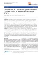
(A) Amplification of standards showing fluorescence versus cycle number concentration of 10
7
– 10
1
molecules are shown from left to rightFigure 1
(A) Amplification of standards showing fluorescence versus
cycle number concentration of 10
7
– 10
1
molecules are
shown from left to right. (B) Standard curve of GI assay R
2
1.00 and a slope of -3.8 was obtained. (C) Melting curve of GI
standards showing melting point at 90°C in descending order
10
7
– 10
1
molecules.
A
B
C
Table 1: Primers and probes used for Lightcycler RT-PCR and
Reverse Line Blot hybridization assay.
Primers Primer Sequence Reference
COG1F CGY TGG ATG CGN TTY CAT GA [7]
G1NVR ACC CAR CCA TTA TAC ATY TG
COG2F CAR GAR BCN ATG TTY AGR TGG ATG
AG
[7]
G2NVR ACC NGC ATA NCC RTT RTA CAT TC
Probes
GI/1 TCT TGC AAT GGA TCC TGT RGC RG
GI/2 GAA CCC GTG GCY GGG CCA AC
GI/3 CCA GAG GCA AAY ACA GCT GAG
GI/4 TGA CCC TGT GGC TGG CTC CTC
GI/5 ATG CTG AAC CAC TGC CWC TTG AT
GI/6 CAA ATT TCA ATG GAY CCT GTT GCG
GI/7 GGT AGT GGG CGC CGC AAC C
GI/8 TGC GGT TGC TAC TGC CGG CCA
GII/1 CGA GAC GAT GGC MCT CGA ACC G
GII/2 TAT AGA CCC TTG GAT TAG AGC A
GII/3 CAA TGG CGC TAG ABC CAG TGG CG
GII/4 GAC GCC AAC CCA TCT GAT GGG TC
GII/5 GGT GGG GGC GTC TTT AGC C
GII/6 CTC AAT CGC AGC TCC TGT YGT
GII/7 GCA TCG CTG GCG ACA CCA GTT G
GII/8 TCA ACC ATG AGG TCA TGG CCA TA
GII/9 CCC CGG GTG AGT TCT TGC TYG A
GII/10 TTC CCC TGG AGA AGT ACT CCT
GII/12 CGA ACT AAA TCC ATA CCT AGC ACAC
GII/13 CAG TGG CGG GAC AAA CCA AC
GII/14 CTC TCC TGG AGA ACT CCT ACT TGA T
GII/15 GAA GTC TTG CCT TTA GAG CCC GTC
GII/16 CAG TTG CGG GAG CTT CAA TCG CT
GII/17 CCT CCC TTT GGA ACC AGT TGC
Virology Journal 2007, 4:86 />Page 4 of 8
(page number not for citation purposes)
described here would be appropriate as a broad range
detection method for NoV infection.
The real-time assay was used to detect and quantify the
presence of 26 NoV positive samples from 56 samples
obtained from two Irish hospitals. The average titre of
virus per gram of stool was found to be 1.02 × 10
9
mole-
cules with the range of titre running from 2.67 × 10
6
per
gram of stool to 7.53 × 10
9
molecules per gram of stool.
These high levels are consistent with numbers reported in
other studies of NoV levels in stools [7,10].
The basis of this quantification was on assumption of
100% RT efficiency. This method allows a calculation of
the minimum amount of NoV present based on cDNA
values. Ideally quantification of an RNA virus involves the
use of an RNA standard. However, RNA standards are not
very stable, thus making standard curve construction dif-
ficult. It is more practical to use plasmids for the construc-
tion of an external standard curve. A recommendation to
this problem would be the generation of an armoured
RNA control for both GI and GII NoV similar to those
available for Hepatitis C [11].
The genotyping of positive NoV obtained from outbreaks
is usually performed by direct sequencing of the PCR
products, a time consuming process. A first generation
line-probe assay was created for genotyping NoV based on
the highly conserved ORF1-ORF2 region. The primer pair
described in this paper contains sufficient sequence varia-
bility between the primer binding sites to allow the design
of specific probes for each genotype. The assay was vali-
dated using the stool panel of 14 different NoV genotypes.
It was found that at an annealing temperature of 57°C,
both the GI and GII probes bound specifically to their
genotypes present in the panel with the exception of GII/
2 which also binds GII/6 (Fig. 3). Analysis of multiple
sequences of GII/2 revealed that it was not possible to
design a probe which would not bind GII/6. Therefore, it
is not possible using this assay to differentiate GII/2 and
GII/6 in an unknown sample.
No cross reactivity was observed between the probes GI/5,
GI/7, GI/8 GII/1, GII/5, GII/7, GII/9, GII/13, GII/14, GII/
15 with the genotypes present in the stool panel (Fig. 3).
The lack of cross reactivity allows these probes to be left
on the membrane and as they are designed to anneal to
PCR products at the same temperature as the validated
probes they should detect their corresponding genotypes
in unknown samples.
Applying this assay to the 14 positives from the 2005 out-
break and the 12 positives from the 2006 outbreak (Fig.
4A and 4B) detected by Real-time RT-PCR revealed that all
26 of the samples were genotype GII/4. This result was
confirmed by sequencing of the RT-PCR products. The
predominance of the GII/4 genotype is consistent with
previous sequence analysis of Irish NoV isolates [12,13]
and with that of other NoV circulating globally [14-17].
This paper describes both a rapid, sensitive and broadly
reactive method for detecting and genotyping Human
NoV. As the same sets of primers are used for both assays
the combination of both methods greatly speeds up ascer-
taining when a NoV outbreak is occurring and which
strain is responsible. A SYBR green mastermix containing
a proof reading enzyme would again speed up the typing
process as this would allow the RT-PCR products to be
applied directly to the membrane by allowing biotin
primers to be used in the initial detection of NoV. Assays
like these have been developed for Human papilloma-
vrius [18]. The genotyping assay would also be useful for
(A) Amplification of GII standards showing fluorescence ver-sus cycle number concentration of 5 × 10
7
– 5 × 10
1
mole-cules are shown from left to rightFigure 2
(A) Amplification of GII standards showing fluorescence ver-
sus cycle number concentration of 5 × 10
7
– 5 × 10
1
mole-
cules are shown from left to right. (B) Standard curve of GII
assay R
2
1.00 and a slope of -3.7 was obtained. (C) Melting
curve of GII standards showing melting point at 88°C in
descending order 5 × 10
7
– 5 × 10
1
molecules.
A
B
C
Virology Journal 2007, 4:86 />Page 5 of 8
(page number not for citation purposes)
investigating outbreaks associated with water or oysters as
it would allow typing of possible mixed infections which
may occur due to the nature of both these contaminants.
Future development of this assay would involve develop-
ing a disposable Line-probe assay based on these probes
similar to those commercially available combined with
automation to further improve the timeframe for geno-
typing a NoV infection.
Conclusion
A real time RT-PCR assay and a RLBH assay were devel-
oped and utilised to identify and genotype the causative
agent of two gastroenteritis outbreaks in two Irish hospi-
tals. The amount of Nov present in infected stool samples
was estimated and the strain of NoV responsible for all
positive cases was genotyped as the GII/4 variant.
Methods
Clinical specimens
A reference panel of various genotypes of NoV was
acquired between January 2003 and December 2006 to
determine the broad reactivity of the primers and probes.
The panel was transported on dry ice and remained frozen
at -20°C until processing This panel was screened by RT-
PCR and the products were TA cloned (Invitrogen) (Table
2). Fifty six stool samples were collected from both Water-
ford Regional hospital and the Mercy Hospital Cork from
January 2004 to March 2006 and stored at 4°C prior to
processing.
Primer and probe design
A multiple alignment was performed using the MEGA-
LIGN programme (DNASTAR). Thirty sequences of GI
and 120 sequences of GII were aligned using this pro-
gram. COG1F and COG2F primers described by [7] were
chosen as forward primers for GI and GII assays respec-
tively. Two reverse primers were designed based on these
alignments and denoted G1NVR and G2NVR (Table 1).
Eight oligonucleotide probes were designed for the detec-
tion of GI NoV and 17 probes were designed for the detec-
tion of GII NoV. The probes were designed based on the
criteria that they were at least 20 nucleotides in length and
that they had a Tm of at least 60°C. The probes were 5'
hexylamine labelled (Operon, Germany).
Extraction of viral RNA
Stools were diluted in a 10% (w/v) Modified Eagles
Medium (Gibco). The suspension was centrifuged at
10000 rpm for 10 min and 200 µl supernatant was
applied to the High Pure Viral nucleic extraction kit
(Roche). The extracted RNA was DNAse treated using
RNAse free DNAse (Ambion).
Reverse Transcription (RT)
RT was performed using a Superscript II Reverse tran-
scriptase kit (Invitrogen™) to a final volume of 20 µl. 10 µl
of extracted RNA and 1 µl of 75 pmole random hexamers
(Roche) are added to a 0.5 ml PCR reaction tube, mixed
and heated to 95°C for 3 min. A master-mix was prepared
according to the manufacturer's instructions and incu-
bated as directed.
Detection of Norovirus
NoV was detected by a Lightcycler assay (Roche Applied
Science) designed in our laboratory based on the COG1F-
GINVR or COG2F-G2NVR primers (Table 1). Quantita-
tive RT-PCR was performed using the LightCycler
®
Fast-
Table 2: Stool panel acquired by this group 2003–2005.
Norovirus panel Norovirus strain Source
Genogroup I
GI/I Hu/NoV/West Chester/2001/USA CDC, USA
GI/2 Hu/NV/SHV/1993UK UK
GI/3 Hu/NV/Stav/1999/Nor Irish isolate
GI/4 Hu/Nv/Saitama T69GI/2002/JP CDC, USA
GI/6 Hu/Nv/Saitama T44GI/2001/JP CDC, USA
Genogroup II
GII/2 Hu/NoV/Melksham/2001/USA CDC, USA
GII/3 Hu/NoV/VannesL169/2000/France HPA, UK
GII/4 Hu/NLV/GII/Carlow/2002/Irl Irish isolate
GII/6 Hu/NoV/SU4-JPN/2002/JP CDC, USA
GII/8 Hu/NoV/Saitama T67GII/2002/JP HPA, UK
GII/10 Hu/NoV/Mc37/2004/JP CDC, USA
GII/12 Hu/NoV/Honolulu/314/1994/US CDC, USA
GII/16 Hu/NoV/Hiram/2000/USA CDC, USA
GII/17 Hu/NoV/CS-E1/2002/USA CDC, USA
CDC : Centres for Disease control
HPA: Health Protection agency
Virology Journal 2007, 4:86 />Page 6 of 8
(page number not for citation purposes)
Start DNA Master SYBR Green I (Roche). A final reaction
volume of 20 µl containing 0.5 µl of cDNA, 2 µl of SYBR
green Mastermix, 2.8 µl of 25 mM MgCl
2
(3.5 mM), 1 µl
of each primer (0.6 µM) and 12.7 µl of PCR grade water.
The reaction was performed using GI primers with a dena-
turation step of 94°C for 8 min followed by 40 cycles at
94°C for 10 s, 45°C for 10 s 72°C for 15 s and a fluores-
cent read step of 85°C for 15 s to melt primer dimers.
The reaction was performed using GII primers with a
denaturation step of 94°C for 8 min followed by 45 cycles
of 94°C for 5 s, 52°C for 10 s, 72°C for 17 s and a fluo-
rescent read step of 84°C for 10 s to melt primer dimers.
For the creation of standard curves, 2 µl containing dilu-
tions of 10
7
to 10
1
molecules of GI/2 plasmid or 5 × 10
7
to
5 × 10
1
molecules of GII/4 plasmid DNA were added to
the reaction tubes. All reactions were run with negative
controls and subjected to melting curve analysis.
Biotinylated RT-PCR
Biotinylated reverse primers G1NVR and G2NVR synthe-
sized by MWG Biotech (Ebersberg, Germany) were used
in the following RT-PCR assay at a final volume of 50 µl.
The reaction contained 4 µl of cDNA from the RT reaction,
5 µl of 10× PCR Buffer, 1.5 mM MgCl
2
, 1 µl of 10 mM
each of dATP, dCTP, dGTP, and dTTP per reaction, 1 µM
each of primers and 2.5 units of Platinum Taq polymerase
(Invitrogen) was performed on a MJ PTC-200 thermocy-
cler (MJ research). The reaction was performed for GI with
a denaturation step at 94°C for 3 min, followed by 40
cycles at 94°C for 1 min, 45°C for 1 min, 68°C for 1 min
and a final extension at 68°C for 7 min. The PCR for GII
was a denaturation at 94°C for 3 min, followed by 40
cycles at 94°C for 30 s, 48°C for 30 s, 72°C for 1 min and
a final extension at 72°C for 7 min. The PCR products
were separated on a 2% agarose gel and visualized by
ethidium bromide staining. The PCR products for Geno-
group I and Genogroup II were 387 bp and 378 bp in
length, respectively.
Reverse Line Blot Hybridization
Twenty-five oligonucleotide probes each corresponding
to a GI or GII genotype (Table 2) which were synthesized
with a 5' hexylamino group (Operon Biotechnologies Ltd,
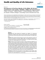
Genotyping of the (A) 14 positive samples from the 2005 outbreak and (B) 12 positive samples from the 2006 out-breakFigure 4
Genotyping of the (A) 14 positive samples from the 2005
outbreak and (B) 12 positive samples from the 2006 out-
break. Control samples containing known RT-PCR products
from the reference panel are shown on the left binding to
there respective probes. Clinical samples binding to probe
GII/4 can be seen on the right of both A and B.
A
B
Probe
GII/2
GII/3
GII/4
GII/6
GII/8
Probe
GII/2
GII/3
GII/4
GII/6
GII/8
Validation of Reverse Line Blot hybridization using stool panel samplesFigure 3
Validation of Reverse Line Blot hybridization using
stool panel samples. Left of diagram indicates where the
25 probes are fixed across the membrane Top of the figure
indicates where denatured PCR products of stool panel have
been applied. Presence of spot indicates probe binding. Gaps
between spots indicate unbound probes for which no refer-
ence samples are available. Lane 1 : GI/1, 2 : GI/2, 3 : GI/3, 4 :
GI/4, 5 : GI/6, 6 : GII/2, 7 : GII/3, 9 : GII/6, 10 : GII/8, 11 : GII/
10, 12 : GII/12, 13 : GII/16, 14 : GII/17.
Probe
GI/1
GI/2
GI/3
GI/4
GI/5
GI/6
GI/7
GI/8
Probe
GII/1
GII/2
GII/3
GII/4
GII/5
GII/6
GII/7
GII/8
GII/9
GII/10
GII/12
GII/13
GII/14
GII/15
GII/16
GII/17
Lane 1 2 3 4 5 6 7 8 9 10 1112 13 14
Virology Journal 2007, 4:86 />Page 7 of 8
(page number not for citation purposes)
Cologne, Germany). The oligonucleotides were cova-
lently bound to a negatively charged nylon membrane
(Biodyne C; Pall Biosupport, Portsmouth, Cambridge,
United Kingdom) by this 5' hexylamino group. Briefly,
the carboxyl groups on the membrane were activated by
incubation for 10 min in 16% (w/v) 1-ethyl-3-(3-dimeth-
ylaminopropyl) carbodimide (EDAC) (Sigma). The mem-
brane was washed with tap water and placed in a
miniblotter system (MN45; Immunetics, Cambridge,
Massachusetts). The slots were filled in parallel with 150
µl of each of the 5'-hexylamine-labeled oligonucleotides
at a final concentration of 1 µM diluted in freshly pre-
pared 0.5 M NaHCO3 [pH 8.4] and after 1 min of incuba-
tion at room temperature, the excess solution was
aspirated and the membrane was removed from the min-
iblotter. The remaining active esters on the membrane
were hydrolyzed by incubation in 0.1 M NaOH for 8 min
at room temperature and rinsed in water. The membrane
was washed twice for 5 min at 60°C in 2 × SSPE (Sigma)
with 0.1% sodium dodecylsulfate (SDS) (BDH, Poole,
United Kingdom). The membrane was used immediately
or washed for 15 min in 20 mM EDTA and stored sealed
in plastic at 4°C.
Prior to use in hybridization, the membrane was washed
for 5 min in 2 × SSPE-0.1% SDS, placed in the miniblot-
ter. The membrane was rotated so that the probes were
perpendicular to the previous position. 15 µl of each PCR
product in 135 µl of 2 × SSPE-0.1% SDS was denatured by
heating to 99°C for 10 min and chilled on ice. The slots
were then filled with 150 µl of PCR product and incubated
for 60 min at 57°C in a hybridization oven. After hybrid-
ization, unbound PCR product was removed by washing
twice with prewarmed 2 × SSPE-0.5% SDS at 60°C for 10
min. The membrane was then incubated at 42°C in 10 ml
of 1:2000 dilution of streptavidin-peroxidase conjugate
(Roche) in prewarmed 2 × SSPE buffer for 1 hr. Unbound
streptavidin-conjugate was removed by washing twice
with 2 × SSPE-0.5% SDS at 42°C for 10 min. lastly the
membrane was washed twice with 2 × SSPE at room tem-
perature for 5 min to remove SDS.
The bound PCR products were detected by a chemilumi-
nescence assay using ECL detection liquid (Roche) and
visualized by exposure of the blot for 10 min to 3 hrs to
an X-ray film (Hyperfilm; Amersham). For repeated use,
the membranes were stripped twice in 1% SDS at 80°C for
30 min and incubated for 15 min at room temperature in
20 mM EDTA solution, the membranes were sealed and
stored at 4°C until further use.
Abbreviations
Norovirus (NoV); Reverse transcription PCR (RT-PCR);
Genogroup I (GI); Genogroup II (GII); Reverse Line Blot
Hybridisation (RLBH).
Competing interests
The author(s) declare that they have no competing inter-
ests.
Authors' contributions
JFM is the corresponding author and main contributing
author of this manuscript. KK contributed to primer
design, the construction of the plasmids used for standard
curve generation and the final review of the manuscript.
Supervision and final review of the manuscript was pro-
vided by JGM. All authors have read and approved the
final manuscript.
Acknowledgements
We are most grateful to Dr. Stephen Monroe, Dr. Suzanne Beard, Dr. Kim
Green, Dr. Ian Clark and Dr Chris Gallimore and their various institutions
for providing us with a Norovirus reference panel. Funding provided by the
Food Institutional Research and Development Measure 3 (ii) Food Sub-Pro-
gramme (Dept. of Agriculture and Food, Republic of Ireland). Thank you to
Mr. Noel Shanaghy and Mrs. Breda Doody of Waterford Regional Hospital,
Mr. Eddie Beggan of Limerick Regional Hospital and Dr. Jim Clair of the
Mercy Hospital Cork for providing us with faecal samples.
References
1. Kapikian AZ: The discovery of the 27-nm Norwalk virus: an
historic perspective. J Infect Dis 2000, 181 Suppl 2:S295-302.
2. Saito H: [Epidemiology on Norwalk virus-related gastroen-
teritis outbreaks among elderly persons living in nursing
homes]. Nippon Rinsho 2002, 60(6):1148-1153.
3. Gallimore CI, Richards AF, Gray JJ: Molecular diversity of norovi-
ruses associated with outbreaks on cruise ships: comparison
with strains circulating within the UK. Commun Dis Public Health
2003, 6(4):285-293.
4. Blanton LH, Adams SM, Beard RS, Wei G, Bulens SN, Widdowson
MA, Glass RI, Monroe SS: Molecular and epidemiologic trends
of caliciviruses associated with outbreaks of acute gastroen-
teritis in the United States, 2000-2004. J Infect Dis 2006,
193(3):413-421.
5. Lang L: Acute gastroenteritis outbreaks on cruise ships linked
to Norwalk-like viruses. Gastroenterology 2003, 124(2):284-285.
6. Zheng DP, Ando T, Fankhauser RL, Beard RS, Glass RI, Monroe SS:
Norovirus classification and proposed strain nomenclature.
Virology 2006, 346(2):312-323.
7. Kageyama T, Kojima S, Shinohara M, Uchida K, Fukushi S, Hoshino FB,
Takeda N, Katayama K: Broadly reactive and highly sensitive
assay for Norwalk-like viruses based on real-time quantita-
tive reverse transcription-PCR. J Clin Microbiol 2003,
41(4):1548-1557.
8. Kojima S, Kageyama T, Fukushi S, Hoshino FB, Shinohara M, Uchida
K, Natori K, Takeda N, Katayama K: Genogroup-specific PCR
primers for detection of Norwalk-like viruses. J Virol Methods
2002, 100(1-2):107-114.
9. O'Neill HJ, McCaughey C, Wyatt DE, Mitchell F, Coyle PV: Gastro-
enteritis outbreaks associated with Norwalk-like viruses and
their investigation by nested RT-PCR. BMC Microbiol 2001,
1:14.
10. Pang X, Lee B, Chui L, Preiksaitis JK, Monroe SS: Evaluation and
validation of real-time reverse transcription-pcr assay using
the LightCycler system for detection and quantitation of
norovirus. J Clin Microbiol 2004, 42(10):4679-4685.
11. WalkerPeach CR, Winkler M, DuBois DB, Pasloske BL: Ribonucle-
ase-resistant RNA controls (Armored RNA) for reverse
transcription-PCR, branched DNA, and genotyping assays
for hepatitis C virus. Clin Chem 1999, 45(12):2079-2085.
12. Waters A, Coughlan S, Dunford L, Hall WW: Molecular epidemi-
ology of norovirus strains circulating in Ireland from 2003 to
2004. Epidemiol Infect 2006, 134(5):917-925.
13. Foley B, O'Mahony J, Hill C, Morgan JG: Molecular detection and
sequencing of "Norwalk-like viruses" in outbreaks and spo-
Publish with BioMed Central and every
scientist can read your work free of charge
"BioMed Central will be the most significant development for
disseminating the results of biomedical research in our lifetime."
Sir Paul Nurse, Cancer Research UK
Your research papers will be:
available free of charge to the entire biomedical community
peer reviewed and published immediately upon acceptance
cited in PubMed and archived on PubMed Central
yours — you keep the copyright
Submit your manuscript here:
/>BioMedcentral
Virology Journal 2007, 4:86 />Page 8 of 8
(page number not for citation purposes)
radic cases of gastroenteritis in Ireland. J Med Virol 2001,
65(2):388-394.
14. Lynch M, Painter J, Woodruff R, Braden C: Surveillance for food-
borne-disease outbreaks United States, 1998-2002. MMWR
Surveill Summ 2006, 55(10):1-42.
15. Vainio K, Myrmel M: Molecular epidemiology of norovirus out-
breaks in Norway during 2000 to 2005 and comparison of
four norovirus real-time reverse transcriptase PCR assays. J
Clin Microbiol 2006, 44(10):3695-3702.
16. Koopmans M, Harris J, Verhoef L, Depoortere E, Takkinen J, Coulom-
bier D: European investigation into recent norovirus out-
breaks on cruise ships: update. Euro Surveill 2006, 11(7):E060706
5.
17. Kearney K, Menton J, Morgan JG: Carlow Virus, a 2002 GII.4 var-
iant Norovirus strain from Ireland. Virol J 2007, 4(1):61.
18. Payan C, Ducancelle A, Aboubaker MH, Caer J, Tapia M, Chauvin A,
Peyronnet D, Le Hen E, Arab Z, Legrand MC, Tran A, Postec E, Tour-
men F, Avenel M, Malbois C, De Brux MA, Descamps P, Lunel F:
Human papillomavirus quantification in urine and cervical
samples by using the Mx4000 and LightCycler general real-
time PCR systems. J Clin Microbiol 2007, 45(3):897-901.