Báo cáo hóa học: " Dengue virus serotype infection specifies the activation of the unfolded protein response" pdf
Bạn đang xem bản rút gọn của tài liệu. Xem và tải ngay bản đầy đủ của tài liệu tại đây (480.21 KB, 10 trang )
BioMed Central
Page 1 of 10
(page number not for citation purposes)
Virology Journal
Open Access
Research
Dengue virus serotype infection specifies the activation of the
unfolded protein response
Indira Umareddy
1
, Olivier Pluquet
2
, Qing Yin Wang
1
,
Subhash G Vasudevan
1
, Eric Chevet
2
and Feng Gu*
1
Address:
1
Novartis Institute for Tropical Diseases, 10-Biopolis Road, #05-01 Chromos, 138670, Singapore and
2
Team AVENIR, GREF INSERM
U899, IFR66, Université Victor Segalen Bordeaux 2, 146 rue Léo Saignat, 33076 Bordeaux, France
Email: Indira Umareddy - ; Olivier Pluquet - ;
Qing Yin Wang - ; Subhash G Vasudevan - ; Eric Chevet - eric.chevet@u-
bordeaux2.fr; Feng Gu* -
* Corresponding author
Abstract
Background: Dengue and Dengue hemorrhagic fever have emerged as some of the most
important mosquito-borne viral diseases in the tropics. The mechanisms of pathogenesis of Dengue
remain elusive. Recently, virus-induced apoptosis mediated by the Unfolded Protein Response
(UPR) has been hypothesised to represent a crucial pathogenic event in viral infection. In an
attempt to evaluate the contribution of the UPR to virus replication, we have characterized each
component of this signalling pathway following Dengue virus infection.
Results: We find that upon Dengue virus infection, A549 cells elicit an UPR which is observed at
the level of translation attenuation (as visualized by the phosphorylation of eIF2alpha) and activation
of specific pathways such as nuclear translocation of ATF-6 and splicing of XBP-1. Interestingly, we
find that specific serotype of virus modulate the UPR with different selectivity. In addition, we
demonstrate that perturbation of the UPR by preventing the dephosphorylation of the translation
initiation factor eIF2alpha using Salubrinal considerably alters virus infectivity.
Conclusion: This report provides evidence that Dengue infection induces and regulates the three
branches of the UPR signaling cascades. This is a basis for our understanding of the viral regulation
and conditions beneficial to the viral infection. Furthermore, modulators of UPR such as Salubrinal
that inhibit Dengue replication may open up an avenue toward cell-protective agents that target
the endoplasmic reticulum for anti-viral therapy.
Background
Dengue virus (DENV) is a member of the Flaviviridae fam-
ily, which include West Nile virus (WNV), yellow fever
virus, Japanese encephalitis virus (JEV), and tick-borne
encephalitis virus (TBEV), among others [1]. Dengue is
caused by four antigenically distinct viruses designated as
Dengue virus type 1–4 (DENV 1–4) and is transmitted
between vertebrate hosts by insect vectors. The most seri-
ous manifestations of the infection are Dengue hemor-
rhagic fever (DHF) and Dengue shock syndrome (DSS).
No effective vaccine or antiviral drug therapy is currently
available against Dengue viruses. The genome of Dengue
virus consists of a single stranded, non segmented, posi-
tive sense ribonucleic acid (RNA) of about 11 kb in length
Published: 24 September 2007
Virology Journal 2007, 4:91 doi:10.1186/1743-422X-4-91
Received: 22 May 2007
Accepted: 24 September 2007
This article is available from: />© 2007 Umareddy et al; licensee BioMed Central Ltd.
This is an Open Access article distributed under the terms of the Creative Commons Attribution License ( />),
which permits unrestricted use, distribution, and reproduction in any medium, provided the original work is properly cited.
Virology Journal 2007, 4:91 />Page 2 of 10
(page number not for citation purposes)
[1]. The genome is translated into a single polypeptide
which is co- and post-translationally processed by host
signalases as well as the virus encoded serine protease into
the three structural and seven non structural proteins (NS)
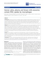
in the order C-prM-E-NS1-NS2A-NS2B-NS3-NS4A-NS4B-
NS5 that traverse the Endoplasmic Reticulum (ER) mem-
brane (Fig. 1). Dengue and other flaviviruses are thought
to replicate in the cytoplasm, mature on intracellular
membranes and egress by exocytosis and in some cases by
budding at the plasma membrane [2]. The host ER is the
primary site of envelope glycoprotein biogenesis,
genomic replication, and particle assembly of flaviviruses.
In the course of productive infection, flaviviruses induce
proliferation and hypertrophy of the ER membranes [3-
5]. Moreover, a large amount of flaviviral proteins are syn-
thesized in infected cells, thus overwhelming the ER fold-
ing capacity. As a natural consequence, we hypothesize
that these events will lead to the activation of the ER stress
response which in turn will modulate various signaling
pathways resulting in cell survival or death decisions.
In mammalian cells, the ER stress response, also called the
Unfolded Protein Response (UPR) is mediated by three
transmembrane proteins that act as sensors: i) the protein
kinase-like ER resident kinase (PERK), ii) the activating
transcription factor 6 (ATF6) and iii) the inositol-requir-
ing enzyme 1 (IRE1) [6]. The activation of PERK and IRE1
is driven by their oligomerization followed by their trans-
auto phosphorylation. Activated PERK phosphorylates
the eukaryotic initiation factor eIF2α thus resulting in
translation attenuation. This is also accompanied by the
activation of negative feed-back transcriptional loops.
This includes the up-regulation of the pro-apoptotic
mRNA CHOP and that encoding GADD34 whose associ-
ation with the phosphatase PP1 leads to the dephosphor-
ylation of eIF2α [6]. Activated IRE-1 cleaves an
unconventional 26-nucleotides intron from X-Box bind-
ing Protein-1 (XBP-1) mRNA which leads to a change in
the open reading frame and leads to the translation of an
active transcription factor [7]. The spliced form encoded
XBP-1 protein (sXBP-1) is involved in the transcriptional
activation of a number of genes including the ER man-
nosidase-like protein EDEM which is involved in protein
degradation. In parallel, upon accumulation of mis-
folded proteins in the ER, ATF-6 exits this compartment to
migrate to the Golgi apparatus where it is cleaved by S1P
and S2P proteases [8]. ATF-6 cytosolic fragment is an
active transcription factor responsible for transcriptional
induction of XBP-1 as well as many ER chaperone encod-
ing genes (reviewed in [6,9]).
Several studies have shown that in some cases virus infec-
tions activate the three branches of the UPR. For instance
the UPR master regulator – BiP is induced in cells infected
with Respiratory syncytial virus [10], hanta viruses [11],
hepatitis C viruses [12] as well as flaviviruses such as cyto-
pathic strains of BVDV [13] and JEV [14]. Activation of
PERK has also been reported in infection with herpes sim-
Dengue viral polyprotein and its predicted membrane topologyFigure 1
Dengue viral polyprotein and its predicted membrane topology. Schematic representation of the membrane topology
of the proteins and their cleavage by host (red and blue arrows) or viral (black arrows) proteases. The 11 kb genome of Den-
gue is translated into a single polypeptide and this polyprotein traverses the ER membrane at several positions. prM, E, NS1
and a part of NS4A and NS4B are thought to localise to the ER lumen via hydrophobic signal sequences whereas the remaining
proteins are thought to be localized on the cytoplasmic side of the ER membrane.
NS3/2B Furin Signalase
Virology Journal 2007, 4:91 />Page 3 of 10
(page number not for citation purposes)
plex virus [15,16], cytomegalovirus [17] and BVDV [13].
The IRE1-XBP1 axis has been recently shown to be acti-
vated in cells infected with JEV and Dengue [18] whereas
the ATF-6 pathway has been reported to be activated upon
HCV infection [19]. It is also becoming increasingly evi-
dent that many viruses have evolved mechanisms to cope
with UPR response or to utilize it to their benefit. Indeed,
the herpes simplex virus genome encodes a GADD34
homolog – γ
1
34.5 protein which leads to the dephosphol-
yation of eIF2α and overcomes the PERK response
[15,20]. The African swine fever virus overcomes the tran-
scriptional activation of CHOP induced by Thapsigargin
[21] and cytomegalovirus overcomes translation inhibi-
tion despite activation of eIF2α phosphorylation [22].
In recent years it has become clear that the ability of
viruses to regulate cellular responses to infection is a key
determinant for the physiological consequences of infec-
tion. Since the activation of the UPR is on the one hand
essential for cell survival during viral infection and on the
other hand detrimental to viral replication, it is therefore
thought that a balance between the two would determine
the outcome of the infection in host cells. Consequently,
it may be advantageous for viruses to modulate the UPR
to its advantage. For instance, replication of hepatitis C
virus has been shown to stimulate the ATF6 pathway [19],
but attenuate the IRE1-XBP1 pathway [19]. Since UPR
induction upon Dengue infection is currently under-
investigated, the aim of this study was to determine the
UPR characteristics under those circumstances. The under-
standing of such response would also yield critical infor-
mation to control Dengue infection.
Results
Dengue infection induces phosphorylation of eIF2
α
Upon virus infection, eukaryotic cells respond in part by
shutting-down translation. This process is mediated by
the phosphorylation of the translation initiation factor
eIF2α [23]. To determine whether eIF2α is phosphor-
ylated upon Dengue infection, A549 cells were infected
for 6 to 72 hours with DENV2 (TSV01) or DENV1 (MY
10245) viruses and harvested at indicated time points
post-infection (Fig. 2). Cell lysates were first analyzed by
immunoblot using an antibody against phospho-eIF2α.
In both cases of Dengue infection, phosphorylated forms
of eIF2α were detected at 24 h post-infection, and accu-
mulated until 72 h (Fig. 2A). This indicates that eIF2α
kinases such as PERK or PKR are activated upon infection
of A549 cells by Dengue virus. Interestingly, by using an
antibody against total eIF2α we showed that eIF2α pro-
tein expression levels increased at 24 h post-infection and
remained elevated up to 72 h compared to mock-infected
cells (Fig. 2A), while thapsigargin (TG, our ER stress posi-
tive control) treatment alone did not modify total eIF2α
protein levels (data not shown). This result suggests that
Dengue virus might be able to overcome or compensate
the UPR response by inducing more elF2α protein for
translation. This is further supported by a recent study
which showed that translation is not attenuated by Den-
gue infection [24] although eIF2α is phosphorylated. The
overall ratio of phospho elF2α and elF2α is quantified for
both DENV 1 and DENV 2 infection (Fig. 2B). The two
serotypes of Dengue showed similar pattern of peak phos-
phorylation at 48 hours, with DENV2 infection slightly
stronger than DENV1.
Dengue infection promotes GADD34 expression
As a recent study showed that translation is not attenuated
by Dengue infection [24] although eIF2α is phosphor-
ylated, we consequently asked whether the regulatory sub-
unit of protein phosphatase (GADD34) that
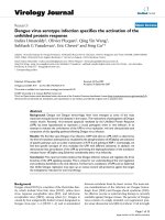
Dengue infection induces phosphorylation of eIF2αFigure 2
Dengue infection induces phosphorylation of eIF2α.
(A) A549 cells were infected with DENV1 or DENV2
viruses at 10 MOI and lysed at indicated time points in lysis
buffer. Protein extracts were subjected to immunoblot analy-
sis with anti-phosphoserine51-eIF2α (top panel), and anti-
eIF2α (bottom panel) antibodies. (B) Phosphoserine51-
eIF2α levels from (A) were quantified and normalized to
eIF2α levels and plotted.
Mock
eIF2α
P-Ser51
-eIF2α
DEN2
DEN1
DEN2
DEN1
DEN2
DEN1
DEN2
DEN1
6h
24h 48h 72h
eIF2α phosphorylation
(Relative intensity)
Hours post-infection
A
B
0
5
10
15
20
0 1224 36486072
DEN1
DEN2
Virology Journal 2007, 4:91 />Page 4 of 10
(page number not for citation purposes)
dephospholyates eIF2α was specifically induced upon
Dengue infection. To this end, A549 cells were infected as
before and RNA extracted for RT PCR analysis to measure
GADD34 mRNA expression. TG, a well-recognized
inducer of ER stress, served as a positive control in these
assays (Fig. 3A). GADD34 mRNA expression levels were
quantified and normalized to actin mRNA levels (Fig. 3B).
When compared to mock-infected cells, Dengue infection
induced the expression of GADD34 at 24 hours post-
infection (Fig. 3A and 3B) most likely to compensate for
the induction of eIF2α phosphorylation. Interestingly,
infection by DENV1 was a more potent inducer of
GADD34 expression than DENV2 (Fig. 3A and 3B). In
order to test whether this was caused by different replica-
tion of the two viruses, we compare the growth of the two
viruses by plaque assay. Figure 3C shows that the DENV2
(MY10245) strain indeed produced slightly more virus at
each time point than the DENV1 (TSV01) strain, but the
two viruses grew at similar rate in A549 cells. Further-
more, it was noted that at 6 h post-infection, GADD34
mRNA expression level was lower than uninfected. This
result may be explained by a virus-induced consumption
of GADD34 mRNA through a translation-dependent
process. These data suggested that Dengue infection led to
increased elF2α phosphorylation, as well as the activation
of downstream feedback loop controlling its dephospho-
rylation, in a strain dependent manner.
Dengue infection activates the ATF6 pathway
We next examined whether the ATF6 pathway was acti-
vated upon Dengue infection. In response to ER stress,
ATF6 exits the ER to traffic to the Golgi apparatus, where
it is processed to its active form which then translocates to
the nucleus. We assessed the presence of this nuclear form
of ATF6 as a marker of its activation during infection by
Dengue virus. To this end, GFP tagged ATF6 was trans-
fected into A549 cells which then were infected with 10
moi of DENV2 (NGC) 24 hours after transfection. The
cells were fixed in cold methanol and immuno-fluores-
cence was performed using anti-E protein antibodies.
ATF6 localized to cytoplasmic ER-like structures in mock
infected cells whereas an intense signal was observed in
the nucleus in DENV2 infected cells. These results suggest
that Dengue virus activates the ATF6 pathway of the UPR.
We next confirmed ATF6 activation by assessing ATF6-
dependent transcriptional activation of XBP1 gene [7,25].
We measured total XBP1 mRNA in Dengue-infected cells
and TG-treated cells by RT-PCR (Fig. 4B). The level of
XBP1 mRNA significantly increased in Dengue-infected
cells at 24 h post-infection. We noticed that DENV2 infec-
tion showed a higher effect on XPB1 mRNA than DENV1.
As expected, TG treatment led to a 3 fold increase in the
amount of XBP1 mRNA compared to control, thus con-
firming ATF6 activation upon Dengue infection. In addi-
tion we detected the XBP-1 hybrid form as previously
reported [26,27].
Dengue infection activates the XBP1 pathway
Upon ER stress, IRE1 processes XBP1 mRNA to result in an
unconventional splicing of a 26-nucleotides intron and a
translational frame shift. The spliced form of XBP1 is
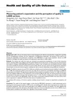
Dengue infection induces the upregulation of GADD34 mRNAFigure 3
Dengue infection induces the upregulation of
GADD34 mRNA. (A) A549 cells were either treated with
increasing concentration of thapsigargin (TG) for 1 hour or
infected with DENV2 or DENV1 viruses (10 MOI) at indi-
cated time points. GADD34 mRNA (top panel) and β-actin
mRNA (bottom panel) levels were determined by semi-quan-
titative RT-PCR with specific primers (see Materials and
Methods). (B) Densitometric quantification of GADD34
mRNA levels from (A) were normalized to β-actin mRNA
levels and plotted in histograms (TG) or graphs (DEN1 and
2). The ratio of the GADD34 to β-actin of the uninfected
(Mock) sample was considered as basal level (0 hour) and
negative value was also represented as basel level. (C) A549
cells were infected with either 1 moi of DENV1 (MY10245)
or DENV2 (TSV01). Virus production after Day 1, Day 2 and
Day 3 of infection were quantified by plaque assay and
expressed by PFU/ml.
Gadd34
β-Actin
DEN2
DEN1
DEN2
DEN1
DEN2
DEN1
DEN2
DEN1
6h 24h 48h 72h
Mock
1 µMTg
2 µMTg
Hours post-infection
GADD34
(Relative intensity)
A
B
C
1
10
100
1000
10000
100000
1000000
10000000
Day 1 Day 2 Day 3
DEN2 (TSV01)
DEN1 (MY10245)
PFU/ml
0
1
2
3
4
5
6
7
8
Mock Tg (1) Tg (2)
0 122436486072
DEN1
DEN2
Virology Journal 2007, 4:91 />Page 5 of 10
(page number not for citation purposes)
translated into a transcription factor. To determine
whether the IRE1 pathway is activated in DENV2 and
DENV1 infected A549 cells, we analyzed the splicing of
XBP1 mRNA by RT-PCR using specific primers (Fig. 5). In
mock-infected cells, only the unspliced form of XBP1
mRNA (uXBP1) was detected. In Dengue infected cells,
the spliced form of XBP1 mRNA (sXBP1) was detected 48
h post-infection as well as in our positive control with TG.
We noticed a hybrid form of XBP1 mRNA (hXBP1) upon
treatment with 2 µM TG and upon Dengue infection (24
h post-infection and thereafter) [26,27]. We also noticed
that cells infected with DENV2 seemed to express higher
levels of XBP1 than mock-infected cells or DENV1
infected cells. These results clearly showed that XBP1
splicing is induced by Dengue replication and that Den-
gue virus activates the IRE-1/XBP-1 pathway of the UPR.
Attenuation of eIF2
α
dephosphorylation modulates
dengue replication
We have demonstrated that upon Dengue infection, all
three branches of the UPR are activated as a host response.
We next determined whether the modulation of the UPR
could have any impact on virus growth. Salubrinal, a
selective inhibitor of the protein complex (containing the
protein phosphatase 1 and its cofactor GADD34) that
dephosphorylates eIF2α was tested in our Dengue virus
infection assay. Salubrinal has previously been reported to
inhibit the replication of HSV [28]. We first tested the
impact of Salubrinal on cellular toxicity. In our assay,
Salubrinal was non-toxic at doses < 5 µM, CC50 (50% of
cell cytotoxicity) was determined to reach approximately
10 µM in A549 cells (Fig. 6B). A549 cells were then treated
with Salubrinal for one hour prior infection with DENV2
virus. Forty-eight hours later, supernatants of infection
were collected and virus production was quantified by
plaque assay. Addition of Salubrinal one hour prior to
infection showed an eighty percent reduction of the virus
at 3.12 µM (Fig 6A). Similar results were found when
Salubrinal was added one hour post infection. (See Addi-
tional file 1). We then used a second method of viral
growth measurement which consists of staining virus
infected cells using antibodies against the Envelop protein
of dengue virus (see Material and Methods, Immunolabe-
ling Assay). Using this assay, we obtained seventy percent
inhibition of the virus at 5 µM, when Salubrinal was
added at the same time as infection (Fig 6B). Together,
these data showed that Salubrinal reduced Dengue virus
growth at micromolar concentrations when added 1 hour
before, at the same time or 1 hour post-infection (salubri-
nal remained for the whole course of infection), thus indi-
cating that modulation of the UPR, in this case, increase
of the elF2α phosphorylation significantly reduced Den-
gue virus infection.
Discussion
Many positive-strand RNA viruses need to modify intrac-
ellular membranes of their host cells in order to create a
compartment suitable for virus replication [29,30].
Although this phenomenon has been well documented,
little is known about the mechanisms triggered by viruses
to induce intracellular membrane proliferation. An
increasing amount of literature supports the hypothesis
that viruses like other ER stress signals may induce mem-
brane proliferation through the activation of specific com-
ponents of the Unfolded Protein Response [31,32]. These
observations are also supported by the occurrence of rep-
lication and maturation of flaviviruses in close association
with the host ER and the membrane rearrangements
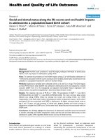
Dengue infection activates the ATF6 pathwayFigure 4
Dengue infection activates the ATF6 pathway. (A)
A549 cells were transiently transfected with GFP-ATF6 plas-
mid. After 24 h, the cells were left untreated or infected with
DENV2 virus at 10 MOI. Twenty four hours post-infection,
immunocytochemistry analysis was performed to detect
GFP-ATF6 (green), the viral E protein (red), and cell nuclei
were detected by DAPI staining (blue). (B) A549 cells were
either treated with thapsigargin (TG) or infected with
DENV1 and DENV2 and total mRNA was extracted and ana-
lyzed for XBP1 expression by RT-PCR as in Figure 5. The
XBP1 mRNA level was quantified by densitometry as the
total of all spliced forms of XBP1 and expressed as fold
increase compared to untreated cells (Ctrl).
E protein Merge
DEN2
Mock
GFP-ATF6
A
B
XBP-1 mRNA expression
(fold increase)
0
1
2
3
4
5
Ctrl
Mock
2 µM
Tg
DEN2 (6h)
DEN1 (6h)
DEN2 (24h)
DEN1 (24h)
DEN2 (48h)
DEN1 (48h)
DEN2 (72h)
DEN1 (72h)
Virology Journal 2007, 4:91 />Page 6 of 10
(page number not for citation purposes)
observed in the course of flavivirus-productive infection
[2]. Moreover, it has been shown that JEV [14], BVDV [13]
and HCV infections [33] induce the Unfolded Protein
Response. Consequently we initiated a study to character-
ize the UPR response to Dengue infection.
The phosphorylation of PERK has been used as an early
marker for ER stress [34]. Although we attempted to deter-
mine the phosphorylation status of PERK in Dengue-
infected A549 cells, we failed to see any PERK signals even
for positive control using the ER stress inducers TG and
DTT. Thus, the potential role of PERK activation in the
Dengue induced UPR is unclear from our present study.
However, microarray analyses described earlier showed
that different strains of DENV2 induced the expression of
PERK and PKR to a different extent (unpublished infor-
mation). Moreover, we could detect phospholyation of
PKR by DENV2 (data not shown) and Dengue virus
induced the phosphorylation of eIF2α in A549 cells. It is
therefore possible that both PKR and PERK kinases might
separately phosphorylate eIF2α in response to Dengue
infection.
Despite this phosphorylation event, translation is not
attenuated in Dengue virus infected cells [24]. We conse-
quently suspected that Dengue virus might activate a com-
pensatory pathway to prevent UPR-mediated translation
attenuation. Because of the deleterious effects of the host's
protein synthesis inhibition, many viruses have evolved
distinct mechanisms to counteract eIF2α phosphorylation
as a means to avoid, at least in part, the antiviral action of
interferons [35]. For instance, the γ
1
34.5 protein of herpes
simplex virus is highly homologous to GADD34 and has
been shown to alleviate translational arrest in cells treated
with TG and DTT [15]. In our study, RT PCR analyses of
GADD34 mRNA expression levels showed that Dengue
induces the expression of GADD34 at approximately the
same time as eIF2α phosphorylation is triggered. This led
us to postulate that Dengue virus may compensate the
eIF2α phosphorylation event by enhancing the expression
of GADD34 which leads to enhanced dephosphorylation
of eIF2α and consequently overcomes the block in trans-
lation. To corroborate our hypothesis, we analyzed the
antiviral effect of Salubrinal on Dengue. Salubrinal was
recently discovered as a small molecule inhibitor of the
protein complex (containing the protein phosphatase 1
and its cofactor GADD34) that dephosphorylates eIF2α
[28] and it has been shown to inhibit the replication of
HSV [28]. Using two methods to score viral growth, we
showed that Salubrinal dramatically reduced Dengue
infection. We therefore conclude that enhancing eIF2α
phosphorylation using Salubrinal helped the host cells to
increase the translational inhibition consequently leading
to reduced Dengue virus production.
The transcriptional activation of ATF6 is critical to the
UPR, since ATF6 induces gene expression products neces-
sary for protein refolding. Our data show that the ATF6
pathway is also activated by Dengue virus infection (Fig.
4A). Moreover, ATF6 has been shown to up regulate the
XBP1 is spliced in Dengue infected A549 cellsFigure 5
XBP1 is spliced in Dengue infected A549 cells. A549 cells were either treated with thapsigargin (TG) or infected with
DENV2 and DENV1 (10 MOI) and harvested at indicated time points. Total mRNA was extracted and analyzed with XBP1
primers (top panel) or β-actin primers (bottom panel) by semi-quantitative RT PCR. The PCR products were run on a 3% aga-
rose gel and the spliced (sXBP1), unspliced (uXBP1) and the hybrid (hXBP1) forms are shown. Thapsigargin was used as a pos-
itive control for induction of XBP1 splicing (sXBP1) and β-actin mRNA levels as loading control.
DEN2
DEN1
DEN2
DEN1
DEN2
DEN1
DEN2
DEN1
6h 24h 48h 72h
Mock
2 µMTg
Ctrl
hXBP-1
sXBP-1
uXBP-1
β-Actin
Virology Journal 2007, 4:91 />Page 7 of 10
(page number not for citation purposes)
level of XBP1 mRNA [7], which, once spliced by IRE1
plays a role in transcriptional activation. In our studies
XBP1 mRNA was increased upon Dengue infection (more
elevated in DENV2 infection as opposed to that of
DENV1) (Fig. 4B). This suggested first that Dengue sero-
types may selectively modulate ATF6 activation to either
inhibit aspects that could be deleterious to the progress of
the viral infection or enhance host's ability to favor it. It
would therefore be exciting to study the mechanism of
activation of ATF6 especially in terms of viral serotypes
and strains and its effect on virus infectivity. XBP1 also
plays a critical role in the UPR as it activates proteins of
the degradation pathway (EDEM) that target mis-folded
or unfolded proteins whereas the unspliced form results
in an inactive protein. Our data show that splicing of
XBP1 mRNA is triggered upon Dengue infection, consist-
ent with the activation of the IRE1 pathway (Fig. 5). Sim-
ilar results were reported earlier with JEV and DENV2
[18]. Furthermore, it was also reported that the XBP1
downstream genes such as EDEM1 and p58 (IPK) were
induced in Dengue infected cells [18]. Interestingly, trans-
activation of XBP1 target genes are suppressed in HCV
[19]. The discrepancy of XBP1 induction between these
viruses might reflect the differences in infection patterns
of these viruses; while HCV usually causes chronic infec-
tion, JEV and Dengue cause acute infection. It is notewor-
thy that knock down of XBP1 had no effect on viral
production suggesting that XBP1splicing is beneficial but
not essential for virus production. Nevertheless, increased
cytopathic effects were noticed in XBP1 knock out cells in
response to Dengue infection indicating that XBP1 allevi-
ates ER stress induced by Dengue infection [18].
Our study is one of the first to report activation of a global
UPR activation upon Dengue virus infection and has
mostly focused on understanding the initial events in this
process. However, the molecular mechanisms by which
Dengue infection activates ER stress remain to be eluci-
dated. In Dengue infected cells, three viral proteins are
glycosylated and accumulated in the ER lumen, namely,
the precursor of membrane protein (prM), the envelope
protein (E), and the non-structural protein NS1 and accu-
mulation of these in the ER may contribute to UPR induc-
tion. These and several non-structural proteins of Dengue
(NS2A, NS2B, 2K-NS4B and NS2B-NS3) have been shown
to induce XBP-1 splicing but none of them to the extent
that whole virus is capable of [18]. Some of the flaviviral
non structural proteins are hypothesized to be viroporins
[36] and may cause homeostasis imbalance of calcium
and other ions in the ER, thereby triggering a more exten-
sive activation of the UPR. Moreover, during virus matu-
ration, virions budding out from the ER appear to
consume the constituents of phospholipid and sterol of
the ER membrane, which may not only activate the UPR
but also induce ER proliferation [14].
Initiation of the UPR is critical for cell survival and conse-
quently for viral replication. However, prolonged/exces-
sive UPR can lead to cell death. Therefore differential
regulation of ER stress by viruses would dictate the bal-
ance between viral pathogenesis and replication.
Although the pathogenesis of Dengue related disease
remains poorly understood, virus-induced cell death by
apoptosis may be a crucial pathogenic event [37]. It has
been suggested that apoptosis is an innate defence mech-
anism, which allows the organism to control virus infec-
tion by elimination of infected cells through phagocytosis
Treatment with Salubrinal modulates Dengue viral replica-tionFigure 6
Treatment with Salubrinal modulates Dengue viral
replication. (A) A549 cells were pre-treated for one hour
with indicated concentrations of Salubrinal and then infected
with DENV2 virus at 10 m.o.i for 48 hours. Salubrinal
remained for the rest of the infection. Supernatants were
collected for plaque assays. Inhibition of virus growth in the
presence of salubrinal is expressed as a percentage of that in
cells without salubrinal. The values represent means +/- SD
from three independent experiments. (B) A549 cells were
infected with DENV2 at 10 m.o.i and Salubrinal was added
with the indicated concentrations at the time of infection for
48 hours. Viral replication (virus) was scored by immunola-
beling using an anti-E antibody. Cell number (cell) was meas-
ured by propidium iodide after fixation. Percentage of viral
replication and cell numbers were calculated using no salubri-
nal as 100%.
0
20
40
60
80
100
120
% virus growth
0 3.12 6.25 12.5 25 50
Salubrinal (µM)
A
B
0
20
40
60
80
100
120
140
0 0.08 0.31 1.25 5 20
virus
cell
Salubrinal (µM)
% virus growth
Virology Journal 2007, 4:91 />Page 8 of 10
(page number not for citation purposes)
[38]. However, several viruses have been shown to induce
apoptosis, which can be detrimental to the host [39-42].
Apoptotic cell death has been implicated as a cytopatho-
logical mechanism in response to Dengue infection both
in vitro and in vivo [38,42-44]. These observations suggest
that virus-induced apoptosis may contribute to the patho-
genesis of Dengue. While the molecular pathways by
which viruses induce apoptosis are not well understood, it
is thought that apoptosis may be initiated in response to
viral proteins or cellular signals and regulated by cellular
proteins such as bcl-2, p53, myc, and c-fos. Several viruses
also induce apoptosis mediated by ER stress. Infection of
JEV exhibits severe cytopathic effects caused by CHOP and
P38
MAPK
mediated apoptosis. Tula virus infection activates
the JNK pathway while BVDV activates caspase-12 to ini-
tiate apoptosis [11,13,14]. It is therefore conceivable that
ER stress response to Dengue infection might play an
important role in Dengue pathogenesis. Further patient-
based studies with various strains of Dengue would be
needed to confirm the role of virus mediated UPR in Den-
gue pathogenesis.
Conclusion
This report provides evidence that Dengue infection
induces and regulates the three branches of the UPR sign-
aling cascades. This is a basis for our understanding of the
viral regulation and conditions beneficial to the viral
infection. Furthermore, modulators of UPR such as Salu-
brinal that inhibit Dengue replication may open up an
avenue toward cell-protective agents that target the endo-
plasmic reticulum for anti-viral therapy.
Methods
Viruses, cell lines and constructs
DENV2 (TSV01, NGC) and DENV1 (MY 10245) were
used in this study. TSV01 was used in most of the DENV2
study except when NGC was indicated. The propagation
of virus was carried out in C6/36 cells utilizing RPMI-
1640 medium containing 10% fetal bovine serum (FBS)
(Gibco). Virus titers (plaque forming unit per ml, PFU/
ml) were determined by a plaque-forming assay on BHK-
21 cells as previously described. Viral infections for ER
stress experiments were done on the A549 cell line propa-
gated in F12 medium (Gibco). ATF6-GFP was constructed
by PCR amplification of the full-length human ATF6α
cDNA followed by Gateway
®
(Invitrogen) cloning into
pDONR201 to generate an Entry clone which was then
recombined into peGFP to generate a N-terminal GFP
fusion protein.
ER stress treatment, preparation of cell lysates, and
immunoblot
Cells were grown to 80% confluence. Thapsigargin (1–2
µM) was added for one hour or cells infected with Dengue
virus for the indicated period of time. Cells were then
washed once in phosphate-buffered saline and lysed on
ice in 150 mM NaCl, 50 mM Tris-HCl, 1% Nonidet P-40,
0.25% Na deoxycholate, 1 mM Na
3
VO
4
, 50 mM NaF, and
Complete protease inhibitors (Roche). Protein concentra-
tion was measured using the Bradford reagent and nor-
malized. Equal amounts of proteins were loaded on SDS-
PAGE and analyzed by immunoblot with specific anti-
bodies.
RNA extraction and RT PCR analysis
Total RNA was isolated using Qiashedder/Rneasy RNA
purification columns (Qiagen). Reverse transcription was
performed using oligodT primer (1
st
Base, Singapore) and
PCR was carried out using the primers indicated below.
Commercially available β-actin primers were ordered
form 1
st
Base, Singapore. PCR products were separated by
electrophoresis on a 3% agarose gel and visualized by
ethidium bromide staining as previously described
[26,45]. The following primers were used XBP1_F AAA
CAG AGT AGC AGC TCA GAC TGC; XBP1_R TCC TTC
TGG GTA GAC CTC TGG GAG, GADD34_F GTG GAA
GCA GTA AAA GGA GCA G, GADD34_R CAG CAA CTC
CCT CTT CCT CG.
Reagents
Salubrinal and Thapsigargin were from Calbiochem. Anti-
phospho eIF2α and eIF2α antibodies were from Cell sig-
naling. Anti-E monoclonal (4G2) antibody was generated
in-house, secondary antibody for ELISA (anti-mouse
HRP) was purchased from Santacruz and secondary anti-
body for immunoflorescence (anti-mouse texas red) was
purchased from Jackson immunoresearch.
Immunolabeling assay
Cells were seeded on the day before infection to reach
approximately 80% confluence. They were then infected
with Dengue virus for 48 hours, washed in PBS, and fixed
for 4 minutes in cold methanol. The fixed cells were incu-
bated with anti-E-antibody 4G2 (hybridoma superna-
tant,1:20) for 1 hour and anti-mouse-HRP antibody
(1:2000, Sigma) for another one hour. After washes, Tetra
methyl benzidine substrate (Sigma) was added and
absorbance readings at 450 nm were used to measure
virus infection. Cells were then washed 3 times with PBS
and Propidium iodide (125 ug/ml) was added and meas-
ured at corresponding fluorescent wave length for cell
number.
Plaque assay
BHK-21 cells were cultured in 24 well plates and incu-
bated with virus in a serial diluted manner (10-fold) for 1
hr before media was aspirated and replaced with 0.5 ml of
0.8% methyl-cellulose medium (with 2% FBS). Plates
were then incubated for 5 days before the media was
removed and cells fixed in 4% formaldehyde for 20 min-
Virology Journal 2007, 4:91 />Page 9 of 10
(page number not for citation purposes)
utes then rinsed in water and stained with crystal violet for
20 min and rinsed again. Plaques were counted manually
and concentrations of plaque forming units per ml (pfu/
ml) of the sample cell culture supernatant calculated.
Competing interests
The author(s) declare that they have no competing inter-
ests.
Authors' contributions
IU was involved in the conception and conducted the
experiments described in this study as well as drafting the
manuscript. QYW did some of the experiments described
in Figure 3 and 6. OP actively participated to the writing.
EC was responsible for initiation and conceptualization of
the project and was involved with writing the manuscript.
SV provided supervision. FG was responsible for the
project and was involved in experiments, analysis and
writing. All authors have read and approved the final
manuscript.
Additional material
Acknowledgements
The authors thank Liu Wei for providing viral seed stocks, Shamala Devi
(University of Malaya) for MY10245 strain, Francis Ng for graphical design
of the figure 1. This work was funded in part by a Marie-Curie International
Reintegration Grant (MIRG-044957) to EC.
References
1. Lindenbach BD, Rice CM: Molecular biology of flaviviruses. Adv
Virus Res 2003, 59:23-61.
2. Chambers TJ, Hahn CS, Galler R, Rice CM: Flavivirus genome
organization, expression, and replication. Annu Rev Microbiol
1990, 44:649-688.
3. Mackenzie JM, Westaway EG: Assembly and maturation of the
flavivirus Kunjin virus appear to occur in the rough endoplas-
mic reticulum and along the secretory pathway, respec-
tively. J Virol 2001, 75(22):10787-10799.
4. Chu PW, Westaway EG: Molecular and ultrastructural analysis
of heavy membrane fractions associated with the replication
of Kunjin virus RNA. Arch Virol 1992, 125(1-4):177-191.
5. Leary K, Blair CD: Sequential events in the morphogenesis of
japanese Encephalitis virus. J Ultrastruct Res 1980, 72(2):123-129.
6. Schroder M, Kaufman RJ: The mammalian unfolded protein
response. Annu Rev Biochem 2005, 74:739-789.
7. Yoshida H, Matsui T, Yamamoto A, Okada T, Mori K: XBP1 mRNA
is induced by ATF6 and spliced by IRE1 in response to ER
stress to produce a highly active transcription factor. Cell
2001, 107(7):881-891.
8. Ye J, Rawson RB, Komuro R, Chen X, Dave UP, Prywes R, Brown MS,
Goldstein JL: ER stress induces cleavage of membrane-bound
ATF6 by the same proteases that process SREBPs. Mol Cell
2000, 6(6):1355-1364.
9. Harding HP, Calfon M, Urano F, Novoa I, Ron D: Transcriptional
and translational control in the Mammalian unfolded protein
response. Annu Rev Cell Dev Biol 2002, 18:575-599.
10. Bitko V, Barik S: An endoplasmic reticulum-specific stress-acti-
vated caspase (caspase-12) is implicated in the apoptosis of
A549 epithelial cells by respiratory syncytial virus. J Cell Bio-
chem 2001, 80(3):441-454.
11. Li XD, Lankinen H, Putkuri N, Vapalahti O, Vaheri A: Tula hantavi-
rus triggers pro-apoptotic signals of ER stress in Vero E6
cells. Virology 2005, 333(1):180-189.
12. Liberman E, Fong YL, Selby MJ, Choo QL, Cousens L, Houghton M,
Yen TS: Activation of the grp78 and grp94 promoters by hep-
atitis C virus E2 envelope protein. J Virol 1999, 73(5):3718-3722.
13. Jordan R, Wang L, Graczyk TM, Block TM, Romano PR: Replication
of a cytopathic strain of bovine viral diarrhea virus activates
PERK and induces endoplasmic reticulum stress-mediated
apoptosis of MDBK cells. J Virol 2002, 76(19):9588-9599.
14. Su HL, Liao CL, Lin YL: Japanese encephalitis virus infection ini-
tiates endoplasmic reticulum stress and an unfolded protein
response. J Virol 2002, 76(9):4162-4171.
15. Cheng G, Feng Z, He B: Herpes simplex virus 1 infection acti-
vates the endoplasmic reticulum resident kinase PERK and
mediates eIF-2alpha dephosphorylation by the
gamma(1)34.5 protein. J Virol 2005, 79(3):1379-1388.
16. Mulvey M, Arias C, Mohr I: Maintenance of endoplasmic reticu-
lum (ER) homeostasis in herpes simplex virus type 1-infected
cells through the association of a viral glycoprotein with
PERK, a cellular ER stress sensor. J Virol 2007, 81(7):3377-3390.
17. Isler JA, Skalet AH, Alwine JC: Human cytomegalovirus infection
activates and regulates the unfolded protein response. J Virol
2005, 79(11):6890-6899.
18. Yu CY, Hsu YW, Liao CL, Lin YL: Flavivirus infection activates
the XBP1 pathway of the unfolded protein response to cope
with endoplasmic reticulum stress. J Virol 2006,
80(23):11868-11880.
19. Tardif KD, Mori K, Kaufman RJ, Siddiqui A: Hepatitis C virus sup-
presses the IRE1-XBP1 pathway of the unfolded protein
response. J Biol Chem 2004, 279(17):17158-17164.
20. He B, Gross M, Roizman B: The gamma(1)34.5 protein of her-
pes simplex virus 1 complexes with protein phosphatase
1alpha to dephosphorylate the alpha subunit of the eukaryo-
tic translation initiation factor 2 and preclude the shutoff of
protein synthesis by double-stranded RNA-activated protein
kinase. Proc Natl Acad Sci U S A 1997, 94(3):843-848.
21. Netherton CL, Parsley JC, Wileman T: African swine fever virus
inhibits induction of the stress-induced proapoptotic tran-
scription factor CHOP/GADD153. J Virol 2004,
78(19):10825-10828.
22. Isler JA, Maguire TG, Alwine JC: Production of infectious human
cytomegalovirus virions is inhibited by drugs that disrupt cal-
cium homeostasis in the endoplasmic reticulum. J Virol 2005,
79(24):15388-15397.
23. Clemens MJ: Initiation factor eIF2 alpha phosphorylation in
stress responses and apoptosis. Prog Mol Subcell Biol 2001,
27:57-89.
24. Edgil D, Polacek C, Harris E: Dengue virus utilizes a novel strat-
egy for translation initiation when cap-dependent transla-
tion is inhibited. J Virol 2006, 80(6):2976-2986.
25. Lee K, Tirasophon W, Shen X, Michalak M, Prywes R, Okada T, Yosh-
ida H, Mori K, Kaufman RJ: IRE1-mediated unconventional
mRNA splicing and S2P-mediated ATF6 cleavage merge to
regulate XBP1 in signaling the unfolded protein response.
Genes Dev 2002, 16(4):452-466.
26. Shang J: Quantitative measurement of events in the mamma-
lian unfolded protein response. Methods 2005, 35(4):390-394.
27. Back SH, Lee K, Vink E, Kaufman RJ: Cytoplasmic IRE1alpha-
mediated XBP1 mRNA splicing in the absence of nuclear
processing and endoplasmic reticulum stress. J Biol Chem 2006,
281(27):18691-18706.
Additional file 1
One hour post-treatment of Salubrinal in infection by plaque assay.
A549 cells were infected with DENV2 at 10 m.o.i for 2 days and treated
with Salubrinal one hour after infection with indicated concentrations for
2 days. Supernatants were collected for plaque assays and expressed by
PFU/ml. The values represent means +/- SD from three independent
experiments.
Click here for file
[ />422X-4-91-S1.pdf]
Publish with BioMed Central and every
scientist can read your work free of charge
"BioMed Central will be the most significant development for
disseminating the results of biomedical research in our lifetime."
Sir Paul Nurse, Cancer Research UK
Your research papers will be:
available free of charge to the entire biomedical community
peer reviewed and published immediately upon acceptance
cited in PubMed and archived on PubMed Central
yours — you keep the copyright
Submit your manuscript here:
/>BioMedcentral
Virology Journal 2007, 4:91 />Page 10 of 10
(page number not for citation purposes)
28. Boyce M, Bryant KF, Jousse C, Long K, Harding HP, Scheuner D,
Kaufman RJ, Ma D, Coen DM, Ron D, Yuan J: A selective inhibitor
of eIF2alpha dephosphorylation protects cells from ER
stress. Science 2005, 307(5711):935-939.
29. Carette JE, Stuiver M, Van Lent J, Wellink J, Van Kammen A: Cowpea
mosaic virus infection induces a massive proliferation of
endoplasmic reticulum but not Golgi membranes and is
dependent on de novo membrane synthesis. J Virol 2000,
74(14):6556-6563.
30. Schlegel A, Giddings TH Jr., Ladinsky MS, Kirkegaard K: Cellular ori-
gin and ultrastructure of membranes induced during poliovi-
rus infection. J Virol 1996, 70(10):6576-6588.
31. He B: Viruses, endoplasmic reticulum stress, and interferon
responses. Cell Death Differ 2006, 13(3):393-403.
32. Kaufman RJ: Orchestrating the unfolded protein response in
health and disease. J Clin Invest 2002, 110(10):1389-1398.
33. Tardif KD, Mori K, Siddiqui A: Hepatitis C virus subgenomic rep-
licons induce endoplasmic reticulum stress activating an
intracellular signaling pathway. J Virol 2002, 76(15):7453-7459.
34. Harding HP, Zhang Y, Ron D: Protein translation and folding are
coupled by an endoplasmic-reticulum-resident kinase. Nature
1999, 397(6716):271-274.
35. Francois C, Duverlie G, Rebouillat D, Khorsi H, Castelain S, Blum HE,
Gatignol A, Wychowski C, Moradpour D, Meurs EF: Expression of
hepatitis C virus proteins interferes with the antiviral action
of interferon independently of PKR-mediated control of pro-
tein synthesis. J Virol 2000, 74(12):5587-5596.
36. Chang YS, Liao CL, Tsao CH, Chen MC, Liu CI, Chen LK, Lin YL:
Membrane permeabilization by small hydrophobic non-
structural proteins of Japanese encephalitis virus. J Virol 1999,
73(8):6257-6264.
37. Courageot MP, Catteau A, Despres P: Mechanisms of dengue
virus-induced cell death. Adv Virus Res 2003, 60:157-186.
38. Despres P, Flamand M, Ceccaldi PE, Deubel V: Human isolates of
dengue type 1 virus induce apoptosis in mouse neuroblast-
oma cells. J Virol 1996, 70(6):4090-4096.
39. Koga Y, Tanaka K, Lu YY, Oh-Tsu M, Sasaki M, Kimura G, Nomoto
K: Priming of immature thymocytes to CD3-mediated apop-
tosis by infection with murine cytomegalovirus. J Virol 1994,
68(7):4322-4328.
40. Geisbert TW, Hensley LE, Gibb TR, Steele KE, Jaax NK, Jahrling PB:
Apoptosis induced in vitro and in vivo during infection by
Ebola and Marburg viruses. Lab Invest 2000, 80(2):171-186.
41. Lewis J, Wesselingh SL, Griffin DE, Hardwick JM: Alphavirus-
induced apoptosis in mouse brains correlates with neurovir-
ulence. J Virol 1996, 70(3):1828-1835.
42. Carrasco L, de Lara FC, Martin de las Mulas J, Gomez-Villamandos JC,
Perez J, Wilkinson PJ, Sierra MA: Apoptosis in lymph nodes in
acute African swine fever. J Comp Pathol 1996, 115(4):415-428.
43. Despres P, Frenkiel MP, Ceccaldi PE, Duarte Dos Santos C, Deubel
V: Apoptosis in the mouse central nervous system in
response to infection with mouse-neurovirulent dengue
viruses. J Virol 1998, 72(1):823-829.
44. Matsuda T, Almasan A, Tomita M, Tamaki K, Saito M, Tadano M, Yag-
ita H, Ohta T, Mori N: Dengue virus-induced apoptosis in
hepatic cells is partly mediated by Apo2 ligand/tumour
necrosis factor-related apoptosis-inducing ligand. J Gen Virol
2005, 86(Pt 4):1055-1065.
45. Iwakoshi NN, Lee AH, Vallabhajosyula P, Otipoby KL, Rajewsky K,
Glimcher LH: Plasma cell differentiation and the unfolded pro-
tein response intersect at the transcription factor XBP-1.
Nat Immunol 2003, 4(4):321-329.