Báo cáo hóa học: " Thiol-reactive reagents inhibits intracellular trafficking of human papillomavirus type 16 pseudovirions by binding to cysteine residues of major capsid protein L1" pdf
Bạn đang xem bản rút gọn của tài liệu. Xem và tải ngay bản đầy đủ của tài liệu tại đây (587.47 KB, 11 trang )
BioMed Central
Page 1 of 11
(page number not for citation purposes)
Virology Journal
Open Access
Research
Thiol-reactive reagents inhibits intracellular trafficking of human
papillomavirus type 16 pseudovirions by binding to cysteine
residues of major capsid protein L1
Yoshiyuki Ishii
1
, Kazunari Kondo
1
, Tamae Matsumoto
1
, Keiko Tanaka
2
,
Fumiko Shinkai-Ouchi
3
, Ken'ichi Hagiwara
3
and Tadahito Kanda*
1
Address:
1
Center for Pathogen Genomics, National Institute of Infectious Diseases, 1-23-1 Toyama, Shinjuku-ku, Tokyo 162-8640, Japan,
2
Department of Pathology, National Institute of Infectious Diseases, 1-23-1 Toyama, Shinjuku-ku, Tokyo 162-8640, Japan and
3
Department of
Biochemistry and Cell Biology, National Institute of Infectious Diseases, 1-23-1 Toyama, Shinjuku-ku, Tokyo 162-8640, Japan
Email: Yoshiyuki Ishii - ; Kazunari Kondo - ; Tamae Matsumoto - ;
Keiko Tanaka - ; Fumiko Shinkai-Ouchi - ; Ken'ichi Hagiwara - ;
Tadahito Kanda* -
* Corresponding author
Abstract
Background: A human papillomavirus (HPV) virion is composed of capsid proteins L1 and L2.
Several cysteine residues are located on L1 of various HPVs at markedly similar relative positions,
suggesting their important functions. Although the authentic virions cannot be studied with
cultured cells, surrogate pseudovirions consisting of capsid and reporter plasmid are available for
studies dealing with infectivity.
Results: HPV type16-pseudovirions (16PVs) were found to lose their infectivity after incubation
with thiol-reactive reagents [biotin polyethyleneoxide iodoacetamide (BPEOIA), 5,5'-dithiobis(2-
nitrobenzoic acid) (DTNB), N-ethylmaleimide (NEM), 4-(N-maleimido)benzyl-trimethylammonium
iodide (MBTA), and [2-(trimethylammonium)ethyl] methanethiosulfonate bromide (MTSET)]. A
labelled streptavidin was detected to bind to the complex of BPEOIA and L1 of the 16PVs incubated
with BPEOIA. The analysis of molecular mass of trypsin-fragments derived from the complex of the
BPEOIA and L1 indicated that BPEOIA bound to at least C146, C225, and C229. No appreciable
change of the 16PVs carrying DTNB or NEM was detected by sedimentation analysis or electron
microscopy. The 16PVs carrying DTNB or NEM were able to bind to and enter HeLa cells but
degraded before they reached the perinuclear region.
Conclusion: HPV16 L1 C146, C225, and C229 have free thiol, which are accessible to BPEOIA,
DTNB, NEM, MBTA, and MTSET. Binding of DTNB or NEM to the thiols may cause
conformational changes that result in the inhibition of the entry and trafficking of the 16PVs.
Background
Human papillomavirus (HPV) is a non-enveloped icosa-
hedral particle (55 nm in diameter) containing an 8-kb
double-strand circular DNA [1]. An HPV-capsid is com-
posed of 360 molecules of major capsid protein L1 and 12
molecules of minor capsid protein L2 [2]. To date more
Published: 26 October 2007
Virology Journal 2007, 4:110 doi:10.1186/1743-422X-4-110
Received: 10 July 2007
Accepted: 26 October 2007
This article is available from: />© 2007 Ishii et al; licensee BioMed Central Ltd.
This is an Open Access article distributed under the terms of the Creative Commons Attribution License ( />),
which permits unrestricted use, distribution, and reproduction in any medium, provided the original work is properly cited.
Virology Journal 2007, 4:110 />Page 2 of 11
(page number not for citation purposes)
than 100 HPV genotypes, which are classified by DNA
homology, have been cloned and are grouped into
mucosal and cutaneous types from the tissue tropism [3].
Among mucosal types 15 HPVs detected in cervical can-
cer, the second most frequent gynaecological malignancy
in the world, are called as high-risk types and those
detected in benign lesions, such as condyloma, are called
as low-risk types [4]. HPV type 16 (HPV16) is believed to
account for 50% of cervical cancer [4].
HPVs infect basal cells of the epithelium through microle-
sions and replicate only in the differentiating cells [5].
These cells are difficult to culture in vitro; hence, no tissue
culture system for the large-scale propagation of HPVs is
available at present. By using surrogate systems the expres-
sion of L1 and L2 in cells harboring episomal copies of
expression plasmid results in packaging of the episomal
DNA into the HPV capsids to produce infectious pseudo-
virions (PVs)[6,7]. These PVs are used as a surrogate virus
to analyse early steps of HPV infection to cells and to
detect neutralizing activity of anti-HPV antibodies [8-13].
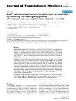
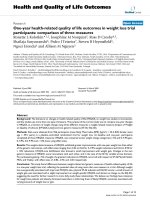
An L1 molecule of various HPVs contains several cysteine
residues at markedly similar relative positions (Fig. 1),
strongly suggesting that these cysteine residues play
important roles in the structure and the function of the
HPV capsids. Previous studies have shown that cysteine
residue at amino acid (aa) 175 (C175) and C428 in
HPV16 L1 (505 amino acids long) are involved in the
intermolecular disulfide bonding that contributes to the
assembly of the capsid [14]. The functions of the other L1
cysteine residues are not known.
In this study we attempted to know whether thiol-reactive
reagents affect infectivity of HPV16 PVs (16Pvs) by bind-
ing to the L1 cysteine residues.
Results
Infectivity of the 16PVs that have bound to thiol-reactive
reagents
The 16PVs was found to lose their infectivity for HeLa cells
after binding to thiol-reactive reagents: biotin polyethyle-
neoxide iodoacetamide (BPEOIA), 5,5'-dithiobis(2-
nitrobenzoic acid) (DTNB), N-ethylmaleimide (NEM), 4-
(N-maleimido)benzyl-trimethylammonium iodide
(MBTA), and [2-(trimethylammonium)ethyl] meth-
anethiosulfonate bromide (MTSET). 16PVs were incu-
bated with BPEOIA (1 mM), DTNB (2 mM), NEM (2
mM), MBTA (2 mM), or MTSET (2 mM) for 2 h at 37°C.
After dilution at 1 to 1,000 the 16PVs were inoculated to
the cells. The number of the infected cells, which
expressed EGFP, was counted 2 days later. The HeLa cells
inoculated with the 16PVs incubated with these thiol-
reactive reagents did not express EGFP (Fig. 2). Like HeLa
cells, SiHa and 293TT cells inoculated with the 16PVs that
had incubated with DTNB did not express EGFP (data not
presented). The data indicate that these thiol-reactive rea-
gents inhibited infectivity of the 16PVs.
DTNB did not affect the cellular susceptibility to 16PVs.
The normal 16PVs infected HeLa cells that had been cul-
tured in the growth medium containing DTNB (2 mM)
for 2 h at 37°C and washed once with fresh growth
medium (data not presented). Furthermore HeLa cells
cultured with growth medium containing 2 mM of DTNB,
MBTA or MTSET for 2 days grew and were maintained
Alignment of L1 amino acid sequences of papillomavirusesFigure 1
Alignment of L1 amino acid sequences of papillomaviruses. Numbers to the left represent human papillomavirus
types. Numbers on the top represent amino acid numbers for cysteines in HPV 16 L1 (positions in L1), starting from the N-
terminus. The number of total amino acids constituting each L1 is shown to the right.
16
31
33
52
58
18
6b
11
1a
2a
5
C
C CCCC CC
CCCC
C
C
C
C
C
C
C
C
C
C
C
C
C
C
C
C
C
CC
C
C
C
C
C
C
C
C
C
C
C
C
C
C
C
C
C
C
C
C
C
C
C
C
C
C
C
C
C
CC
CCC
CCC
CCC
CCC
CC
CC
CCC
CC
C
CCC
C
C
C
C C
C CCCC
C CCCC
C CCCC
C CCCC
CCCC
CCCC
CCCC
CCC
CCC
CCC C
102
146
157
161
175
185
225
229
324
345
379
428
1
505
504
499
529
524
566
500
501
508
510
516
1
1
1
1
1
1
1
1
1
1
Virology Journal 2007, 4:110 />Page 3 of 11
(page number not for citation purposes)
normally, strongly suggesting the reagents were not harm-
ful to HeLa cells at the concentration of 2 mM.
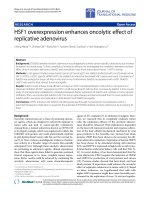
Binding of BPEOIA to the L1 cysteine residues of 16PV
BPEOIA, capable of making a complex with streptavidin,
was found to bind to the free thiol of the cysteine residues
of L1 of 16PV, which was produced by packaging of a
reporter plasmid into an HPV16 capsid. Purified 16PVs
were incubated with 1 mM BPEOIA at 37°C for 2 h. The
resultant 16PVs were electrophoresed on an SDS-polyacr-
ylamid gel and the separated proteins were stained by
SYPRO Ruby (Fig. 3A) or transferred to a membrane. The
membrane was probed by horseradish peroxidase (HRP)
conjugated streptavidin (Fig. 3B). After the incubation of
16PVs with BPEOIA, the molecular mass of L1 shifted
from 55 kDa to 57 kDa (Fig. 3A). The molecular mass of
L2 (68 kDa) was not affected by the incubation. The
streptavidin made a complex with only 57 kDa L1 (Fig.
3B). The data indicate that BPEOIA bound to the free thiol
of cysteine residue(s) of L1.
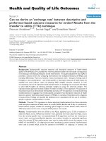
The 57 kDa L1/BPEOIA complex was digested with
trypsin and the fragments complexing with BPEOIA were
selectively obtained by column chromatography with the
streptavidin-resin. The molecular mass of the fragments
was measured by liquid chromatography-electrospray
ionization-tandem mass spectrometry (LC-ESI-MS/MS)
(Fig. 4). The mass of the three fragments, ECISMDYK,
SEVPLDICTSICK, and SEVPLDICTSICK, matched with the
calculated mass, indicating that BPEOIA bound to the
thiol of C146, C225, and C229. Some of the large tryptic
fragments that bound to BPEOIA may not be detected
because of their low recovery from LC and/or inefficiency
in the ionization.
Binding of BPEOIA to L1 of the 16PVFigure 3
Binding of BPEOIA to L1 of the 16PV. (A) The 16PVs
were incubated with 1 mM biotin-PEO-iodoacetamide (1
mM) in DMEM at 37°C for 2 h, electrophoresed on an SDS-
polyacrylamide gel, and stained with SYPRO Ruby. (B) The
proteins in the gel were transferred to a polyvinylidene diflu-
oride membrane and probed with Streptavidin-HRP.
BPEOIA - + - +
Avidin
-HRP
SYPRO
Ruby
L2
L1
250K
150K
100K
75K
50K
37K
AB
Infectivity of the 16PVs that have bound to BPEOIA, NEM, DTNB, MBTA, or MTSETFigure 2
Infectivity of the 16PVs that have bound to BPEOIA,
NEM, DTNB, MBTA, or MTSET. The 16PVs were incu-
bated with the thiol-reactive reagent indicated at 37°C for 2
h. The samples were diluted by 1000-fold and added to HeLa
cells. The cells were incubated for 2 days and harvested. The
cells expressing EGFP were counted by a FACS. BPEOIA:
biotin polyethyleneoxide iodoacetamid, NEM: N-ethylmale-
imide, DTNB: 5,5'-dithiobis(2-nitrobenzoic acid), MBTA: 4-
(N-maleimido)benzyl-trimethylammonium iodide, MTSET:
[2-(trimethylammonium)ethyl] methanethiosulfonate bro-
mide.
0
100
200
300
400
500
600
-
+ BPEOIA
+ DTNB
+ NEM
+ MBTA
+ MTSET
Thiol reactive reagents
Number of cells positive for EGFP
Virology Journal 2007, 4:110 />Page 4 of 11
(page number not for citation purposes)
Analysis of L1-cysteine residues that have bound to BPEOIA by mass spectrometryFigure 4
Analysis of L1-cysteine residues that have bound to BPEOIA by mass spectrometry. (A) Chromatogram of the
tryptic peptides of L1 that bound to BPEOIA. The upper box shows calculated monoisotopic mass value of mono (1+) and
doubly (2+) charged masses of three peptides. The asterisk denotes a cysteine bound with BPEOIA. (B and D) Full scan mass
spectra corresponding to the EC
146
*ISMDYK (B) and SEVPLDIC
225
*TSICK (D). (C, E and F) MS/MS spectra of corresponding
to EC
146
*ISMDYK (C), SEVPLDIC
225
*TSICK (E), PLDIC
225
*TSICK and PLDICTSI C
229
*K (F).
0 5 10 15 20 25 30 35 40 45 50
Time (min)
20
40
60
80
100
Relative signal intensity
941.84
702.44
EC
146
*ISMDYK
SEVPLDIC
225
*TSICK
20
40
60
80
100
Relative signal intensity
703.08
702.35
A
B
C
D
200 400 600 800 1000 1200
m/z
EC
146
*ISMDYK
941.91
783.36
20
40
60
80
100
Relative signal intensity
1400 1600 1800 2000600 800 1000 1200
m/z
SEVPLDIC
225
*TSICK
PLDIC
225
*TSICK
PLDICTSIC
229
*K
m/z
Sequence 1+ 2+
EC*
146
ISMDYK 1403.9029 702.4515
SEVPLDIC*
225
TSICK 1880.1987 940.5994
SEVPLDICTSIC*
229
K 1880.1987 940.5994
E
F
E-C
146
*-I-S-M-D-Y-K
Virology Journal 2007, 4:110 />Page 5 of 11
(page number not for citation purposes)
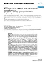
Sedimentation and morphology of the 16PVs that have
bound to DTNB or NEM
The 16PVs that had been incubated with DTNB (2 mM) or
NEM (2 mM) sedimented through the sucrose gradient
(5–40%) as the normal 16PVs did (Fig. 5A). The 16PVs
pre-incubated with MBTA or MTSET sedimented similarly
(data not presented). These results strongly suggest that
the 16PVs that had bound to these reagents were morpho-
logically similar to the normal 16PVs and did not make
aggregates.
The 16PVs that had bound to NEM (NEM-16PVs) were
not distinguishable from the normal 16PV by an electron
microscopy (Fig. 5B). The 16PVs that had been incubated
with NEM (2 mM) at 37°C for 2 h were negatively stained
with 4% uranylacetate and examined under a transmis-
sion electron microscope. Any morphological abnormali-
ties of the NEM-16PVs were not detectable at a
magnification of 1:200,000.
Binding to HeLa cells, internalization, and trafficking of
the 16PVs that have bound to DTNB or NEM
The DTNB-16PVs and the NEM-16PVs were found to bind
to HeLa cells less efficiently than the normal 16PVs did.
The 16Pvs that had been incubated with DTNB (2 mM) or
NEM (2 mM) were inoculated to HeLa cells with incuba-
tion at 4°C for 1 h. After a wash with cold PBS to remove
the unbound 16Pvs, the cells were lysed immediately.
Proteins in the lysate were separated by SDS-polyacryla-
mid gel electrophoresis (PAGE) and transferred to a mem-
brane. L1 on the membrane was detected with mouse
anti-HPV16L1 antibody and goat anti-mouse IgG-HRP
(Fig. 6A). The levels of L1 from the DTNB-16PVs and the
NEM-16PVs were 50–60 % of that of L1 from the normal
Sedimentation and morphology of the 16PVs that have bound to DTNB or NEMFigure 5
Sedimentation and morphology of the 16PVs that have bound to DTNB or NEM. (A) The 16PVs were incubated
with DTNB (2 mM) or NEM (2 mM) at 37°C for 2 h. The sample was loaded on the top of a linear sucrose-density gradient (5
to 40%) and centrifuged. L1 in the fractions obtained by a bottom puncture was detected by immunoblotting with mouse anti-
HPV16L1 antibody. (B) The 16PVs were incubated with NEM (2 mM) at 37°C for 2 h and observed under a transmission elec-
tron microscope.
16PV 16PV + NEM
A
B
Sucrose gradient
16PV
16PV+DTNB
16PV+NEM
Virology Journal 2007, 4:110 />Page 6 of 11
(page number not for citation purposes)
Binding, trafficking, and degradation of the 16PVs that have bound to DTNB or NEMFigure 6
Binding, trafficking, and degradation of the 16PVs that have bound to DTNB or NEM. (A) The 16PVs were incu-
bated with DTNB(2 mM) or NEM (2 mM) at 37°C for 2 h and added to HeLa cells. After incubation at 4°C for 1 h, the cells
were washed by PBS and lysed. The lysate was electrophoresed on an SDS-polyacrylamide gel. L1 was detected by immunob-
lotting with anti-HPV16L1 antibody. (B) The 16PVs incubated with DTNB or NEM were added to HeLa cells and incubated for
1 h at 4°C. The cells were cultured at 37°C for 2, 4, 8 or 20 h and fixed. L1 was detected by rabbit anti-HPV16L1 antibody and
goat anti-rabbit IgG conjugated with Alexa Fluor 546 (red). DNA was stained with DAPI (blue). (C) The 16PVs incubated with
DTNB were added to HeLa cells and incubated for 1 h at 4°C. The cells were harvested with PBS containing 2.5 mM EDTA
(for trypsin – sample at 0 h) or with trypsin (for trypsin + sample at 0 h). The rest of cells were cultured at 37°C for 2, 4, 8 or
20 h and harvested with trypsin. The cells were lysed and the lysates were electrophoresed on an SDS-polyacrylamide gel. L1
was detected by immunoblotting with anti-HPV16L1 antibody.
A
16PV + DTNB
16PV
-
L1
16PV + NEM
16PV
-
B
C
2h 8h 20h
16PV
16PV
+DTNB
20h
16PV
16PV
+NEM
Trypsin
- +++++ - +++++
Incubation (h)
0024820
0024820
16PV 16PV+DTNB
132K
90K
55K
45K
34K
25K
L1
L1
12 4567389101112
Virology Journal 2007, 4:110 />Page 7 of 11
(page number not for citation purposes)
16 PVs, indicating that DTNB and NEM reduced the cell-
binding ability of the 16PVs. But the reduction of the
binding efficiency does not fully account for the inhibi-
tion of the infectivity of the DTNB-16PVs and the NEM-
16PVs.
We found that the DTNB-16PVs and the NEM-16PVs dis-
appeared on their way to the nucleus (Fig. 6B). The 16PVs
incubated with DTNB (2 mM) or NEM (2 mM) were inoc-
ulated to HeLa cells with incubation at 4°C for 1 h. After
a wash with the growth medium the cells were incubated
further with the growth medium for 2, 8 or 20 h, fixed
with paraformaldehyde (4 %), and permeated by Triton-
X100. L1 was stained with rabbit anti-HPV16-L1 antibody
[12] and anti-rabbit-IgG goat antibody conjugated with
Alexa Fluor 546. Nuclear DNA was stained with DAPI. The
localization of L1 and DNA was observed under a confo-
cal microscope (Fig. 6B). Although the normal 16PVs
reached the perinuclear region and accumulated there at
20 h, the DTNB-16PVs and the NEM-16PVs became unde-
tectable at 20 h.
Consistent with the above observation by confocal micro-
scopy, the immunoblotting to detect L1 showed that the
DTNB-16PVs were rapidly degraded in the cells (Fig. 6C).
HeLa cells were inoculated with the 16PVs pre-incubated
with DTNB (2 mM), and the infected cells were incubated
at 4°C for 1 h. After a wash with cold PBS the cells were
incubated with trypsin to digest the 16PVs that had not
entered the cells. Then, the cells were lysed and subjected
to PAGE. The intracellular L1 levels were analysed by
immunoblotting with mouse anti-HPV16L1 antibody.
The cell-bound 16PVs (lanes 1 and 7) were sensitive to the
trypsin digestion (lanes 2 and 8) and became resistant
after the incubation at 37°C for 2 h (lanes 3 and 9). The
cells inoculated with the 16PVs were further incubated
with the growth medium at 37°C for 2, 4, 8, and 20 h,
digested with trypsin, and analysed. Fig. 6C clearly
showed that the DTNB-16PVs were degraded rapidly and
became almost undetectable at 8 h (lane 11).
Thus, the DTNB-16PVs had reduced capability of binding
to the cells and enhanced sensitivity to the cellular mech-
anisms controlling the degradation of foreign proteins.
Infectivity of the 16PVs composed of mutant L1s having a
replacement of cysteine with alanine
A mutational analysis was unsuccessful to identify one
particular cysteine residue having the free thiol essential
for the infectivity of the 16PV. We newly constructed the
L1 mutants by replacement of the cysteine, except for
C175 and C428, with alanine. C161A (C161 was replaced
with A) was extremely unstable in 293TT cells and was not
available for the analysis. The yields of C157A, C229A,
C342A, and C379A were very low. Therefore, the number
of HeLa cells infected with the mutant PVs was normal-
ized to the content of L1 (Table 1). The infectivity of the
mutant PVs, including C146A, C225A, and C229A,
ranged between 58 and 187 % of the infectivity of the nor-
mal 16PV.
Discussion
In this study we found that the thiol-reactive reagents
bound to C146, C225, and C229 of the 16PVs. In the 3-
dimentional-structure model of L1 [15], C146 is involved
in forming the DE-loop and C225 and C229 are involved
in forming the EF-loop. Because the loops are generally
flexible and because C146 and C225 are located at the sur-
face region of the loops and C229 is located near the sur-
face region, it is likely that thiol-reactive reagents easily
access the thiols of these cysteine residues. Although it is
difficult to examine experimentally whether all or part of
the cysteine residues have free thiols, we presume the great
majority of C146, C225, and C229 may have free thiols,
Table 1: Infectiviy of mutant 16PVs
L1 in PV stock Infectious units
ng of L1/µl % units/µg of L1 % SD
WT 82.0 100 1.77.E+05 100 17.5
C102A 29.3 35.7 1.05.E+05 59.3 1.40
C146A 54.9 66.9 1.64.E+05 92.4 3.37
C157A 3.46 4.22 8.60.E+04 48.1 2.04
C161A ND - NT - -
C185A 33.8 41.2 1.62.E+05 91.4 3.86
C225A 81.2 99.0 2.82.E+05 159 4.46
C229A 8.53 10.4 9.60.E+04 54.6 5.59
C324A 5.85 7.14 8.50.E+04 50.2 2.47
C345A 44.3 54.0 3.30.E+05 187 4.03
C379A 13.1 16.0 1.62.E+05 91.4 4.43
ND: not detected NT: not tested
Virology Journal 2007, 4:110 />Page 8 of 11
(page number not for citation purposes)
because the incubation of the 16PVs with the thiol-reac-
tive reagents induced a large effect on their infectivity. The
16PVs lost their infectivity after binding of the thiol-reac-
tive reagents to the free thiols. The DTNB-16PVs and the
NEM-16PVs, whose C146, C225, and C229 carried DTNB
and NEM as additional side chains, respectively, bound to
HeLa cells less efficiently and were degraded rapidly in the
cells. Although the 16PV mutants with two or three Ala
substitutions for C146, C225, or C229 were too unstable
to be used in the infectivity analysis, we obtained the three
mutants with an Ala substitution (C146A, C225A, and
C229A) and found that the substitution did not affect the
infectivity much. Therefore, it is likely that the steric bulk
of DTNB or NEM occludes a neighboring portion of the
virion involved in the entry and trafficking processes. But
there remains a possibility that the disulfide bonding
between an unidentified cellular protein(s) and the two
remaining cysteine residues in the mutants plays an addi-
tive role in the viral entry and trafficking.
It has been reported that cell-surface protein disulfide iso-
merase (PDI) is required for the entry process of several
viruses including mouse polyomavirus. The siRNA-medi-
ated down regulation of PDI of HeLa cells prevents the
cells from being infected with mouse polyomavirus [16].
Inactivation of the cell-surface PDI by adding the thiol-
reactive reagents, such as DTNB, which do not permeate
the membrane, to the culture medium results in the inhi-
bition of entry of HIV1 and Newcastle disease virus
[17,18], suggesting that the modified envelope conforma-
tion induced by reforming the disulfide-bonding is
required for the membrane fusion, which is the essential
step for the virus entry [18,19]. However, pre-incubation
of the cells with DTNB did not inhibit infection with the
16PV, indicating that modification of the disulfide-bond-
ing in the capsid by cell-surface PDI is not involved in the
early steps of HPV infection. The data are consistent with
the recent report that reducing agents, such as DTT and 2-
ME, do not inhibit HPV infection [20].
Because the thiol-reactive reagents tested in this study
bound to the free thiol of the 16PVs at a concentration not
toxic for HeLa cells, these reagents might function as prac-
tical inhibitors of HPV infection. It would be necessary to
test the efficacy and safety of the reagents in animal mod-
els.
Conclusion
HPV16 L1 C146, C225, and C229 have free thiol, which
is accessible by the thiol-reactive reagents, such as
BPEOIA, DTNB, and NEM. The HPV16 pseudovirions car-
rying these thiol-reactive reagents lost infectivity by
mainly the rapid degradation in the cytoplasm.
Methods
Cells
293TT cells, a human cell line expressing a high level of
SV40 T antigen, was a kind gift from J. T. Schiller
(National Cancer Institute, USA). The cells were cultured
in Dulbecco's modified minimal essential medium
(DMEM) (No. 21063, Invitrogen Corp., Carlsbad, CA)
supplemented with 10% heat-inactivated fetal bovine
serum (FBS), 1% non-essential amino acids (Invitrogen
Corp.), 1% GlutaMax-I (Invitrogen Corp.), penicillin G
potassium (100 units/ml) (Meiji seika Ltd., Tokyo,
Japan), kanamycin sulfate (60 µg/ml) (Wako pure chem-
ical industries Ltd. Tokyo, Japan) (growth medium) and
hygromycine B (400 µg/ml) (Invitrogen Corp.) in 5%
CO2 at 37°C. HeLa cells and SiHa cells were cultured in
DMEM supplemented 10% FBS, penicillin G potassium,
and kanamycin sulfate.
Plasmids
pYSEAP, ph16L1, and ph16L2 were gift from J. T. Schiller.
pEF1a-EGFP was newly constructed by an insertion of
EGFP gene derived from pCMS-EGFP (Clonthech labora-
tories Inc., Mountain View, CA) into the backbone of
pYSEAP. Plasmid expressing mutant L1 with a substitu-
tion of alanine for cysteine was constructed by overlap
extension PCR method [21] using KOD plus polymerase
(TOYOBO Corp., Osaka, Japan) and ph16L1 as template.
5'-GCTGGTGTGGGCCGCCGTGGGCGTGGAG-3' and 5'-
CCTCCACGCCCACGGCGGCCCACACCAGCC-3' were
used as forward (F) and reverse (R) primers to replace
C102 with A, respectively. Following oligonucleotides
were used as primers to introduce the other mutantions:
5'-CGACAACAGGGAGGCCATCAGCATGGACTACAAG-
3' (F for C146A), 5'-GTAGTCCATGCTGATGGCCTCCCT-
GTTGTCCAC-3' (R for C146A), 5'-CAAGCAGAC-
CCAGCTGGCCCTGATCGGCTGCAAG-3' (F for C157A),
5'-CTTGCAGCCGATCAGGGCCAGCTGGGTCTGCTTG-3'
(R for C157A), 5'-CTGTGCCTGATCGGCGCCAAGCCCC
CCATCG-3' (F for C161A), 5'-CGATGGGGGGCTTGGCG
CCGATCAGGCACAG-3' (R for C161A), 5'-AACCCCG-
GCGACGCCCCCCCCCTGGAGCTG-3' (F for C185A), 5'-
CAGCTCCAGGGGGGGGGCGTCGCCGGGGTTC-3' (R
for C185A), 5'-GTGCCCCTGGACATCGCCACCAGCATC
TGCAAG-3' (F for C225A), 5'-CTTGCAGATGCTGGTGGC
GATGTCCAGGGGCAC-3' (R for C225A), 5'-ACCAG-
CATCGCCAAGTACCCCGACTACATC-3' (F for C229A),
5'-ATGTAGTCGGGGTACTTGGCGATGCTGGTGCAGAT
G-3' (R for C229A), 5'-CAACAACGGCATCGCCTGGGGC
AACCAGCTGTTC-3' (F for C324A), 5'-GAACAGCTGGTT-
GCCCCAGGCGATGCCGTTGTTGTG-3' (R for C324A),
5'-CCAACATGAGCCTGGCCGCCGCCATCAGCAC-3' (F
for C345A), 5'-GTGCTGATGGCGGCGGCCAGGCTCAT-
GTTGGTGCTCC-3' (R for C345A), 5'-CATCTTCCAGCT-
GGCCAAGATCACCCTGAC-3' (F for C379A), 5'-
GTCAGGGTGATCTTGGCCAGCTGGAAGATGAACTG-3'
Virology Journal 2007, 4:110 />Page 9 of 11
(page number not for citation purposes)
(R for C379A). 5'-TGCCTTTACTTCTAGGCCTGTACG-3'
and 5'-TGCTCCTGGTGGTGTCCACCACGGTC-3' were
used as primers to join the part containing the mutation
back to the rest of the entire L1 gene of C146A, C157A,
C161A, C185A, C225A, C229A. 5'-AACCTGGCCAGCAG-
CAACTACTTCCC-3' and 5'-AACTAGAAGGCACAGTC-
GAGGCTG-3' were used similarly to produce C324A,
C345A, C379A. The resultant DNA fragments were
inserted into ph16L1 after digestion with NotI and ApaI
(for C146A, C157A, C161A, C185A, C225A, and C229A)
or with ApaI and HindIII (for C324A, C345A, and
C379A).
Thiol-reactive reagents
Biotin polyethyleneoxide iodoacetamide (BPEOIA) was
purchased from SIGMA-ALDRICH Corp. (Saint Luis,
MO). N-ethylmaleimide (NEM) was purchased from
Nakarai Tesque. Inc. (Kyoto, Japan). 5,5'-dithiobis(2-
nitrobenzoic acid) (DTNB) was purchased from SIGMA-
ALDRICH Corp (Saint Luis, MO). 4-(N-maleimido)ben-
zyl-trimethylammonium iodide (MBTA), and [2-(tri-
methylammonium)ethyl] methanethiosulfonate
bromide (MTSET) were purchased from Toronto Research
Chemicals Inc. (Toronto, Canada)
Preparation of 16PV
The 16PV, an HPV16 capsid containing a reporter plasmid
expressing EGFP, was produced by the previously
described procedure [6,7,12] with minor modification.
293TT cells (40% confluent in 10-cm culture dish) were
transfected with mixture of ph16L1 (13.5 µg), ph16L2 (3
µg), and pEF1a-EGFP (13.5 µg) by using Optifect (Invitro-
gen Corp.) in OPTI-MEMII (Invitrogen Corp.). After incu-
bation for 3 days, the cells were scraped off and suspended
in 0.5 ml lysis buffer (PBS containing 9.5 mM MgCl
2
,
0.35% Brij 58, [Sigma-Aldrich Inc., St. Louis, MO], 0.1%
Benzonase [Sigma-Aldrich Inc.], 0.1% Plasmid Safe ATP
dependent-DNase [EPICENTRE Corp. Madison, WI], 1
mM ATP) and incubated at 37°C for 20–24 h with slow
rotation. The lysate was cooled on ice for 5 min, mixed
with 1/4 volume of 5 M NaCl solution, and kept on ice for
10 min, then, centrifuged at 5,000 × g at 4°C for 10 min.
The resultant supernatant was laid on an Optiprep gradi-
ent composed of 27%, 33%, and 39% in PBS containing
1 mM CaCl
2
, 0.5 mM MgCl
2
, 2.1 mM KCl, and 0.8 M
NaCl and centrifuged at 47,900 rpm at 16°C for 3 h with
SW50.1 rotor (Beckman Coulter Inc. Fullerton, CA). The
fraction containing the purified 16PVs was collected by
puncturing the bottom and used as the stock.
Infectivity assay
The 16PV stock was diluted at 10-fold with DMEM and
received BPEOIA (1 mM); the 16PV stock was diluted at
10-fold with the growth medium and received DTNB (2
mM), NEM (2 mM), MBTA (2 mM), or MTSET (2 mM).
The mixtures were incubated at 37°C for 2 h and diluted
with the growth medium at 1,000-fold. HeLa cells (1.5 ×
10
5
) in a well of a 24-well culture-plate were inoculated
with the sample and cultured for 2 days. The cells were
harvested with trypsin. EGFP-positive cells were counted
by a fluorescence activated cell sorting (FACS Calibar, Bec-
ton Dickinson and Company Ltd., San Joe, CA).
Binding of BPEOIA to 16PVs
BPEOIA was dissolved in H
2
O to 18.4 mM. The 16PV
stock was diluted at 10-fold with DMEM (No. 21063, Inv-
itrogen Corp.) containing BPEOIA (1 mM) and incubated
at 37°C for 2 h. Then, DTT (100 mM), which reacted with
remaining excess BPEOIA, was added to the mixture and
incubated at 37°C for 30 m (BPEOIA+ sample). The 16PV
stock was diluted at 10-fold with DMEM, incubated at
37°C for 2 h, mixed with DTT (100 mM), and further
incubated at 37°C for 30 m. Then, BPEOIA (1 mM) was
added to the mixture and incubated at 37°C for 30 m
(BPEOIA- sample). The 16PVs were concentrated by using
a PAGEprep Advance Kit (PIERCE Biotechnology Inc.,
Rockford, IL) and suspended in the SDS sample buffer (50
mM Tris-HCl pH 6.8, 5% glycerol, 2 % SDS, and
bromphenol blue) containing 100 mM DTT. The sample
was boiled and electrophorased on an SDS-polyacryla-
mide gel. The proteins in the gel were stained with SYPRO
Ruby (Invitrogen Corp.) or transferred to membrane
Hybond-P (GE Healthcare Bio-Science AB, Uppsala, Swe-
den). The membrane was blocked with skim milk and
incubated with the horseradish peroxidase (HRP) conju-
gated-streptavidin (GE Healthcare Bio-Science AB). The
HRP activity was detected by using an ECL plus western
blotting detection system (GE Healthcare Bio-Science AB)
and Typhoon 9410 (GE Healthcare Bio-Science AB).
Analysis by liquid chromatography electrospray
ionization-tandem mass spectrometry (LC-ESI-MS/MS)
The HPV16 pseudovirions that bound to BPEOIA were
separated by SDS-PAGE and stained by SYPRO Ruby as
described above. The gel pieces containing the L1/BPEOIA
complex were excised and the L1 in the gel was digested
with trypsin (Trypsin Gold Mass Spectrometry Grade,
Promega Corp., Madison WI) as previously described
[22]. The digested peptides were extracted from the gel
pieces by one change of NH
4
HCO
3
(20 mM) and 4
changes of acetonitrile (50 %). The peptides were sus-
pended in PBS by adding equal volume of 2× concen-
trated PBS and then incubated with monomeric avidin
beads (Ultralink Immobilized Monomeric Avidin,
PIERCE Biotechnolog Inc.). The beads were washed with
PBS twice, with methanol (20 %) in NH
4
HCO
3
(50 mM)
twice, and with water twice. The peptides that bound to
the beads were eluted with acetonitrile (30 %) containing
TFA (0.4 %). After volatilization of acetonitrile the pep-
tides were analyzed by a liquid chromatography (MAGIC
Virology Journal 2007, 4:110 />Page 10 of 11
(page number not for citation purposes)
2002 system, Michrome Bioresources Inc., Auburn, CA)
equipped with C18 column (Inertsil EX-Nano ODS-3, 0.1
mm i.d. × 50 mmL, GL Sciences Inc, Tokyo, Japan) cou-
pled with a nano spray apparatus (AMR Inc. Tokyo,
Japan) for electrospray ionization-iontrap mass spectrom-
etry (LC-ESI-IT-MS) (LCQ-decaXP, Thermo electron corp.,
San Jose, CA). The data were collected by data-dependent
mode and the MS/MS sequence were analysed by using
software Bioworks (Ver.3.1, Thermo electron corp.) with
variable modification option of a mass unit of 414.19 for
the biotin polyethleneoxide moiety.
Sedimentation assay
The 16PV stock was diluted at 10-fold with the growth
medium, and DTNB (2 mM) was added to the medium.
The 16PVs were then incubated for 2 h at 37°C. The sam-
ple was loaded on a linear sucrose-density gradient (5 to
40%) in PBS. After centrifugation at 120,000 × g at 4°C
for 2.5 h with and SW50.1 rotor, aliquots (400 µl) were
collected. Ten µl of the aliquot was mixed with an equal
volume of the 2× concentrated SDS-sample buffer con-
taining 100 mM DTT, boiled, and electrophorased on an
SDS-polyacrylamide gel. The proteins were transferred to
a Hybond-P membrane (GE Healthcare Bio-Science AB).
The membrane was blocked with skim milk, incubated
with mouse anti-HPV type 16L1 antibody (BD Bio-
sciences Pharmingen Com., San Diego, CA), and then
incubated with anti-mouse IgG-HRP (Santa Cruz Biotech-
nology Inc., Santa Cruz, CA). The HRP activity was
detected by using an ECL plus western blotting detection
system (GE Healthcare Bio-Science AB) and Typhoon
9410 (GE Healthcare Bio-Science AB).
Electron microscopy
The 16PV stock was mixed with NEM (2 mM) and incu-
bated at 37°C for 2 h. The excess NEM was removed by
using a Bio-Spin 30 column (Bio-Rad Laboratories Inc.,
Hercules, CA) equilibrated with phosphate buffer con-
taining 0.5 M NaCl. The 16PVs were concentrated with a
Microcon YM-100 (Millipore Corp., Bedford, MA) and
then settled on carbon-coated copper grids. The 16PVs
were negatively stained with 4% uranylacetate and exam-
ined in a transmission electron microscope (Hitachi
model H-7650, Hitachi corp., Tokyo, Japan).
Binding assay
The 16PV stock was diluted at 20-fold with the growth
medium, and DTNB (2 mM) was added to the medium.
The stock was incubated at 37°C for 2 h and used for the
binding assay. The 16PV stock was mixed with NEM (2
mM) and incubated at 37°C for 2 h. Then, the excess NEM
was removed by using a Bio-Spin 30 column as described
above was used for the binding assay. These samples were
diluted with the growth medium at 20-fold and added to
HeLa cells (1.5 × 10
5
). The cells were incubated at 4°C for
1 h, washed with PBS, harvested with PBS containing 2.5
mM EDTA, and lysed. The lysate was electrophorased on
an SDS-polyacrylamide gel. The separated proteins were
transferred to a polyvinylidene difluoride membrane and
L1 was detected by immunoblotting with mouse anti-
HPV16 L1 anitbody and anti-mouse IgG-HRP.
Immunofluoresence microscopy
HeLa cells (1.5 × 10
5
) were seeded onto a well of a 4-
chamber glass slide (BD Biosciences Falcon, Bedford, MA)
with the growth medium. The cells were inoculated with
the 16PV samples similarly prepared as the samples for
the biding assay and incubated at 4°C for 1 h. The cells
were washed with the growth medium and incubated at
37°C for 2, 8 or 20 h. The cells were fixed with PBS con-
taining paraformaldehyde (4%) at room temperature
(RT) for 10 m and washed with PBS. The cells were made
permeable with PBS containing Triton X-100 (1%) at RT
for 10 m and washed with PBS. The cells were incubated
with rabbit anti-HPV16 L1 serum [12] in PBS containing
BSA (3%) at RT for 1 h, washed with PBS containing
Tween-20 (0.2%), incubated with Alexa Fluor 546 goat
anti-rabbit IgG (H+L) (Invitrogen Corp.) in PBS contain-
ing BSA (3%), and washed with PBS containing Tween-20
(0.2%). The cells were coated with a ProLong Gold anti-
fade reagent with DAPI (Invitrogen Corp.) and imaged in
a FLUOVIEW FV1000 confocal microscope (OLYMPUS,
Tokyo, Japan).
Internalizaiton assay
HeLa cells (1.5 × 10
5
) in a well of a 24-well culture plate
were inoculated with the 16PVs that had been preincu-
bated with DTNB as done for the binding assay. The cells
were incubated at 4°C for 1 h. The cells for the samples at
0 h were washed with the growth medium and immedi-
ately harvested with PBS containing 2.5 mM EDTA or with
PBS containing trypsin. The other cells were incubated
with the growth medium at 37°C for 2, 4, 8 and 20 h and
harvested with PBS containing trypsin. The cells were
lysed and electrophorased on an SDS-polyacrylamide gel.
The separated proteins were transferred to a polyvinyli-
dene difluoride membrane. L1 was detected by immuno-
blotting with mouse anti-HPV16 L1 anitbody and anti-
mouse IgG-HRP.
Competing interests
The author(s) declare that they have no competing inter-
ests.
Authors' contributions
YI conceived of the study, carried out the biological exper-
iments, and drafted the manuscript. KK supported the
preparation of the 16PV stock. TM constructed the
reporter plasmid. KT conducted electron microscopy. FSO
Publish with BioMed Central and every
scientist can read your work free of charge
"BioMed Central will be the most significant development for
disseminating the results of biomedical research in our lifetime."
Sir Paul Nurse, Cancer Research UK
Your research papers will be:
available free of charge to the entire biomedical community
peer reviewed and published immediately upon acceptance
cited in PubMed and archived on PubMed Central
yours — you keep the copyright
Submit your manuscript here:
/>BioMedcentral
Virology Journal 2007, 4:110 />Page 11 of 11
(page number not for citation purposes)
and KH conducted the analysis by LC-ESI-IT-MS. TK
supervised the study and helped to draft the manuscript.
Acknowledgements
We thank Dr. Kunito Yoshiike for critical reading of the manuscript. This
work was supported by a grant-in-aid from the Ministry of Health, Labour
and Welfare for the Third-Term Comprehensive 10-year Strategy for Can-
cer Control.
References
1. Howley PM, Lowy DR: Papillomaviruses and their replication.
In Fields Virology Volume 2. Forth edition. Edited by: Lnipe DM, Howley
PM. Philadelphia, Lippincott Williams&Wilkins; 2001:2197-2229.
2. Volpers C, Schirmacher P, Streeck RE, Sapp M: Assembly of the
major and the minor capsid protein of human papillomavirus
type 33 into virus-like particles and tubular structures in
insect cells. Virology 1994, 200:504-512.
3. de Villiers EM, Fauquet C, Broker TR, Bernard HU, zur Hausen H:
Classification of papillomaviruses. Virology 2004, 324:17-24.
4. Munoz N, Bosch FX, Castellsaque X, Diaz M, de Sanjose S, Ham-
mouda D, Shah KV, Meijer CJ: Against which human papilloma-
virus types shall we vaccinate and screen? The international
perspective. Int J Cancer 2004, 111:278-285.
5. zur Hausen H: Papillomaviruses and cancer: from basic studies
to clinical application. Nat Rev Cancer 2002, 2:342-350.
6. Buck CB, Pastrana DV, Lowy DR, Schiller JT: Efficient intracellular
assembly of papillomaviral vectors. J Virol 2004, 78:751-757.
7. Buck CB, Thompson CD, Pang YY, Lowy DR, Schiller JT: Matura-
tion of papillomavirus capsids. J Virol 2005, 79:2839-2846.
8. Day PM, Baker CC, Lowy DR, Schiller JT: Establishment of papil-
lomavirus infection is enhanced by promyelocytic leukemia
protein (PML) expression. Proc Natl Acad Sci USA 2004,
101:14252-14257.
9. Buck CB, Day PM, Thompson CD, Lubkowski J, Lu W, Lowy DR,
Schiller JT: Human alpha-defensins block papillomavirus infec-
tion. Proc Natl Acad Sci USA 2006, 103:1516-1521.
10. Richards RM, Lowy DR, Schiller JT, Day PM: Cleavage of the pap-
illomavirus minor capsid protein, L2, at a furin consensus
site is necessary for infection. Proc Natl Acad Sci USA 2006,
103:1522-1527.
11. Pastrana DV, Gambhira R, Buck CB, Pang YY, Thompson CD, Culp
TD, Christensen ND, Lowy DR, Schiller JT, Roden RB: Cross-neu-
tralization of cutaneous and mucosal Papillomavirus types
with anti-sera to the amino terminus of L2. Virology 2005,
337:365-372.
12. Kondo K, Ishii Y, Ochi H, Matsumoto T, Yoshikawa H, Kanda T: Neu-
tralization of HPV16, 18, 31, and 58 pseudovirions with antis-
era induced by immunizing rabbits with synthetic peptides
representing segments of the HPV16 minor capsid protein
L2 surface region. Viroloy 2007, 358:266-272.
13. Slupetzky K, Gambhira R, Culp TD, Shafti-Keramat S, Schellenbacher
C, Christensen ND, Roden RB, Kirnbauer R: A papillomavirus-like
particle (VLP) vaccine displaying HPV16 L2 epitopes induces
cross-neutralizing antibodies to HPV11. Vaccine 2007,
25:2001-10.
14. Ishii Y, Tanaka K, Kanda T: Mutational analysis of human papil-
lomavirus type 16 major capsid protein L1: the cysteines
affecting the intermolecular bonding and structure of L1-
capsids. Virology 2003, 308:128-136.
15. Chen XS, Garcea RL, Goldberg I, Casini G, Harrison SC: Structure
of small virus-like particles assembled from the L1 protein of
human papillomavirus 16. Mol Cell 2000, 5:557-567.
16. Gilbert J, Ou W, Silver J, Benjamin T: Downregulation of protein
disulfide isomerase inhibits infection by the mouse polyoma-
virus. J Virol 2006, 80:10868-10870.
17. Ryser HJ, Levy EM, Mandel R, DiSciullo GJ: Inhibition of human
immunodeficiency virus infection by agents that interfere
with thiol-disulfide interchange upon virus-receptor interac-
tion. Proc Natl Acad Sci USA 1994, 91:4559-4563.
18. Jain S, McGinnes LW, Morrison TG: Thiol/disulfide exchange is
required for membrane fusion directed by the Newcastle
disease virus fusion protein. J Vriol 2007, 81:2328-2339.
19. Ryser HJ, Fluckiger R: Progress in targeting HIV-1 entry. Drug
Discov Today 2005, 10:1085-1094.
20. Buck CB, Thompson CD, Roberts JN, Muller M, Lowy DR, Schiller JT:
Carrageenan is a potent inhibitor of papillomavirus infec-
tion. PLoS Pathog 2006, 2:e69.
21. Abbe NV, Robert JP, Larry RP: Mutagenesis and Synthesis of
Novel Recombinant Genes Using PCR. In PCR PRIMER A Labo-
ratory Manual Edited by: Carl WD, Gabriela SD. Cold Spring Harbor
Laboratory Press; 1995:603-612.
22. Shevchenko A, Wilm M, Vorm O, Mann M: Mass spectrometric
sequencing of proteins silver-stained polyacrylamide gels.
Anal Chem 1996, 68:850-858.