Báo cáo hóa học: " Evidence that the Nijmegen breakage syndrome protein, an early sensor of double-strand DNA breaks (DSB), is involved in HIV-1 post-integration repair by recruiting the ataxia telangiectasia-mutated kinase in a " pptx
Bạn đang xem bản rút gọn của tài liệu. Xem và tải ngay bản đầy đủ của tài liệu tại đây (653.45 KB, 12 trang )
BioMed Central
Page 1 of 12
(page number not for citation purposes)
Virology Journal
Open Access
Research
Evidence that the Nijmegen breakage syndrome protein, an early
sensor of double-strand DNA breaks (DSB), is involved in HIV-1
post-integration repair by recruiting the ataxia
telangiectasia-mutated kinase in a process similar to, but distinct
from, cellular DSB repair
Johanna A Smith
1,5
, Feng-Xiang Wang
1,5
, Hui Zhang
1,5
, Kou-Juey Wu
2,5
,
Kevin Jon Williams
1,3,5
and René Daniel*
1,4,5
Address:
1
Division of Infectious Diseases – Center for Human Virology, Kimmel Cancer Center, Thomas Jefferson University, Philadelphia, PA,
USA,
2
Institute of Biochemistry and Molecular Biology, National Yang-Ming University, Taipei, Taiwan,
3
Division of Endocrinology, Thomas
Jefferson University, Philadelphia, USA,
4
Kimmel Cancer Center, Immunology Program, Thomas Jefferson University, Philadelphia, PA, USA and
5
704G Abramson Research Center, 3615 Civic Center Boulevard, Philadelphia, PA 19104, USA
Email: Johanna A Smith - ; Feng-Xiang Wang - ; Hui Zhang - ;
Kou-Juey Wu - ; Kevin Jon Williams - ; René Daniel* -
* Corresponding author
Abstract
Retroviral transduction involves integrase-dependent linkage of viral and host DNA that leaves an
intermediate that requires post-integration repair (PIR). We and others proposed that PIR hijacks
the host cell double-strand DNA break (DSB) repair pathways. Nevertheless, the geometry of
retroviral DNA integration differs considerably from that of DSB repair and so the precise role of
host-cell mechanisms in PIR remains unclear. In the current study, we found that the Nijmegen
breakage syndrome 1 protein (NBS1), an early sensor of DSBs, associates with HIV-1 DNA,
recruits the ataxia telangiectasia-mutated (ATM) kinase, promotes stable retroviral transduction,
mediates efficient integration of viral DNA and blocks integrase-dependent apoptosis that can arise
from unrepaired viral-host DNA linkages. Moreover, we demonstrate that the ATM kinase,
recruited by NBS1, is itself required for efficient retroviral transduction. Surprisingly, recruitment
of the ATR kinase, which in the context of DSB requires both NBS1 and ATM, proceeds
independently of these two proteins. A model is proposed emphasizing similarities and differences
between PIR and DSB repair. Differences between the pathways may eventually allow strategies to
block PIR while still allowing DSB repair.
Introduction
Post-integration repair (PIR) is an essential step in the ret-
roviral lifecycle, and yet it remains incompletely under-
stood. PIR occurs after the retroviral integrase has
removed two nucleotides from the 3'-ends of viral DNA
and then joined the newly exposed hydroxyl groups to
staggered phosphates in complementary strands of the
host chromosomal DNA, through non-blunt cleavage of
host DNA in concert with the ligation reaction [1,2]. This
initial integrase-mediated linkage between viral and host
Published: 22 January 2008
Virology Journal 2008, 5:11 doi:10.1186/1743-422X-5-11
Received: 16 November 2007
Accepted: 22 January 2008
This article is available from: />© 2008 Smith et al; licensee BioMed Central Ltd.
This is an Open Access article distributed under the terms of the Creative Commons Attribution License ( />),
which permits unrestricted use, distribution, and reproduction in any medium, provided the original work is properly cited.
Virology Journal 2008, 5:11 />Page 2 of 12
(page number not for citation purposes)
DNA produces an intermediate, in which the proviral
DNA is flanked by short, single-stranded gaps in the host-
cell DNA. PIR completes integration through four distinct
steps: trimming the 2-bp flaps from the 5'-ends of the pro-
viral DNA, filling in the single-stranded gaps that arose
from the original staggered cleavage of host DNA, ligation
of the trimmed 5' viral DNA ends to the filled-in host
DNA strands, and reconstitution of appropriate chroma-
tin structure at the integration site.
It has been proposed that that the virus exploits host-cell
double-strand DNA break (DSB) repair pathways to com-
plete the integration process, and initial evidence suggests
that it involves the NHEJ (non-homologous end joining)
pathway, as well as the ATM (ataxia telangiectasia
mutated) and ATR (ATM and Rad3 related) kinases [3-9].
Nevertheless, several key issues remain. First, the earliest
known sensor of DSBs, the Nijmegen breakage syndrome-
1 protein (NBS1), has not been examined in the context
of retroviral PIR. NBS1 is the crucial initiating component
of the MRN complex, which comprises three proteins:
MRE11 (meiotic recombination 11 homologue), a com-
bined exo- and endo-nuclease [10]; RAD50, which binds
DNA duplexes and may function as an anchor to hold the
DNA ends together at a DSB [11]; and NBS1 itself. NBS1
associates with DSBs immediately after the DNA damage
occurs [12] and recruits MRE11 and RAD50 [13,14]. In
addition, NBS1 recruits the ATM kinase to DSB sites [15],
and NBS1 [15] and ATM [16] are then both required to
recruit the ATR kinase [16]. Activation of the ATM and
ATR kinases allows them to phosphorylate several DNA
repair and checkpoint proteins, including NBS1 itself [17-
21]. Nijmegen breakage syndrome (NBS), which is caused
by a hypomorphic mutation in the NBS1 gene, and ataxia
telangiectasia (A-T), which is caused by mutations in the
ATM gene, highlight the significance of NBS1 in DSB
repair [22,23]. NBS and A-T cells exhibit similar DNA
repair deficiencies, including hypersensitivity to γ-irradia-
tion, which causes DSBs, and defective cell-cycle check-
points that fail to arrest cell proliferation when unrepaired
DSBs are present [21,24]. Because of the central role of
NBS1 in DSB repair, we now hypothesize that this protein
might initiate cellular responses leading to retroviral PIR
as well.
The second key issue in understanding retroviral PIR con-
cerns conflicting data in the literature about the roles of
the ATM and ATR kinases. Although many publications
demonstrated the participation of other NHEJ proteins in
PIR [3,5,6,8,25,26], the precise roles for ATM and ATR
remain less clear. For example, we reported only a minor
function for the ATM protein, which became apparent
mainly in the absence of other NHEJ components [5]. In
contrast, some laboratories reported that ATM is required
for efficient PIR even in the presence of NHEJ [8,27],
whereas others reported efficient transduction even in the
absence of ATM [28,29]. One explanation is that these dis-
crepancies arose from the use of different immortalized
cell lines in these studies. Therefore, in the current study
we addressed the role of ATM in PIR in primary human
cells.
Third, although a great deal is known about DSB repair,
details of PIR have yet to be delineated. Retroviruses
hijack numerous DSB repair proteins [3,5,6,8,25,26,30],
but the geometry of retroviral integration differs consider-
ably from DSB repair, which is limited to linking two
blunt ends together. We now hypothesize that the two
repair processes may crucially diverge. Initial supportive
evidence comes from our recent finding that phosphoryla-
tion of the histone H2AX on its Ser 139 residue is crucial
to DSB repair, but not for efficient PIR [31]. Importantly,
differences between the two repair processes might allow
strategies to inhibit PIR while still allowing NHEJ.
Therefore, we now sought to examine the presence, inter-
actions, and function of several DSB repair proteins in ret-
roviral PIR, namely, the initial DSB sensor NBS1 and the
ATM and ATR kinases. Our comparisons of PIR with DSB
repair continue to reveal fundamental similarities and dif-
ferences.
Experimental procedures
Primary human fibroblasts and lymphoid cell lines
All human cells were purchased from the Coriell Cell
Repository (Camden, New Jersey): primary NBS fibroblast
cells (deficient in the wild type NBS1 protein – GM07166)
and matched control cells (GM04506); A-T primary
fibroblasts (deficient in the ATM protein – GM02052)
and matched controls (GM01661); EBV transformed NBS
B-Lymphocytes (GM15818) and matched control EBV
transformed cells (GM15817). All cells were maintained
in RPMI-1640 medium in the presence of 10% fetal
bovine serum (FBS), 5 × 10
-6
M 2-mercaptoethanol, non-
essential amino acids, and 1% Pen/Strep.
HIV-1-based vectors
All VSV G-pseudotyped HIV-1-based vectors were pre-
pared as described previously [3,32], and carried either a
lacZ or EGFP reporter gene. A multiply attenuated vector
(lacking the accessory proteins vpr, nef, vpu and vif) carry-
ing the lacZ reporter is denoted as MAV [33].
Infections
Primary fibroblasts were plated at a density of 2 × 10
4
cells/well in 24-well plates, 10
5
cells/60-mm dish, or 3 ×
10
5
/100-mm dish. B-Lymphocytes were plated at a den-
sity of 3 × 10
5
/ml in 24-well plates. Cells were infected the
next day for 6 hours or overnight in the presence of 5 or
10 μg of DEAE-dextran per ml. Cultures were then assayed
Virology Journal 2008, 5:11 />Page 3 of 12
(page number not for citation purposes)
for reporter gene expression at multiple time points from
two to seven days post-infection (dpi). Cells infected with
lacZ-encoding viruses were stained overnight to detect β-
galactosidase activity directly in dishes (Stratagene proto-
col) and blue cells were counted the following day. EGFP
reporter gene expression was detected by flow cytometry.
As a control to rule out non-specific effects of NBS1 defi-
ciency on transient expression of lacZ, NBS1-deficient and
control primary fibroblasts were plated at a density of 2 ×
10
4
cells/well in a 24-well plate. The following day, cells
were transfected with the lacZ plasmid, which encodes the
lacZ reporter under control of the CMV promoter [32]
using a ProFection Mammalian Calcium Chloride Trans-
fection system (Promega). Cells were stained three days
later for β-galactosidase activity (Stratagene protocol). To
evaluate the effect of re-introduction of wild-type NBS1
on HIV-1 transduction, NBS1-deficient cells were plated at
a density of 3 × 10
4
cells/well in a 24-well plates. The fol-
lowing day, cells were transfected with either an NBS1
expression plasmid or an empty vector, using the ProFec-
tion Mammalian Calcium Chloride Transfection system
(Promega). One day post-transfection cells were infected
with the HIV-1-based vector at a multiplicity of infection
(m.o.i.) of 0.005. Cells were stained eight days later using
a β-galactosidase assay as described above.
Chromatin Immunoprecipitation
Chromatin Immunoprecipitation (ChIP) assays were per-
formed as described previously [34]. 3 × 10
5
NBS1-defi-
cient primary fibroblasts or control fibroblast cells were
infected with our HIV-1-based vector (lacZ reporter) at
m.o.i. 1. At the time points indicated, viral DNA and inter-
acting proteins were cross-linked by the addition of for-
maldehyde (1% final concentration) to the cultures,
which were then incubated for 30 min at room tempera-
ture. In the reconstitution experiment described in Figure
1B, cells were transfected with 50 μg of the NBS1 expres-
sion plasmid or the empty vector using the Lipo-
fectamine™ 2000 reagent (Invitrogen, Cat no. 11668-
027). 48 hrs after transfection, cells were infected with the
HIV-1-based vector under conditions described above.
Crosslinking was performed 24 hrs after addition of the
virus. The cross-linking reaction was quenched with gly-
cine (0.125 M final concentration). Plates were then
washed with cold phosphate-buffered saline, and then
scraped into phosphate-buffered saline containing pro-
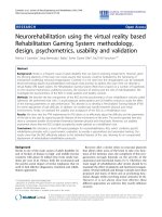
NBS1 associates with viral DNA and is required for recruitment of ATM but not ATRFigure 1
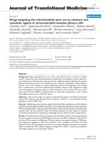
NBS1 associates with viral DNA and is required for recruitment of ATM but not ATR. (A) Chromatin immuno-
precipitation of infected NBS1-deficient and control cells. To establish if NBS1, ATM, and/or ATR associate with viral DNA,
normal and NBS1-deficient cells were infected with the HIV-1-based vector at an m.o.i. of 0.1 and chromatin immunoprecipita-
tion was performed with anti-NBS1, anti-ATM and anti-ATR antibodies as described in the Experimental Procedures. m –
mock, uninfected cells. The immunoprecipitating antibody is indicated on the left side of the photograph of the gel. (B) Chro-
matin immunoprecipitation of infected NBS1-deficient and control cells, which were transfected with the normal NBS1 gene.
Control and NBS1-deficient cells were transfected with the NBS1-coding plasmid or an empty vector. 48 hrs post-transfection,
cells were infected with the HIV-1-based vector at an m.o.i. of 0.1 and chromatin immunoprecipitation was performed 24 hrs
later with anti-NBS1 and anti-ATM antibodies as described in the Experimental Procedures. m – uninfected cells, v – cells
infected with the HIV-1-based vector, N – cells transfected with the normal NBS1 gene and infected with the HIV-1-based vec-
tor, c – cells transfected with the empty plasmid vector and infected with the HIC-1-based vector.
AB
ATM
ATR
PI-3K
NBS1
ATM
m
NBS1
8
12
24
48
m
812
24
48
Hrs
post-infection
Hrs
post-infection
m
N
vcm
N
vc
Control cells NBS1 (-) cells
Control cells NBS1 (-) cells
Virology Journal 2008, 5:11 />Page 4 of 12
(page number not for citation purposes)
tease inhibitors, and washed and lysed by addition of
0.5% Nonidet P-40, 5 mM PIPES, pH 8.0, 85 mM KCL
and protease inhibitors. The intact nuclei were isolated by
centrifugation at 5000 rpm at 4°C. Nuclei were then
resuspended in a lysis buffer (1% SDS, 50 mM Tris-Cl, pH
8.1, 10 mM EDTA, protease inhibitors). Chromatin was
sonicated to obtain DNA fragments of approximately 600
bp. Samples were subjected to centrifugation to remove
debris and were precleared by shaking for 1 hr with
salmon sperm DNA/protein A-agarose (Upstate, Temec-
ula, CA, cat. no. 16–157), which were then removed and
supernatants were diluted 10-fold with a dilution buffer
(0.01% SDS, 1.1% Triton X-100, 1.2 mM EDTA, 16.7 mM
Tris-Cl, pH 8.1, 167 mM NaCl, protease inhibitors). Chro-
matin fragments were immunoprecipitated overnight
with antibodies against ATM (Santa Cruz Biotechnology,
sc-15392), ATR (Santa Cruz Biotechnology, sc-1887),
NBS1 (Santa Cruz Biotechnology, sc-8580), or, as a con-
trol, the irrelevant protein PI-3K 110δ (Santa Cruz Bio-
technology, sc-55589). Protein-DNA-antibody complexes
were isolated by the addition of salmon sperm DNA/pro-
tein A-agarose. After 1 hr, complexes were collected by
centrifugation and washed three times with buffer (100
mM Tris, pH 8, 500 mM LiCl, 1% Nonidet P-40, 1% deox-
ycholic acid). Pellets were eluted from salmon sperm
DNA/protein A-agarose with 50 mM NaHCO3, 1% SDS
for 15 min at room temperature. Clarified samples were
incubated with RNase and 5 M NaCl at 67°C for 4–5 hr to
reverse cross-links and then precipitated overnight with
ethanol. Following centrifugation, pellets were resus-
pended in proteinase K buffer and treated with proteinase
K to digest residual proteins. After phenol/chloroform
extraction, the DNA was precipitated with ethanol. Viral
sequences in these fractions were detected by PCR using
primers targeting the HIV-1 long terminal repeats: M667,
5'-GGC TAA CTA GGG AAC CCA CTG-3'; AA55, 5'-CTG
CTA GAG ATT TTC CAC ACT GAC-3'[35]. The PCR reac-
tion was done as follows: 94C for 5 min, then 30 cycles of
94C – 1 min, 55C – 1 min, 72C – 1 min. Final extension
was run for 5 min at 72C. PCR products were resolved on
an ethidium bromide-stained 2% agarose gel.
Alu-PCR
To detect and quantify fully integrated proviral DNA, a
two-step nested PCR technique was conducted. Primary
NBS1-deficient fibroblasts and control cells were infected
with HIV-1-based vector (lacZ reporter) at m.o.i 1, m.o.i.
0.01, or mock infected. Three days post-infection genomic
DNA was extracted (Promega kit A1120). First round of
Alu-PCR employed a primer targeting the cellular Alu
sequence 5'-GCC TCC CAA AGT GCT GGG ATT ACA G-3'
as well as the M661 primer targeting the HIV-1 LTR/gag
region, 5'-CCT GCG TCG AGA GAG CTC CTC TGG-3'.
This initial amplification step used 150 ng of genomic
DNA as template. Samples were subjected to 30 PCR
cycles of 95C – 30 s, 60C – 45 s, and 72C – 5 min, and
after the final round, samples were kept at 72°C for 10
min. Products of the first round were diluted 1/1,000 and
used in the 30-cycle second round (nested) with viral LTR
primers: 5'-GGA TTG TGC TAC AAG CTA GTA CC-3'; and
5'-TGA GGG ATC TCT AGT TAC CAG AGT-3'. Second-
round PCR was cycled as follows: 95°C for 5 min; 30
cycles of 95°C for 40 s, 55°C for 45 s, 72°C for 60 s, and
the last round was followed by 72°C for 10 min. PCR
products from the second round were resolved by electo-
phoresis on an agarose gel and subjected to Southern blot-
ting with an HIV-1- LTR probe.
Statistics
Quantitative data are displayed as means ± standard devi-
ations. Comparisons between two groups were performed
using the two tailed Student t-test.
Results
The NBS1 protein is required for association of ATM, but
not ATR, with viral DNA
Normal and NBS1-deficient primary human fibroblasts
were infected with the pseudotyped HIV-1-based vector
(lacZ reporter) at an m.o.i. of 0.1 and harvested at the
indicated time points (Figure 1A). ChIP analysis was used
to identify accumulation of NBS1, ATM, and ATR at sites
of proviral DNA integration. Nuclear DNA and its associ-
ated proteins were crosslinked, immunoprecipitated with
the indicated antibodies (anti-NBS1, ATM, or ATR), and
associated viral DNA was amplified by PCR. Figure 1A
shows an agarose gel of the amplified PCR products. In
normal primary fibroblasts, the presence of viral DNA in
NBS1, ATM, and ATR immunoprecipitates was first
detected 12 hrs post-infection. In NBS1-deficient cells,
however, we did not observe any association of viral DNA
at any timepoint with NBS1, as expected, nor with ATM
(Figure 1A). Surprisingly, NBS1 deficiency and the failure
to recruit ATM did not block the association of viral DNA
with ATR (Figure 1A, third row), even though NBS1 and
ATM are each required for recruitment of ATR to DSB sites
[15,16]. As a negative control, no viral DNA was detected
in any sample immunoprecipitated with the irrelevant
anti-PI-3K kinase antibody (Figure 1A, bottom row).
To verify that the failure of ATM association with viral
DNA in NBS1-deficient cells arises specifically from the
mutation in the NBS1 gene, rather than from some other
difference between these and control cells, we performed
NBS1 reconstitution studies. Normal and NBS1-deficient
fibroblasts were transfected with either an expression plas-
mid for wild-type NBS1 or an empty vector [36]. Trans-
fected cells were then infected with the HIV-1-based
vector. The right half of Figure 1B shows ATM association
with viral DNA in NBS1-deficient cells that were trans-
fected with the NBS1 expression plasmid, but not in
Virology Journal 2008, 5:11 />Page 5 of 12
(page number not for citation purposes)
NBS1-deficient cells transfected with the empty vector,
thereby confirming the essential role of NBS1. The NBS1
expression plasmid brought the amount of viral DNA
associated with ATM to roughly the same level as in nor-
mal control cells (Figure 1B). Interestingly, overexpres-
sion of NBS1 in normal cells enhanced the association of
viral DNA with ATM (Figure 1B, left half), suggesting that
the NBS1 protein could be a limiting factor for ATM-medi-
ated PIR even in normal cells. Taken together, our results
demonstrate that NBS1 is required for association of ATM,
but not ATR, with vector DNA.
The NBS1 protein is required for efficient stable
transduction of human fibroblasts by HIV-1-based vectors
Given our finding of NBS1 association with DNA of the
HIV-1-based vector, we sought to determine its role in the
life-cycle of the HIV-1-based vectors. Normal and NBS1-
deficient primary fibroblasts were infected with the HIV-
based vector carrying the lacZ reporter at an m.o.i. of
0.025, and the infected cells were counted by staining for
β-galactosidase activity at late timepoints, indicative of
stable retroviral transduction (5–7 days post-infection,
dpi). Of note, we observed that the NBS1-deficient pri-
mary fibroblasts in this study grew at a rate close to that of
normal cells and exhibited the same plating efficiency as
normal cells. As shown in Figure 2A, the infection effi-
ciency of NBS1-deficient fibroblasts was only 35% of that
of the control cells at 5 dpi and decreased to 24% of the
control value at 7 dpi. Figure 2B shows typical micro-
scopic images used to generate the quantitative data in
Figure 2A. To verify that NBS1 deficiency does not directly
affect the lacZ reporter, control and NBS1-deficient cells
were transfected with the non-viral lacZ plasmid, and β-
galactosidase activity was quantified in cells 3 days later
by staining. As shown in Figure 2C, NBS1 deficiency did
not alter CMV-driven lacZ expression.
To test whether the transduction deficiency of NBS1-defi-
cient cells can be observed using another reporter gene,
control and NBS1-deficient primary human fibroblasts
were infected with an HIV-1-based vector carrying the
EGFP reporter [3]. At an m.o.i. of 0.1, 13.45% of control
cells expressed the reporter gene whereas EGFP expression
was detected in only about one third as many NBS1-defi-
cient fibroblasts (4.79%, Figure 2D). Based on the results
of these different assays, we conclude that NBS1 defi-
ciency substantially decreases stable retroviral transduc-
tion of primary human fibroblasts. We note that a drop of
transduction efficiency of NBS1-deficient cells was noted
previously (about two fold), but the data were not further
analyzed [37].
The transduction deficiency of the NBS1-deficient cells
can be rescued by expression of normal NBS1
The transduction deficiency of NBS1-deficient cells could
be conceivably due to an additional mutation gained by
these cells, instead of the NBS1 mutation. To test this
hypothesis, NBS1-deficient fibroblasts were transfected
with the expression plasmid for wild-type NBS1 or the
empty control vector [36]. Transfected cells were then
infected with the lacZ-carrying HIV-1-based vector. As
shown in the Figure 3, transduction efficiency of NBS1-
deficient cells reconstituted with the NBS1 expression
plasmid was more than twice the level in cells that
received the empty vector. Thus, the deficiency in retrovi-
ral transduction in NBS1-deficient cells arises directly
from the mutation in the NBS1 gene.
The NBS1 protein is required for efficient transduction of
human lymphoid cells by HIV-1-based vectors
To determine if retroviral transduction depends on NBS1
in cells other than primary human fibroblasts, EBV-trans-
formed B-lymphoid cells from normal and NBS subjects
were infected with the HIV-1-based vector carrying the
EGFP marker. Transduction efficiency was measured 7 dpi
by flow cytometry. At an m.o.i. of 0.1, 7.29% of control
cells were infected, while NBS EBV-transformed B-lym-
phocytes were infected at approximately one-third the rate
(2.64%, Figure 4). Thus, similar to NBS1-deficient fibrob-
lasts, NBS1-deficient B-lymphoid cells exhibit substan-
tially decreased transduction efficiency by HIV-1-based
vectors, indicating that efficient HIV-1 transduction
requires NBS1 in other cell types as well.
The transduction deficiency of NBS cells does not result
from a defect in vpr-mediated cell-cycle arrest
An HIV-1 accessory gene, vpr, was reported to induce G2
cell-cycle arrest by triggering the ATR-dependent check-
point cascade [38]. Hypothetically, vpr could increase
HIV-1 transduction by inducing the growth arrest, thereby
giving the cell additional time to complete PIR. Since
NBS1 is involved in cell-cycle checkpoint activation as
well [39], it is conceivable that NBS1 deficiency could
result in loss of vpr-induced growth arrest and it this way
lead to reduced HIV-1 transduction. If this were the case,
then the NBS1 deficiency should not affect HIV-1 trans-
duction in the absence of vpr. To test this hypothesis, we
infected control and NBS1-deficient fibroblasts with a
multiply attenuated HIV-1-based vector (MAV) that is
missing the vpr gene. Separate cells were infected in paral-
lel with the original non-attenuated HIV-1-based vector,
which contains vpr. Figure 5 shows that control cells were
infected with both vectors at approximately 6-fold higher
rates than NBS1-deficient cells. Because MAV does not
contain the viral vpr gene, reduced transduction of NBS1-
deficient cells cannot be attributed to a lack of vpr-medi-
ated cell cycle arrest.
Virology Journal 2008, 5:11 />Page 6 of 12
(page number not for citation purposes)
The NBS1 protein is required for efficient joining of viral
DNA to host cell DNA, but does not affect other steps in
the HIV-1 life cycle
To determine which step of the vector life-cycle involves
NBS1, we infected primary NBS1 and control fibroblasts
with the HIV-1-based vector carrying the lacZ reporter and
analyzed viral DNA synthesis, nuclear import of viral
DNA, and completed DNA joining events by Alu-PCR at 3
dpi. NBS1 deficiency did not measurably decrease viral
DNA synthesis or nuclear import as measured by forma-
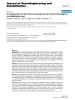
NBS1 is required for efficient HIV-1 transduction of primary cellsFigure 2
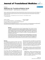
NBS1 is required for efficient HIV-1 transduction of primary cells. (A) NBS1-deficient fibroblasts (GM07166) and
matched controls (GM04506) were infected with HIV-1-based vector carrying the lacZ reporter at an m.o.i. of 0.25. Five and
seven days post infection (dpi) cells were stained using a β-galactosidase assay (Stratagene protocol) and transduced (blue) cells
were counted under a light microscope the following day. Light grey – NBS1-deficient cells; dark grey – normal cells. The error
bars represent standard deviation, p = 0.029 for 5 dpi and 0.021 for 7 dpi. (B) Light microscopic images from the same exper-
iment as in A. (C) The effect of the NBS1 deficiency on expression of the lacZ marker. The NBS1-deficient and control cells
were transfected with the lacZ plasmid and lacZ-expressing cells were counted three days post transfection. Six randomly
selected fields were counted per each point. The error bars represent standard deviation. The differences were not statistically
significant (p > 0.2) (D) Transduction with the HIV-1-based vector carrying the EGFP marker. NBS1-deficient and control
fibroblasts were infected with the vector and transduced cells were counted by flow cytometry at multiple time points (2–7
dpi). Results from 7 dpi are shown. Histograms of mock infected cells (top) and cells infected at an m.o.i. of 0.1 (bottom) are
shown. As seen in the gated regions, 13.45% of control fibroblasts were stably transduced, whereas transduction of NBS
fibroblasts was only 4.79%.
Control 5 dpi
Control 7 dpi
NBS1(-) 5 dpi
0.03%
Control Mock
0.01%
NBS1(-) Mock
Control Infected NBS1(-) Infected
4.79%
13.45%
NBS1(-) 7 dpi
GFP
GFPGFP
0
100
200
300
400
500
600
5 dpi 7 dpi
Stably Transduced Cells
Control Cells
NBS Cells
NBS1(-) Cells
BA
C
Control cells NBS1 (-) cells
lacZ lacZmm
5
10
15
20
25
30
Blue cells per field
D
GFP
Virology Journal 2008, 5:11 />Page 7 of 12
(page number not for citation purposes)
tion of 2-LTR circles (data not shown), the latter finding
being consistent with Kilzer et al. Importantly, the
number of completed joining events was reduced by
approximately two-thirds in NBS1-deficient fibroblasts
relative to the control cells (Figure 6A). Thus, NBS1 is
involved in the joining of viral to host DNA.
Retroviral infection triggers apoptosis of NBS1-deficient
cells in an integrase-dependent manner
The decreased amount of viral DNA that is joined to host
cell DNA in NBS1-deficient cells presumably results from
a failure of PIR. As a theoretical alternative, the NBS1 pro-
tein might be required for the initial integrase-mediated
joining reaction. To distinguish between these possibili-
ties, we took advantage of the fact that failure of PIR after
integrase-mediated joining of viral and host DNA in other
contexts triggers apoptosis through activation of cell-cycle
checkpoint proteins by the unrepaired intermediate, lead-
ing to a loss of infected cells from the population
[3,5,6,8,25,26]. Thus, normal and NBS1-deficient fibrob-
lasts were infected at a high m.o.i. (4.0) with an integra-
tion-competent HIV-1-based vector or a vector carrying
the enzymatically inactive D64V mutation in the integrase
protein. Cells were further analyzed by Western blotting
for the presence of the 85-kDa PARP fragment, an apop-
totic marker generated by caspase-mediated cleavage of
the PARP protein [40]. As shown in the Figure 6B, only
one infection condition stimulated PARP cleavage,
namely, infection of NBS1-deficient cells with the inte-
grase-competent HIV-1-based vector. Neither cell type
underwent apoptosis after infection with the integrase-
deficient virus, and neither viral construct induced PARP
cleavage in the normal cells. Thus, HIV-1 infection
induces apoptosis of NBS1-deficient cells in an integrase-
dependent manner. This finding is consistent with the
failure of PIR rather than a defect in the initial integrase-
mediated joining.
The ATM kinase is required for efficient HIV-1
transduction of primary human cells
As noted in the Introduction, ATM was proposed as an
essential host factor for PIR, but the literature contains
conflicting data [5,8,27,28], possibly owing to the use of
different transformed cell lines by different laboratories.
Moreover, DSB requires both NBS1 [15] and ATM [16] for
the recruitment of ATR, yet our ChIP studies demon-
strated that ATR robustly localizes to sites of PIR without
either of these proteins (Figure 1A, third row). Thus, cells
specifically deficient in ATM, despite their defect in DSB
repair [21,24], would still exhibit localization of both
NBS1 and ATR to sites of viral integration, and it is possi-
ble that these proteins would then mediate PIR independ-
ently of ATM. To test this possibility in non-transformed
cells, we infected normal and ATM-deficient (A-T) pri-
mary human fibroblasts with our HIV-1-based vectors.
ATM-deficient cells reproducibly demonstrated a decrease
of transduction efficiency by 60–80% compared to nor-
mal cells, regardless of the readout method or the trans-
duced reporter (Fig 7). These results agree with Lau et al.
[8] and support the hypothesis that ATM is required for
efficient PIR in primary human cells, despite the inde-
pendent recruitment of ATR (Figure 1A).
Discussion
In this study, we demonstrated that NBS1, an early sensor
of DSBs, associates with viral DNA, is required for the
association of ATM – but not ATR – with viral DNA, medi-
ates efficient integration of viral DNA, promotes stable
retroviral transduction, and blocks integrase-dependent
apoptosis that can arise from unrepaired viral-host link-
ages. These data support a key role for the NBS1 protein in
PIR. We and others proposed that retroviral PIR employs
the NHEJ pathway, including the ATM and ATR kinases
[3-9]. Our current results extend that work, by demon-
strating the dependence of PIR on NBS1, an interaction
between NBS1 and ATM, and a dependence on ATM for
PIR in primary, non-transformed cells. All of these fea-
tures are shared with cellular DSB repair.
Reintroduction of normal NBS1 cDNA into NBS1-deficient cells restores HIV-1 transduction efficiencyFigure 3
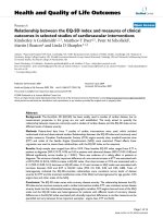
Reintroduction of normal NBS1 cDNA into NBS1-
deficient cells restores HIV-1 transduction efficiency.
NBS1-deficient cells were transfected with a plasmid encod-
ing normal NBS1 cDNA or an empty vector plasmid. One
day post-transfection, cells were infected with the HIV-1-
based vector carrying the lacZ reporter. Cells were then
stained eight days post-infection using a β-galactosidase assay
and transduced (blue) cells were counted. c – control (cells
transfected) with the empty vector, NBS1 – cells transfected
with the plasmid carrying the normal NBS1 gene. The error
bars represent standard deviation, p = 0.037.
Virology Journal 2008, 5:11 />Page 8 of 12
(page number not for citation purposes)
Nevertheless, the integration intermediate structurally dif-
fers from a DSB (see the Introduction), and so we now
revised our model to include the concept that the two
repair processes may diverge in key aspects. Initial evi-
dence that PIR uses somewhat different cellular machin-
ery than DSB repair came from our recent study where we
observed that phosphorylation of the histone H2AX iso-
form, which is mediated by both the ATM and ATR
kinases and is required for DSB repair, appears to be dis-
pensable for PIR, although it can be detected at the inte-
gration sites [31]. Importantly, our current results
establish the surprising finding that recruitment of ATR,
which in the context of DSB requires both NBS1 and ATM,
proceeds independently of these two proteins. In this con-
text, we note that some HIV-1 transduction occurs even in
the absence of normal NBS1 or ATM (see Results section).
It is possible that this residual transduction is mediated by
ATR.
One possible explanation for the difference in ATR recruit-
ment in PIR vs. DSB repair could be that the single-
stranded DNA gaps, which flank the integration site, are
sufficient to recruit the ATR protein. In contrast, MRN-
dependent processing of DSBs, which may generate sin-
gle-stranded DNA through the nuclease activity of
MRE11, appears necessary for accumulation of ATR at the
DSB sites [16]. Additional differences between these DNA
repair processes may exist, and might guide the develop-
ment of therapeutic strategies to selectively inhibit PIR
without blocking DSB repair.
NBS1 is required for efficient HIV-1 transduction of lymphoid cellsFigure 4
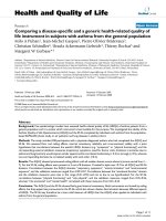
NBS1 is required for efficient HIV-1 transduction of lymphoid cells. EBV-transformed NBS1-deficient B-lymphoid
cells (GM15818) and matched control EBV-transformed cells (GM15817) were infected with the HIV-1-based vector carrying
the EGFP reporter and then assayed 7 dpi by FACS to quantify reporter gene expression. At an m.o.i. of 0.1, control lym-
phocytes were infected at a rate of 7.29%, while only 2.64% of NBS lymphocytes were infected.
Control B-Lymphocytes Infected
7.29%
2.64%
NBS1(-) B-Lymphocytes Infected
Control B-Lymphocytes Mock
NBS1(-) B-Lymphocytes Mock
0.00%
0.03%
Virology Journal 2008, 5:11 />Page 9 of 12
(page number not for citation purposes)
NBS1 facilitates HIV-1 transduction independently of the vpr geneFigure 5
NBS1 facilitates HIV-1 transduction independently of the vpr gene. Cells were infected with either the normal ("n")
or MAV vector at an m.o.i. of 0.1, and then stained overnight using a β-galactosidase assay at seven dpi. (A) Stably transduced
cells per dish. (B) Pictures under the light microscope from the same experiment.
0
100
200
300
400
500
600
700
800
"wt" vector MAV vector
Stably Transduced cells
A
Control Cells
NBS1(-) Cells
“n” vector MAV vector
Stably Transduced Cells
“n”
MAV
Control 7 dpi NBS1(-) 7 dpi
B
NBS1 is required to efficiently complete the integration of viral DNA and to avoid integrase-dependent apoptosisFigure 6
NBS1 is required to efficiently complete the integration of viral DNA and to avoid integrase-dependent apop-
tosis. (A) Completed integration in NBS1-deficient vs. normal control cells. Alu-PCR was performed to detect viral-host
DNA junctions. In this nested PCR technique, genomic DNA was extracted from HIV-1-infected NBS1-deficient and control
cells at 3 dpi. The first round of PCR was performed with one primer targeting the virus LTR region, and the other primer tar-
geting cellular Alu sequences. The second round utilized two LTR primers. Top – the amplified viral sequences were detected
by southern blotting. Bottom – Southern was quantified by densitometry. (B) PARP cleavage in infected cells. Normal and
NBS1-deficient cells were infected as described in the Experimental Procedures. Two days post-infection, cells were harvested,
lysed and cell lysates subjected to western blotting with an anti-PARP antibody. wt – cells infected with an integration-compe-
tent HIV-1-based vector, D64V – cells infected with the vector carrying the D64V mutation in the integrase protein.
Virology Journal 2008, 5:11 />Page 10 of 12
(page number not for citation purposes)
ATM is required for efficient transduction of primary fibroblastsFigure 7
ATM is required for efficient transduction of primary fibroblasts. (A) HIV-1 transduction of the lacZ marker as meas-
ured by detecting lacZ reporter activity in infected A-T fibroblast and control fibroblast cells. Cells were infected with an HIV-
1-based vector carrying the lacZ reporter at an m.o.i. of 0.3. Infected cells were stained overnight using a β-galactosidase assay
at five and seven dpi and transduced cells counted. (B) Light microscopic images from the same experiment as in A, taken 4
dpi. (C) A-T and control fibroblast infections with the HIV based vector carrying the EGFP marker. Cells were evaluated by
flow cytometry at multiple time points (2–7 dpi). Results from 7 dpi are shown. Histograms of mock-infected control cells
(left), control cells infected at m.o.i. of 0.1 (middle), and infected A-T cells are shown. The error bars represent standard devi-
ation, p = 0.0014 for 5 dpi and 0.027 for 7 dpi.
Control Mock Control Infected A-T Infected
0.00% 9.87%
3.28%
AB
0
50
100
150
200
250
300
350
400
450
5 dpi 7 dpi
Stably Transduced cells
Control
A-T
C
A-TControl
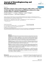
Model for the role of NBS1 in post-integration repairFigure 8
Model for the role of NBS1 in post-integration repair. Integrase catalyzes formation of the integration intermediate (1).
NBS1 and ATR are recruited independently to the integration sites (2). NBS1 then recruits MRE11, RAD50 and ATM. (3). The
5'-end DNA flaps of the viral DNA are trimmed, possibly by MRN. ATM phosphorylates H2AX. However, H2AX phosphoryla-
tion is not required for post-integration repair. (4). We speculate that Artemis and NHEJ proteins are recruited to the integra-
tion site (5). These proteins, and likely other factors, then mediate the other steps of post integration repair, which require
possibly further end processing, gap filling, ligation and chromatin remodeling (6).
INTEGRATED RETROVIRAL DNA
INTEGRATION
INTERMEDIATE
RETROVIRAL DNA
5'
5'
(REPAIRED)
phosphorylation
5'
5'
ATM
5'
5'
Other (unknown)
factors?
ATR
Flap resection?
H2AX
5'
5'
ATM
MRN
ATR
P
P
R = Artemis
1
2
3
4
5
Further end processing
Gap filling
Ligation
Chromatin remodeling
6
H2AX
5'
5'
ATM
MRN
ATR
NHEJ
R
P
P
NBS1
ATR
NBS1
RAD50
MRE11
Virology Journal 2008, 5:11 />Page 11 of 12
(page number not for citation purposes)
Our discovery of a crucial role for NBS1 in PIR opens sev-
eral possibilities with regards to the molecular mecha-
nism of PIR. First, the simplest model is that NBS1 acts
primarily through its recruitment of the ATM kinase to
integration sites, as suggested by our results. ATM, in turn,
may phosphorylate other proteins at integration sites and
thus regulate their activity. Interestingly, it was recently
shown that ATM phosphorylates the DNA repair protein
Artemis, and ATM is required for Artemis-dependent
processing of damaged ends of DNA [41]. Artemis is a crit-
ical component of the cellular non-homologous end join-
ing (NHEJ) DNA repair pathway and these data thus
provide a link between the ATM kinase and NHEJ path-
way. We and others have presented extensive evidence
indicating that NHEJ is involved in PIR [3,5-8]. One could
thus imagine that NBS1 exerts its effect on PIR by regulat-
ing the ATM-Artemis-NHEJ pathway. However, NBS1 is
also a component of the MRN complex, and recruits the
MRE11 nuclease of this complex to the sites of DNA
breaks [13,14]. The process of PIR involves trimming of
5'-viral DNA ends prior to joining of viral and host DNA
ends. An intriguing role for MRE11 in the MRN complex
would be trimming of these short flaps of viral DNA (Fig-
ure 8).
As we and others have suggested, cellular co-factors con-
stitute an attractive target for anti-HIV-1 therapy, since
development of resistance against inhibitors of these pro-
teins is unlikely [6,8,9,27,42]. NBS1 and its interactive
partners, being such co-factors, are thus potential targets
for anti-HIV-1 therapeutics, particularly at steps where PIR
differs from DSB repair.
Competing interests
The author(s) declare that they have no competing inter-
ests.
Authors' contributions
JAS carried out the HIV-1 transduction experiments and β-
galactosidase and EGFP assays. FW carried out the Alu-
PCR assay. KW provided the NBS1 and control vectors
and participated in designing the NBS1 reconstitution
experiment and revising the manuscript. HZ and KJW
extensively participated in drafting the manuscript and
experimental design. RD conceived of the study, carried
out the chromatin immunoprecipitations and western
blotting experiments and wrote the manuscript. All
authors read and approved the final manuscript.
Acknowledgements
We thank Dr. David Horn (TJU-Infectious Diseases) for reading the manu-
script and helpful comments. This work has been supported by NIH grants
CA98090 and CA125272 (R.D.) and MH70279 (K.J.W.) and a W.W. Smith
Foundation AIDS Research Award (R.D.).
References
1. Craigie R, Fujiwara T, Bushman F: The IN protein of Moloney
murine leukemia virus processes the viral DNA ends and
accomplishes their integration in vitro. Cell 1990, 62:829-837.
2. Katz RA, Merkel G, Kulkosky J, Leis J, Skalka AM: The avian retro-
viral IN protein is both necessary and sufficient for integra-
tive recombination in vitro. Cell 1990, 63:87-95.
3. Daniel R, Greger JG, Katz RA, Taganov KD, Wu X, Kappes JC, Skalka
AM: Evidence that stable retroviral transduction and cell sur-
vival following DNA integration depend on components of
the nonhomologous end joining repair pathway. J Virol 2004,
78:8573-8581.
4. Daniel R, Kao G, Taganov K, Greger JG, Favorova O, Merkel G, Yen
TJ, Katz RA, Skalka AM: Evidence that the retroviral DNA inte-
gration process triggers an ATR-dependent DNA damage
response. Proc Natl Acad Sci USA 2003, 100:4778-4783.
5. Daniel R, Katz RA, Merkel G, Hittle JC, Yen TJ, Skalka AM: Wort-
mannin potentiates integrase-mediated killing of lym-
phocytes and reduces the efficiency of stable transduction by
retroviruses. Mol Cell Biol 2001, 21:1164-1172.
6. Daniel R, Katz RA, Skalka AM: A role for DNA-PK in retroviral
DNA integration. Science 1999, 284:644-647.
7. Downs JA, Jackson SP: Involvement of DNA end-binding pro-
tein Ku in Ty element retrotransposition. Mol Cell Biol 1999,
19:6260-6268.
8. Lau A, Swinbank KM, Ahmed PS, Taylor DL, Jackson SP, Smith GC,
O'Connor MJ: Suppression of HIV-1 infection by a small mole-
cule inhibitor of the ATM kinase. Nat Cell Biol 2005, 7:493-500.
9. Skalka AM, Katz RA: Retroviral DNA integration and the DNA
damage response. Cell Death Differ 2005, 12(Suppl 1):971-978.
10. Paull TT, Gellert M: The 3' to 5' exonuclease activity of Mre 11
facilitates repair of DNA double-strand breaks. Mol Cell
1998,
1:969-979.
11. D'Amours D, Jackson SP: The Mre11 complex: at the crossroads
of dna repair and checkpoint signalling. Nat Rev Mol Cell Biol
2002, 3:317-327.
12. Lukas C, Falck J, Bartkova J, Bartek J, Lukas J: Distinct spatiotem-
poral dynamics of mammalian checkpoint regulators
induced by DNA damage. Nat Cell Biol 2003, 5:255-260.
13. Kobayashi J, Tauchi H, Sakamoto S, Nakamura A, Morishima K, Mat-
suura S, Kobayashi T, Tamai K, Tanimoto K, Komatsu K: NBS1
localizes to gamma-H2AX foci through interaction with the
FHA/BRCT domain. Curr Biol 2002, 12:1846-1851.
14. Tauchi H, Kobayashi J, Morishima K, Matsuura S, Nakamura A, Shirai-
shi T, Ito E, Masnada D, Delia D, Komatsu K: The forkhead-associ-
ated domain of NBS1 is essential for nuclear foci formation
after irradiation but not essential for hRAD50[middle
dot]hMRE11[middle dot]NBS1 complex DNA repair activ-
ity. J Biol Chem 2001, 276:12-15.
15. Falck J, Coates J, Jackson SP: Conserved modes of recruitment
of ATM, ATR and DNA-PKcs to sites of DNA damage. Nature
2005, 434:605-611.
16. Jazayeri A, Falck J, Lukas C, Bartek J, Smith GC, Lukas J, Jackson SP:
ATM- and cell cycle-dependent regulation of ATR in
response to DNA double-strand breaks. Nat Cell Biol 2006,
8:37-45.
17. Lim DS, Kim ST, Xu B, Maser RS, Lin J, Petrini JH, Kastan MB: ATM
phosphorylates p95/nbs1 in an S-phase checkpoint pathway.
Nature 2000, 404:613-617.
18. Stiff T, Reis C, Alderton GK, Woodbine L, O'Driscoll M, Jeggo PA:
Nbs1 is required for ATR-dependent phosphorylation
events. Embo J 2005, 24:199-208.
19. Wu X, Ranganathan V, Weisman DS, Heine WF, Ciccone DN, O'Neill
TB, Crick KE, Pierce KA, Lane WS, Rathbun G, et al.: ATM phos-
phorylation of Nijmegen breakage syndrome protein is
required in a DNA damage response. Nature 2000,
405:477-482.
20. Zhao S, Weng YC, Yuan SS, Lin YT, Hsu HC, Lin SC, Gerbino E, Song
MH, Zdzienicka MZ, Gatti RA, et al.: Functional link between
ataxia-telangiectasia and Nijmegen breakage syndrome
gene products. Nature 2000, 405:473-477.
21. Difilippantonio S, Celeste A, Fernandez-Capetillo O, Chen HT, Reina
San Martin B, Van Laethem F, Yang YP, Petukhova GV, Eckhaus M, Fei-
genbaum L, et al.: Role of Nbs1 in the activation of the Atm
kinase revealed in humanized mouse models. Nat Cell Biol
2005, 7:675-685.
Publish with BioMed Central and every
scientist can read your work free of charge
"BioMed Central will be the most significant development for
disseminating the results of biomedical research in our lifetime."
Sir Paul Nurse, Cancer Research UK
Your research papers will be:
available free of charge to the entire biomedical community
peer reviewed and published immediately upon acceptance
cited in PubMed and archived on PubMed Central
yours — you keep the copyright
Submit your manuscript here:
/>BioMedcentral
Virology Journal 2008, 5:11 />Page 12 of 12
(page number not for citation purposes)
22. De la Torre C, Pincheira J, Lopez-Saez JF: Human syndromes with
genomic instability and multiprotein machines that repair
DNA double-strand breaks. Histol Histopathol 2003, 18:225-243.
23. Shiloh Y: ATM and related protein kinases: safeguarding
genome integrity. Nat Rev Cancer 2003, 3:155-168.
24. Kang J, Bronson RT, Xu Y: Targeted disruption of NBS1 reveals
its roles in mouse development and DNA repair. Embo J 2002,
21:1447-1455.
25. Jeanson L, Subra F, Vaganay S, Hervy M, Marangoni E, Bourhis J, Mous-
cadet JF: Effect of Ku80 depletion on the preintegrative steps
of HIV-1 replication in human cells. Virology 2002, 300:100-108.
26. Waninger S, Kuhen K, Hu X, Chatterton JE, Wong-Staal F, Tang H:
Identification of cellular co-factors for human immunodefi-
ciency virus replication via a ribozyme-based genomics
approach. Journal of Virology 2004, 78:12829-12837.
27. Daniel R, Pomerantz RJ: ATM: HIV-1's Achilles heel? Nat Cell Biol
2005, 7:452-453.
28. Ariumi Y, Turelli P, Masutani M, Trono D: DNA damage sensors
ATM, ATR, DNA-PKcs, and PARP-1 are dispensable for
human immunodeficiency virus type 1 integration. J Virol
2005, 79:2973-2978.
29. Dehart JL, Andersen JL, Zimmerman ES, Ardon O, An DS, Blackett J,
Kim B, Planelles V: The ataxia telangiectasia-mutated and
Rad3-related protein is dispensable for retroviral integra-
tion. J Virol 2005, 79:1389-1396.
30. Li L, Olvera JM, Yoder KE, Mitchell RS, Butler SL, Lieber M, Martin SL,
Bushman FD: Role of the non-homologous DNA end joining
pathway in the early steps of retroviral infection. EMBO Jour-
nal 2001, 20:3272-3281.
31. Daniel R, Ramcharan J, Rogakou E, Taganov KD, Greger JG, Bonner
W, Nussenzweig A, Katz RA, Skalka AM: Histone H2AX is phos-
phorylated at sites of retroviral DNA integration but is dis-
pensable for postintegration repair. J Biol Chem 2004,
279:45810-45814.
32. Naldini L, Blomer U, Gallay P, Ory D, Mulligan R, Gage FH, Verma IM,
Trono D:
In vivo gene delivery and stable transduction of non-
dividing cells by a lentiviral vector. Science 1996, 272:263-267.
33. Zufferey R, Nagy D, Mandel RJ, Naldini L, Trono D: Multiply atten-
uated lentiviral vector achieves efficient gene delivery in
vivo. Nat Biotechnol 1997, 15:871-875.
34. Boyd KE, Farnham PJ: Identification of target genes of onco-
genic transcription factors. Proc Soc Exp Biol Med 1999, 222:9-28.
35. Zack JA, Arrigo SJ, Weitsman SR, Go AS, Haislip A, Chen IS: HIV-1
entry into quiescent primary lymphocytes: molecular analy-
sis reveals a labile, latent viral structure. Cell 1990, 61:213-222.
36. Chiang YC, Teng SC, Su YN, Hsieh FJ, Wu KJ: c-Mycdirectly regu-
lates the transcription of the NBS1 gene involved in DNA-
double-strand break repair. J Biol Chem 2003, 278:19286-19291.
37. Kilzer JM, Stracker T, Beitzel B, Meek K, Weitzman M, Bushman FD:
Roles of host cell factors in circularization of retroviral dna.
Virology 2003, 314:460-467.
38. Roshal M, Kim B, Zhu Y, Nghiem P, Planelles V: Activation of the
ATR-mediated DNA damage response by the HIV-1 viral
protein R. J Biol Chem 2003, 278:25879-25886.
39. Zhang Y, Zhou J, Lim CU: The role of NBS1 in DNA double
strand break repair, telomere stability, and cell cycle check-
point control. Cell Research 2006, 16:45-54.
40. Kim TW, Pettingell WH, Jung YK, Kovacs DM, Tanzi RE: Alterna-
tive cleavage of Alzheimer-associated presenilins during
apoptosis by a caspase-3 family protease. Science 1997,
277:373-376.
41. Riballo E, Kuhne M, Rief N, Doherty A, Smith GC, Recio MJ, Reis C,
Dahm K, Fricke A, Krempler A, et al.: A pathway of double-strand
break rejoining dependent upon ATM, Artemis, and pro-
teins locating to gamma-H2AX foci. Mol Cell 2004, 16:715-724.
42. Hauber I, Bevec D, Heukeshoven J, Kratzer F, Horn F, Choidas A,
Harrer T, Hauber J: Identification of cellular deoxyhypusine
synthase as a novel target for antiretroviral therapy. J Clin
Invest
2005, 115:76-85.