Báo cáo hóa học: " Marine mimivirus relatives are probably large algal viruses" docx
Bạn đang xem bản rút gọn của tài liệu. Xem và tải ngay bản đầy đủ của tài liệu tại đây (303.29 KB, 8 trang )
BioMed Central
Page 1 of 8
(page number not for citation purposes)
Virology Journal
Open Access
Research
Marine mimivirus relatives are probably large algal viruses
Adam Monier
1
, Jens Borggaard Larsen
2
, Ruth-Anne Sandaa
2
,
Gunnar Bratbak
2
, Jean-Michel Claverie
1
and Hiroyuki Ogata*
1
Address:
1
Structural and Genomic Information Laboratory, CNRS-UPR 2589, IBSM, Parc Scientifique de Luminy, 163 avenue de Luminy, Case
934, 13288 Marseille Cedex 9, France and
2
Department of Biology, University of Bergen, PO Box 7800, N-5020 Bergen, Norway
Email: Adam Monier - ; Jens Borggaard Larsen - ; Ruth-
Anne Sandaa - ; Gunnar Bratbak - ; Jean-Michel Claverie - jean-
; Hiroyuki Ogata* -
* Corresponding author
Abstract
Background: Acanthamoeba polyphaga mimivirus is the largest known ds-DNA virus and its 1.2
Mb-genome sequence has revealed many unique features. Mimivirus occupies an independent
lineage among eukaryotic viruses and its known hosts include only species from the Acanthamoeba
genus. The existence of mimivirus relatives was first suggested by the analysis of the Sargasso Sea
metagenomic data.
Results: We now further demonstrate the presence of numerous "mimivirus-like" sequences using
a larger marine metagenomic data set. We also show that the DNA polymerase sequences from
three algal viruses (CeV01, PpV01, PoV01) infecting different marine algal species (Chrysochromulina
ericina, Phaeocystis pouchetii, Pyramimonas orientalis) are very closely related to their homolog in
mimivirus.
Conclusion: Our results suggest that the numerous mimivirus-related sequences identified in
marine environments are likely to originate from diverse large DNA viruses infecting
phytoplankton. Micro-algae thus constitute a new category of potential hosts in which to look for
new species of Mimiviridae.
Background
The discovery of Acanthamoeba polyphaga mimivirus was a
significant breakthrough in the recent history of virology.
Both mimivirus particle size (~750 nm) and its genetic
repertoire (1.2 Mb-genome encoding 911 protein coding
genes) are comparable to those of many parasitic cellular
organisms [1,2]. This giant virus exhibits several genes for
translation system components [3], and its particle con-
tains both DNA and RNA molecules [2]. These features
both quantitatively and qualitatively challenge the
boundary between viruses and cells, and reignited a smol-
dering debate about the origin of viruses and their role in
the emergence of eukaryotes [4-9].
Mimivirus belongs to Nucleocytoplasmic large DNA
viruses (NCLDVs) [10]. From its basal position in the phy-
logenetic trees based on conserved NCLDV core genes
[1,2], the new "Mimiviridae" family was proposed for
mimivirus [11]. NCLDVs now include Mimiviridae, Phy-
codnaviridae, Iridoviridae, Asfarviridae and Poxviridae. Mim-
ivirus is the sole member of the Mimiviridae family. The
lack of known close relatives of mimivirus makes it diffi-
Published: 23 January 2008
Virology Journal 2008, 5:12 doi:10.1186/1743-422X-5-12
Received: 9 November 2007
Accepted: 23 January 2008
This article is available from: />© 2008 Monier et al; licensee BioMed Central Ltd.
This is an Open Access article distributed under the terms of the Creative Commons Attribution License ( />),
which permits unrestricted use, distribution, and reproduction in any medium, provided the original work is properly cited.
Virology Journal 2008, 5:12 />Page 2 of 8
(page number not for citation purposes)
cult to build the evolutionary history of its surprising fea-
tures. Is mimivirus one of many eccentric creatures in
nature such as Rafflesia, a parasitic plant in southeastern
Asia known for its gigantic flower [12]? Are the mimivirus
extraordinary characteristics linked to the origin of
eukaryotes [5]? Clearly, appraising the actual biological
significance of this exceptional virus requires the isolation
and characterization of additional members of the Mimi-
viridae family.
Mimivirus was initially isolated in amoebae sampled
from the water of a cooling tower. Following the circum-
stances of its discovery, mimivirus was suspected to be a
causative agent of pneumonia [13]. The presence of anti-
bodies recognizing mimivirus in the sera of patients with
community or hospital-acquired pneumonia was
reported [14,15]. However, no serological evidence of
mimivirus infection was found in hospitalized children in
Austria [16] and mimivirus has never been isolated from
an infected patient despite numerous attempts. In the lab-
oratory, mimivirus appears to infect only species of Acan-
thamoeba [17]. Acanthamoeba are ubiquitous in nature and
they have been isolated from diverse environments
including freshwater lakes, river waters, salt water lakes,
sea waters, soils and the atmosphere [18,19]. Mimivirus
relatives might thus exist everywhere.
Ghedin and Claverie identified sequences similar to mim-
ivirus genes in the environmental sequence library from
the Sargasso Sea [20]. This strongly suggested the exist-
ence of mimivirus relatives in the sea. More recently, we
found numerous additional "mimivirus-like" sequences
in the much larger metagenomic data set generated by the
Global Ocean Sampling Expedition (hereafter referred to
as GOS data; [21]) (Monier et al., manuscript in prepara-
tion). However, the analysis of metagenomic data (i.e.
short sequences from unknown and mixed organisms)
provides no insights into the hosts susceptible to harbor
the putative new species of Mimiviridae corresponding to
these sequences.
While continually monitoring the new occurrences of
mimivirus-like sequences in public databases, we recently
noticed that the type B DNA polymerase (hereafter
referred to as PolB) sequences of three lytic viruses from
Norwegian coastal waters were very similar to the PolB
sequence of mimivirus. The three viruses [CeV01 (Gen-
Bank accession: ABU23716
), PpV01 (ABU23718), PoV01
(ABU23717
)] were isolated from diverse marine unicellu-
lar algae: Chrysochromulina ericina, Phaeocystis pouchetii and
Pyramimonas orientalis, respectively [22,23]. C. ericina and
P. pouchetii are both haptophytes but phylogenetically dis-
tant and classified in different orders, i.e. Prymnesiales and
Phaeocystales. P. pouchetii forms dense and almost mono-
specific spring blooms while C. ericina thrive in mixed
flagellate communities and at cell densities usually not
attaining bloom levels [24,25]. P. orientalis is a prasino-
phyte belonging to the green algae. It has a worldwide dis-
tribution but the abundance is most often low with no
significant contribution to the overall phytoplankton bio-
mass [26,27]. The three algal viruses infecting these phy-
toplankters have all been classified as phycodnaviruses.
In this report, we first analyzed the distribution of mimi-
virus-like sequences found in the GOS data and mapped
them on the mimivirus genome. We then performed phy-
logenetic analyses which indicated a very close relation-
ship between the PolB sequences of mimivirus and the
three algal viruses (CeV01, PpV01, PoV01), as well as with
their homologs from the metagenomic data set.
Results
We first examined the presence of "mimivirus-like"
sequences in the GOS data composed of 7.7 million
sequencing reads. Based on a protocol similar to the one
used by Ghedin and Claverie [20], we identified 5,293
open reading frames (ORFs; ≥ 60 aa) that are closely
related to protein sequences encoded in the mimivirus
genome. Of 911 mimivirus protein coding genes, 229
(25%) showed closely related sequences in the GOS data.
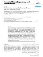
The distribution of the number of GOS matches for each
of the 229 mimivirus genes is highly variable ranging
from 1 to 249 (ex. 249 hits for MIMI_R555 DNA repair
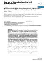
protein). These 229 mimivirus genes are distributed
widely along the chromosome, with an apparently higher
concentration in the central part of the genome (Fig. 1).
This part of the genome encodes many conserved genes
including most of the NCLDV core genes [2]. Mimivirus
possesses 26 NCLDV core genes (class I, II and III), of
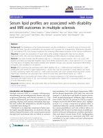
which 17 had close homologs in the GOS data (Table 1
and Additional File 1). Phylogenetic trees for the
homologs of two class I core genes (L437, VV A32-type
virion packaging ATPase; L206/L207, VV D5-type ATPase)
confirmed the separate grouping of the mimivirus
sequences with their closest homologs found in the GOS
data (Fig. 2) Among the translation related genes of mim-
ivirus, mRNA cap binding protein gene (MIMI_L496) and
translation initiation factor eEF-1 gene (MIMI_R624) had
close homologs in the GOS data. Remarkably, 55 of the
229 mimivirus genes exhibiting a strong similarity in the
GOS data, correspond to ORFans (i.e. ORFs lacking
homologs in known species), further suggesting that their
GOS homologs belong to viruses closely related to mimi-
virus.
We next selected fourteen mimivirus PolB-like GOS-ORF
sequences that are long enough to be fully aligned with
homologs from different viruses including three algal
viruses, CeV01, PpV01 and PoV01. PolB sequences from
CeV01 (GenBank: ABU23716), mimivirus [28] and Heter-
Virology Journal 2008, 5:12 />Page 3 of 8
(page number not for citation purposes)
Table 1: A selected list of mimivirus genes with closely related sequences in the GOS data.
Mimivirus ORF Annotation Number of "mimivirus-like" sequences in
the GOS data
NCLDV class I core genes
MIMI_L206 * Helicase III/VV D5-type ATPase (C-term) 139
MIMI_L207 * Helicase III/VV D5-type ATPase (N-term) 90
MIMI_R322 DNA polymerase (B family) 185
MIMI_R350 putative transcription termination factor, VV
D6R helicase
90
MIMI_L396 VV A18 helicase 138
MIMI_R400 S/T protein kinase 32
MIMI_L425 Major capsid protein 7
MIMI_L437 VV A32 virion packaging ATPase 71
MIMI_R450 A1L transcription factor 28
MIMI_R596 Thiol oxidoreductase E10R 7
NCLDV class II core genes
MIMI_R339 TFII-like transcription factor 3
MIMI_R493 Proliferating Cell Nuclear Antigen 45
NCLDV class III core genes
MIMI_L244 Rpb2 1
MIMI_L364 SW1/SNF2 helicase (MSV224) 54
MIMI_R382 mRNA Capping Enzyme 189
MIMI_R429 PBCV1-A494R-like, 9 paralogs 145
MIMI_R480 Topoisomerase II 1
MIMI_R501 Rpb1 14
Translation
MIMI_L496 Translation initiation factor 4E, (mRNA cap
binding)
11
MIMI_R624 GTP binding elongation factor eF-Tu 3
DNA repair
MIMI_L315 Hydrolysis of DNA containing ring-opened N7
methylguanine
58
MIMI_L359 DNA mismatch repair ATPase MutS 44
MIMI_R406 Alkylated DNA repair 3
MIMI_L687 Endonuclease for the repair of UV-irradiated
DNA
2
MIMI_R693 Methylated-DNA-protein-cysteine
methyltransferase
9
Other genes with more than 100
matches
MIMI_L250 putative transcription initiation factor IIB 143
MIMI_L251 Lon domain protease 110
MIMI_R303 NAD-dependent DNA ligase 163
MIMI_R325 Metal-dependent hydrolase (Chilo iridescent
virus 136R)
136
MIMI_R354 Lambda-type exonuclease 147
MIMI_R355 Unknown 150
MIMI_L375 Unknown 130
MIMI_L377 putative NTPase I 133
MIMI_R409 Unknown 155
MIMI_L434 Unknown 103
MIMI_R453 TATA-box binding protein (TBP) 131
MIMI_L454 Unknown 119
MIMI_R555 putative DNA repair protein 249
MIMI_R563 Contains helicase conserved C-terminal
domain (PFAM)
118
* Two ORFs (L206, L207) have been recently merged into a single ORF after the re-sequencing of the genomic region (SWISS-PROT: Q5UQ22,
Stéphane Audic, personal communication).
Virology Journal 2008, 5:12 />Page 4 of 8
(page number not for citation purposes)
osigma akashiwo virus [29] contain an intein element at
the same location. These intein sequences were removed
to obtain a canonical multiple alignment of the PolB
sequences. This alignment confirmed the conservation of
all the known catalytic residues [28] of the polymerase
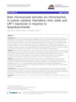
domain. A maximum likelihood tree obtained from the
alignment strongly supported the grouping of the mimivi-
rus PolB sequence, its homologs from the metagenomic
data and the PolB sequences from CeV01, PpV01 and
PoV01 (bootstrap value = 98%; Fig. 3). Similar levels of
bootstrap support were obtained by neighbor joining and
maximum parsimony approaches (99% and 80%, respec-
tively). Certain of the GOS-ORFs (nine GOS-ORFs) are
more closely related to PolB's from CeV01 and/or PpV01
(bootstrap value = 100%), while others appear to be more
closely related to PolB's from PoV01 and/or mimivirus.
The percentage of identical amino acid residues between
mimivirus PolB sequence and its GOS homologs in Figure
3 varies from 37% to 48%, suggesting a substantial level
of genetic diversity of the mimivirus relatives in the sea.
Mimivirus PolB sequence exhibits 41%, 31%, 45% iden-
tity with the PolB sequence of the three algal viruses
CeV01, PpV01, and PoV01, respectively. The phylogenetic
tree presented in Figure 3 supports the monophyletic
grouping for iridoviruses (100%) as well as for poxviruses
(75%). In contrast, the inclusion of the new mimivirus-
like PolB sequences in the phylogenetic analysis appar-
ently breaks the monophyletic grouping of viruses previ-
ously classified as member of the phycodnavirus family,
robustly clustering the CeV01, PpV01, and PoV01 viruses
with mimivirus.
Discussion
CeV01, PpV01 and PoV01 were initially isolated from
Norwegian coastal waters. An electron cryomicroscopic
analysis revealed the icosahedral capsid of PpV01 particles
with a maximum diameter of 220 nm [23]. Icosahedral
morphology was also suggested for CeV01 (160 nm) and
PoV01 (220 × 180 nm) from the observations by trans-
mission electron microscopy [22]. The genomes of these
viruses are composed of double-stranded DNA, with esti-
mated sizes being 510-kb for CeV01, 485-kb for PpV01
and 560-kb for PoV01 [22,30]. The genome sizes are sub-
stantially larger than the currently sequenced largest phy-
codnavirus genome (i.e. 407-kb for EhV-86, [31]. Electron
microscopy observations of infected cells indicate that
viral assembly takes place in the cytoplasm of all three
host cells [22,32]. Given these features, these three lytic
algal viruses are tentatively classified as phycodnaviruses.
Previous studies have indicated a relatively close phyloge-
netic relationship [2] and a similarity in gene composition
[10] between phycodnaviruses and mimivirus. Several
phycodnaviruses exhibit the largest genome sizes (>300-
kb) after mimivirus [4]. Claverie et al. have hypothesized
that Phycodnaviridae is a promising source of giant viruses
[4]. In this study, we present phylogenetic evidence for a
close relationship between the PolB sequences of three
algal viruses (CeV01, PpV01, PoV01) and mimivirus, and
for the segregation of these from homologs of other
known viruses. PolB is one of the NCLDV core genes, and
serves as a phylogenetic marker for the classification of
large DNA viruses [33,34]. There now seems to be a con-
tinuum between the giant mimivirus and some algal
viruses at least with respect to the sequence of this essen-
tial viral enzyme. The large genome sizes of CeV01,
PpV01, and PoV01 might be another indication of their
close evolutionary relationship with mimivirus. Phyloge-
netic classification of phycodnaviruses and mimiviruses
(including the split of Phycodnaviridae or merging of Mim-
iviridae and Phycodnaviridae) may have to be revisited
based on sequence information from other genetic mark-
ers such as major capsid proteins (Larsen et al. manuscript
in preparation) and other NCLDV core genes.
Our discovery of the close relationships among PolB
sequences of mimivirus and the three algal viruses as well
as their homologs from metagenomic data now sheds
Mimivirus-like sequences in the GOS metagenomic dataFigure 1
Mimivirus-like sequences in the GOS metagenomic data.
0
50
100
150
200
250
300
1 101 201 301 401 501 601 701 801 901
Number of Mimivirus-like GOS-ORFs
Mimivirus 911 CDSs
Virology Journal 2008, 5:12 />Page 5 of 8
(page number not for citation purposes)
new light on the nature of the mimivirus relatives in the
sea. The mimivirus-like sequences in the metagenomic
data are likely to originate from large DNA viruses closely
related to mimivirus, CeV01, PpV01 and PoV01. Proba-
bly, there is a substantial genetic variation among these
putative viruses. The fact that the host algae of CeV01,
PpV01 and PoV01 have worldwide distributions, suggests
that these putative viruses might not be necessarily associ-
ated with marine amoebae, but rather to algal species
closely related to C. ericina, P. pouchetii or P. orientalis.
Mimivirus was proposed to be a human pathogen causing
pneumonia. However, the close relationship of mimivirus
with viruses infecting phytoplankton does not favor this
hypothesis, as eukaryotic large DNA virus groups (e.g. at
the level of genus) usually correspond to a relatively nar-
row hosts range. Given the strong cytopathic effect of
mimivirus on its amoebal host and its phylogenetic affin-
ity with certain algal viruses, we now begin to suspect that
the natural reservoir of mimivirus might be some algae.
Indeed, algae are frequently found together with acan-
thamoeba, in anthropogenic ecosystems such as air-con-
ditioning units.
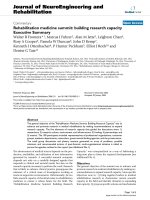
Maximum likelihood trees for two NCLDV class I core genesFigure 2
Maximum likelihood trees for two NCLDV class I core genes. (A) Homologs for the mimivirus L437 (VV A32-type virion pack-
aging ATPase). (B) Homologs for the mimivirus L206/L207 (VV D5-type ATPase). Nodes with rectangle marks correspond to
the sequences from the GOS data. These trees are unrooted.
JCVI-SCAF-1101668193166
JCVI-SCAF-1096627283011
JCVI-SCAF-1101668312069
JCVI-SCAF-1096627013160
JCVI-SCAF-1101668015449
A.polyphaga mimivirus Q5UQ22
JCVI-SCAF-1101668242113
Invertebrate iridescent virus 6 NP_149647
Invertebrate iridescent virus 3 YP_654693
Infectious spleen and kidney necrosis virus NP_612331
Ambystoma tigrinum virus YP_003852
Frog virus 3 YP_031600
Singapore grouper iridovirus YP_164147
Lymphocystis disease virus 1 NP_078717
Lymphocystis disease virus YP_073585
African swine fever virus NP_042765
E huxleyi virus 86 YP_294217
E.siliculosus virus 1 NP_077594
A.turfacea chlorella virus 1 YP_001426547
P.bursaria chlorella virus FR483 YP_001426306
P.bursaria chlorella virus 1 NP_048813
P.bursaria chlorella virus AR158 YP_001498643
P.bursaria chlorella virus NY2A YP_001497819
63
61
81
99
100
92
100
54
100
100
88
100
100
100
100
100
97
1
Poxviridae
African swine fever virus NP_042772
E.huxleyi virus 86 YP_293826
H akashiwo virus 1 Q91DI0
E siliculosus virus 1 NP_077511
P.bursaria chlorella virus 1 NP_048749
P.bursaria chlorella virus NY2A YP_001497732
P.bursaria chlorella virus AR158 YP_001498560
A.turfacea chlorella virus 1 YP_001426918
P.bursaria chlorella virus FR483 YP_001426221
Invertebrate iridescent virus 6 NP_149538
Invertebrate iridescent virus 3 YP_654660
Frog virus 3 YP_031593
Singapore grouper iridovirus YP_164229
Infectious spleen and kidney necrosis virus NP_612345
Lymphocystis disease virus YP_073620
Lymphocystis disease virus 1 NP_078656
JCVI-SCAF-1096626882244
JCVI-SCAF-1096627549470
JCVI-SCAF-1096626854560
JCVI-SCAF-1096626921870
JCVI-SCAF-1101668346786
A.polyphaga mimivirus YP_142791
JCVI-SCAF-1101668147028
JCVI-SCAF-1101668297249
JCVI-SCAF-1101668307373
JCVI-SCAF-1101668097837
100
51
51
100
96
100
84
88
99
99
89
97
97
50
89
100
100
90
0.5
AB
Virology Journal 2008, 5:12 />Page 6 of 8
(page number not for citation purposes)
If horizontal transfer of viral PolB genes does occur, it
would become difficult to interpret the PolB phylogeny as
representing the true relationships between viruses. How-
ever, to the best of our knowledge, no instance of lateral
transfer of PolB genes between distantly related eukaryotic
large DNA viruses has been documented. The determina-
tion of the whole genome sequences of CeV01, PpV01
and PoV01 would definitely help clarifying their evolu-
tionary relationship with mimivirus.
Conclusion
Three algal viruses (CeV01, PpV01 and PoV01) possess
DNA polymerase genes that are closely related to the DNA
polymerase from the giant mimivirus. This suggests that
Maximum likelihood tree of the PolB sequences from NCLDV and the GOS dataFigure 3
Maximum likelihood tree of the PolB sequences from NCLDV and the GOS data. Nodes with rectangle marks correspond to
the sequences from the GOS data. This tree is rooted by phage sequences.
JCVI-SCAF-1101668738707
P.pouchetii virus
JCVI-SCAF-1101668711727
C.ericina virus
JCVI-SCAF-1101668138124
JCVI-SCAF-1101668537640
JCVI-SCAF-1096627004132
JCVI-SCAF-1101668140135
JCVI-SCAF-1101668214945
JCVI-SCAF-1096626877081
JCVI-SCAF-1096626927911
JCVI-SCAF-1101668142153
JCVI-SCAF-1096626875531
A.polyphaga mimivirus
JCVI-SCAF-1096626853699
P.orientalis virus
JCVI-SCAF-1101668008794
JCVI-SCAF-1096626895945
H.akashiwo virus 1
E.siliculosus virus 1
Feldmannia irregularis virus a
P.bursaria chlorella virus 1
P.bursaria chlorella virus CVK2
P.bursaria chlorella virus NY2A
E.huxleyi virus 86
Phycodnaviruses
Lymphocystis virus 1
A.tigrinum virus
Infectious spleen and kidney necrosis virus
Invertebrate iridescent virus 6
Iridoviridae
Asfarviridae
African swine fever virus
Swinepox virus
Myxoma virus
Yaba-like disease virus
Variola virus
Molluscum contagiosum virus
Canarypox virus
M.sanguinipes entomopoxvirus
A.moorei entomopoxvirus 'L'
Poxviridae
63
98
60
100
71
56
69
68
96
94
100
100
55
59
100
68
74
54
77
100
97
75
0.2
Mimivirus
³Phycodnaviruses´
Mimi-like metagenomic
sequences
Virology Journal 2008, 5:12 />Page 7 of 8
(page number not for citation purposes)
the numerous "mimivirus-like" sequences detected in
marine metagenomic data might originate from viruses
infecting phytoplankton species related to C. ericina, P.
pouchetii or P. orientalis, rather than marine amoebae.
These results imply new approaches in attempting the iso-
lation of additional, and eventually closer, relatives of
mimivirus.
Methods
The scaffold sequences for the combined assembly of the
GOS metagenomic data were downloaded from the CAM-
ERA web site [35]. We extracted 21,406,171 ORFs (≥ aa)
from the scaffolds using the EMBOSS/getorf program
[36].
We defined "mimivirus-like ORFs" based on the follow-
ing two-way BLASTP searches [37]. First, the amino acid
sequences of the ORFs were searched against the UniProt
sequence database release 11.3 (as of July 2007, [38])
using BLASTP (E-value < 0.001). This search resulted in
6,212 ORFs with its best hit to a mimivirus protein in the
database. For each of the 6,212 ORFs, we extracted a seg-
ment of the mimivirus sequence that was aligned with the
ORF by BLASTP. Next, this partial mimivirus sequence
was searched against the UniProt database (excluding
mimivirus entries in the database). If the best score
obtained by this second BLASTP search is lower than the
BLASTP score obtained by the first BLASTP search, we kept
the ORF as "mimivirus-like". Accordingly, we obtained
5,293 mimivirus-like ORFs. The UniProt database does
not contain the three entries used for the phylogenetic
study (i.e. ABU23716, ABU23717, ABU23718).
Mimivirus ORFans were defined by the lack of detectable
homologs in the UniProt database using BLASTP with an
E-value threshold of 0.001.
Multiple sequence alignment was constructed using MUS-
CLE [39]. All the gap-containing sites in the alignment
were excluded in the phylogenetic analysis. We used only
the polymerase domain sequences, and removed exonu-
clease domain sequences. The delineation of the polymer-
ase domains were performed using the Pfam entry
PF00136 [40]. Intein sequences were also removed from
Mimivirus, HaV, CeV01 PolB sequences. Maximum likeli-
hood phylogenetic analysis was performed using PhyML
[41] with JTT substitution model and 100 bootstrap repli-
cates. Neighbor joining analysis was performed using
BIONJ [42]. The above methods are available from the
Phylogeny.fr server [43]. Maximum parsimony analysis
was performed using PHYLIP/PROTPARS [44].
List of abbreviations used
CeV: Chrysochromulina ericina virus; PpV: Phaeocystis pou-
chetii virus; PoV: Pyramimonas orientalis virus; NCLDV:
Nucleocytoplasmic large DNA virus; GOS: Global Ocean
Sampling Expedition; PolB: type B DNA polymerase; ORF:
open reading frame.
Competing interests
The author(s) declare that they have no competing inter-
ests.
Authors' contributions
AM performed the phylogenetic analyses. JBL and RAS
contributed new sequence data. HO performed the analy-
ses of the metagenomic data set. GB, JMC and HO con-
tributed to the writing of the manuscript. All authors have
read and approved the final document.
Additional material
Acknowledgements
AM is partially supported by the EuroPathoGenomics European network of
excellence. This work was partially supported by Marseille-Nice Genopole
and the French National Network (RNG).
References
1. La Scola B, Audic S, Robert C, Jungang L, de Lamballerie X, Drancourt
M, Birtles R, Claverie JM, Raoult D: A giant virus in amoebae. Sci-
ence 2003, 299(5615):2033.
2. Raoult D, Audic S, Robert C, Abergel C, Renesto P, Ogata H, La Scola
B, Suzan M, Claverie JM: The 1.2-megabase genome sequence of
Mimivirus. Science 2004, 306(5700):1344-1350.
3. Abergel C, Rudinger-Thirion J, Giege R, Claverie JM: Virus-encoded
aminoacyl-tRNA synthetases: structural and functional char-
acterization of mimivirus TyrRS and MetRS. J Virol 2007,
81(22):12406-12417.
4. Claverie JM, Ogata H, Audic S, Abergel C, Suhre K, Fournier PE:
Mimivirus and the emerging concept of "giant" virus. Virus
Res 2006, 117(1):133-144.
5. Claverie JM: Viruses take center stage in cellular evolution.
Genome Biol 2006, 7(6):110.
6. Forterre P: Three RNA cells for ribosomal lineages and three
DNA viruses to replicate their genomes: a hypothesis for the
origin of cellular domain. Proc Natl Acad Sci U S A 2006,
103(10):3669-3674.
7. Koonin EV, Senkevich TG, Dolja VV: The ancient Virus World
and evolution of cells. Biology direct 2006, 1:29.
8. Bell PJ: Sex and the eukaryotic cell cycle is consistent with a
viral ancestry for the eukaryotic nucleus. J Theor Biol 2006,
243(1):54-63.
9. Monier A, Claverie JM, Ogata H: Horizontal gene transfer and
nucleotide compositional anomaly in large DNA viruses.
BMC Genomics 2007, 8(1):456.
10. Iyer LM, Balaji S, Koonin EV, Aravind L: Evolutionary genomics of
nucleo-cytoplasmic large DNA viruses. Virus Res 2006,
117(1):156-184.
11. Mayo MA, Haenni AL: Report from the 36th and the 37th meet-
ings of the Executive Committee of the International Com-
Additional file 1
Number of Mimivirus-like sequences in the GOS metagenomic data set.
The file shows the number of "mimivirus-like" ORFs that we found in the
GOS metagenomic data set for each mimivirus ORF.
Click here for file
[ />422X-5-12-S1.xls]
Publish with BioMed Central and every
scientist can read your work free of charge
"BioMed Central will be the most significant development for
disseminating the results of biomedical research in our lifetime."
Sir Paul Nurse, Cancer Research UK
Your research papers will be:
available free of charge to the entire biomedical community
peer reviewed and published immediately upon acceptance
cited in PubMed and archived on PubMed Central
yours — you keep the copyright
Submit your manuscript here:
/>BioMedcentral
Virology Journal 2008, 5:12 />Page 8 of 8
(page number not for citation purposes)
mittee on Taxonomy of Viruses. Archives of virology 2006,
151(5):1031-1037.
12. Davis CC, Latvis M, Nickrent DL, Wurdack KJ, Baum DA: Floral
gigantism in Rafflesiaceae. Science 2007, 315(5820):1812.
13. Khan M, La Scola B, Lepidi H, Raoult D: Pneumonia in mice inoc-
ulated experimentally with Acanthamoeba polyphaga mim-
ivirus. Microb Pathog 2007, 42(2-3):56-61.
14. La Scola B, Marrie TJ, Auffray JP, Raoult D: Mimivirus in pneumo-
nia patients. Emerg Infect Dis 2005, 11(3):449-452.
15. Berger P, Papazian L, Drancourt M, La Scola B, Auffray JP, Raoult D:
Ameba-associated microorganisms and diagnosis of nosoco-
mial pneumonia. Emerg Infect Dis 2006, 12(2):248-255.
16. Larcher C, Jeller V, Fischer H, Huemer HP: Prevalence of respira-
tory viruses, including newly identified viruses, in hospital-
ised children in Austria. Eur J Clin Microbiol Infect Dis 2006,
25(11):681-686.
17. Suzan-Monti M, La Scola B, Raoult D: Genomic and evolutionary
aspects of Mimivirus. Virus Res 2006, 117(1):145-155.
18. Khan NA: Acanthamoeba: biology and increasing importance
in human health. FEMS Microbiol Rev 2006, 30(4):564-595.
19. Lorenzo-Morales J, Ortega-Rivas A, Foronda P, Martinez E, Valladares
B: Isolation and identification of pathogenic Acanthamoeba
strains in Tenerife, Canary Islands, Spain from water
sources. Parasitology research 2005, 95(4):273-277.
20. Ghedin E, Claverie JM: Mimivirus relatives in the Sargasso sea.
Virol J 2005, 2:62.
21. Rusch DB, Halpern AL, Sutton G, Heidelberg KB, Williamson S,
Yooseph S, Wu D, Eisen JA, Hoffman JM, Remington K, Beeson K,
Tran B, Smith H, Baden-Tillson H, Stewart C, Thorpe J, Freeman J,
Andrews-Pfannkoch C, Venter JE, Li K, Kravitz S, Heidelberg JF,
Utterback T, Rogers YH, Falcon LI, Souza V, Bonilla-Rosso G, Eguiarte
LE, Karl DM, Sathyendranath S, Platt T, Bermingham E, Gallardo V,
Tamayo-Castillo G, Ferrari MR, Strausberg RL, Nealson K, Friedman
R, Frazier M, Venter JC: The Sorcerer II Global Ocean Sampling
expedition: northwest Atlantic through eastern tropical
Pacific.
PLoS Biol 2007, 5(3):e77.
22. Sandaa RA, Heldal M, Castberg T, Thyrhaug R, Bratbak G: Isolation
and characterization of two viruses with large genome size
infecting Chrysochromulina ericina (Prymnesiophyceae)
and Pyramimonas orientalis (Prasinophyceae). Virology 2001,
290(2):272-280.
23. Yan X, Chipman PR, Castberg T, Bratbak G, Baker TS: The marine
algal virus PpV01 has an icosahedral capsid with T=219 qua-
sisymmetry. J Virol 2005, 79(14):9236-9243.
24. Hansen PJ, Nielsen TG, H. K: Distribution and growth of protists
and mesozooplankton during a bloom of Chrysochromulina
spp. (Prymnesiophyceae, Prymnesiales). Phycologia 1995,
34(5):409-416.
25. Schoemann V, Becquevort S, Stefels J, Rousseau V, Lancelot C: Phae-
ocystis blooms in the global ocean and their controlling
mechanisms: a review. J Sea Res 2005, 53:43-66.
26. Daugbjerg N, Moestrup O: Four new species of Pyramimonas
(Prasinophyceae) from arctic Canada including a light and
electron microscopic description of Pyramimonas quadrifo-
lia sp. nov. Eur J Phycol 1993, 28(1):3-16.
27. Aure J, Rey F: Oceanographic conditions in the Sandsfjord sys-
tem, western Norway, after a bloom of the toxic prymnesi-
ophyte Prymnesium parvum Carter in August 1990. Sarsia
1992, 76(4):247-254.
28. Ogata H, Raoult D, Claverie JM: A new example of viral intein in
Mimivirus. Virol J 2005, 2(1):8.
29. Nagasaki K, Shirai Y, Tomaru Y, Nishida K, Pietrokovski S: Algal
viruses with distinct intraspecies host specificities include
identical intein elements. Appl Environ Microbiol 2005,
71(7):3599-3607.
30. Castberg T, Thyrhaug R, Larsen A, Sandaa RA, Heldal M, Van Etten JL,
Bratbak G: Isolation and characterization of a virus that
infects Emiliania huxleyi (Haptophyta). J Phycol 2002,
38(4):767-774.
31. Wilson WH, Schroeder DC, Allen MJ, Holden MT, Parkhill J, Barrell
BG, Churcher C, Hamlin N, Mungall K, Norbertczak H, Quail MA,
Price C, Rabbinowitsch E, Walker D, Craigon M, Roy D, Ghazal P:
Complete genome sequence and lytic phase transcription
profile of a Coccolithovirus. Science 2005,
309(5737):1090-1092.
32. Jacobsen A, Bratbak G, Heldal M: Isolation and characterization
of a virus infecting Phaeocystis pouchetii (Prymnesiophyc-
eae). J Phycol 1996, 32(6):923-927.
33. Chen F, Suttle CA: Evolutionary relationships among large
double-stranded DNA viruses that infect microalgae and
other organisms as inferred from DNA polymerase genes.
Virology 1996, 219(1):170-178.
34. Villarreal LP, DeFilippis VR: A hypothesis for DNA viruses as the
origin of eukaryotic replication proteins. J Virol 2000,
74(15):7079-7084.
35. Seshadri R, Kravitz SA, Smarr L, Gilna P, Frazier M: CAMERA: a
community resource for metagenomics. PLoS Biol 2007,
5(3):e75.
36. Rice P, Longden I, Bleasby A: EMBOSS: the European Molecular
Biology Open Software Suite. Trends Genet 2000,
16(6):276-277.
37. Altschul SF, Madden TL, Schaffer AA, Zhang J, Zhang Z, Miller W, Lip-
man DJ: Gapped BLAST and PSI-BLAST: a new generation of
protein database search programs. Nucleic Acids Res 1997,
25(17):3389-3402.
38. Wu CH, Apweiler R, Bairoch A, Natale DA, Barker WC, Boeckmann
B, Ferro S, Gasteiger E, Huang H, Lopez R, Magrane M, Martin MJ,
Mazumder R, O'Donovan C, Redaschi N, Suzek B: The Universal
Protein Resource (UniProt): an expanding universe of pro-
tein information. Nucleic Acids Res 2006, 34(Database
issue):D187-91.
39. Edgar RC: MUSCLE: a multiple sequence alignment method
with reduced time and space complexity. BMC Bioinformatics
2004, 5(1):113.
40. Bateman A, Birney E, Durbin R, Eddy SR, Finn RD, Sonnhammer EL:
Pfam 3.1: 1313 multiple alignments and profile HMMs match
the majority of proteins. Nucleic Acids Res 1999, 27(1):260-262.
41. Guindon S, Gascuel O: A simple, fast, and accurate algorithm
to estimate large phylogenies by maximum likelihood. Sys-
tematic biology
2003, 52(5):696-704.
42. Gascuel O: BIONJ: an improved version of the NJ algorithm
based on a simple model of sequence data. Mol Biol Evol 1997,
14(7):685-695.
43. Phylogeny.fr: [
].
44. Felsenstein J: PHYLIP (Phylogeny Inference Package) version
3.6b. Distributed by the author. Department of Genome Sci-
ences, University of Washington, Seattle. 2004.