Báo cáo hóa học: " Development and evaluation of one step single tube multiplex RT-PCR for rapid detection and typing of dengue viruses" pdf
Bạn đang xem bản rút gọn của tài liệu. Xem và tải ngay bản đầy đủ của tài liệu tại đây (266.61 KB, 5 trang )
BioMed Central
Page 1 of 5
(page number not for citation purposes)
Virology Journal
Open Access
Methodology
Development and evaluation of one step single tube multiplex
RT-PCR for rapid detection and typing of dengue viruses
Parag Saxena, Paban Kumar Dash, SR Santhosh, Ambuj Shrivastava,
Manmohan Parida and PV Lakshmana Rao*
Address: Division of Virology, Defence Research & Development Establishment, Jhansi Road, Gwalior 474 002, MP, India
Email: Parag Saxena - ; Paban Kumar Dash - ;
SR Santhosh - ; Ambuj Shrivastava - ;
Manmohan Parida - ; PV Lakshmana Rao* -
* Corresponding author
Abstract
Background: Dengue is emerging as a major public health concern in many parts of the world.
The development of a one-step, single tube, rapid, and multiplex reverse transcription polymerase
chain reaction (M-RT-PCR) for simultaneous detection and typing of dengue virus using serotype
specific primers during acute phase of illness is reported.
Results: An optimal assay condition with zero background was established having no cross-
reaction with closely related members of flavivirus (Japanese encephalitis, West Nile, Yellow fever)
and alphavirus (Chikungunya). The feasibility of M-RT-PCR assay for clinical diagnosis was validated
with 620 acute phase dengue patient sera samples of recent epidemics in India. The comparative
evaluation vis a vis conventional virus isolation revealed higher sensitivity. None of the forty healthy
serum samples screened in the present study revealed any amplification, thereby establishing
specificity of the reported assay for dengue virus only.
Conclusion: These findings clearly suggested that M-RT-PCR assay reported in the present study
is the rapid and cost-effective method for simultaneous detection as well as typing of the dengue
virus in acute phase patient serum samples. Thus, the M-RT-PCR assay developed in this study will
serve as a very useful tool for rapid diagnosis and typing of dengue infections in endemic areas.
Background
Dengue is the most important mosquito borne viral infec-
tion and is prevalent in most parts of the tropics. Cur-
rently dengue fever causes more illness and death than
any other arboviral disease in humans. An estimated 2.5
billion people live in the areas at risk for epidemic dengue
virus transmission. One hundred million cases of dengue
fever (DF) and 450,000 cases of dengue hemorrhagic
fever/dengue shock syndrome (DHF/DSS) are reported
annually [1-3]. The most challenging problem associated
with dengue is patient management, which is possible
through rapid diagnosis of early infection. In dengue
infection, serotyping is very important because of the fact
that secondary infection with a heterologous serotype
often leads to life threatening dengue haemorrhagic fever
(DHF) and dengue shock syndrome (DSS) [4].
Published: 30 January 2008
Virology Journal 2008, 5:20 doi:10.1186/1743-422X-5-20
Received: 27 November 2007
Accepted: 30 January 2008
This article is available from: />© 2008 Saxena et al; licensee BioMed Central Ltd.
This is an Open Access article distributed under the terms of the Creative Commons Attribution License ( />),
which permits unrestricted use, distribution, and reproduction in any medium, provided the original work is properly cited.
Virology Journal 2008, 5:20 />Page 2 of 5
(page number not for citation purposes)
Laboratory diagnosis of dengue infection is primarily
achieved through virus isolation from acute phase serum,
serodiagnosis and molecular assays for detection of viral
RNA [5]. Virus isolation though considered 'gold stand-
ard' is technically demanding and time consuming. The
most rapid serological technique, such as IgM ELISA with
a single serum sample, does not furnish information
about the serotype of the virus. The plaque reduction neu-
tralization technique (PRNT) allows typing but is costly,
slow and difficult to perform. The molecular methods
based on PCR technique offer a suitable alternative to
conventional isolation technique. The RT-PCR targeting
the conserved regions of C-prM gene junction is widely
employed for precise confirmation of an infection [6]. In
routine practice, a two-step method with a RT-PCR fol-
lowed by a nested PCR is used for serotyping of dengue
viruses. However, this method is expensive, time consum-
ing, and also suffers from carryover contamination prob-
lems. To improve on this, we report here the development
of a one-step single-tube rapid multiplex PCR assay for
rapid detection and differentiation of dengue serotypes in
acute phase serum samples.
Results
All the viruses were propagated in C6/36 cells prior to
extraction of RNA. The isolation of virus on C6/36 cells
led to successful isolation of Dengue type 2, 3 and 4
viruses (Table 1). RNA was extracted from culture super-
natant and used as template in RT-PCR and nested PCR.
The RT-PCR and nested PCR revealed desired amplifica-
tion as reported earlier [6]. The multiplex PCR was stand-
ardized as a single tube method by incorporating all the
four-serotype specific primers, along with a dengue con-
sensus forward primer. The assay was standardized by
optimizing the annealing temperature (55°C), primer
(20pmol) concentration that resulted in generation of dif-
ferently sized serotype specific amplicons viz., Dengue 1
(482 bp), Dengue 2 (119 bp), Dengue 3 (290 bp), and
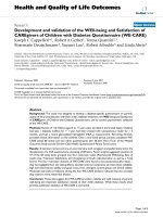
Dengue 4 (389 bp) as shown in the Figure 1. Following
standardization, the assay was further optimized for
screening RNA extracted from clinical samples.
Sensitivity and Specificity of multiplex RT-PCR
The detection limit of both dengue complex and the Mul-
tiplex RT-PCR assay was determined through 10 fold serial
dilution of RNA copies. The RNA copies were generated
using in vitro transcription. The sensitivity of dengue com-
plex and multiplex RT-PCR was found to be 500 and 2500
RNA copies respectively. The multiplex PCR was also com-
pared with gold standard virus isolation and the sensitiv-
ity was found to be much higher (96 compared to 57 in
isolation) (Table 2). The serotypes of all these isolates
Table 1: Distribution of serotypes among confirmed dengue positive cases by multiplex RT-PCR (M-RTPCR)
Dengue virus serotypes Number of patients found infected with dengue viruses (serotype-wise distribution)
M-RTPCR (n = 96) Cell culture supernatant (n = 57)
DEN-1 20
DEN-2 47 31
DEN-3 42 25
DEN-4 51
Total 96 57
1.5% Agarose gel electrophoresis demonstrating the cor-rectly sized amplicons generated by the single tube dengue multiplex RT-PCRFigure 1
1.5% Agarose gel electrophoresis demonstrating the cor-
rectly sized amplicons generated by the single tube dengue
multiplex RT-PCR. Lane 1: 100 bp DNA ladder (Fermentas),
lanes 2–6: Dengue 1 (482 bp), Dengue 2 (119 bp), Dengue 3
(290 bp), Dengue 4 (389 bp) and negative control.
Virology Journal 2008, 5:20 />Page 3 of 5
(page number not for citation purposes)
were confirmed using both multiplex RT-PCR and two
step RT-PCR followed by nested PCR, which revealed
100% concordance (Details not shown). The isolation
results was further confirmed by sequencing of the respec-
tive amplicons, which revealed perfect matching with the
respective serotype specific sequences, confirming the spe-
cificity of this assay (data not shown). The specificity of
the multiplex RT-PCR assay was compared with closely
related flavi- and alphaviruses. It was observed that this
assay is highly specific for dengue serotypes with no cross
reactivity with Japanese encephalitis, West Nile, Yellow
fever and Chikungunya viruses. Furthermore, all the
serum samples from a panel of 40 healthy individuals
analyzed in this study revealed no amplification, thereby
establishing the specificity of this assay.
Evaluation of Multiplex RT-PCR
The feasibility of the assay for clinical diagnosis was vali-
dated by evaluating with serum samples from 620 acute-
phase suspected patients and 40 healthy individuals from
the same area. On comparative evaluation with RT-PCR,
the multiplex RT-PCR was found to be equally sensitive
for detection of dengue viral RNA in patient sera.
Discussion
Dengue cases are increasing over the years in many parts
of the world. Now it is endemic in India, with circulation
of multiple serotypes [7,8]. There is also an increased inci-
dence of fatal DHF and DSS, which requires urgent medi-
cal intervention [9]. Early diagnosis of dengue is critical in
the absence of any licensed antiviral therapy and prophy-
laxis. The diagnosis is achieved either serologically by
detecting dengue-specific IgM and IgG antibodies, which
generally appear 7–8 days after the onset of illness [10].
However, it is of less value for early diagnosis. In addition,
persistent circulation of IgM antibodies for more than 90
days also is a limiting factor in confirmatory diagnosis.
The detection of IgG is generally not considered authentic,
due to cross reactivity with other closely related members
of Flaviviruses. This needs to be confirmed with paired
sera, which is not practical in most cases [11,12].
The detection of dengue serotype is very important due to
fact that in secondary cases, infection with a heterologous
serotype often leads to fatal DHF and DSS [13]. So, early
typing is a pre-requisite for proper patient management. A
large number of molecular methods have been reported
for serotyping of dengue viruses [6,14-18]. However, most
of these methods are four tube based RT-PCR followed by
nested PCR or four serotype specific RT-PCR. The multi-
plex PCR assay described by Harris et al., (1998) [16] also
suffers from lack of proper evaluation with clinical sam-
ples. To improve on the existing assay systems, we stand-
ardized a one step single tube protocol for rapid
serotyping of dengue viruses. This assay can be performed
rapidly with in a period of 4 hours compared to 8 hours
in two-step typing assays. This can provide faster informa-
tion to the clinicians leading to initiation of suitable
symptomatic therapy. This single tube protocol also
reduces the cost by four fold, resulting in an economical
way of serotyping [16]. The two-step assays are always
more prone to contamination due to opening of tubes
between the steps. All these advantages make this assay a
user friendly, rapid, cost effective diagnostic tool and can
be utilized in many developing countries, where dengue is
endemic.
The sensitivity and specificity of the single-tube multiplex
RT-PCR was also verified. It could correctly serotype all
the four respective dengue serotypes. No cross-reaction
was observed when other related flaviviruses and alpha
viruses were included. The processing of all the 96 PCR
positive samples for virus isolation, led to 57 dengue iso-
lates. This isolation was confirmed by both nested PCR
and multiplex PCR, which revealed similar results. This
indicates the lower sensitivity of isolation compared to
RT-PCR, which may be attributed to either inactivation of
virus during transportation or failure in maintenance of
cold chain [7]. The sensitivity was further checked by
developing RNA transcripts. The detection limit of multi-
plex PCR was found to be 2500 copies. Though it is 5 fold
less sensitive compared to group specific RT-PCR, how-
ever, its importance can be judged from the fact that this
assay gives precise information regarding the serotype of
the virus.
Conclusion
The development of a suitable effective vaccine and thera-
peutics for dengue is not yet achieved and it is presumed
that it will not be available for coming few years. In these
circumstances, rapid diagnosis can help in timely patient
management. The multiplex PCR assay developed in this
study will be extremely useful for rapid diagnosis and
serotyping of viruses in dengue infections.
Table 2: Comparison of Multiplex PCR and Virus isolation for the detection and serotyping of dengue viruses from acute phase serum
samples (n = 620)
M-RT-PCR Virus isolation
Positive 96 (15.48%) 57 (9.20%)
Negative 524 (84.52%) 563 (90.80%)
Virology Journal 2008, 5:20 />Page 4 of 5
(page number not for citation purposes)
Methods
Virus
Virus strains of the four standard dengue serotypes were
obtained from the National Institute of Virology, Pune,
India (Den-1, Hawaii; Den-2, P-23085; Den-3, 633798
and Den-4, 642069). The dengue viruses isolated during
this study from dengue outbreak in India since 2001 were
also included. To check the cross reactivity, related flaviv-
iruses viz., Japanese encephalitis (JaOArS982), West Nile
(G22886), Yellow fever (17D vaccine strain) virus and
one alphavirus (Chikungunya (S27) were also included in
this study. These viruses were propagated in C6/36 cells.
Clinical Samples
A total of 620 human serum samples from febrile patients
clinically suspected of having dengue fever were collected
within 0 to 4 days from the time of onset of symptoms.
These samples were collected from different parts of India
during various outbreaks from 2001–2007 [7,8]. In addi-
tion, a panel of 40 serum samples collected from healthy
volunteers from the same area were included as negative
control in this study.
Virus isolation
C6/36 cells [19] were grown in Eagle's minimum essential
medium (EMEM) (Sigma, USA) supplemented with 10%
Tryptose Phosphate Broth (TPB) (DIFCO, USA), 10%
Fetal bovine serum (FBS) (Sigma, USA), 3% L-glutamine
(Sigma, USA) and gentamicin (80 mg/l) (Nicholas-
Piramal, India) at 28°C. Isolation of viruses from acute
phase dengue suspected samples were attempted follow-
ing the standard virus adsorption technique [20]. Briefly,
preformed manolayer of cells were washed with plain
medium prior to infection. The virus/suspected serum was
allowed to adsorb to the cells for 1 hr at 37°C. Following
adsorption, the inoculum was replenished with 2 ml of
maintenance medium (EMEM with 2% FBS). Suitable cell
controls were also kept along side. The cells were har-
vested on appearance of cytopathic effects or on 6
th
day
post inoculation (dpi), whichever is earlier. The identifi-
cation of the virus isolates obtained from the clinical sam-
ples was carried out by RT-PCR as described below.
RNA Extraction
RNA was extracted from standard viruses, virus isolates,
sera of suspected dengue patients and healthy volunteers
using QIAquick viral RNA mini Kit, following the manu-
facturer's protocol. Finally, the RNA was eluted in 60 μl of
diethyl pyrocarbonate (DEPC) treated water (Sigma,
USA).
Dengue complex Reverse transcription polymerase chain
reaction (RT-PCR)
Conventional Dengue complex RT-PCR assays were per-
formed according to the protocol [6] with slight modifica-
tions. Briefly, the RT-PCR was performed with RNA from
standard dengue viruses and confirmed dengue virus-
infected patient serum samples initially in a 50 ul reaction
volume using Access quick RT-PCR kit (Promega, USA)
with dengue virus group-specific consensus primers (D1:
5' TCAATATGCTAAAACGCGCGAGAAACCG 3' and D2:
5' TTGCACCAACAGTCAATGTCTTCAGGTTC 3').
Dengue Nested PCR
The nested PCR assay was performed according to the pro-
tocol [6] with slight modifications. Briefly, the 1: 10 dilu-
tion of RT-PCR amplicon was used as template in the
nested PCR in a 50 μl reaction volume using master mix
of Access quick RT-PCR kit, with dengue virus group-spe-
cific consensus forward primer (D1), and four serotype
specific reverse primers (Ts1: 5' CGTCTCAGTGATCCG-
GGGG 3', Ts2: 5'CGCCACAAGGGCCATGAACAG 3', Ts3:
5' TAACATCATCATGAGACAGAGC 3' and Ts4:
5'TGTTGTCTTAAACAAGAGAGGTC3'), as reported earlier
[6].
Single-step Dengue multiplex RT-PCR (M-RT-PCR)
A one-step single tube serotype-specific multiplex PCR
was performed with RNA from standard dengue viruses
and confirmed dengue virus-infected patient serum sam-
ples using a multiplex RT-PCR protocol. The amplifica-
tion was carried out in a 50 μl total reaction volume with
Access quick RT-PCR kit according to the manufacturer's
protocol, along with five primers viz., forward D1 and
four serotype specific reverse primers (Ts1, Ts2, Ts3 and
Ts4).
Evaluation of multiplex RT-PCR
The evaluation of the multiplex RT-PCR assay was carried
out with 620 serum samples collected over a period of six
years from India.
Preparation of RNA standard
The detection limit of this assay was determined using
RNA standards. The RNA standard was produced using T7
transcription kit (MBI Fermentas, USA) following the
manufacturer's protocol. Initially PCR amplicons of
respective dengue serotypes were generated using a modi-
fied D1 primer (T7 promoter sequence (TAATACGACT-
CACTATAGG) was added at the 5' end of D1 primer) and
normal D2 primer. These amplicons (530 bp) of all the
four serotypes were gel purified, quantitated, before being
used as template in transcription reaction. The purified
template was subjected to in vitro transcription (IVT) at
37°C for 1 h. The IVT products were then treated with 1 U
of DNase I and incubated at 37°C for 15 min to remove
the remaining DNA followed by inactivation of DNase I at
70°C for 15 min. The IVT products were ethanol precipi-
tated and resuspended in DEPC treated water. The
amount of respective dengue serotype specific RNA tran-
Publish with BioMed Central and every
scientist can read your work free of charge
"BioMed Central will be the most significant development for
disseminating the results of biomedical research in our lifetime."
Sir Paul Nurse, Cancer Research UK
Your research papers will be:
available free of charge to the entire biomedical community
peer reviewed and published immediately upon acceptance
cited in PubMed and archived on PubMed Central
yours — you keep the copyright
Submit your manuscript here:
/>BioMedcentral
Virology Journal 2008, 5:20 />Page 5 of 5
(page number not for citation purposes)
scripts were determined spectrophotometrically and con-
verted to molecular copies by using the following formula
21.
Y (molecules/μl) = [X(g/μl)/transcript length (nucle-
otides) × 340] × 6.023 × 10
23
Sensitivity and specificity of the assay
The sensitivity of both the dengue complex and serotype
specific multiplex RT-PCR assay was determined through
serial dilutions of the RNA transcripts. The specificity was
determined on comparison with related Flaviviruses (JE,
West Nile and Yellow fever viruses), alphavirus (Chikun-
gunya virus) and a panel of 40 serum samples from
healthy volunteers.
Competing interests
The author(s) declare that they have no competing inter-
ests.
Authors' contributions
PS carried out the standardization of all RT-PCR experi-
ments, virus isolation, and evaluation of the M-RT-PCR
assay. PKD carried out the nested PCR and In vitro tran-
scription and sequencing. SSR carried out the RT-PCR, IVT
assay. AS carried out the virus isolation and evaluation
with clinical samples. MMP conceived the study and
planned the work. PVLR helped out to design and draft
the manuscript and also revised it critically. All authors
read and approved the final manuscript.
Acknowledgements
The authors are thankful to Director, Defence Research and Development
Establishment (DRDE), Ministry of Defence, Govt. of India for his support,
constant inspiration, and providing the necessary facilities for this study.
References
1. Monath TP: Dengue: The risk to developed and developing
countries. Proc Natl Acad Sci, USA 1994, 91:2395-400.
2. Gubler DJ: Dengue and dengue hemorrhagic fever. Clin Micro-
biol Rev 1988, 11:480-96.
3. World Health Organization: Dengue and dengue haemorrhagic
fever. Fact sheet 2002, 117:.
4. McBride WJH, Belefeldt-Ohmann H: Dengue viral infections;
pathogenesis and epidemiology. Microbes and Infection 2000,
2:1041-50.
5. Guzman MG, Kouri G: Advances in dengue diagnosis. Clin Diagn
Lab Immunol 1996, 3:621-7.
6. Lanciotti RS, Calisher CH, Gubler DJ, Chang GJ, Vorndam AV: Rapid
detection and typing of dengue viruses from clinical samples
by using reverse transcriptase-polymerase chain reaction. J
Clin Microbiol 1992, 30:545-51.
7. Dash PK, Parida MM, Saxena P, Abhyankar A, Singh CP, Tewari KN,
Jana AM, Sekhar K, Rao PVL: Reemergence of dengue virus type-
3 (subtype-III) in India: Implications for increased incidences
of DHF & DSS. Virology J 2006, 3:55. (Highly accessed).
8. Dash PK, Parida MM, Saxena P, Kumar M, Rai A, Pasha ST, Jana AM:
Emergence and continued circulation of Dengue-2 (Geno-
type IV) virus strains in northern India. J Med Virol 2004,
74:314-22.
9. Gore MM: Need for constant monitoring of dengue infections.
Ind J Med Res 2005, 121:9-12.
10. Parida MM, Upadhyay C, Saxena P, Dash PK, Jana AM, Seth P: Evalu-
tion of a Dipstick ELISA and a rapid Immunochromato-
graphic test for diagnosis of dengue virus infection. Acta
Virologica 2001, 45:299-304.
11. Kuno G, Gomez I, Gubler DJ: An ELISA procedure for the diag-
nosis of dengue infections. J Virol Methods 1991, 33:101-13.
12. Satish N, Vijaykumar TS, Abraham P, Sridharan G: Dengue fever:
it's laboratory diagnosis with special emphasis on IgM detec-
tion.
WHO Dengue Bull 2003, 27:116-125.
13. Halstead SB: Pathogenesis of dengue: challenges to molecular
biology. Science 1998, 239:476-81.
14. Morita K, Tanaka M, Igarashi A: Rapid identification of dengue
virus serotypes by using polymerase chain reaction. J Clin
Microbiol 1991, 29:2107-10.
15. Henchal E, Polo S, Vorndam V, Yaemsiri C, Innis B, Hoke C: Sensi-
tivity and specificity of a universal primer set for the rapid
diagnosis of dengue virus infections by polymerase chain
reaction and nucleic acid hybridization. Am J Trop Med Hyg
1991, 45:418-28.
16. Harris E, Roberts TG, Smith L, Selle J, Kramer LD, Valle S, Sandovan
E, Balmaseda A: Typing of dengue viruses in clinical specimens
and mosquitoes by single tube multiplex reverse tran-
scriptase PCR. J Clin Microbiol 1998, 36:2634-9.
17. De Paula SO, Lima DM, da Fonseca BA: Detection and identifica-
tion of dengue-1 virus in clinical samples by a nested-PCR fol-
lowed by restriction enzyme digestion of amplicons. J Med
Virol 2002, 66:529-34.
18. Parida MM, Horioke K, Ishida H, Dash PK, Saxena P, Jana AM, Islam
MA, Inoue S, Hosaka N, Morita K: Rapid detection and differen-
tiation of dengue virus serotypes by a real-time reverse tran-
scription-loop-mediated isothermal amplification assay. J
Clin Microbiol 2005, 43:2895-903.
19. Igarashi A: Isolation of Singh's Aedes albopictus cell clone sen-
sitive to dengue and chikungunya viruses. J Gen Virol 1978,
40:581-4.
20. Gould EA, Clegg TCS: Growth, assay and purification of
Alphaviruses and Flaviviruses. In Virology: A Practical Approach
Edited by: BW J. Mahy, Oxford University Press; 1991:43-78.
21. Krieg P: Improved synthesis of full length RNA probe at
reduced incubation temperatures. Nucleic Acids Res 1991,
18:6463.