Báo cáo hóa học: " Changes in viral load and HBsAg and HBeAg status with age in HBV chronic carriers in The Gambia" pdf
Bạn đang xem bản rút gọn của tài liệu. Xem và tải ngay bản đầy đủ của tài liệu tại đây (274.59 KB, 8 trang )
BioMed Central
Page 1 of 8
(page number not for citation purposes)
Virology Journal
Open Access
Research
Changes in viral load and HBsAg and HBeAg status with age in HBV
chronic carriers in The Gambia
Maimuna E Mendy*
1
, Samuel J McConkey
1,2
, Marianne AB Sande van der
1,3
,
Sarah Crozier
1,4
, Steve Kaye
1,5
, David Jeffries
1
, Andrew J Hall
6
and
Hilton C Whittle
1
Address:
1
Medical Research Council Laboratories, Atlantic Boulevard, PO Box 273, Banjul, The Gambia,
2
Royal College of Surgeons in Ireland,
123 St. Stephen's Green, Dublin 2, Ireland,
3
National Institute for Public Health and the Environment, Bilthoven, The Netherlands,
4
Medical
Research Council Epidemiology Resource Centre, University of Southampton, Southampton, UK,
5
Imperial College, London, UK and
6
London
School of Hygiene and Tropical Medicine, Keppel Street, London, UK
Email: Maimuna E Mendy* - ; Samuel J McConkey - ; Marianne AB Sande van
der - ; Sarah Crozier - ; Steve Kaye - ;
David Jeffries - ; Andrew J Hall - ; Hilton C Whittle -
* Corresponding author
Abstract
Background: Little is known about changes in hepatitis B viral load (HBV DNA) in relation to age
in Africa. The aim of this study is to determine the natural course of HBV chronic infection,
particularly in relation to sequential changes in serum HBV DNA levels and hepatitis B surface
(HBsAg) antigen/hepatitis e antigen (HBeAg) status by age.
Methods: The study was conducted on 190 HBV chronic carriers, aged 1–19 years who were
followed for 19 years. 160, 99 and 123 were traced at 5, 9 and 19 years later. All available samples
were tested for HBsAg and HBeAg, whilst 170, 61, 63 and 81 were tested for HBV DNA at the
baseline, and at 5, 9 and 19 years following recruitment.
Results: In general HBeAg which correlated with high levels of HBV DNA was lost at a much faster
rate than HBsAg. 86% of the carriers who were recruited at the age of 1–4 yrs lost HBeAg by the
age of 19 years compared to 30% who lost HBsAg. HBeAg negative carriers had serum HBV DNA
levels of < 10
5
copies per mL, HBV DNA positivity declined from 100% in 1–4 yrs old carriers at
recruitment to 62.5%,60% and 88% at 5, 9 and 19 years respectively following recruitment.
Conclusion: After 19 years of follow up, the majority of HBV surface antigen carriers had lost
HBeAg positivity and had low levels of viral replication. However small proportions (10–20%)
retained HBeAg and continue to have high levels of viral replication.
Introduction
Hepatitis B virus (HBV) is the leading cause of viral hepa-
titis in humans worldwide. Currently over two billion
people have evidence of previous HBV infection and 350
million have become chronic carriers of the virus, 60 mil-
lion of them residing in Africa [1]. There seem to be differ-
ing risks for HCC by geographical location, with higher
risk recorded in countries in sub Saharan Africa and Asia
Published: 16 April 2008
Virology Journal 2008, 5:49 doi:10.1186/1743-422X-5-49
Received: 3 October 2007
Accepted: 16 April 2008
This article is available from: />© 2008 Mendy et al; licensee BioMed Central Ltd.
This is an Open Access article distributed under the terms of the Creative Commons Attribution License ( />),
which permits unrestricted use, distribution, and reproduction in any medium, provided the original work is properly cited.
Virology Journal 2008, 5:49 />Page 2 of 8
(page number not for citation purposes)
compared to Europe [2-4]. Chronic HBV infection is a
major cause of liver disease and is strongly associated with
the development of hepatocellular carcinoma (HCC) [4].
Other risk factors for HCC include, male gender, co-infec-
tion with hepatitis C virus (HCV) [5], alcohol abuse [6],
aflatoxin exposure [7,8] and HBV genotype [9].
Approximately one third of HBV carriers will progress to
cirrhosis and 25% will develop HCC [10]. The risk of
HCC is six times higher in patients who are persistently
HBeAg positive than in HBeAg negative patients [11] and
twelve times higher in patients with high DNA viral load
(> 10
6
copies per mL)[12]. Most of the studies of the asso-
ciation of HBeAg and viral load with chronic liver diseases
and HCC have been conducted in Southeast Asia. There is
no comparable data in Sub Saharan Africa.
HBV infection is endemic in The Gambia with 15–20%
unvaccinated Gambians chronically infected [13]. Liver
cancer remains the commonest cause of death in adult
males in The Gambia [14]. Studies of HBV infection con-
ducted in two neighbouring villages of Keneba and Man-
duar (situated in West Kiang district) in the period before
1986–1990, when the national vaccination programme in
The Gambia commenced, have shown that infection was
uncommon in infants under the age of 6 months and sib-
ling-to-sibling transmission rather than perinatal or verti-
cal transmission was of major importance [13,15].
Other studies have shown that early in the carrier state,
HBV infected individuals test positive for hepatitis B sur-
face antigen (HBsAg) and hepatitis B e antigen (HBeAg)
and have high levels of HBV DNA in their serum [16,17].
However the serum of older carriers, have detectable anti-
body to HBeAg (anti-HBe), which appears following
clearance of HBeAg and is associated with low levels of
HBV DNA [18,19].
The natural history of HBV infection is complex and vari-
able and is greatly influenced by the age at infection and
the level of HBV replication. The diversity of clinical out-
come of HBV, either resolution (acute infection) or per-
sistence (chronic carriage) of infection is dependent on
the host immune response to the virus [20].
Important changes are occurring which are impacting on
the natural history of HBV infection. These changes
include vaccination programmes [21,22] and the develop-
ment of mutant forms of the virus mainly pre core (pre-C)
and basal core promoter (BCP) mutants [23].
The aim of the present study is to determine the natural
history of chronic hepatitis B infection, particularly the
changes in viral load, in The Gambia, where horizontal
transmission occurs in childhood. In this manuscript we
report HBV DNA levels and HBsAg and HBeAg status in
chronic carriers who were identified at various ages and
followed for a period of 19 years.
Materials and methods
Study population
Between 1972 and 1974 and in 1984, sero-surveys of HBV
infections were conducted in inhabitants of the two vil-
lages of Keneba and Manduar and the pattern of child-
hood hepatitis was described [24-26]. The two villages are
situated 8 kilometres apart and the inhabitants are of sim-
ilar ethnic and religious background. Accurate demo-
graphic data have been kept since 1951, date of births
have been recorded for every individual born after 1951.
During the 1984 survey 82 chronic carriers were identified
in Keneba and 108 in Manduar. They have been followed
at 3 time points 5 (in 1989), 9 (in 1993)and 19 (in 2003)
years after recruitment [21,22,26]. The number and age
distribution of the carriers at baseline was 68 age 1–4
years, 66 age 5–9, 38 age 10–14 and 18 age 15–19 years.
Blood samples collected at baseline in 1984 were tested
for HBV marker (HBsAg, HBeAg if HBsAg is positive and
anti HB core antibody), levels of HBV DNA and liver func-
tion tests (AST and ALT). During the follow up surveys
blood samples were retested for HBV markers and HBV
DNA levels. Liver enzymes were measured at baseline
only.
Because the study is in the format of a cross sectional sur-
vey we were unable to establish the actual time of initial
infection of the older children. However previous studies
suggest that the majority were infected before the age of
10 years [13,15].
HBV serology
Samples collected in 1984, 1989 and 1993 were tested for
HBsAg by reverse passive haemaglutination assay (RPHA)
(MUREX, Dartfort, UK) and by radioimmunoassay (RIA)
(DiaSorin Biomedica, Sallugia, Italy). In 2003 following
the discontinuation of RPHA and RIA assays, the samples
were tested for HBsAg by Determine™ (Abbott laborato-
ries), a visually read, qualitative immunochromatography
assay. There was good correlation between the deter-
mine™ test and the RPHA and RIA assays. Samples posi-
tive for HBsAg were tested for HBeAg by RIA between
1984 and 1993 and later by enzyme immunoassay (EIA)
(DiaSorin, Biomedica, Sallugia, Italy) after the discontin-
uation of RIA assay by DiaSorin. There was good correla-
tion between the EIA method and the RIA assay.
HBV DNA quantification by real time PCR
HBV DNA was measured by quantitative real-time PCR
according to a previously described method [16]. Blood
Virology Journal 2008, 5:49 />Page 3 of 8
(page number not for citation purposes)
samples were collected and transferred into plain tubes
and serum was separated and stored immediately at -
20°C (for sample collected in 1984 and 1989) or -70°C
(for samples collected in 1993 and 2003) until needed.
DNA was extracted from stored sera which have been kept
frozen for a period of 1 to 20 years. The assay was carried
out using commercial SYBR-Green reaction mix (Qiagen,
Hilden, Germany) and primers specific to the S gene
designed to amplify a 98 base pair product. Thermal
cycling was performed in an ABi 5700 sequence detection
system (PE Applied Biosystems, Warrington, UK). The
detection limit of the qPCR assay was 40 copies per mL
and quantified accurately samples with greater than 2.6 ×
10
2
DNA copies per mL. Test samples falling above the top
of the standard curve were re-assayed at a dilution of
1:100. The assay was 100% specific when tested against
HBV seronegative sera from ten subjects and coefficient of
variation obtained from intra-and inter assay was 1.08
and 1.72 respectively. We considered serum HBV DNA
levels greater than 10
5
copies/mL as high viral load and
less than 10
5
copies as low viral load.
Measurement of liver enzymes
Sera from blood samples were tested for ALT and AST ami-
notransferases by use of a Roche Cobas Mira Autoana-
lyser.
Data management and statistics
Demographic data, subject's identification, date of birth,
serology and results from biochemical tests were
imported from the various databases developed during
previously longitudinal studies, into an excel database cre-
ated for the present study. The data for HBV DNA results
were exported as an Excel spreadsheet into an Access data-
base from the ABi real time machine after the PCR ampli-
fication and quantification.
Linear trend was tested by fitting a generalized estimating
equation to allow for multiple responses within subjects
using a logistic link with interchangeable correlation
structure. For those analysis with trends having significant
curvature, the rate difference between the first two time
points was tested. Log rank tests were used to test the sig-
nificance of relationships found. Analysis was performed
using Stata 8.0.
Results
Prevalence of HBeAg and changes in viral load in HBsAg
chronic carriers aged 1–4 years
Sixty-eight HBsAg carriers aged 1–4 yrs were identified in
1984. Forty-six (67.6%) of the 68 carriers were males and
22 were females, 10 (16.2%) had raised AST (> 44 mIU/
mL) and 4 (5.9%) had raised ALT (> 44 mIU/mL) at
recruitment. In 3 carriers both indices were raised.
Table 1a shows changes in HBV serological markers and
HBV DNA detection in the 68 children identified at age
1–4 yrs. The proportion positive for HBsAg and HBV DNA
fell sharply over the first five years then stabilized there
after. In contrast, HBeAg prevalence dropped continually
over the years (p vaue for trend < 0.001).
HBV DNA was detectable in the majority (85–100%) of
HBeAg positive carriers and the majority of them had
DNA levels greater than 10
5
copies/mL (Table 1b).
Although HBV DNA was detected in 100% HBeAg nega-
tive carriers at baseline, the prevalence of HBVDNA posi-
tivity decreased at subsequent years following
recruitment. DNA levels were mostly below 1.0 × 10
5
cop-
ies/mL in HBeAg negative carriers, only 7.1% to 18% of
them have serum HBV DNA levels greater than 10
5
DNA
copies/mL (Table 1c).
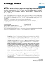
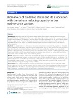
The median viral load in the 68 younger carriers decreased
significantly (p < 0.001) with time in this cohort (Figure
1). There was 1.0 log drop in viral load after 5 years and a
further 4.0 log drop over the next 4 years thereafter levels
stabilize. The difference in median viral load between car-
riers at age 5–9 yrs and at subsequent ages was highly sig-
nificant (p < 0.001). Similar results were observed in the
10 carriers who had DNA test on each of the four time
points (data not shown). However as the proportion of
HBeAg positive carriers fall when we separate values for
HBeAg positive carriers from HBeAg negative carriers we
found no significant difference in serum HBV DNA levels
between the different age groups.
High HBV DNA concentrations (> 10
5
copies/mL) were
detected in only 3/31 (9.6%) HBeAg negative carriers
compared to 5/5 (100%) HBeAg positive carriers aged
20–24 yrs (Tables 1b and 1c). Four of the HBeAg positive
carriers had maintained high levels of HBV DNA over the
period of 19 years.
Prevalence of HBsAg and HBeAg and level of viral load
with age in chronic carriers identified at older ages (5–19
yrs old)
Tables 2, 3 and 4 show data on the 122 older chronic car-
riers of all ages and recruited over the age of 5 years.
Although this is a cross sectional study, it includes a lon-
gitudinal evaluation of carriers recruited between the ages
of 1–19 yrs who were tested at 3 follow-up points. We
were therefore able to compare longitudinal data using
results from the younger carriers (aged 1–4 ys) with cross
sectional data using results from the older carriers (5–19
yrs). The prevalence of HBsAg, HBeAg and HBV DNA pos-
itivity in the 122 older carriers followed similar patterns to
that reported in the 1–4 years old cohort. Estimates of age
specific seroconversion rates are shown in tables 5a–c. The
comparison between the two cohorts revealed similar pat-
Virology Journal 2008, 5:49 />Page 4 of 8
(page number not for citation purposes)
tern of HBsAg and HBeAg seroconversion and serum HBV
DNA clearance. Whilst the HBsAg seroconversion rates
was high over the first 5 years in the each of the study
groups there was little or no change in the subsequent
periods following recruitment, HBeAg clearance rates
were maintained at high levels in the different age groups.
However the rate of serum HBVDNA clearance declined
steadily with age.
We compared the HBeAg data with data from a cross sec-
tional study conducted in Taiwan[27]. Similar patterns of
rates of HBeAg seropositivity was obtained in young carri-
ers (5–9 yrs) from the two countries, 64.3% reported in
the Taiwanese study compared to 58.4% in our cross sec-
tional study and 59.5% in the longitudinal study. How-
ever the rates of HBeAg positivity between the two cohorts
are different in the older (> 10 years old) carriers. You et
al., reported higher HBeAg prevalence of 50.8%, 26.7%
and 23.3% in age groups 10–19 yrs, 29–29 yrs and 30–39
yrs respectively compared to 38.6%, 8.1% and < 5% in
similar age groups of Gambian carriers.
Discussion
Understanding the natural history of HBV is of public
health importance since it will inform decision making in
relation to the adoption of treatment strategies. Despite
the vast amount of evidence on the role of HBV chronic
carriage on the risk of development of HCC, there is little
documented information on the natural history of HBV
chronic carriage particularly in relation to viral load in
people from sub Saharan Africa who have predominantly
acquired HBV in early childhood, rather than during par-
turition or in adult life.
We showed that one third of young carriers clear HBsAg
and progressed to 'immune clearance' phase in the first 10
years after infection; thus resulting to a short lived
'immune tolerance' phase. The remaining two thirds per-
sistently test positive for HBsAg and continue to tolerate
the virus. In contrast to HBsAg, clearance of HBeAg occurs
at steady rates over the years. By the age of 24 years only
13.6% Gambian HBsAg carriers are positive for HBeAg.
Our data supports previous cross-sectional studies, that
the majority of chronically infected adult Gambians have
undetectable HBeAg [17]. It was noted then and con-
Table 1: a. Changes in HBsAg, HBeAg and HBV DNA prevalence with time in carriers detected between 1–4 years of age
Years after recruitment
Serological markers 0 5 9 19
HBsAg sero positivity (%) 68/68* (100) 42/60 (70.0%) 33/47 (70.2%) 37/53 (70.0%)
HBeAg seropositivity (%) 57/68* (83.8%) 25/42 (59.5%) 12/33 (36.3%) 5/53 (13.6%)
HBV DNA positive 62/62* (100%) 15/24 (62.5%) 20/39 (60.6%) 30/34 (88.2%)
b)
HBeAg positive carriers
05 919
Those with detectable HBV DNA 46/46 (100%) 9/9 (100%) 6/7 (85.7%) 5/5 (100%)
Those with viral load > 10
5
copies/mL) 46/46 (100%) 9/9 (100%) 6/7 (85.7%) 5/5 (100%)
Geometric Mean HBV DNA (copies/mL) 4.5 × 10
8
1.1 × 10
8
4.4 × 10
8
8.4 × 10
8
Median HBV DNA (copies/mL) 1.0×10
9
3.5×10
8
1.5×10
9
5.0×10
8
IQR (copies/mL) 5.0 × 10
8
–2.0 × 0
9
5.0 × 10
7
–6.5 × 10
8
5.5 × 10
8
–6.5 × 10
9
4.0 × 10
8
–2.5 × 10
9
c)
HBeAg negative carriers
05 919
Those with detectable HBV DNA 11/11 (100%) 2/7 (28.6%) 10/14 (71.4%) 28/31 (90.3%)
Those with high viral load (> 10
5
DNA copies/mL) 2/11 (18.2%) 1/7 (14.2%) 1/14 (7.1%) 3/31 (9.6%)
Geometric mean HBV DNA (copies/mL) 7.7 × 10
7
2.1 × 10
8
1.8 × 10
3
4.2 × 10
3
Median HBV DNA IQR (copies/mL) 2.0 × 10
4
2.0 × 10
5
2.0 × 10
3
1.0 × 10
3
IQR (copies/mL) 5.0 × 10
3
–8.0 × 10
4
**3.6 × 10
4
–5.2 × 10
8
1.0 × 10
3
–1.0 × 10
4
4.0x × 10
2
-7.5 × 10
3
*The p-values for difference in prevalence between the first two time points are < 0.006 for HBsAg, < 0.001 for HBeAg and HBV DNA
respectively.**One carrier who is HBeAg negative had high serum DNA level (5.2 × 10
8
) at the second follow up point.
Virology Journal 2008, 5:49 />Page 5 of 8
(page number not for citation purposes)
firmed in the present study that HBeAg tend to wane with
increasing age and the majority of adult carriers have
maintained an inactive status. It is still not clear what fac-
tors are responsible for the loss of HBsAg or HBeAg which
can occur spontaneously or following treatment with
interferon or nucleoside analogues [28,29]. However, the
risk for cirrhosis and HCC is low in inactive carriers with
non-replicating HBV and these carriers have similar rates
of liver-related morbidity and mortality when compared
to uninfected individuals [3]. The lower rates of HBeAg
seropositivity in older carriers in the present study com-
pared to carriers from Taiwan relates to the reported dif-
ferences in the epidemiology of HBV between sub-
Saharan Africa and South east Asia. Perinatal transmission
is the commonest route of transmission in Asia and affects
young infants (< 6 months) whilst in Africa, horizontal or
sibling to sibling transmission is the most important route
of transmission and affects young children between 6–12
months of age [13,30].
Although our data suggests that HBV DNA levels decline
with age among asymptomatic carriers it is beyond the
scope of this study, because we did not have disease as an
outcome, to determine whether low levels are maintained
in people without liver disease or whether increased viral
replication is restored in persons with liver disease. We
were also unable to determine whether persistent high
levels of HBV DNA are associated with liver disease.
Chen et al, were able to evaluate HBV DNA levels in a Tai-
wanese HBV cohort of asymptomatic carriers and HCC
disease prior to HCC diagnosis [12]. They showed signifi-
cant increased risk of HCC among those persons with lev-
els > 10
5
copies/ml. They suggest that persistently elevated
HBV DNA levels are the best predictor of HCC develop-
ment. A prospective longitudinal study by Hui et al.,
showed that although a high serum HBV DNA may be
associated with a lifelong higher chance of developing
HCC or cirrhosis, carriers in the immune-tolerant phase
who have high levels of HBV DNA, have low disease activ-
ity and low rate of disease progression. The rate of progres-
Change in serum HBV DNA with time in HBsAg carriers detected between 1–4 years of ageFigure 1
Change in serum HBV DNA with time in HBsAg carriers detected between 1–4 years of age. Horizontal bars
represent median values. HBeAg positive carriers are represented by crosses and HBsAg negative carriers by the open circles.
0 5 10 15 20
2
3
4
5
6
7
8
9
10
11
log
10
(copies per mL serum DNA)
year after recruitment
Virology Journal 2008, 5:49 />Page 6 of 8
(page number not for citation purposes)
sion of fibrosis increased once the carriers progressed into
the immune clearance phase [31].
It has been suggested by a liver cancer case control study
conducted in The Gambia, that even a low level viremia
(HBV DNA < 1.0 × 10
5
copies/ml) is significantly associ-
ated with HCC but risk for cirrhosis did not increase until
serum HBV DNA levels reaches ≥ 1.0 × 10
5
copies/ml
(Mendy et al., -in press). There is a small proportion of
carriers in our study, who maintained high levels of HBV
DNA (> 1.0 × 10
5
) they would have benefited from anti-
viral therapy. Although treatment is not recommended in
carriers who are in the immune-tolerant phase, not only
because of little benefit to the carriers but will encourage
the development of viral resistance [20]. However such
individuals would benefit from regular screening pro-
grammes and they should be monitored closely for pro-
gression to the immune clearance phase.
Replicative HBV infection is the stimulus for host immune
responses leading to the chronic process of hepatocyte
destruction and regeneration with development of fibro-
sis and eventually cirrhosis. As such, it may be that a cer-
tain threshold level of HBV replication is required to lead
to development of cirrhosis. Serial sampling of HBV-
infected persons with lengthy longitudinal follow-up,
similar to the current study but with disease end points
will allow investigators to fill in the gaps in our knowl-
edge of the time sequence between HBV DNA levels meas-
ured remotely and proximately to development of
cirrhosis or HCC.
HBV core mutations, particularly in the basal core pro-
moter (BCP) region have been reported in asymptomatic
carriers from The Gambia [23]. BCP mutants were impli-
cated in the development of advanced liver diseases in the
Gambia (Mendy et al.,-in press). Further studies are
needed to determine the extent of the involvement of
these mutants in development of HCC in The Gambia.
Apart from viral replication, HBeAg status and viral muta-
tions, several other factors, including HBV genotypes, co
infections with HIV or HCV and exposure to aflatoxin
have been suggested to influence natural history of
chronic carriage [4,32,33]. HIV and HCV infection are of
low prevalence in The Gambia. (1.0% for HIV-1, 0.8% for
HIV-2 and 2.0% of HCV) [32], Although HCV is present
in only 2.0% of the Gambian population it contributes to
19.0% HCC cases mainly in the older age groups [4].
In conclusion, we charted the level of viral load and HBV
serology marker in carriers over a period of 19 years. Our
data confirms previous findings that viral load declined
with age [17]. We further conclude that HBsAg clearance
was not affected by the rate of reduction in concentration
of viral load.
Despite immunotolerance, older HBV carriers clear
HBeAg and partially control viral replication. Most Gam-
bian chronic carriers are in the inactive HBsAg carrier state
and once cleared; reactivation of HBeAg is uncommon.
Table 2: Changes in HBsAg prevalence with time in HBsAg positive carriers aged 5 years or older
Years after recruitment
Age at recruitment 0 5 9 19
5–9 (n = 66) 65/65 (100%) 49/62 (79%) 29/38 (76.3%) 38/43 (88.4%)
*10–14 (n = 38) 39/39 (100%) 21/28 (75%) 9/12 (75.0%) 16/20 (80%)
*15–19 (n = 18) 18/18 (100%) 6/10 (60%) 0/1 4/7 (57%)
*The p-values for the difference in prevalence between the first two time points for HBsAg is < 0.001
Table 3: Changes in HBeAg with age in HBsAg positive carriers aged 5 years or older
Subjects who remained HBeAg positive
Years after recruitment
Age at recruitment 0 5 9 19
*5–9 38/65 (58.4%) 19/46 (41.3%) 10/29 (34.4%) 2/37 (5.4%)
*10–14 18/39 (46.0%) 9/21 (42.8%) 2/9 (22.2%) 0/15 (0%)
*15–19 6/18 (33.3%) 1/6 (16.6%) - 0/4 (0%)
*The p-values for trend with time are 0.001, 0.001 and 0.376 for 5–9, 10–14 and 15–19 years old carriers respectively.
Virology Journal 2008, 5:49 />Page 7 of 8
(page number not for citation purposes)
Authors' contributions
MM, SM, MVDS, AH and HW participated in the design of
the study; HW and SM obtained funding support; MM
and SK participated in the laboratory analysis; MM, SC
and DJ Performed the data analysis; MM, HW, SM and
MVDS and AH contributed to manuscript preparation. All
authors read and approved the final manuscript.
Acknowledgements
We are grateful for the cooperation with the subjects of this study and for
agreeing to participate in such a long period of follow-up. We thank the
field supervisors and fieldworkers for their excellent communication and
follow-up skills, Adam Jeng-Barry and Alasana Bah; the laboratory staff in
the hepatitis virology lab for dedicated sample processing and analysis, the
data entry staff for efficient data management. We also thank laboratory
and clinical staff of Dunn nutrition unit, Keneba; Pauline A Waight, Paul
Snell, Aveika Akum and Timothy Awine of computer centre for their assist-
ance. We would like to also thank Sarah Rowland-Jones, and Matt Cotten
for their support. This study was funded by the Medical Research Council,
UK.
This work is supported by Medical Research Council, UK and Fajara, The
Gambia,
References
1. Lee WM: Hepatitis B virus infection. N Engl J Med 1997,
337:1733-1745.
2. Villeneuve JP, Desrochers M, Infante-Rivard C, Willems B, Raymond
G, Bourcier M, Cote J, Richer G: A long-term follow-up study of
asymptomatic hepatitis B surface antigen-positive carriers
in Montreal. Gastroenterology 1994, 106:1000-1005.
3. Manno M, Camma C, Schepis F, Bassi F, Gelmini R, Giannini F, Miselli
F, Grottola A, Ferretti I, Vecchi C, De Palma M, Villa E: Natural his-
tory of chronic HBV carriers in northern Italy: morbidity and
mortality after 30 years. Gastroenterology 2004, 127:756-763.
4. Kirk GD, Lesi OA, Mendy M, Akano AO, Sam O, Goedert JJ, Hainaut
P, Hall AJ, Whittle H, Montesano R: The Gambia Liver Cancer
Study: Infection with hepatitis B and C and the risk of hepa-
tocellular carcinoma in West Africa. Hepatology 2004,
39:211-219.
5. Ikeda K, Arase Y, Saitoh S, Kobayashi M, Someya T, Hosaka T, Akuta
N, Suzuki Y, Suzuki F, Sezaki H, Kumada H, Tanaka A, Harada H: Pre-
diction model of hepatocarcinogenesis for patients with hep-
atitis C virus-related cirrhosis. Validation with internal and
external cohorts. J Hepatol 2006, 44:1089-1097.
6. Seitz HK, Stickel F: Risk factors and mechanisms of hepatocar-
cinogenesis with special emphasis on alcohol and oxidative
stress. Biol Chem 2006, 387:349-360.
7. Kirk GD, Lesi OA, Mendy M, Szymanska K, Whittle H, Goedert JJ,
Hainaut P, Montesano R: 249(ser) TP53 mutation in plasma
DNA, hepatitis B viral infection, and risk of hepatocellular
carcinoma. Oncogene 2005, 24:5858-5867.
8. Kirk GD, Turner PC, Gong Y, Lesi OA, Mendy M, Goedert JJ, Hall AJ,
Whittle H, Hainaut P, Montesano R, Wild CP: Hepatocellular car-
cinoma and polymorphisms in carcinogen-metabolizing and
DNA repair enzymes in a population with aflatoxin exposure
and hepatitis B virus endemicity. Cancer Epidemiol Biomarkers
Prev 2005, 14:373-379.
9. Cougot D, Neuveut C, Buendia MA: HBV induced carcinogene-
sis. J Clin Virol 2005, 34 (Suppl 1):S75-8.
Table 5: The proportion of subjects who seroconverted or
cleared serum HBV DNA by age of recruitment and interval
after recruitment
a)
HBsAg seroconversion
Age (yrs) at recruitment Interval after recruitment
First 5 years 5–9 years 10–19 years
*1–4 18/60 (30%) 1/33 (3.3%) 1/29 (3%)
*5–9 13/62 (21%) 0/27 0/22
10–14 7/23 (30%) 0/4 0/5
15–19 4/15 (27%) 0/0 0/0
b)
HBeAg seroconversion
Age (yrs) at recruitment First 5 years 5–9 years 10–19 years
*1–4 12/36 (33%) 6/16 (37%) 7/10 (70%)
*5–9 10/29 (34%) 4/12 (33%) 5/7 (71%)
10–14 4/11 (36%) 1/2 (50%) 1/1 (100%)
15–19 1/4 (25%) 0/0 0/0
c)
Serum HBV DNA clearance
Age (yrs) at recruitment First 5 years 5–9 years 10–19 years
*1–4 8/23 (34.7%) 4/12 (33.3%) 2/16 (12.5%)
*5–9 5/29 (17.2% 3/11 (27.2%) 1/12 (8.3%)
10–14 1/9 (11.1%) 0/2 (0%) 0/4 (0%)
15–19 1/3 (33.3%) 0/0 0/0
*The p-values for the test of trend over time for the first five years
after recruitment is < 0.01, < 0.01 and < 0.05 for HBsAg, HBeAg and
HBV DNA respectively.
Table 4: Changes in median HBV DNA (copies/mL) in HBsAg positive carriers aged 5 years or over
DNA copies/mL at different time points
Years after recruitment
Age at recruitment 0 5 9 19
5–9 3.4 × 10
8
(n = 60) 4.1 × 10
4
(n = 26) *3.2 × 10
7
(n = 18) 1.70 × 10
3
(n = 32)
10–14 2.1 × 10
6
(n = 32) 9.0 × 10
7
(n = 9) 2.0 × 10
3
(n = 6) 2.8 × 10
3
(n = 11)
15–19) 6.9 × 10
5
(n = 16) *2.5 × 10
8
(n = 2) (n = 0) 3.4 × 10
3
(n = 4)
The (n) in brackets represent the number of subjects tested in the different age group category.
*The median values in this group is affected by
Publish with BioMed Central and every
scientist can read your work free of charge
"BioMed Central will be the most significant development for
disseminating the results of biomedical research in our lifetime."
Sir Paul Nurse, Cancer Research UK
Your research papers will be:
available free of charge to the entire biomedical community
peer reviewed and published immediately upon acceptance
cited in PubMed and archived on PubMed Central
yours — you keep the copyright
Submit your manuscript here:
/>BioMedcentral
Virology Journal 2008, 5:49 />Page 8 of 8
(page number not for citation purposes)
10. Moyer LA, Mast EE: Hepatitis B: virology, epidemiology, dis-
ease, and prevention, and an overview of viral hepatitis. Am
J Prev Med 1994, 10 (Suppl):45-55.
11. Yang HI, Lu SN, Liaw YF, You SL, Sun CA, Wang LY, Hsiao CK, Chen
PJ, Chen DS, Chen CJ: Hepatitis B e antigen and the risk of
hepatocellular carcinoma. N Engl J Med 2002, 347:168-174.
12. Chen CJ, Yang HI, Su J, Jen CL, You SL, Lu SN, Huang GT, Iloeje UH:
Risk of hepatocellular carcinoma across a biological gradient
of serum hepatitis B virus DNA level. Jama 2006, 295:65-73.
13. Whittle HC, Bradley AK, McLauchlan K, Ajdukiewicz AB, Howard
CR, Zuckerman AJ, McGregor IA: Hepatitis B virus infection in
two Gambian villages. Lancet 1983, 1:1203-1206.
14. Bah E, Parkin DM, Hall AJ, Jack AD, Whittle H: Cancer in the Gam-
bia: 1988-97. Br J Cancer 2001, 84:1207-1214.
15. Whittle HC, Inskip H, Bradley AK, McLaughlan K, Shenton F, Lamb
W, Eccles J, Baker BA, Hall AJ: The pattern of childhood hepatitis
B infection in two Gambian villages. J Infect Dis 1990,
161:1112-1115.
16. Mendy ME, Kaye S, van der Sande M, Rayco-Solon P, Waight PA, Ship-
ton D, Awi D, Snell P, Whittle H, McConkey SJ: Application of real-
time PCR to quantify hepatitis B virus DNA in chronic carri-
ers in The Gambia. Virol J 2006, 3:23.
17. Mendy ME, Fortuin M, Hall AJ, Jack AD, Whittle HC: Hepatitis B
virus DNA in relation to duration of hepatitis B surface anti-
gen carriage. Br J Biomed Sci 1999, 56:34-38.
18. Alter HJ, Seeff LB, Kaplan PM, McAuliffe VJ, Wright EC, Gerin JL, Pur-
cell RH, Holland PV, Zimmerman HJ: Type B hepatitis: the infec-
tivity of blood positive for e antigen and DNA polymerase
after accidental needlestick exposure. N Engl J Med 1976,
295:909-913.
19. Beasley RP, Trepo C, Stevens CE, Szmuness W: The e antigen and
vertical transmission of hepatitis B surface antigen. Am J Epi-
demiol 1977, 105:
94-98.
20. Hui CK, Lau GK: Current issues and future directions in treat-
ment. Semin Liver Dis 2006, 26:192-197.
21. Whittle HC, Maine N, Pilkington J, Mendy M, Fortuin M, Bunn J, Alli-
son L, Howard C, Hall A: Long-term efficacy of continuing hep-
atitis B vaccination in infancy in two Gambian villages [see
comments]. Lancet 1995, 345:1089-1092.
22. Whittle HC, Inskip H, Hall AJ, Mendy M, Downes R, Hoare S: Vacci-
nation against hepatitis B and protection against chronic
viral carriage in The Gambia. Lancet 1991, 337:747-750.
23. Dumpis U, Mendy M, Hill A, Thursz M, Hall A, Whittle H, Karayiannis
P: Prevalence of HBV core promoter/precore/core muta-
tions in Gambian chronic carriers. J Med Virol 2001, 65:664-670.
24. Whittle HC, McLauchlan K, Bradley AK, Ajdukiewicz AB, Howard
CR, Zuckerman AJ, McGregor IA: HBV infection in The Gambia.
Lancet JID - 2985213R 1983, 2:733-Whittle, H.
25. Whittle HC, Maine N, Pilkington J, Mendy M, Fortuin M, Bunn J, Alli-
son L, Howard C, Hall A: Long-term efficacy of continuing hep-
atitis B vaccination in infancy in two Gambian villages. Lancet
1995, 345:1089-1092.
26. van der Sande MA, Waight P, Mendy M, Rayco-Solon P, Hutt P, Ful-
ford T, Doherty C, McConkey SJ, Jeffries D, Hall AJ, Whittle HC:
Long-term protection against carriage of hepatitis B virus
after infant vaccination. J Infect Dis 2006, 193:1528-1535.
27. You SL, Yang HI, Chen CJ: Seropositivity of hepatitis B e antigen
and hepatocellular carcinoma. Ann Med 2004, 36:215-224.
28. Liaw YF, Chu CM, Huang MJ, Chen TJ, Lin DY: The etiology of
acute viral hepatitis in an endemic area of hepatitis A and B.
Am J Trop Med Hyg 1983, 32:1401-1406.
29. Fattovich G, Brollo L, Pontisso P, Pornaro E, Rugge M, Alberti A,
Realdi G: Levamisole therapy in chronic type B hepatitis.
Results of a double-blind randomized trial. Gastroenterology
1986, 91:692-696.
30. Chen CJ, Wang LY, Yu MW: Epidemiology of hepatitis B virus
infection in the Asia-Pacific region.
J Gastroenterol Hepatol 2000,
15 (Suppl):E3-6.
31. Hui CK, Leung N, Yuen ST, Zhang HY, Leung KW, Lu L, Cheung SK,
Wong WM, Lau GK: Natural history and disease progression in
Chinese chronic hepatitis B patients in immune-tolerant
phase. Hepatology 2007, 46:395-401.
32. Schim van der Loeff MF, Sarge-Njie R, Ceesay S, Awasana AA, Jaye P,
Sam O, Jaiteh KO, Cubitt D, Milligan P, Whittle HC: Regional dif-
ferences in HIV trends in The Gambia: results from sentinel
surveillance among pregnant women. Aids 2003, 17:1841-1846.
33. Kirk GD, Camus-Randon AM, Mendy M, Goedert JJ, Merle P, Trepo
C, Brechot C, Hainaut P, Montesano R: Ser-249 p53 mutations in
plasma DNA of patients with hepatocellular carcinoma from
The Gambia. J Natl Cancer Inst 2000, 92:148-153.