Báo cáo hóa học: " N-methylisatin-beta-thiosemicarbazone derivative (SCH 16) is an inhibitor of Japanese encephalitis virus infection in vitro and in vivo" ppt
Bạn đang xem bản rút gọn của tài liệu. Xem và tải ngay bản đầy đủ của tài liệu tại đây (769.42 KB, 12 trang )
Virology Journal
BioMed Central
Open Access
Research
N-methylisatin-beta-thiosemicarbazone derivative (SCH 16) is an
inhibitor of Japanese encephalitis virus infection in vitro and in vivo
Liba Sebastian1, Anita Desai*1, Madhusudana N Shampur1,
Yogeeswari Perumal2, D Sriram2 and Ravi Vasanthapuram1
Address: 1Department of Neurovirology, National Institute of Mental Health and Neuro Sciences, Bangalore-560029, India and 2Department of
Pharmacy, Birla Institute of Technology and Sciences, Pilani-333031, India
Email: Liba Sebastian - ; Anita Desai* - ;
Madhusudana N Shampur - ; Yogeeswari Perumal - ; D Sriram - ;
Ravi Vasanthapuram -
* Corresponding author
Published: 22 May 2008
Virology Journal 2008, 5:64
doi:10.1186/1743-422X-5-64
Received: 22 January 2008
Accepted: 22 May 2008
This article is available from: />© 2008 Sebastian et al; licensee BioMed Central Ltd.
This is an Open Access article distributed under the terms of the Creative Commons Attribution License ( />which permits unrestricted use, distribution, and reproduction in any medium, provided the original work is properly cited.
Abstract
Background: During the early and mid part of 20th century, several reports described the
therapeutic effects of N-methylisatin-β-Thiosemicarbazone (MIBT) against pox viruses, Maloney
leukemia viruses and recently against HIV. However, their ability to inhibit flavivirus replication has
not been investigated. Hence the present study was designed to evaluate the antiviral activity of 14
MIBT derivatives against Flaviviruses that are prevalent in India such as Japanese Encephalitis Virus
(JEV), Dengue-2 (Den-2) and West Nile viruses (WNV).
Results: Amongst the fourteen Mannich bases of MIBT derivatives tested one compound – SCH
16 was able to completely inhibit in vitro Japanese encephalitis virus (JEV) and West Nile virus
(WNV) replication. However no antiviral activity of SCH 16 was noted against Den-2 virus
replication. This compound was able to inhibit 50% of the plaques (IC50) produced by JEV and WNV
at a concentration of 16 µgm/ml (0.000025 µM) and 4 µgm/ml (0.000006 µM) respectively.
Furthermore, SCH 16 at a concentration of 500 mg/kg body weight administered by oral route
twice daily was able to completely (100%) prevent mortality in mice challenged with 50LD50 JEV by
the peripheral route. Our experiments to understand the mechanism of action suggest that SCH
16 inhibited JEV replication at the level of early protein translation.
Conclusion: Only one of the 14 isatin derivatives -SCH 16 exhibited antiviral action on JEV and
WNV virus infection in vitro. SCH 16 was also found to completely inhibit JEV replication in vivo in
a mouse model challenged peripherally with 50LD50 of the virus. These results warrant further
research and development on SCH 16 as a possible therapeutic agent.
Background
Flaviviruses are considered to be important pathogens
responsible for significant human morbidity and mortality. The World Health Organization estimated that more
than 50 million Dengue viral infections and 50,000 cases
of Japanese encephalitis occur annually worldwide [1].
Severe manifestations of flavivirus disease include hemorrhagic fever, encephalitis and neurological sequelae.
Despite the major clinical and public health impact of flaviviruses, there are no drugs available for chemoprophyPage 1 of 12
(page number not for citation purposes)
Virology Journal 2008, 5:64
laxis or chemotherapy of these infections. The advent of
potent combination antiretroviral therapy has been an
important breakthrough in the treatment of HIV-1 infection, resulting in marked reductions in HIV-1-related
morbidity and mortality [2]. This has rekindled interest in
the search for antiviral agents for a variety of viral infections.
Earlier reports have described antiviral activity of some
compounds against flaviviruses [3]. However, only a few
of them have described both in vitro and in vivo activity of
antiviral agents against flaviviruses [3]. Thiosemicarbazones were the first antiviral compounds recognized to
have a broad-spectrum antiviral activity against a range of
DNA and RNA viruses [4,5]. The use of N-methylisatin-βthiosemicarbazone (methisazone/marboran) as an effective antiviral drug in the chemoprophylaxis of small pox
was demonstrated in human volunteers in South India as
early as 1965 [6]. In several trials during Indian epidemics
methizasone proved its value by reducing the attack rates
by 75 to 95% [6]. Similarly, other studies have shown that
Methyl isatin-β-diethylthiosemicarbazone inhibits replication of Moloney Leukemia Virus by interfering with the
early phase of viral life cycle [7]. However, the antiviral
activity of isatin thiosemicarbazone derivatives has not
been evaluated against flaviviruses. Therefore, this study
was undertaken to investigate if any of the N-methylisatin-β-thiosemicarbazone derivatives could suppress
common flavivirus infections encountered in South India
such as Japanese Encephalitis, Dengue and West Nile viral
infections. The aim was not to develop a clinical protocol
for therapy of these infections but rather to investigate the
possibility of identifying antiviral agents that could target
flavivirus multiplication.
Results
Antiviral screening of compounds in vitro by cytopathic
inhibition assay
Initially, the 50% Cytotoxic Concentration (CC50) of the
14 MIBT derivatives and Ribavirin were determined on
Porcine Stable kidney (PS) and Baby hamster kidney
(BHK 21) cell lines and the results are depicted in Table 1.
The antiviral activity of the 14 MIBT derivatives were initially evaluated against JEV, WNV and Den-2 using Cytopathic Effect (CPE) inhibition assay and it was observed
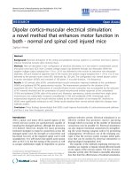
that only SCH 16 showed inhibition of CPE. The structure
of this MIBT derivative is depicted in Figure 1. Ribavarin,
a known inhibitor of flavivirus was used as a control in all
the experiments. Although there is no structural similarity
between Ribavarin and SCH 16, we opted to use Ribavarin
as a positive control in all experiments so that we have a
reference value for comparing the results of SCH 16. These
two compounds were then subjected to evaluation by the
plaque reduction assay at non-cytotoxic concentrations
(
/>
Table 1: List of Methylisatin-β-thiosemicarbazone (MIBT)
derivatives and the CC50 on PS and BHK-21 cells
Compounds
*CC50 on PS cell line
*CC50 on BHK-21 cell line
1 SF3
2 SF7
3 SCH16
4 SF17
5 SCH17
6 SC18
7 SCH19
8 SC18
9 SB18
10 SF24
11 SF27
12 SC27
13 SC28
14 SB29
15.Ribavirin
47 µg/ml
43 µg/ml
76 µg/ml
21 µg/ml
41 µg/ml
17 µg/ml
18 µg/ml
31.5 µg/ml
22.5 ug/ml
25.5 ug/ml
22.5 ug/ml
21 ug/ml
21 ug/ml
25 ug/ml
50 ug/ml
86 ug/ml
200 ug/ml
126 ug/ml
42 ug/ml
46 ug/ml
82 ug/ml
141 ug/ml
140 ug/ml
36 ug/ml
16.8 ug/ml
51 ug/ml
46 ug/ml
94 ug/ml
21.5 ug/ml
200 ug/ml
*CC50 = The concentration of the compound that reduced the
viability of cells to 50% of the control. Note: The 14 MIBT
compounds belonged to four different categories based on the
halogen or methyl group substituted at the position R and are
designated as SB group with bromine, SC group with chlorine, SF
group with fluorine and SCH group with -CH3 group substituted at R.
R' has N-substituted aromatic side chain attached to the -CH2
moiety. Cytotoxicity concentration (CC50) of synthesized compounds
was evaluated on exponentially growing PS and BHK-21 cells. It can
be observed that the CC50 of MIBT derivatives ranged from ≥ 76 ug/
ml to ≥ 17 ug/ml, while CC50 on BHK-21 cell line ranged from ≥ 200
ug/ml to ≥ 16.8 ug/ml.
ited a dose depended reduction of plaques formed by JEV
and WNV (Figure 2, Panels A and B) with an IC50 of 16 µg/
ml (0.000025 µM) and 4 ug/ml (0.000006 µM) for JEV
and WNV respectively. On the contrary the IC50 of Ribavirin was 3.9 µg/ml (0.000016 µM) and 1.7 µg/ml
(0.000007 µM) for JEV and WNV respectively. No antiviral activity of SCH 16 was noted against Den-2 although
Ribavarin showed a dose dependent inhibition of Den-2
plaque formation (Figure 2, Panels C and D).
The specificity of the action of an antiviral compound is
determined by calculating the Therapeutic Index (TI),
which is the ratio of CC50 to IC50. The TI of SCH 16 was
5 and 19 for JEV and WNV respectively while for Ribavirin
it was 13 and 29 respectively. This suggests that SCH 16 is
moderately active against JEV and highly active against
WNV.
The kinetics of action of SCH 16 in relation to the
replicative cycle of JEV in vitro
As a first step to understand JEV and SCH 16 interactions,
experiments were designed to determine the kinetics of
JEV replication in vitro. It was noted that the earliest
appearance of JEV antigen in infected PS cells was at 10
hours post-adsorption as detected by IFA (data not presented). However, the first infectious progeny of virus was
Page 2 of 12
(page number not for citation purposes)
Virology Journal 2008, 5:64
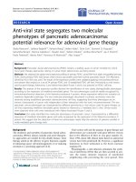
Butterfly structure of N-Methylisatin-β-Thiosemicarbazone
Figure 1
derivative SCH 16
Butterfly structure of N-Methylisatin-β-Thiosemicarbazone derivative SCH 16.
detected in the supernatant medium at 14 hours postadsorption thereby suggesting that a single replicative
cycle of JEV in vitro in PS cell line requires 14 hours for
completion (data not presented).
The antiviral activity of SCH 16 was subsequently investigated in relation to the kinetics of JEV replication. Nontoxic concentration of SCH 16 was added at various time
points following entry of JEV into PS cells and the experiments terminated following 48 hours incubation. The
compound at a concentration of 76 µg/ml (0.00012 µM)
was able to completely inhibit JEV replication when
added to the infected monolayer at 2, 4, 6 and 8 hours
post-infection evidenced by the absence of viral RNA, viral
antigen and inhibition of virus yield (Figure 3, Panel A to
C). However, addition of SCH 16 beyond 8 hours post
infection did not completely inhibit JEV replication since
JEV antigen, RNA and infectious virus were detected at
subsequent time points (Figure 3, Panels A to C).
In order to determine the minimum contact period
required for SCH 16 to exert its antiviral effect on JEV replication in vitro, a series of experiments were performed.
SCH 16 was added to JEV infected cell cultures at '0' hour
post-infection and removed at 4 hourly time points up to
14 hours and the monolayers were further incubated for
48 hours at 37°C under 5% CO2. It was observed that
there was complete inhibition of virus replication when
SCH 16 was allowed to be in contact with infected cultures for more than 8 hours post-infection. However,
when SCH16 was withdrawn at earlier time points there
was no inhibition of virus replication as confirmed by the
detection of viral antigen, viral RNA and infectious virus
yield (Figure 3, Panels D to F).
/>
Effect of SCH 16 on viral translation
To understand the probable action of SCH 16 on the viral
replicative cycle and to study the extent of damage caused
by the compound on the viral RNA that might result in the
inhibition of viral events such as protein synthesis (translation), an in vitro translation experiment was carried out
as described in materials and methods. RNA was extracted
from drug treated (4 hours and 10 hours post infection)
and untreated monolayers of JEV infected cells and subjected to Real Time PCR analysis to confirm the presence
of JEV RNA. Subsequently, the viral RNA was subjected to
in vitro translation. It was observed that RNA extracted
from JEV infected cells treated with SCH 16 for 4 hours
failed to translate into JEV proteins in vitro. On the contrary, viral RNA extracted from infected cells treated with
SCH 16 at 10 hours as well as RNA from infected cells that
were not treated with SCH 16 showed the presence of JEV
proteins (Figure 4).
In vivo evaluation of compounds against JEV using mouse
model
After ascertaining the in vivo non-toxic concentrations in
preliminary experiments, the therapeutic potential of
SCH 16 was evaluated in mice using intracerebral and
intraperitoneal challenge routes. In the intracerebral challenge model, mice that were treated with 100 and 200 mg/
kg body weight of SCH 16 showed no protection. However, it was interesting to note that all the mice that were
treated with SCH 16 remained healthy up to day 6 postinfection without showing any apparent symptoms of JEV
infection (data not presented). The symptoms started
appearing in these mice from day 7 post-infection. There
was a gradual progression of the symptoms and death
occurred on day 9. On the other hand, untreated mice
appeared sick by day 3 and succumbed by day 5. This suggests that there was a prolonged survival time of 3 days
between the treated and untreated mice.
The prolonged survival time observed in the intracerebral
challenge experiments prompted us to make use of a
peripheral challenge model (JEV 50LD50) using a multiple
dosage regimen wherein 200, 400 and 500 mg/kg body
weight of SCH 16 was administered by oral route. It was
observed that, there was 25% protection in the group of
mice administered with 200 mg/kg body weight of SCH
16, 50% protection observed in the group that received
400 mg/kg body weight and complete protection was
observed in the group that were given with 500 mg/kg
body weight of SCH 16 (Figure 5). Mice that survived the
challenge post treatment were sacrificed; brains harvested
and subjected to virus isolation, detection of viral antigen
and viral RNA. Viable virus could not be isolated from the
brain tissue of these mice. Further, no viral antigen could
be demonstrated in the brain smears by immunofluorescent staining using monoclonal antibodies to JEV. How-
Page 3 of 12
(page number not for citation purposes)
Virology Journal 2008, 5:64
/>
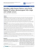
Figure 2activity of Ribavirin and SCH 16 against JEV, WNV and Den-2 evaluated using the plaque reduction assay
Antiviral
Antiviral activity of Ribavirin and SCH 16 against JEV, WNV and Den-2 evaluated using the plaque reduction
assay. Panel A: Represents the dose dependent reduction in JEV (yellow bars) and WNV (green bars) plaques obtained in PS
cells with the standard antiviral agent Ribavirin (represented as bars). The X axis represents the various concentrations of the
compound, Y' axis represents the percent reduction in plaques. The viability of cells is represented as line graph superimposed
on the bar diagram on the Y axis. Panel B: Represents the dose dependent reduction in JEV (yellow bars) and WNV (green
bars) plaques obtained in PS cells with the SCH 16 (represented as bars). The X axis represents the various concentrations of
the compound, Y' axis represents the percent reduction in plaques. The viability of cells is represented as line graph superimposed on the bar diagram on the Y axis. Panel C: Represents the dose dependent reduction in Den-2 plaques (orange bars)
obtained in BHK 21 cells with the Ribavirin (represented as bars). The X axis represents the various concentrations of the
compound, Y' axis represents the percent reduction in plaques. The viability of cells is represented as line graph superimposed
on the bar diagram on the Y axis. Panel D: Note that there was no reduction of in Den-2 plaques was obtained with the SCH
16 in BHK 21 cells. The X axis represents the various concentrations of the compound, Y' axis represents the percent reduction in plaques. The viability of cells is represented as line graph superimposed on the bar diagram on the Y axis.
ever, the RT-PCR products amplified from the brain
homogenate suggested that viral RNA was present in the
brain of animals that survived JEV infection following
treatment with 400 and 500 mg/kg body weight of SCH
16.
Discussion
There is currently no specific antiviral treatment available
for Japanese encephalitis, West Nile and Dengue virus
infections. Recently there has been renewed interest in the
search for antiviral compounds active against a variety of
viral infections. For instance, there are several reports
describing the in vitro inhibitory effect of compounds such
as ribavirin, mycophenolic acid, imino sugars, inhibitors
of serine protease, RNA interference and non-steroidal
anti-inflammatory drugs against flaviviruses [8-13]. NMethylisatin-β-thiosemicarbazone (MIBT) was one of the
first antiviral compounds to be discovered. It exhibits
Page 4 of 12
(page number not for citation purposes)
Virology Journal 2008, 5:64
/>
Figure of
Kinetics 3 action of SCH 16 in relation to the replicative cycle of JEV in PS cells
Kinetics of action of SCH 16 in relation to the replicative cycle of JEV in PS cells.Panel A represents the results of
the experiments wherein the addition of the drug SCH 16 to virus infected PS cells was staggered (refer to Material & Methods
for details). X axis represents the various time points at which SCH 16 was added after adsorption of JEV onto PS cells. Note
that there was no virus yield (represented as Log TCID50/ml on Y axis) in drug treated cells (blue triangle) until 8 hours post
infection after which virus yield steadily increased to attain levels similar to that obtained in untreated cells (pink sphere). The
Y' axis represents the optical density values obtained in the JEV antigen capture ELISA. Soluble JEV antigen was measured in the
supernatant fluids obtained at 48 hrs after the experiment (refer to Material & Methods for details) in both drug treated (black
square) and untreated (red diamond) cells. Panel B depicts the detection of JEV specific antigen using an immunofluorescent
assay. Note the presence of bright immunofluorescence in the JEV infected monolayers (virus control). It can also be observed
that JEV infected mono layers treated with SCH 16 were positive for viral antigen at 10, 12 and 14 hours post infection whilst
viral antigen was undetectable by immunofluorescence at 0, 2, 4, 6 and 8 hrs post infection respectively (400×). Panel C: The
amplification plots obtained in Real Time PCR depicting the detection of JEV RNA in the untreated cells and SCH 16 treated
cells at varying time points post-infection. Panel C-1 depicts the typical amplification plot (fluorescence vs cycle number)
obtained by the real time PCR with the RNA extracted from the virus infected untreated cells at varying time points. Note that
JEV RNA was detected at all time points. In contrast JEV RNA was undetectable at 0, 2,4, and 8 hrs in the SCH 16 treated cells
(Panel C2). Panel D represents the results of the experiments wherein the minimum time required for SCH 16 to exert antiviral activity was evaluated (refer to Materials & Methods for details). SCH 16 was added to all monolayers 2 hrs post virus
adsorption and removed from the mono layers at periodic intervals. X-axis represents the various time points when SCH 16
was removed after JEV entry into PS cells. Note that virus yield (represented as Log TCID50/ml on Y axis) in drug treated cells
(blue triangle) steadily declined from 0 hrs post infection until 8 hours post infection after which there was no virus production
noted in drug treated cells. On the contrary virus yields continued to be high in untreated cells (pink sphere) at all time points.
The Y' axis represents the optical density values obtained in the JEV antigen capture ELISA. Soluble JEV antigen was measured
in the supernatant fluids obtained at 48 hrs after infection (refer to Materials & Methods for details) in both drug treated (black
square) and untreated (red diamond) cells. Panel E: Effect of duration of antiviral action of SCH 16 on JEV replication postinfection. SCH 16 was added to JEV infected PS cell monolayer at 0 hours post – adsorption and the inoculums were removed
at different time points post-infection (0 to 14 hrs). The monolayer was stained using JEV specific monoclonal antibodies by IFA
at 48 hours (400×). Presence of cell bound antigen can be appreciated upon the removal of SCH 16 in the early hours (up to 4
hours) of viral replicative cycle, while viral antigen was not detected when SCH 16 was retained with the infected monolayer
for longer duration (8 hours andmore). Panel F: The amplification plots obtained in Real Time PCR depicting the detection of
JEV RNA in the infected cells treated with SCH 16 at 0 hours and inoculums removed at varying time points (refer to Materials
and methods for details). Panel F-1 depicts the typical amplification plot (fluorescence vs cycle number) obtained by the real
time PCR with the RNA extracted from the virus infected untreated cells at varying time points. Note that JEV RNA was
detected at all time points. In contrast JEV RNA was detectable only at 0 and 4 hrs in the SCH 16 treated cells and undetectable beyond 8 hrs (Panel F2).
Page 5 of 12
(page number not for citation purposes)
Virology Journal 2008, 5:64
/>
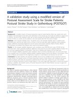
Figure 4
Western blot illustrating the effect of SCH 16 on JEV translation using an in vitro translation kit
Western blot illustrating the effect of SCH 16 on JEV translation using an in vitro translation kit. Lane 1-uninfected cell control, lanes 2 and 4 – in vitro translation products of RNA obtained form JEV infected PS cells (untreated) at 4 and
10 hours post infection respectively. Lanes 3 and 5 – in vitro translation product of RNA obtained from JEV infected PS cells
treated with SCH 16 for 4 hours and 10 hours respectively. Note that SCH 16 treatment of JEV infected cells did not show any
in vitro translation product at 4 hours post treatment (Lane 3) whilst at 10 hours (Lane 5) a 50 Kda in vitro translation product
was obtained. Lane M represents molecular weight markers.
antiviral activity against a variety of RNA and DNA viruses
[14-17]. Recent studies have demonstrated that thiosemicarbazone and Mannich bases of thiosemicarbazone
derivatives exhibit anti-HIV activity in vitro [18-22]. Therefore this study was designed to investigate the antiviral
property of isatin β thiosemicarbazone derivatives against
JEV, WNV and Den-2 viruses.
In the present study, fourteen Mannich bases of MIBT
derivatives were synthesized and evaluated for their ability to inhibit flaviviral replication. However, only one
compound (SCH 16) showed antiviral activity against JEV
and WNV in vitro with a therapeutic index of 5 and 16
respectively. This compound did not exhibit any virus
inactivating property. SCH 16 (Figure 1) is a mannich
base of N-Methylisatin-β-thiosemicarbazone possessing
an isatin backbone with modifications made at the side
chains. Chemically isatins are diketonic compounds. It
has been earlier noted that, heteroaromatic thioamides
containing N-substitution at more than one position per
heterocyclic ring are worthy of investigation due to its
increased antiviral property [4]. It is therefore likely that
the antiviral activity of SCH 16 may be due to the N sub-
stitution at the 8th position in the heterocyclic benzene
ring and a NO2 group attached to the aromatic side chain.
Although SCH16 exhibited antiviral activity against WNV,
we did not pursue further experiments with it since WNV
is not a public health concern in India. In contrast, JEV is
a major public health problem in India and hence we set
about to investigate in detail the mechanism of antiviral
activity of SCH 16 against JEV. Two crucial questions pertaining to the antiviral activity of SCH16 against JEV were
addressed; (i) how long after virus infection can addition
of drug be delayed in vitro in order to achieve inhibition
of virus replication? and (ii) what is the minimum time
required for SCH16 to exert its antiviral activity?. For this
purpose we used an experimental approach similar to that
described earlier by Baginiski et al and Lammarre et al
[23,24]. Our results showed that when the drug was
added to infected cells at various time points post virus
entry, neither viral antigen (Figure 3 Panel A & B) nor viral
nucleic acid (Figure 3, Panel C) was detected up to 8 hours
post infection. Beyond this time point however, viral antigen, nucleic acid and infectious virus was detectable in the
cultures. Indeed viral antigen, viral RNA and virus yields
were comparable to those obtained with untreated cells
Page 6 of 12
(page number not for citation purposes)
Virology Journal 2008, 5:64
/>
Survival 5
Figure graphs depicting the in vivo effect of SCH 16 against a lethal JEV challenge
Survival graphs depicting the in vivo effect of SCH 16 against a lethal JEV challenge. SCH 16 was administered to
swiss albino mice (n = 4 per group) per orally twice daily at 12 hour intervals to three groups of mice. Each group of mice
received the drug at 200 (red square), 400 (blue triangle) and 500 (black sphere) mg/kg body weight respectively. A forth group
of mice (n = 4) served as virus control (green diamond) and did not receive the drug. All the groups of mice were challenged
with 50LD50JEV (P20778) by intraperitoneal route as described in materials and methods. The survival of mice was monitored
for 20 days post-challenge. X-axis depicts the days post challenge. Y-axis depicts the percentage survival of mice treated with
various concentrations of the drug as wells untreated control mice. Each data point depicts the mean survival rate of four mice
in the respective group. Note that all mice in the virus control group succumbed by 7 days post challenge.
beyond the 8 hour time point thereby suggesting that SCH
16 did not inhibit normal cellular functions (Figure 3,
Panels A to C). This suggests that the drug was not toxic to
cells and did not inhibit the ability of cells to support virus
replication at later time points. To ascertain the minimum
time required for SCH16 to exert its antiviral activity, the
compound was added at 2 hours post infection and
removed at various time points post viral entry. The
results revealed that, SCH 16 probably acted as an inhibitor of early protein synthesis. Had SCH 16 been an uncoating inhibitor or a polymerase inhibitor, the drug
would have required a contact time of less than 4 hours to
bring about its inhibitory effect. Similarly if it were a protease inhibitor the minimum contact period for SCH 16 to
bring about inhibition of virus replication would have
been greater than 8–10 hours. Since we observed that the
minimum contact period of 8 hours was required for
SCH16 to completely inhibit virus replication, it probably
indicates that the drug is acting at the level of translation.
Cooper et al [25] in an earlier study with vaccinia virus
had demonstrated that the specific antiviral effect of MIBT
was noted 6 hours post-infection thereby indicating inhibition of viral protein synthesis. In order to ascertain
whether this was indeed also true for SCH 16 we adopted
another approach to investigate the precise role of SCH 16
on translation events in JEV replication. We obtained RNA
samples from the experiments that treated JEV infected
monolayer's with SCH 16 for 4 hours and SCH 16 added
at 10 hours post infection from infected cells treated with
SCH 16 as well as cells that were untreated using identical
extraction protocols. Subsequently we performed Real
Time SYBR Green I PCR using JEV specific primers to confirm the presence of JEV RNA in samples obtained from
both drug treated as well as untreated cells. The viral RNA
Page 7 of 12
(page number not for citation purposes)
Virology Journal 2008, 5:64
thus obtained, was then subjected to in vitro translation
experiments which clearly showed that there were no
translation products obtained with RNA obtained from
drug treated cells at 4 hours post infection (Figure 4, lane
3). On the contrary, RNA obtained from drug treated cells
at 10 hours post infection (Figure 4, lane5) as well as RNA
obtained from untreated cells at both 4 hours and 10
hours post infection (Figure 4, lanes 2 and 4). This result
demonstrates that SCH16 is able to selectively suppress
translation of JEV RNA at early time points in the life
cycle. Similar observations have been made earlier by
Ronen et al on other RNA virus [26] who investigated the
inhibitory action of N-methyl isatin beta-diethylthiosemicarbazones on Moloney Leukemia virus replication.
The therapeutic potential of SCH 16 against JEV was evaluated in vivo in mice using the intracerebral and intraperitoneal challenge studies. The mice that were evaluated in
the intracerebral challenge route did not show any protection although there was a delay in appearance of symptoms and death in drug treated mice. The lack of
protection by this route may be due to (i) the direct introduction of large amount of infectious virus (50LD50) into
the CNS which might have compromised the inhibitory
action of SCH 16 and/or (ii) inability to achieve therapeutic concentrations of the drug in the brain either due to
delay in the compound reaching the brain from the intraperitoneal compartment or poor penetration of the drug
into the brain parenchyma. On the contrary, the drug
treated mice challenged by the intraperitoneal route
showed a dose dependent reduction in mortality, whilst
all the untreated mice succumbed to the challenge with
50LD50 of JEV by day seven (Figure 5). Furthermore, neither viable virus nor viral antigen could be demonstrated
in the brains of the mice that survived the challenge. However, viral RNA was detected by real-time RT-PCR in all the
brain tissues. Since flavivirus RNA dependent RNA
polymerases are active within three hours of viral entry
this is not a surprise finding [27]. Because, SCH 16 is primarily an early translation inhibitor, it appears that this
drug does not interfere with RNA polymerization resulting in accumulation of viral RNA in the brains of drug
treated mice that survived the challenge. Alternatively,
SCH 16 treatment could have curtailed JEV replication in
the periphery resulting in a very small amount of JEV
entering the brain. Consequently the virus was unable to
establish a productive infection in the brain and the presence of viral RNA could be as a result of residual virus in
brains of mice that survived the challenge. In an experimental rat model, with post-encephalitic Parkinsonism
induced by JEV infections [28,29] it was observed that,
administration of isatin improved the motor neuron
activities significantly. Indeed, they attributed that the
improvement in the motor weakness was probably due to
the MAO inhibitory activity of isatin and suggested that
/>
isatin could possibly serve as a new therapeutic agent for
Parkinsonism. However, these studies were not designed
to address the antiviral action of isatin against JEV but
aimed at investigating the neurotransmitter inhibitory
effect. It may be argued therefore that the in vivo effect of
SCH 16 against JEV noted in this study may also be attributed to the immunomodulating or neuroprotective property of SCH 16.
An intriguing observation in this study was the differential
ability of SCH 16 to suppress JEV, WNV and Den-2 multiplication in vitro. It is difficult to hypothesize the differential antiviral property of SCH 16 noted against JEV and
WNV in this study as they are structurally similar and we
have used the same cell system (PS cells) for evaluating
the drug. On the contrary, we used BHK 21 cells for assaying the antiviral activity of SCH 16 against Den-2 virus,
which could have contributed to the lack of anti-Dengue
activity of SCH 16. Protein synthesis consists of an intricate series of events requiring components that are too
numerous to be encoded by viral genomes [30,31]. It has
been observed that Den-2 and other flaviviruses, such as
WNV, yellow fever, JEV, and Kunjin viruses, are presumed
to undergo cap-dependent translation [32,33]. However,
evidence exists that under certain conditions that inhibit
cap dependent translation, Den-2 viruses can switch to
more efficient cap independent translation. Further,
mammalian cellular stress response and immune functions, such as the interferon antiviral response [34,35],
may compel viral translation by one mechanism over the
other. Since we used PS cells for evaluation of JEV and
WNV and BHK 21 cells for Den-2 it is possible that the
translation pathway adopted by Den-2 against SCH 16
may be due to the presence of certain BHK 21 cell specific
factors. However, strong experimental evidence is needed
to support this hypothesis and it would be interesting to
investigate whether SCH 16 is indeed a cap dependent
translation inhibitor.
Conclusion
In conclusion, the findings of this study unequivocally
demonstrate that SCH 16 has antiviral activity against JEV
and WNV in vitro. Furthermore, SCH 16 was also found to
completely inhibit JEV replication in vivo in a mouse
model challenged peripherally with 50LD50 of the virus in
a dose dependent manner. This necessitates further investigation into the pharmacokinetcis of the compound. Its
moderate therapeutic index (TI = 5) may be a concern.
However, further investigation on structure – activity relationships and appropriate modification in the aryl ring of
the isatin moiety could provide more effective JEV-inhibitors with improved efficacy in future.
Page 8 of 12
(page number not for citation purposes)
Virology Journal 2008, 5:64
Materials and methods
Viruses
Standard strains of JEV (P20778), Den-2 virus (P23085)
and WNV (G22886) were obtained from National Institute of Virology (NIV), Pune, India.
Cells and animals
Aedes albopictus (C6/36) mosquito cell line and Porcine
Stable kidney (PS) cells were maintained in Minimum
Essential Medium (MEM) with 10% fetal calf serum while
Baby Hamster Kidney (BHK-21) cells were maintained in
Dulbecco's MEM with 10% fetal calf serum (NCCS, Pune,
India). Random bred Swiss albino mice (4–5 week old)
were obtained from Central Animal Research Facility,
NIMHANS, Bangalore, India, and used for the in vivo evaluation. All animal experiments were conducted after
obtaining permission from Institutional Animal Ethics
Committee.
N-Methylisatinisatin-β-Thiosemicarbazone (MIBT)
derivatives
Fourteen mannich bases of isatin-β-thiosemicarbazone
derivatives (Table 1) were obtained from Dr. Sriram, Birla
Institute of Technology and Science (BITS), Pilani, India.
The compounds were synthesized by Schiff reaction. N, Ndiethyl thiosemicarbazide was condensed with isatin in
the presence of glacial acetic acid to form 1H-indole-2, 3dione -3-N, N-diethyl thiosemicarbazone (Schiff base).
The N-Mannich bases were further condensed using acidic
imino group along with formaldehyde and various secondary amines to obtain isatin thiosemicarbazone derivatives. Ribavirin, which is a known inhibitor of flavivirus
replication, was obtained from commercial sources
(Sigma, USA) and used as a control drug in this study.
Cytotoxicity of Ribavirin and MIBT derivatives
Cytotoxicity of the antiviral compounds was evaluated
using the Trypan blue exclusion assay [36]. Briefly, PS and
or BHK-21 cells grown to semi-confluence in 24-well
plates were exposed to different concentrations of the
compounds for 4 days at 37°C. Following this, the cells
were harvested by trypsinization and re-suspended in 0.5
ml of MEM containing 10% FCS. A 100 µl of the cell suspension was mixed with 50 µl of 2.5% Trypan blue and
the number of viable cells was enumerated using a hemocytometer. The concentration of compound that reduced
cell growth by 50% was estimated as the 50% cytotoxic
concentration (CC50). The effect of the compounds on
cellular proliferation was also studied. Briefly, the drug
treated cells and untreated cells were seeded at a rate of 2
× 104 cells per well into 24-well plates and allowed to proliferate for 3 days in MEM, containing 10% FCS. The proliferations of cells were monitored every day
microscopically by recording signs of toxicity such as
/>
altered morphology presence or absence of vacuoles and/
or dead cells.
Screening for inhibition of virus induced cytopathic effect
in vitro
The antiviral activity assay of the Ribavarin and MIBT
derivatives against JEV, Den-2 virus or WNV were screened
in vitro using the cytopathic effect (CPE) inhibition assay
carried out in a 96 well plate. Briefly, monolayers of PS
and/or BHK-21 were inoculated with 100 µl of appropriate virus suspension containing 1 MOI of virus and
adsorbed for two hours at 37°C. At the end of incubation
period, the virus (JEV, Den-2 or WNV) was removed and
the monolayers were rinsed with MEM to remove
unbound virus. Doubling dilutions of different concentrations of Ribavirin and MIBT derivatives (beginning
with CC50) were prepared in MEM, added to the monolayer (100 µl) and incubated at 37°C for 3 days under 5%
CO2. The experiment was terminated when the virus control showed maximum CPE. The presence or absence of
CPE was recorded microscopically every day and the
plates were stained using crystal violet at the termination
of experiment and compared with the untreated virus controls and drug controls. All the experiments were run in
triplicates to ensure reproducibility.
Confirmation of antiviral activity by plaque reduction
assay
The compounds that showed inhibition of virus replication in the CPE inhibition assay were further evaluated
using plaque-reduction assay. Briefly, PS (4 × 10⁴ cells/
well) cells were grown to a confluent monolayer in a 24
well plate and infected with 100 µl of virus suspension
containing 1 MOI of JEV and incubation was carried out
for 2 hours at 37°C. At the end of adsorption, monolayers
were rinsed with sterile PBS and 100 µl MEM containing
varying concentrations of the compounds were added.
The monolayer was then overlaid with maintenance
medium containing 0.2% molten agarose (Sigma-Aldrich,
USA). Appropriate controls were included in each run of
the assay. Incubation was carried out at 37°C for 3 days.
At the end of incubation period monolayers were fixed in
10% formal saline, the agarose was gently removed and
the cells were stained using 1% crystal violet. Two independent observers counted the plaques using a hand lens.
All the experiments were run in triplicates. Percentage
inhibitions of plaques were determined using the formula
given below.
% Inhibition =
Number of plaques in virus control-Number of plaques in drug treated
× 100
Number of plaques in virus control
The antiviral activity was expressed as 50% inhibitory concentration (IC50) of the compound, which is the concentration of the compound required to inhibit viral plaques
by 50% as compared to virus control. The therapeutic
Page 9 of 12
(page number not for citation purposes)
Virology Journal 2008, 5:64
potential and specificity of action was determined by calculating the Therapeutic Index (TI), which is the ratio of
CC50 to IC50 (CC50/IC50) [37].
Understanding the mechanism of action of SCH 16 in
relation to JEV replication
To understand the possible mechanism of action in relation to the replicative cycle of JEV, the compounds that
showed 100% inhibition of viral plaques were evaluated
by in vitro experiments detailed below.
Determining kinetics of JEV replication in PS cells
A 24 well plate containing sterile cover slips in each well
was seeded with 4 × 10⁴ cells/well and incubated at 37°C
overnight. When the cells were a confluent monolayer,
they were infected with JEV (MOI = 1) for 1 hour at 37°C.
The monolayer was rinsed thoroughly with sterile PBS
and replenished with medium containing 1% FCS. This
time point was considered as '0' hour post-infection. Subsequently at 2, 4, 6, 8, 10, 12, 14, 16 and 24 hours postinfection, the medium was harvested to determine the
amount of extracellular virus released into the supernatant. At each time point, the cover slip containing cells was
also removed, fixed in chilled acetone and stained by
Immunofluorescent Assay (IFA) using a monoclonal antibody to envelope protein of JEV to detect the cell bound
antigen [38].
Understanding the kinetics of the antiviral activity of SCH 16
A 24 well plate was seeded with 4 × 10⁴ cells/well and
incubated at 37°C overnight. To this monolayer JEV was
added (MOI = 1) and incubated for 1 hour at 37°C. At the
end of adsorption, the virus was removed, the monolayer
was rinsed 3 – 4 times using sterile PBS and replenished
with MEM containing 1% FCS. This time point was considered as 0 hour post-infection. Starting from 0 hour
time point, 76 ug/ml (IC50) of the compound was added
at 2, 4, 6, 8, 10, 12, 14, 16, and 24 hours post-infection
and incubated at 37°C. The supernatant fluid was harvested from the respective wells at 48 hours post-infection. The fluid was divided into two parts. One part was
used to determine the virus yield in the supernatant fluid
(TCID50/ml) and the second part of the fluid was used to
detect the presence of soluble JEV antigen using an antigen capture ELISA described elsewhere [39]. In order to
detect cell bound antigen the cover slip cultures were fixed
in chilled acetone for 30 minutes at 4°C and stained using
monoclonal antibody to JEV (Clone F2C2) and antimouse IgG FITC conjugate by indirect IFA as described
earlier. The cells in each well were treated with 750 µl of
TRIzol (Invitrogen, USA) for RNA extraction and reverse
transcription was carried out using cDNA archive kit
(Applied Biosystems, USA) as described below.
/>
Real Time PCR using Syber Green I chemistry
Detection of viral RNA was carried out by Real Time PCR
using Syber Green I chemistry as described by Shu et al
[40] with minor modifications. Briefly, a 120 base pair
product of the PreM gene of JEV was amplified using the
forward primer F1 (gga gcc atg aag ttg tca aat ttc) and
reverse primer R1 (ttg ccc gga ccc aac at) based on the prototype standard strain of JEV (P20778) Gen Bank
Ac.No.7080251.
A second set of experiments was designed to estimate the
minimum time required for the compound to bring about
complete inhibition of JEV replication. A 24 well plate was
seeded with 4 × 10⁴ cells/well in quadruplicates and incubated at 37°C overnight. Confluent PS monolayers were
infected with JEV (MOI = 1) and adsorbed for 1 hour at
37°C. Following this, the monolayer was rinsed with sterile PBS and replenished with plain medium containing
non-toxic concentration of SCH16. Control wells received
plain medium. This time point was considered as '0' hour
post-infection. Starting from 0 hour time point, medium
containing the compound was removed at 0, 4, 8, 12, and
14 hours post-infection and replenished with MEM containing 1% FCS. At the end of 48 hours incubation, the
fluid harvested from one of the quadruplicate set of wells,
was evaluated for presence of extracellular virus by titration while soluble antigen was detected using an antigen
capture ELISA described earlier. Cells in a second set of
wells were trypsinised, re-suspended in maintenance
medium and subjected to three freeze thaw cycles to
release intracellular virus, which was quantitated by titration. Cells from the third set of wells were stained by an
IFA to detect cell bound antigen. The cells in the fourth set
of wells were treated with 750 µl of TRIzol (Invitrogen,
USA) for RNA extraction and reverse transcription was carried out using cDNA archive kit (Applied Biosystems,
USA).
Effect of SCH 16 on the translation of JEV
In order to understand the probable action of SCH 16 on
the events of viral replication, an in vitro translation experiment was carried out using commercially available Transcend™ non-radioactive translation detection system and
rabbit reticulocyte lysate kit (Promega, USA). A 24 well
plate was seeded with PS cells (4 × 10⁴/ml), incubated at
37°C for 18 to 24 hrs and the monolayer formed was
adsorbed with JEV (MOI = 1) for 1 hour. The infected
monolayer was rinsed with sterile PBS to remove the
unbound virus. To one set of JEV infected monolayer cultures, SCH 16 at non-toxic concentration was added at '0'
hour and incubated for 4 hours. Medium containing SCH
16 was removed at 4 post-infection and replenished. To a
second set of monolayer cultures, SCH 16 at the same
concentration was added at 10 hours post adsorption. The
plates were further incubated for 48 hours at 37°C.
Page 10 of 12
(page number not for citation purposes)
Virology Journal 2008, 5:64
Appropriate virus and cell controls were included in parallel to the test. At the end of incubation, the cells were
treated with 750 µl of TRIzol and the viral RNA was
extracted as described earlier. The viral RNA thus
obtained, was divided in to two equal parts. One part was
subjected to RT-PCR using JEV specific primers to confirm
the presence of JEV RNA. The second part was subjected to
in vitro translation carried out using a commercial kit
(Promega, USA) and manufacturer's guidelines. Briefly, a
50 µl reaction containing 35 µl of rabbit reticulocyte
lysate, 10 µl of nuclease free water, 1 µl of RNasin (40 U/
µl), 1 µl of complete amino acid mixture (1 mM), 1 µl of
Transcend ™ tRNA and 2 µl of RNA template was set up at
30°C and incubated for 60 minutes. After the completion
of translation reaction, 1 ul of the product was subjected
to SDS – PAGE, electroblotted on to a PVDF membrane,
blocked with skimmed milk powder solution, reacted
with JEV specific monoclonal antibody to visualize the
bands.
In vivo evaluation of SCH 16 against JEV
Evaluation of non-toxic concentration of the compounds in mice
In order to determine the in vivo non-toxic concentrations,
the compound SCH 16 (100, 200, 400 and 500 mg/kg
body weight) were administered either per orally, or intraperitoneally into four different groups of 4 – 5 weeks old
Swiss albino mice (n = 4). Two groups of mice served as
normal controls that received plain medium per orally or
intraperitoneally. All mice were observed for a period of
45 days for loss or gain in weight, and other evidences of
toxicity as compared to the untreated normal mice.
/>
neally, starch intracerebrally and plain MEM orally while
a fifth group of mice (n = 4) served as sham controls and
received MEM intraperitoneally and starch intracerebrally.
Mice were observed every day for 20 days post-infection
for the appearance of symptoms and death. At the end of
the observation period the mice that survived the infection were sacrificed, brains harvested and subjected to JEV
antigen detection by IFA, JEV nucleic acid detection by
real-time PCR and virus isolation using shell vial method
[41].
Competing interests
The authors declare that they have no competing interests.
Acknowledgements
The authors thank Mr. Sunil Gowekar, research scholar, Department of
Neurovirology, NIMHANS, Bangalore, for making the computer generated
structure of SCH 16
References
1.
2.
3.
4.
5.
6.
7.
In vivo evaluation of SCH 16 by intracerebral challenge
To determine the in vivo antiviral potential of SCH 16,
three groups of mice (4 mice per group) were injected
intracerebrally with 30 µl of virus suspension containing
50 LD50 of JEV. This was followed by intraperitoneal
administration of SCH16 (100 and 200 mg/Kg body
weight) into two groups of mice twice daily for 10 days. A
third group of mice served as control animals that
received virus intracerebrally and plain MEM intraperitoneally. Animals were monitored for the appearance of
symptoms for JEV infections such as paralysis and death.
In vivo evaluation of SCH 16 by peripheral challenge
The therapeutic potential of SCH 16 was also evaluated
using a peripheral challenge model wherein JEV (50LD50)
was injected (200 µl) intraperitoneally into four groups of
4–5 weeks old mice (n = 4). Two hours later, each group
received 30 µl of 1% sterile starch intracerebrally to facilitate virus entry into the brain. This was followed by oral
administration of SCH 16 in a dose of 200, 400 or 500
mg/kg body weight twice daily into the three respective
groups for 12 days. A fourth group of mice (n = 4) served
as 'no drug controls' which received virus intraperito-
8.
9.
10.
11.
12.
13.
14.
15.
Ray Shi: Recent advances in Flavivirus Antiviral Drug Discovery and Vaccine Development. Recent Patents on Anti-Infective
Drug Discovery 2006, 1:45-55.
Simon Ho: HIV dynamics in vivo: implications for therapy.
Nature Review Microbiology 2003, 13:181-190.
Shi PY: Strategies for the identification of inhibitors of West
Nile virus and other flaviviruses. Current Opinion in Investigational
Drugs 2002, 3:1567-1573.
Bauer DJ, Sadler PW: The structural activity relationships of
the antiviral chemotherapeutic activity of isatin β-thiosemicarbazone. British Journal of Pharmacology 1960, 15:101-110.
Glover V, Bhattacharya SK, Sandler M: Isatin – A new biological
factor. Indian Journal of Experimental Biology 1991, 29:1-5.
Bauer DJ: Clinical experience with the antiviral drug marboran (1-methylisatin3-thiosemicarbazone). Annals of New York
Academy of Sciences 1965, 130:110-117.
Ronen D, Nir E, Teitz Y: Effect of N-methylisatin-β-4': 4'diethylthiosemicarbazones on intracellular Moloney Leukemia virus constituents. Antiviral Research 1985, 5:249-254.
Leyssen P, Balzarini J, De Clercq E, Neyts J: The predominant
mechanism by which ribavirin exerts its antiviral activity in
vitro against flaviviruses and paramyxoviruses is mediated by
inhibition of IMP dehydrogenase. Journal of Virology 2005,
79:1943-1947.
Diamond MS, Zachariah M, Harris E: Mycophenolic acid inhibits
Dengue virus infection by preventing replication of viral
RNA. Virology 2002, 304:211-221.
Wu SF, Lee CJ, Lia CL, Dwek RA, Zitzmann N, Lin YL: Antiviral
effects of an iminosugar derivative on flavivirus infections.
Journal of Virology 2002, 76:3596-3604.
Genesh VK, Muller N, Judge K, Luan CH, Padmanabhan R, Murthy
KH: Identification and characterization of non-substrate
based on inhibitors of the essential dengue and West Nile
virus proteases. Bioorganic Medicinal Chemistry 2005, 13:257-264.
Geiss BJ, Peirson TC, Diamond MS: Actively replicating West
Nile Virus is resistant to cytoplasmic delivery of SiRNA. Virology Journal 2005, 2:53.
Chen CJ, Raung SL, Kuo MD, Wang Y-M: Suppression of Japanese
encephalitis virus infection by non-steroidal anti-inflammatory drugs. Journal of General Virology 2002, 83:1897-1905.
Logan JC, Fox MP, Morgan JH, Makohon AM, Pfau CJ: Arenavirus
inactivation on contact with N-substituted isatin beta-thiosemicarbazones and certain cations. Journal of General Virology
1975, 28:271-83.
Teiz Y, Ronnen D, Vansover A, Stematsky T, Riggs JL: Inhibition of
human immunodeficiency virus by N-Methyl isatin β4':4' diethyl thiosemicarbazone and N-allyl isatin β4':4' diallylthiosemicarbzone. Antiviral Research 1994, 24:305-314.
Page 11 of 12
(page number not for citation purposes)
Virology Journal 2008, 5:64
16.
17.
18.
19.
20.
21.
22.
23.
24.
25.
26.
27.
28.
29.
30.
31.
32.
33.
34.
Levinson W, Woodson B, Jackson J: Inactivation of Rous sarcoma
virus on contact with N-Ethyl Isatin beta thiosemicarbazone.
Nature new biology 1971, 232:116-118.
Woodson B, Joklik WK: The inhibition of Vaccinia virus multiplication by Isatin-beta-thiosemicarbazone. Proceedings of
National Academy of Sciences USA 1965, 54:946-953.
Pandeya SN, Yogeeswari P, Sriram D, de Clercq E, Pannecouque C,
Witvrouw M: Synthesis and screening for anti-HIV activity of
some N-Mannich bases of isatinderivatives. Chemotherapy
1999, 4:192-6.
Sriram D, Yogeeswari P, Srichakravarty N, Bal TR: Synthesis of stavudine amino acid ester prodrugs with broad-spectrum
chemotherapeutic properties for the effective treatment of
HIV/AIDS.
Bioorganic Medicinal Chemistry Letters 2004,
8,14(5):1085-1087.
Sriram D, Bal TR, Yogeeswari P: Aminopyrimidinimino isatin
analogues: Design of novel nonnucleoside HIV-1 reverse
transcriptase inhibitors with broadspectrum chemotherapeutic properties. Journal of Pharmacy and Pharmaceutical Sciences
2005, 8:565-577.
Sriram D, Yogeeswari P, Meena K: Synthesis of anti-HIV and antitubercular activities of isatin derivatives. Pharamzie 2006,
61:274-277.
Bal TR, Anand B, Yogeeshawri P, Sriram D: Synthesis and evaluation of anti-HIV activity of isatin β-thiosemicarbazone derivatives. Bioorganic Medicinal Chemistry Letters 2005, 15:4451-4455.
Baginiski SB, Pavear DC, Seipel M, Chang Sun SC, Benetatos CA,
Chunduru SK, Rice CM, Collett MS: Mechanism of action of a
Pestivirus antiviral compound. Proceedings of National Academy of
Sciences 2000, 97:7981-7986.
Lamarre D, Croteau G, Wardrop E, Bourgon L, Thibeault D, Clouette C, Vaillancourt M, Cohen E, Pargelis C, Yoakim C, Anderson PC:
Antiviral properties of Palinavir, a potent inhibitor of the
Human Immunodeficiency virus type-1 protease. Antimicrobial
Agents Chemotherapy 1997, 41:965-971.
Cooper JA, Moss B, Katz E: Inhibition of Vaccinia virus late protein synthesis by isatin thiosemicarbazone: characterization
and in vitro translation of viral mRNA. Journal of Virology 1979,
96:381-392.
Ronen DL, Sherman S, Bar-nun , Teitz Y: N-Methyl-β-4',4'-diethylthiosemicarbazone, an inhibitor of Moloney Leukemia Virus
protein production: Characterization and in vitro translation
of viral mRNA.
Antimicrobial Agents Chemotherapy 1987,
31:1798-1802.
Gubler DJ, Kuno G, Markoff L: Flaviviruses. In Fields Virology 5th edition. Philadelphia: Lippincot Willims and Wilkins; 2007:1153-1225.
Ogata A, Hamaue N, Terado M, Minami M, Nagashima K, Tashiro K:
Isatin, an endogenous MAO inhibitor, improves bradykinesia
and Dopamine levels in rat model of Parkinson's disease
induced by Japanese encephalitis virus. Journal of Neurological
Sciences 2003, 206:79-83.
Hamaue N, Minami M, Terado m, Hirafuji M, Endo T, Machida M,
Heroshige T, Ogata A, Tashiro K, Saito H, Parvez SH: Comparative
study of the effects of isatin, an endogenous MAO-inhibitor,
and seleginine on bradykinasia and dopamine levels in a rat
model Parkinson's disease induced by the Japanese encephalitis virus. Neurotoxicology 2004, 25:205-213.
Brown DM, Kauder SE, Cornell CT, Jang GM, Racaniello VR, Semler
BL: Cell-dependent role for the poliovirus 3' noncoding
region in positive-strand RNA synthesis. Journal of Virology 2004,
78:1344-1351.
Dobrikova E, Florez P, Bradrick S, Gromeier M: Activity of a type
1 picornavirus internal ribosomal entry site is determined by
sequences within the 3' nontranslated region. Proceedings of
National Academy of Sciences, USA 2003, 100:15125-15130.
Edgil DM, Diamond S, Holden KL, Paranjape SM, Harris E: Differences in cellular infection among dengue virus type 2 strains
correlate with efficiency of translation.
Virology 2003,
317:275-290.
Edgil D, Polaecek C, Harris E: Dengue viruses utilize a novel
strategy for translation initiaition when cap-dependent
translation is inhibited. Journal of Viology 2006, 80:2976-2986.
Gil J, Alcami J, Esteban M: Induction of apoptosis by double
stranded-RNA-dependent protein kinase (PKR) involves the
alpha subunit of eukaryotic translation initiation factor 2 and
NF-κ-B. Molecular Cell Biology 1999, 19:4653-4663.
/>
35.
36.
37.
38.
39.
40.
41.
Egloff MP, Benarroch D, Selisko B, Romette JL, Canard B: An RNA
cap (nucleoside-2_-O-)-methyltransferase in the flavivirus
RNA polymerase NS5: crystal structure and functional characterization. EMBO Journal 2002, 21:2757-2768.
Crance JM, Scarmozzino N, Jouan A, Garin D: Interferon, Ribavirin, 6-Azauridine and Glycyrrhizin: antiviral compounds
active against pathogenic flavivirus. Antiviral Research 2003,
57:73-79.
Briolant S, Garin D, Scaramozzino N, Jouan A, Crance JM: In vitro
inhibition of Chikungunya and Semliki forest virus replication by antiviral compounds: Synergistic effect of interferonα and ribavirin combination. Antiviral Research 2004, 61:111-117.
Desai A, Ravi V, Guru SC, Shankar SK, Kaliaperumal VG, Chandramuki A, Gourie-Devi M: Detection of autoantibodies to neural
antigens in the CSF of Japanese encephalitis patients and corelation of findings with the outcome. Journal of Neurological Sciences 1994, 51:1-8.
Whitby K, Pierson TC, Geiss B, Lane K, Engle M, Zhou Y, Doms RW,
Diamond MS: Castanospermine, a potent inhibitor of dengue
virus infection in vitro and in vivo. Journal of Virology 2005,
79:8698-706.
Shu PY, Chang SF, Kuo YC, Yueh YY, Chien LJ, Sue CL, Lin TH, Huang
JH: Development of group and serotype specific one-step
Syber Green-I based real time reverse transcription PCR
assay for Dengue virus. Journal of Clinical Microbiology 2003,
41:2408-2416.
Rafique A, Tahir , Goyal SM: Rapid detection of pseudorabies
virus by shell vial technique. Journal of veterinary diagnostic investigation 1995, 7:173-176.
Publish with Bio Med Central and every
scientist can read your work free of charge
"BioMed Central will be the most significant development for
disseminating the results of biomedical researc h in our lifetime."
Sir Paul Nurse, Cancer Research UK
Your research papers will be:
available free of charge to the entire biomedical community
peer reviewed and published immediately upon acceptance
cited in PubMed and archived on PubMed Central
yours — you keep the copyright
BioMedcentral
Submit your manuscript here:
/>
Page 12 of 12
(page number not for citation purposes)