Báo cáo hóa học: " Complete genome of a European hepatitis C virus subtype 1g isolate: phylogenetic and genetic analyses" pptx
Bạn đang xem bản rút gọn của tài liệu. Xem và tải ngay bản đầy đủ của tài liệu tại đây (281.88 KB, 7 trang )
BioMed Central
Page 1 of 7
(page number not for citation purposes)
Virology Journal
Open Access
Research
Complete genome of a European hepatitis C virus subtype 1g
isolate: phylogenetic and genetic analyses
Maria A Bracho*
1,4
, Verónica Saludes
2,4
, Elisa Martró
2,4
, Ana Bargalló
3
,
Fernando González-Candelas
1,4
and Vicent Ausina
2,5
Address:
1
Institut "Cavanilles" de Biodiversitat i Biologia Evolutiva, Universitat de València, Paterna (València), Spain,
2
Servei de Microbiologia,
Hospital Universitari Germans Trias i Pujol, Departament de Genètica i Microbiologia, Universitat Autònoma de Barcelona, Badalona, Barcelona,
Spain,
3
Servei d'Aparell Digestiu, Hospital Universitari Germans Trias i Pujol, Departament de Genètica i Microbiologia, Universitat Autònoma de
Barcelona, Badalona, Barcelona, Spain,
4
CIBER en Epidemiología y Salud Pública (CIBERESP), Spain and
5
CIBER en Enfermedades Respiratorias
(CIBERES), Spain
Email: Maria A Bracho* - ; Verónica Saludes - ;
Elisa Martró - ; Ana Bargalló - ; Fernando González-
Candelas - ; Vicent Ausina -
* Corresponding author
Abstract
Background: Hepatitis C virus isolates have been classified into six main genotypes and a variable
number of subtypes within each genotype, mainly based on phylogenetic analysis. Analyses of the
genetic relationship among genotypes and subtypes are more reliable when complete genome
sequences (or at least the full coding region) are used; however, so far 31 of 80 confirmed or
proposed subtypes have at least one complete genome available. Of these, 20 correspond to
confirmed subtypes of epidemic interest.
Results: We present and analyse the first complete genome sequence of a HCV subtype 1g isolate.
Phylogenetic and genetic distance analyses reveal that HCV-1g is the most divergent subtype among
the HCV-1 confirmed subtypes. Potential genomic recombination events between genotypes or
subtype 1 genomes were ruled out. We demonstrate phylogenetic congruence of previously
deposited partial sequences of HCV-1g with respect to our sequence.
Conclusion: In light of this, we propose changing the current status of its subtype-specific
designation from provisional to confirmed.
Background
Hepatitis C virus (HCV), a single-stranded positive-sense
RNA virus belonging to the Flaviviridae family, is the lead-
ing etiologic agent of chronic liver disease. According to
WHO, about 180 million people, an estimated 3% of the
world population, are infected with HCV [1]. Its genome,
which is approximately 9600 nucleotide (nt) long, con-
tains two short untranslated regions at each end (5'UTR
and 3'UTR) and a single ORF of about 9000 nt, known as
polyprotein, encoding three structural (core, E1 and E2)
and seven non-structural proteins (P7, NS2, NS3, NS4A,
NS4B, NS5A and NS5B). Based mainly on phylogenetic
analyses, all HCV isolates are currently grouped into six
genotypes (from 1 to 6) [2], and within each genotype,
closely related isolates cluster in a varying number of sub-
types (designated with letters a, b, c and so on) [3]. Provi-
Published: 5 June 2008
Virology Journal 2008, 5:72 doi:10.1186/1743-422X-5-72
Received: 30 January 2008
Accepted: 5 June 2008
This article is available from: />© 2008 Bracho et al; licensee BioMed Central Ltd.
This is an Open Access article distributed under the terms of the Creative Commons Attribution License ( />),
which permits unrestricted use, distribution, and reproduction in any medium, provided the original work is properly cited.
Virology Journal 2008, 5:72 />Page 2 of 7
(page number not for citation purposes)
sional designation of subtypes requires rigorous
phylogenetic analysis of sequences from both the core/E1
region and the NS5B region obtained from three or more
different infections. Confirmed designation status is
acquired after intensive phylogenetic analysis including,
at least, one complete genome sequence of the candidate
subtype. Before a new subtype is confirmed, rigorous
recombination and phylogenetic analyses should pre-
clude both recombination events between subtypes and
significant grouping within any of the confirmed subtypes
[3].
Thirteen subtypes of HCV genotype 1 have been described
so far (from 1a to 1m). However, only three subtypes (1a,
1b and 1c), for which the complete genome sequence has
been obtained, have the status of confirmed subtype. The
remaining subtypes (from 1d to 1m), from which only
partial sequences are known, have been denoted as provi-
sional. In addition, a complete genotype 1 sequence from
an Equatorial Guinea isolate with unassigned subtype is
also available [4].
The HCV genotype infecting a patient is important as it
influences dose and duration of current antiviral therapy
(pegylated alpha interferon plus ribavirin); patients
infected with genotype 2 or 3 respond better than those
infected with genotype 1 or 4 [5,6]. Apart from being an
excellent method for reliable genotyping, phylogenetic
and genetic analysis of appropriate sequence data, is an
important tool for epidemiological surveys, including
deep outbreak studies [7], novel transmission risks [8],
viral evolution [9,10] and origin and spread of HCV epi-
demics [11-14].
In the present study, viral RNA was isolated from a speci-
men (serum) obtained from a 56-year-old Spanish female
patient, who seroconverted to HCV after undergoing sur-
gery and receiving a blood transfusion in 1996. No other
recognizable risk factor could be identified for acquiring
HCV infection. Serum was obtained before pegylated
alpha interferon plus ribavirin treatment, to which the
patient did not respond. Initially, HCV genotype was
determined by means of two genotyping assays. First, an
assay using the Trugene
®
5'NC genotyping kit (TRUGENE
5'NC; Bayer HealthCare) based on the sequencing of a
fragment of the 5'UTR, led to an ambiguous subtype 1a/
1c. Secondly, use of the Abbott Real Time HCVTM kit
(Abbott Diagnostics), which targets the NS5B region for
genotype 1 but only distinguishes subtypes a and b, led to
an unambiguous subtype 1a. Accurate identification as
subtype 1g could only be determined after partial
sequencing of the NS5B gene followed by both sequence
comparison against sequence databases and phylogenetic
analysis. Many authors have pointed out some discordant
subtyping results on comparing the results obtained using
different genotyping assays based on the 5'UTR [15-17] or
on comparing results from these assays with results from
genotyping in-house methods, based on NS5B sequences
[18,19]. Furthermore, a more relevant point has defini-
tively been demonstrated concerning the intrinsic limita-
tions of the 5'UTR. Due to this region's high level of
conservation, its power to reproduce phylogenetic trees
obtained using complete genome is limited, and conse-
quently, it fails to discriminate subtypes or even geno-
types [20]. As a result of inefficient genotyping and
subtyping in most commercial assays, the presence of
some subtypes could have been underestimated, or some
of them even ignored, in epidemiological investigations
of circulating HCV variants. An important consequence of
accurate assignation of HCV subtypes based on appropri-
ate sequence data is that it turns routine genotyping into
a reliable tool for molecular epidemiology studies in
which, apart from a clear description of circulating sub-
types, putative new subtypes and/or genotypes can be
detected [21].
Results and Discussion
Here we report the first complete genome of a hepatitis C
virus subtype 1g isolate. To demonstrate this we have both
performed phylogenetic analysis with representative com-
plete genomes of all genotypes, including the confirmed
subtypes 1a, 1b and 1c, and also with all of the partial sub-
type 1g sequences deposited in sequence databanks.
The complete subtype 1g genome (9490 nt) was obtained
by direct sequencing of ten overlapping RT-PCR frag-
ments, and includes the complete coding region and par-
tial sequences from both 5'UTR and 3'UTR. A codon-
based nucleotide alignment of the coding region of the
new sequence was used in phylogenetic analyses, along
with 29 representative sequences of all six HCV genotypes.
In order to better represent genotype 1 subtypes, two or
more sequences of subtypes 1a, 1b and 1c, were chosen.
The best evolutionary model for this multiple alignment
was determined according to the procedure implemented
in Modeltest 3.8. This model, GTR+G+I, was used to
obtain the unrooted maximum-likelihood phylogenetic
tree shown in Fig. 1, in which the six well-defined clades
corresponding to the six established genotypes were
found, each containing all their known subtypes. It is
worth noticing, in the tree showed, the significant group-
ing of genotypes 1 and 4 with a maximum bootstrap sup-
port. The close relationship between these two clades is
only recognized in phylogenies using complete genomes
where the nucleotide substitution model that best fits the
data is taken into account [10,22]. Our subtype 1g
sequence groups within the well-supported genotype 1
clade as a separate basal branch, which joins the group
that includes all described HCV-1 subtypes. This indicates
Virology Journal 2008, 5:72 />Page 3 of 7
(page number not for citation purposes)
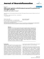
Maximum likelihood phylogenetic tree of complete genome sequencesFigure 1
Maximum likelihood phylogenetic tree of complete genome sequences. Phylogenetic tree was obtained with PHYML
using GTR+G+I for the new sequence and 29 complete genomes (coding region) representative of all 6 HCV genotypes. Geno-
type and subtype labels (in bold) are next to accession numbers. The sequence obtained in this study is underlined. Bar repre-
sents 0.1 substitutions per nucleotide position. Support value of nodes was estimated by bootstrap (1000 replicates using
neighbour-joining with the maximum likelihood distance). Only values >75% are shown.
AM910652 1g
L02836 1b
D11168 1b
AF483269 1b
AJ000009 1b
AJ851228 1
D10749 1a
M62321 1a
AF009606 1a
EU155214 1a
D14853 1c
AY051292 1c
DQ418788 4a
DQ418786 4d
AF064490 5a
D63822 6g
D84263 6d
DQ835766 6m
D84264 6k
D84265 6h
DQ835770 6i
D84262 6b
Y12083 6a
D63821 3k
D49374 3b
AF046866 3a
D50409 2c
AB031663 2k
AF169005 2a
D10988 2b
100
99
99
100
100
100
87
100
100
100
97
100
100
100
100
100
100
100
75
100
100
100
100
77
0.1
Virology Journal 2008, 5:72 />Page 4 of 7
(page number not for citation purposes)
that divergence of subtype 1g occurred before evolution-
ary divergence of subtypes 1a, 1b and 1c.
Potential recombination events between genotypes or
subtype 1 genomes were investigated following two
approaches and, finally, ruled out. Firstly, phylogenetic
reconstructions using the same representative sequences
as in Fig. 1 were performed separately for the 10 protein-
coding genes (from core to NS5B). With respect to the
grouping of HCV-1g within HCV-1 subtypes, all the phyl-
ogenetic trees congruently reproduced the same topology
obtained from the complete genome analysis. Moreover,
subtype 1g still retained its basal position with respect to
the other HCV-1 sequences in topologies based on E1, E2,
NS2, NS4A, NS4B and NS5A genes (data not shown). Sec-
ondly, potential recombination events using the complete
sequence alignment were investigated using the RDP
3.0b03 software [[23] and references therein]. This pro-
gram implements several methods to identify of recom-
binant sequences and recombination breakpoints. All the
recombination analyses based on the complete genome
alignment showed no evidence that our subtype 1g
sequence had participated in recombination events (data
not shown).
The mean genetic distances between genotype 1 subtypes
based on 26 representative sequences of subtypes 1a, 1b,
1c, an unassigned subtype 1 and our subtype 1g sequence
were calculated (Table 1). The appropriate nucleotide sub-
stitution model was determined for this genotype 1 lim-
ited codon-based nucleotide alignment as described as
above. The four mean genetic distances between the HCV-
1g sequence and the other subtypes fall within the highest
five of all comparisons. These results, based on complete
genomes, suggest that the genotype 1g sequence is the
most divergent genome with respect to the rest of the
HCV-1 subtypes.
Finally, we carried out phylogenetic analyses with our
subtype 1g sequence and all available deposited
sequences provisionally designated as subtype 1g (Fig. 2).
These partial sequences, corresponding to four genomic
regions (5'UTR, core, core/E1 and NS5B), were retrieved
from the HCV sequence database in Los Alamos [24]. In
all four analysed regions, the subtype 1g sequence
described here always grouped within the sequences pro-
visionally designated as subtype 1g. In eight cases, HCV
genome from the same patient (the patient code appears
in parenthesis in the corresponding trees) was partially
sequenced in three different regions: 4 cases from Egypt
(5'UTR, core and NS5B), 3 cases also from Egypt (5'UTR,
core/E1 and NS5B) and 2 cases from Canada (5'UTR,
core/E1 and NS5B). In all cases, we observed phylogenetic
congruence of partial sequences of different regions
obtained from the same specimen with respect to our sub-
type 1g sequence.
In the analyses of the NS5B region, three short deposited
sequences were not included, because after nucleotide
alignment the overlapping region was too short to be ana-
lysed. These three early deposited sequences, then consid-
ered subtype 1c and later assigned as subtype 1g, (Z70375,
Z70392 and X88710) were obtained from sera dated
between 1994 and 1995 in Germany [25] and would rep-
resent the first subtype 1g isolates detected in Europe.
Although birthplace of these three patients could not be
checked, the authors mentioned that some patients partic-
ipating in the study had recently emigrated from Egypt
and Sudan. The phylogenetic tree obtained using the
NS5B region also includes 2 sequences from Lebanon
[26], deposited in 1993 as subtype 1c and later assigned
to subtype 1g (in fact, the two first subtype 1g sequences
detected worldwide). The tree also includes one sequence
from a Sudanese individual, detected in a study of unpaid
blood donors in the Netherlands [27]. Interestingly, the
patient in our study was born and resided in Spain, which
is evidence of local transmission of subtype 1g.
Conclusion
In summary, we have determined the complete genome
sequence of an HCV-1g isolate, we have verified its group-
ing within HCV-1 and differentiation from other subtypes
Table 1: Mean genetic distances among HCV subtype 1 representative sequences
subtype 1a subtype 1b subtype 1c Unassigned subtype 1 subtype 1g
subtype 1a (n = 10) 0.021 0.019 0.013 0.018
subtype 1b (n = 12) 0.690 0.019 0.014 0.012
subtype 1c (n = 2) 0.563 0.715 0.006 0.022
Unassigned subtype 1 (n = 1) 0.627 0.480 0.656 NA
subtype 1g (n = 1) 0.709 0.726 0.729 0.711
NA, not applicable.
Mean genetic distances (lower-left matrix) among the HCV subtype 1g and representative sequences from subtypes 1a, 1b, 1c and an unassigned
subtype 1 sequence. A codon-based nucleotide alignment containing the polyprotein of 26 complete genomes (9066 nucleotides) was used for
distance estimates. Standard deviations are indicated in the upper-right matrix. Distance model was GTR+I+G with assumed proportion of
invariable sites of 0.42 and a shape parameter (alpha) of 1.02 for the gamma distribution of substitution rates at variable sites. The number of
sequences included in each subtype group is indicated in parenthesis.
Virology Journal 2008, 5:72 />Page 5 of 7
(page number not for citation purposes)
of this group by rigorous phylogenetic analyses, we have
verified that this genome does not result from recombina-
tion events and that it is the most basal subtype among
those belonging to HCV-1 for which a complete genome
sequence is currently available. Taking this into account,
we propose changing the status of subtype 1g from pro-
posed to confirmed subtype.
Methods
Viral purification, RT-PCR and sequencing
Viral RNA was extracted from 200 μl of serum using High
Pure Viral RNA Kit (Roche). Retrotranscription of viral
RNA was performed in a final volume of 20 μl containing
10 μl of eluted RNA, 4 μl retrotranscription buffer, 500
μM of each dNTP, either 0.5 μg of random hexadeoxynu-
cleotides (Promega) or 1 μM antigenomic sense primer,
100 U of M-MLV reverse transcriptase (Promega) and 20
U of rRNasin
®
Ribonuclease Inhibitor (Promega). The
mixture was incubated at 42°C for 60 min, followed by 3
min at 95°C. Table showed in additional file 1, lists the
oligonucleotide primers used to obtain and/or sequence
the overlapping RT-PCR products, which covered almost
the whole genome. Primers denoted with "g" or "a" indi-
cate genomic or antigenomic sense, respectively. Primers
named with "h" refer to primers used in first round PCR
followed by a hemi-nested PCR. Sequence primers named
with "R" were directly designed from our sequence of sub-
type 1g. The genome was covered by 10 overlapping frag-
ments using primer pairs: H28g-COA1a, COS2g-E1E2a,
E1E2A2g-NS1a, NS1g2R-NS3a3, NS3g2R-305a2R, 1503g-
577a, 5600gR-NS5a2R, KUg2-NS5B1a, NS5B1g-1279a,
1327gR-3utra2. When necessary, additional PCR frag-
ments were also obtained by using combinations of the
primers listed in additional file 1.
First round and hemi-nested amplifications were per-
formed in a 50 μl volume containing either 5 μl of the RT
product (in the case of first round PCR) or 1 μl of the first
round PCR product (in the case of hemi-nested PCR), 5 μl
of 10× PCR buffer, 100 μM of each dNTP, 200 nM of the
genomic sense primer, 200 nM of the antigenomic sense
primer and 5 U of Taq DNA Polymerase (Amersham). All
PCRs were performed in a GeneAmp
®
PCR system 2700
(Applied Biosystems) thermal cycler with the following
profile: 95°C for 2 min, then 35 cycles at 95°C for 30 sec,
50–65°C (depending on the primers used) for 30 sec and
72°C for 3 min, and a final extension at 72°C for 10 min.
Amplified products were purified with High Pure PCR
Products Purification Kit (Roche). These purified DNAs
were sequenced using the ABI PRISM BigDye Terminator
Cycle Sequencing Ready Reaction Kit v 3.1 in a 3700 auto-
mated sequencer (Applied Biosystems). Sequencing prim-
ers are also listed in additional file 1. Chromatogram files
were assembled, verified and edited using the Staden
Package [28]. The newly characterised sequence has been
deposited in EMBL with accession number AM910652
.
Phylogenetic reconstructions and genetic distances
Two sets of nucleotide sequences were analysed: one cor-
responding to the complete polyprotein (Fig. 1) and the
other corresponding to all sequences provisionally
designed as subtype 1g and deposited at the HCV
sequence database in Los Alamos [24] (Fig. 2). In the first
set, the nucleotide sequence coding for the polyprotein
(Fig. 1) was included in phylogenetic reconstructions
along with 29 homologous complete genome sequences
representative of the main HCV genotypes and subtypes
(see accession numbers, genotypes and subtypes in Fig.
Maximum likelihood phylogenetic trees including partial HCV-1g sequencesFigure 2
Maximum likelihood phylogenetic trees including partial HCV-1g sequences. Four regions were studied (a) 5'UTR,
(b) core, (c) E1 and (d) NS5B. Only genotype 1 clade is shown. Support value of nodes was estimated by bootstrap (1000 rep-
licates using neighbour joining with maximum likelihood distance). Only values >75% are shown. Bar represents in each case
number of substitutions per nucleotide position. Patient code label (in parenthesis), genotype and subtype labels (in bold) are
next to accession numbers. The sequence obtained in this study is underlined. Country names are CA, Canada; EG, Egypt; ES,
Spain; LB, Lebanon and SD, Sudan.
AY548687 (EG_036) 1g EG
AF271888 (3464) 1g EG
AY548686 (EG_024) 1g EG
EF115546 (QC71) 1g CA
AF271889 (1382) 1g EG
AM910652 1g ES
EF115549 (QC78) 1g CA
AF009606 1a
AY051292 1c
AJ851228 1
D11168 1b
AF271890 (2004) 1g EG
AY548684 (HCC_EG_016) 1g EG
AY548685 (HCC_EG_015) 1g EG
83
0.01
Outgroup
sequences
83
(a)
AF271824 1g EG
EF115761 (QC71) 1g CA
AY767465 1g
EF115764 (QC78) 1g CA
AY766715 1g
AF271823 1g EG
AF271822 (2152) 1g EG
AY767654 1g
AY766923 1g
AY768008 1g
AY766760 1g
AY767031 1g
AY766916 1g
AF271821 (2004) 1g EG
AY767956 1g
AY766729 1g
AM910652 1g ES
AY767725 1g
AF271820 (1382) 1g EG
AY051292 1c
D11168 1b
AJ851228 1
AF009606 1a
79
0.5
Outgroup
sequences
(c)
L23447 1g LB
AF271797 (1382) 1g EG
EF115983 (QC71) 1g CA
AY548713 (EG_036) 1g EG
AY548726 (HCC_EG_015) 1g EG
AM910652 1g ES
DQ911176 1g EG
L23446 1g LB
AF271799 (3464) 1g EG
AY548727 (HCC_EG_016) 1g EG
EF115986 (QC78) 1g CA
AF271798 (2152) 1g EG
AY548709 (EG_024) 1g EG
DQ238693 1g SD
AF009606 1a
AY051292 1c
AJ851228 1
D11168 1b
75
100
79
89
0.1
Outgroup
sequences
(d)
AY548628 (EG_036) 1g EG
AY548639 (HCC_EG_016) 1g EG
AY548638 (HCC_EG_015) 1g EG
AM910652 1g ES
AY548538 (EG_024) 1g EG
AY051292 1c
AJ851228 1
AF009606 1a
D11168 1b
0.05
Outgroup
sequences
(b)
93
Virology Journal 2008, 5:72 />Page 6 of 7
(page number not for citation purposes)
1). Selected sequences fulfil the condition of containing
less than 15 ambiguities. In the second set, partial
sequences belonging to four regions of HCV genome
(5'UTR, core, core/E1 and NS5B) were analysed separately
along with the corresponding homologous fragment of
our subtype 1g complete genome. Alignments of partial
sequences of HCV-1g used in phylogenetic reconstruc-
tions included (number of nucleotides in parenthesis):
nine 5'UTR sequences (186 nt), four core sequences (217
nt), eighteen core/E1 sequences (220 nt) and thirteen
NS5B sequences (222 nt). (see accession numbers, speci-
men name, and subtypes in Fig. 2). In addition, represent-
ative sequences used in the complete genome analysis for
subtypes 2a, 3a, 4a, 5a and 6a (Fig. 1) and referred as out-
group were also included in the phylogenetic analyses.
ClustalW [29] implemented in MEGA version 4 [30] was
used to obtain a multiple alignment of the corresponding
amino acid sequences from which a codon-based nucle-
otide alignment was derived, except for the 5'UTR align-
ment. All phylogenetic trees were constructed by
maximum likelihood in PHYML with the nucleotide sub-
stitution model that best fit the data according to Akaike
Information Criterion (AIC) [31] for which we used the
procedure implemented in Modeltest 3.8 [32]. The
robustness of the tree topology was assessed by bootstrap
analysis with 1000 replicates implemented in PHYML
[33].
Estimates of mean distances between subtypes of HCV
genotype 1 and between these and the new subtype 1g
sequence were obtained with the maximum likelihood
distance (see above) with PAUP*4.0b10 [34]. For this, we
used 26 complete genomes from EMBL: our sequence for
subtype 1g [EMBL: AM910652
], ten sequences represent-
ing subtype 1a [EMBL: D10749
, EMBL: M62321, EMBL:
M67463
, EMBL: AF009606, EMBL: AF011751, EMBL:
AF011752
, EMBL: AF290978, EMBL: AF271632, EMBL:
AJ278830
, EMBL: EU155214], twelve sequences for sub-
type 1b [EMBL: D11168
, EMBL: D14484, EMBL: D45172,
EMBL: L02836
, EMBL: AB080299, EMBL: AB016785,
EMBL: AB049095
, EMBL: AF139594, EMBL: AF165048,
EMBL: AF333324
, EMBL: AJ000009, EMBL: AY045702),
two sequences for subtype 1c [EMBL: D14853
, EMBL:
AY051292
] and one sequence that corresponds to an
unassigned subtype 1 [EMBL: AJ851228
].
Competing interests
The authors declare that they have no competing interests.
Authors' contributions
MAB, VS, and EM co-conceived, designed and coordi-
nated the study, participated in the molecular studies,
sequence alignment, phylogenetic and genetic analyses,
interpreted data, and co-drafted the manuscript; AB and
VA performed the clinical work, recruitment of the
patient, procurement of specimens and participated in
proofreading of the manuscript; FG-C coordinated the
study, interpreted data, co-performed phylogenetic and
genetic analyses and participated in proofreading of the
manuscript. All authors read and approved the final man-
uscript
Additional material
Acknowledgements
This work is supported by Conselleria de Sanitat i Consum, Generalitat
Valenciana (Spain) and project BFU2005-00503 from Ministerio de Edu-
cación y Ciencia (Spain).
This work is also partially supported by grant PI051131 from Instituto de
Salud Carlos III-Fondo de Investigaciones Sanitarias, grant CD05/00258
(EM) (contratos postdoctorales de perfeccionamiento) from the Ministerio
de Sanidad y Consumo, within the Plan Nacional de Investigación científica,
Desarrollo e Innovación Tecnológica (I+D+I); and by grant 2007FIC00550
(VS) from Comissionat per a Universitats i Recerca del Departament
d'Innovació, Universitats i Empresa de la Generalitat de Catalunya i del Fons
Social Europeu (Spain).
References
1. WHO: Hepatitis C – global prevalence (update). Weekly Epide-
miological Record 1999, 49:425-427.
2. Robertson B, Myers G, Howard C, Brettin T, Bukh J, Gaschen B,
Gojobori T, Maertens G, Mizokami M, Nainan O, Netesov S, Nishioka
K, Shin-I T, Simmonds P, Smith D, Stuyver L, Weiner A: Classifica-
tion, nomenclature, and database development for hepatitis
C virus (HCV) and related viruses: proposals for standardiza-
tion. Arch Virol 1998, 143:2493-2503.
3. Simmonds P, Bukh J, Combet C, Deleage G, Enomoto N, Feinstone S,
Halfon P, Inchauspe G, Kuiken C, Maertens G, Mizokami M, Murphy
DG, Okamoto H, Pawlotsky JM, Penin F, Sablon E, Shin-I T, Stuyver
LJ, Thiel HJ, Viazov S, Weiner AJ, Widell A: Consensus proposals
for a unified system of nomenclature of hepatitis C virus gen-
otypes. Hepatology 2005, 42:962-973.
4. Bracho MA, Carrillo-Cruz FY, Ortega E, Moya A, González-Candelas
F: A new subtype of hepatitis C virus genotype 1: complete
genome and phylogenetic relationships of an Equatorial
Guinea isolate. J Gen Virol 2006, 87:1697-1702.
5. Carrat F, Bani-Sadr F, Pol S, Rosenthal E, Lunel-Fabiani F, Benzekri A,
Morand P, Goujard C, Pialoux G, Piroth L, Salmon-Céron D, Degott
C, Cacoub P, Perronne C, ANRS HCO2 RIBAVIC Study Team:
Pegylated interferon alfa-2b vs standard interferon alfa-2b,
plus ribavirin, for chronic hepatitis C in HIV-infected
patients: a randomized controlled trial. JAMA 2004,
292:2839-2848.
6. Heathcote EJ: Antiviral therapy: chronic hepatitis C. J Viral
Hepat 2007:82-88.
7. Wróbel B, Torres-Puente M, Jiménez N, Bracho MA, García-Robles I,
Moya A, González-Candelas F: Analysis of the overdispersed
clock in the short-term evolution of hepatitis C virus: Using
the E1/E2 gene sequences to infer infection dates in a single
source outbreak. Mol Biol Evol 2006, 23:1242-1253.
Additional file 1
Oligonucleotide primers used for amplification and sequencing. List of oli-
gonucleotide primers including name, sequence, position and sense.
Click here for file
[ />422X-5-72-S1.doc]
Publish with BioMed Central and every
scientist can read your work free of charge
"BioMed Central will be the most significant development for
disseminating the results of biomedical research in our lifetime."
Sir Paul Nurse, Cancer Research UK
Your research papers will be:
available free of charge to the entire biomedical community
peer reviewed and published immediately upon acceptance
cited in PubMed and archived on PubMed Central
yours — you keep the copyright
Submit your manuscript here:
/>BioMedcentral
Virology Journal 2008, 5:72 />Page 7 of 7
(page number not for citation purposes)
8. Bronowicki JP, Venard V, Botté C, Monhoven N, Gastin I, Choné L,
Hudziak H, Rihn B, Delanoë C, LeFaou A, Bigard MA, Gaucher P:
Patient-to-patient transmission of hepatitis C virus during
colonoscopy. N Engl J Med 1997, 337:237-240.
9. Jiménez-Hernández N, Torres-Puente M, Bracho MA, García-Robles
I, Ortega E, del Olmo J, Carnicer F, González-Candelas F, Moya A:
Epidemic dynamics of two coexisting hepatitis C virus sub-
types. J Gen Virol 2007, 88:123-133.
10. Salemi M, Vandamme AM: Hepatitis C virus evolutionary pat-
terns studied through analysis of full-genome sequences. J
Mol Evol 2002, 54:62-70.
11. Njouom R, Nerrienet E, Dubois M, Lachenal G, Rousset D, Vessière
A, Ayouba A, Pasquier C, Pouillot R: The hepatitis C virus epi-
demic in Cameroon: genetic evidence for rapid transmission
between 1920 and 1960. Infect Genet Evol 2007, 7:361-367.
12. Pybus OG, Charleston MA, Gupta S, Rambaut A, Holmes EC, Harvey
PH: The epidemic behavior of the hepatitis C virus. Science
2001, 292:2323-2325.
13. Pybus OG, Cochrane A, Holmes EC, Simmonds P: The hepatitis C
virus epidemic among injecting drug users. Infect Genet Evol
2005, 5:131-139.
14. Simmonds P: Genetic diversity and evolution of hepatitis C
virus – 15 years on. J Gen Virol 2004, 85:3173-3188.
15. Germer JJ, Majewski DW, Rosser M, Thompson A, Mitchell PS, Smith
TF, Elagin S, Yao JDC: Evaluation of the TRUGENE HCV 5'NC
genotyping kit with the new GeneLibrarian module 3.1.2 for
genotyping of hepatitis C virus from clinical specimens. J Clin
Microbiol 2003, 41:4855-4857.
16. Martró E, González V, Buckton AJ, Saludes V, Fernández G, Matas L,
Planas R, Ausina V: Evaluation of a new assay for hepatitis C
virus (HCV) genotyping targeting both 5'NC and NS5b
genomic regions, in comparison with reverse hybridization
and sequencing methods. J Clin Microbiol 2008, 46:192-197.
17. Schutzbank TE, Sefers SE, Kahmann N, Li H, Tang YW: Compara-
tive evaluation of three commercially available methodolo-
gies for hepatitis C virus genotyping. J Clin Microbiol
2006,
44:3797-3798.
18. Cantaloube JF, Laperche S, Gallian P, Bouchardeau F, de Lamballerie
X, de Micco P: Analysis of the 5' noncoding region versus the
NS5b region in genotyping hepatitis C virus isolates from
blood donors in France. J Clin Microbiol 2006, 44:2051-2056.
19. Laperche S, Lunel F, Izopet J, Alain S, Dény P, Duverlie G, Gaudy C,
Pawlotsky JM, Plantier JC, Pozzetto B, Thibault V, Tosetti F, Lefrère
JJ: Comparison of hepatitis C virus NS5b and 5' noncoding
gene sequencing methods in a multicenter study. J Clin Micro-
biol 2005, 43:733-739.
20. Hraber PT, Fischer W, Bruno WJ, Leitner T, Kuiken C: Compara-
tive analysis of hepatitis C virus phylogenies from coding and
non-coding regions: the 5' untranslated region (UTR) fails to
classify subtypes. Virol J 2006, 3:103.
21. Murphy DG, Willems B, Deschênes M, Hilzenrat N, Mousseau R, Sab-
bah S: Use of sequence analysis of the NS5B region for rou-
tine genotyping of hepatitis C virus with reference to C/E1
and 5' untranslated region sequences. J Clin Microbiol 2007,
45:1102-1112.
22. Lu L, Li C, Fu Y, Thaikruea L, Thongswat S, Maneekarn N, Apichart-
piyakul C, Hotta H, Okamoto H, Netski D, Pybus OG, Murphy D,
Hagedorn CH, Nelson KE: Complete genomes for hepatitis C
virus subtypes 6f, 6i, 6j and 6m: viral genetic diversity among
Thai blood donors and infected spouses. J Gen Virol 2007,
88:1505-1518.
23. Martin D, Rybicki E: RDP: detection of recombination amongst
aligned sequences. Bioinformatics 2000, 16:562-563.
24. Kuiken C, Hraber P, Thurmond J, Yusim K: The hepatitis C
sequence database in Los Alamos. Nucleic Acids Res 2007,
36:D512-D516.
25. Feucht HH, Schröter M, Zöllner B, Polywka S, Nolte H, Laufs R: The
influence of age on the prevalence of hepatitis C virus sub-
types 1a and 1b. J Infect Dis 1997, 175:685-688.
26. Simmonds P, Holmes EC, Cha TA, Chan SW, McOmish F, Irvine B,
Beall E, Yap PL, Kolberg J, Urdea MS: Classification of hepatitis C
virus into six major genotypes and a series of subtypes by
phylogenetic analysis of the NS-5 region. J Gen Virol 1993,
74:
2391-2399.
27. Laar TJ van de, Koppelman MH, Bij AK van der, Zaaijer HL, Cuijpers
HT, Piel CL van der, Coutinho RA, Bruisten SM: Diversity and ori-
gin of hepatitis C virus infection among unpaid blood donors
in the Netherlands. Transfusion 2006, 46:1719-1728.
28. Staden R, Beal KF, Bonfield JK: The Staden Package, 1998. Meth-
ods Mol Biol 2000, 132:115-130.
29. Thompson JD, Higgins DG, Gibson TJ: CLUSTAL W: improving
the sensitivity of progressive multiple sequence alignment
through sequence weighting, positions-specific gap penalties
and weight matrix choice. Nucl Acids Res 1994, 22:4673-4680.
30. Tamura K, Dudley J, Nei M, Kumar S: MEGA4: Molecular Evolu-
tionary Genetics Analysis (MEGA) software version 4.0. Mol
Biol Evol 2007, 24:1596-1599.
31. Akaike H: A new look at the statistical model identification.
IEEE Trans Automatic Control 1974, 19:716-723.
32. Posada D, Crandall KA: Selecting the best-fit model of nucle-
otide substitution. Syst Biol 2001, 50:580-601.
33. Guindon S, Gascuel O: A simple, fast, and accurate algorithm
to estimate large phylogenies by maximum likelihood. Syst
Biol 2003, 52:696-704.
34. Swofford DL: PAUP*. Phylogenetic analysis using parsimony (* and other
methods) 4th edition. Sunderland, MA, Sinauer Associates; 2002.