Báo cáo hóa học: " Could FIV zoonosis responsible of the breakdown of the pathocenosis which has reduced the European CCR5-Delta32 allele frequencies?" pptx
Bạn đang xem bản rút gọn của tài liệu. Xem và tải ngay bản đầy đủ của tài liệu tại đây (1.11 MB, 19 trang )
BioMed Central
Page 1 of 19
(page number not for citation purposes)
Virology Journal
Open Access
Research
Could FIV zoonosis responsible of the breakdown of the
pathocenosis which has reduced the European CCR5-Delta32 allele
frequencies?
Eric Faure
Address: LATP, CNRS-UMR 6632, IFR48 Infectiopole, Evolution biologique et modélisation, case 5, Université de Provence, Place Victor Hugo,
13331 Marseille cedex 3, France
Email: Eric Faure -
Abstract
Background: In Europe, the north-south downhill cline frequency of the chemokine receptor
CCR5 allele with a 32-bp deletion (CCR5-
Δ
32) raises interesting questions for evolutionary
biologists. We had suggested first that, in the past, the European colonizers, principally Romans,
might have been instrumental of a progressively decrease of the frequencies southwards. Indeed,
statistical analyses suggested strong negative correlations between the allele frequency and
historical parameters including the colonization dates by Mediterranean civilisations. The gene
flows from colonizers to native populations were extremely low but colonizers are responsible of
the spread of several diseases suggesting that the dissemination of parasites in naive populations
could have induced a breakdown rupture of the fragile pathocenosis changing the balance among
diseases. The new equilibrium state has been reached through a negative selection of the null allele.
Results: Most of the human diseases are zoonoses and cat might have been instrumental in the
decrease of the allele frequency, because its diffusion through Europe was a gradual process, due
principally to Romans; and that several cat zoonoses could be transmitted to man. The possible
implication of a feline lentivirus (FIV) which does not use CCR5 as co-receptor is discussed. This
virus can infect primate cells in vitro and induces clinical signs in macaque. Moreover, most of the
historical regions with null or low frequency of CCR5-
Δ
32 allele coincide with historical range of
the wild felid species which harbor species-specific FIVs.
Conclusion: We proposed the hypothesis that the actual European CCR5 allelic frequencies are
the result of a negative selection due to a disease spreading. A cat zoonosis, could be the most
plausible hypothesis. Future studies could provide if CCR5 can play an antimicrobial role in FIV
pathogenesis. Moreover, studies of ancient DNA could provide more evidences regarding the
implications of zoonoses in the actual CCR5-
Δ
32 distribution.
Background
As infection is the greatest killer in human history [1], the
strongest evidence for selection in the human genome has
been obtained for genes involved in immune defense,
including those which encode receptors. One of the most-
celebrated examples of adaptive selection is the 32-bp
coding sequence deletion, CCR5-
Δ
32, of the chemokine
receptor CCR5. This is probably the more recent and com-
Published: 16 October 2008
Virology Journal 2008, 5:119 doi:10.1186/1743-422X-5-119
Received: 26 August 2008
Accepted: 16 October 2008
This article is available from: />© 2008 Faure; licensee BioMed Central Ltd.
This is an Open Access article distributed under the terms of the Creative Commons Attribution License ( />),
which permits unrestricted use, distribution, and reproduction in any medium, provided the original work is properly cited.
Virology Journal 2008, 5:119 />Page 2 of 19
(page number not for citation purposes)
plete example of a gene studied from clinical, epidemio-
logical and evolutionary genetics. CCR5 function as co-
receptors for the cell entry of HIV-1 and the deletion
which leads to a frame shift and generates an inactive
CCR5 receptor. Homozygosity for the CCR5-
Δ
32 allele
confers almost complete, mendelian resistance to R5-
tropic HIV-1 while HIV-infected individuals heterozygous
for this allele were delayed in progression to AIDS [2,3].
The CCR5-
Δ
32 allele is mainly present in Europeans (10%
on average) and the allele frequency exhibits a north-
south cline with frequencies ranging from 16% in North-
ern Europe to 4% or less in Greece and in most of the
Mediterranean islands (Figure 1A and [4,5]). The broadest
area of high frequency is located in North-Eastern Europe,
particularly in the Baltic and White Sea regions. From
these maximum, the frequency gradually decreases in all
directions across Europe [4]; however, some additional
peaks of frequency are found in France or Russian areas
[4,6-8]. Moreover, Ashkenazi Jews have high frequencies
of CCR5-
Δ
32, but this is likely due to founder effects
unique to their history rather than the general process of
dispersal that spread the allele in other populations [9].
Outside Europe, the mutation can be found at low fre-
quencies in neighbouring regions (North Africa, Middle
East, Central Asia); it is absent in Sub-Saharan Africa, East
and South-East Asia and in indigenous populations of
America and Oceania (Figure 1A).
Because the AIDS pandemic is too recent to change allele
frequencies, other infectious diseases have been suggested
as the agent causing the selection of the null allele
increase, such as resistance to plague and smallpox infec-
tions [10]. However, analyses of Scandinavian Mesolithic
DNA which have pushed the date of the first occurrence
back to around 5000 BC [11] and genomic analyses [12]
have weakened the evidence for recent selection of the
null allele. Due to the north-south spatial gradient, it has
been proposed that the actual allele distribution could be
explained by migrations of Northern populations. As sug-
gested by Lucotte [13] in its seminal article in the field and
by Balanovsky et al. [4] Vikings and Uralic speaking peo-
ple, respectively, could have brought the deletion in some
Southern populations. Moreover, these migrations and/or
gene flow cannot explain, according to us, the whole of
the European allele frequency distribution. Also, we have
proposed an alternative hypothesis in which the actual
allele frequency distribution might not be due to the
genes spreading, but to a negative selection resulting in
the spread of pathogens principally during principally
Roman expansion [5]. This hypothesis is supported by
several facts.
The idea that bottlenecks and founder effects could lead to
an increase in damaging alleles in human populations
was historically reserved for isolated populations that
experienced severe founder effects (for example,
Ashkenazi Jews [14] and Finns [15]). However, recently
signs of a population bottleneck in variability data
obtained for a number of genomic loci in European pop-
ulations were described and also led to the conclusion
that a severe bottleneck occurred after the appearance of
the anatomically modern human in Africa, and thus pre-
sumably during, or after, the emigration out of Africa [16-
18] and references therein). Moreover, the earlier Euro-
pean population of hunter-gatherers could suffer severe
bottlenecks during the latest ice age (Pleistocene) [19]. As
there is strong evidence for the unitary origin of the CCR5-
Δ
32 mutation [20,21], the null allele could have been
already present in the ancestors of the European popula-
tions (in spite of their present language differences) at a
relatively high frequency, probably >10% as suggested by
analysis of ancient DNA from Bronze age [22] and Neo-
lithic [11], similarly to many other polymorphisms found
in Europeans but not in the other populations [23].
Previous statistical analyses showed strong negative corre-
lations in Europe between the allele frequency and two
historical parameters, i.e. the first colonization dates by
the great ancient Mediterranean civilisations, and the dis-
tances from the frontiers of the Roman Empire in its great-
est expansion [5]. However, the possible decrease of the
ancestral CCR5-
Δ
32 allele frequency was not due directly
to the colonizers, as the gene flows to European native
populations were extremely low [19]. This suggests that
the role of colonizers were indirect. As evolutionary biol-
ogists have shown several evidences that infectious dis-
eases, as a leading cause of human morbidity and
mortality, have exerted important selective forces on our
genomes [24,25], the cause of the decrease of the CCR5-
Δ
32 allele frequency in Southern European populations is
probably due to infectious agent(s). It has been suggested
that the most important infectious diseases of modern
food-producing human populations also include diseases
that could have emerged only within the past 11,000
years, following the rise of agriculture [1,25,26]. The sec-
ond great historical transitions occurred when great
ancient conquering Eurasian civilizations (such as the
Greek and Roman empires) came into military and com-
mercial contact, ca. 3000–2000 years ago, swapping their
dominant infections [27]. It is either a human disease or
a zoonosis transmittable to humans. Moreover, studies on
the West Nile virus have shown that host genetic factors
are highly pathogen-specific and can therefore be benefi-
cial in one context and harmful in another [28]. Which
agree that the possible decrease of the CCR5-
Δ
32 allele fre-
quency in the South of the Europe could be due to para-
sites. The introduction of parasites in naive colonized
populations could have induced a breakdown of the
pathocenosis and a new equilibrium has been reached
Virology Journal 2008, 5:119 />Page 3 of 19
(page number not for citation purposes)
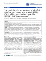
Geographic distribution of the CCR5-
Δ
32 allele (A) compared with historical range of felids carrying species-specific FIVs (B)Figure 1
Geographic distribution of the CCR5-
Δ
32 allele (A) compared with historical range of felids carrying species-
specific FIVs (B). In (A), only the frequencies of Native populations have been evidenced in America, Asia, Africa and Oce-
ania. Map redrawn and modified from [4,5]. In (B), the black areas correspond to the range of wild individuals bearing species-
specific FIVs in a given continent, America: bobcat, jaguarundi, ocelot and puma; Asia: Pallas cat; Africa: cheetah, leopard and
lion. The pale grey areas correspond to the range where individuals of these species have been found seronegative or when
their serological status is unknown in a given continent (Asia: cheetah, leopard and lion; Europe: leopard and lion). Areas where
these last three species lived in sympatry with Pallas cat are in dark grey. The historical ranges are approximate by 500 BC for
Europe, North Africa and Western Asia; since the European settlement in America, and during the 1500's to the beginning of
the 1900's in the remainder of Africa, Asia and Oceania. These data were principally inferred from [65-71].
Virology Journal 2008, 5:119 />Page 4 of 19
(page number not for citation purposes)
through a decrease of the CCR5-
Δ
32 allele frequency. The
theoretical framework of pathocenosis, first coined by
Grmek [29,30]) and developed by Biraben [31], offers a
synthetic approach to the history of disease. Drawing on
the concept of biocenosis, Grmek defines pathocenosis as
"the ensemble of pathological states present in a specific
population at a given moment in time" and suggests that
"the frequency and overall distribution of each disease
depends on the frequency and distribution of all the other
diseases within a given population". The concept of
pathocenosis attempts to offer a synthetic view of disease
ecology, which, in our context is defined as all interde-
pendences within pathogens, their hosts (including their
genetic responses) and their environment.
The aim of this article is to critically discuss the possible
nature of this (or these) parasite(s) responsible of the
decrease of the CCR5-
Δ
32 allele frequency in the Southern
European populations.
Results
Putative role of cats as host-parasite
Previous, statistical analyses suggested a decrease of the
ancestral CCR5-
Δ
32 allele frequency in European popula-
tion due principally to Roman expansion [5]. However,
this negative selection was not directly due to the military
or colonisation spreads, as the gene flows from colonisa-
tors to European native populations were too low [19].
Moreover, statistical analyses suggested that factor(s)
responsible for the decrease of null allele frequency had
partly diffused beyond the borders of the Roman Empire
[5]. The diffusion of one or more factor(s) excludes the
role of climatical changes, the change in allele frequency
could be due to the spread of human or animal parasites
that affect human populations.
More than any other civilizations, the Romans have cre-
ated links between Mediterranean basin and Western and
Central Europe and the great routes of infectious diseases
went straight through it [32]. Not only did the first great
historical pestilences pass through the Empire, but also
the slow insidious penetration of endemic disease (like
tuberculosis, leprosy and malaria) has invaded Europe
[30]. Moreover, conquerors and invading armies brought
also with them insect and rodent vectors that could intro-
duce or sustain infectious diseases in nonendemic Euro-
pean areas. As, to our knowledge, no known human
diseases could explain the decrease of the null allele in
Europe, zoonoses might be implicated. Indeed, most of
the infectious diseases affecting human populations are
considered zoonotic in origin [33-35]. Close contact with
animals is a risk for humans to acquire infectious diseases
and it is well known that the domestication of animals has
facilitated the passage of animals parasites to human
[36,37]. Many of the major human infectious diseases,
including some now confined to humans and absent from
animals, have arisen only after the origins of agriculture
(11,000 years BP) [1,25,26]. The five animal species (cow,
sheep, goat, pig and dog) which have had probably the
most epidemic impact on the human populations are
explicitly named the Pandora's pentad [38]. Moreover,
few tropical but many temperate diseases arose from
domestic animals, because these live mainly in the tem-
perate zones, and their concentration there was formerly
even more lopsided [35]. In Europe, the Romans were the
cause of some permanent changes in the distribution of
birds and beasts [39]; several animals, such as cat, donkey,
mule and pheasant have been voluntarily introduced
throughout Europe [40] and others involuntarily, such as
malaria vector mosquito species [30]. If we consider that
the most impact on the decrease of the CCR5-
Δ
32 allele
frequency could be principally due to Roman expansion,
according to us, among all the domestic animals, cat
could be the best candidate. Indeed, once the cat had
arrived in Rome; this animal would have spread through-
out Europe, quite likely as a camp follower and compan-
ion to the constantly travelling Roman armies. Moreover,
several parasitic, bacterial and viral zoonoses diagnosed in
cats could be transmitted to man [41,42]. To support this
view, before investigate the type of disease which could be
transmitted, the major steps of the spread of the domestic
cat in Europe are summarized.
Cat's origin is yet little uncertain; however, several analy-
sis revealed that cats were domesticated in the Near East;
wildcats of Near East (F. s. lybica) are the closest group to
all domestic cats [43,44], and likely coincide with agricul-
tural village development in the Fertile Crescent. This is
congruent with archaeological studies, the earliest evi-
dence of cat-human association involves their co-occur-
rence in Cyprus deposits aged at 9,500 years ago [45].
Similarly, in all the other islands of the Mediterranean
Basin far beyond continent (Sardinia, Corsica and Crete),
felids originated from African or Middle East wildcats
which were voluntarily introduced by Neolithic naviga-
tors about 6,000–8,000 years ago [46-50]. Interestingly,
the populations of these areas have the lower level of
CCR5-
Δ
32 allele frequency (references therein [5]. The
earliest records of probably tamed or domestic cats in con-
tinental Europe would be in Greece by 1000 BC; however,
at that time, cats were very extremely rare until 6
th
–5
th
cen-
turies BC [51-53]. In the Italian Peninsula, first historical
evidence of tamed or early-domesticated cats was found
on archaeological sites from the beginning of the 5
th
–4
th
centuries BC [50,54]. Interestingly, in numerous parts of
the Roman Empire, generally the oldest remains of the
domestic cat (for example in Belgium, Netherlands, Hun-
gary and Switzerland) dated to the Roman period [55-59];
moreover, remains of cats have been found in many of the
Roman settlements excavated extensively suggesting that
Virology Journal 2008, 5:119 />Page 5 of 19
(page number not for citation purposes)
the spread of domesticated cats throughout continental
Europe and Great Britain is principally due to Romans
[40,60]. Moreover, as contrarily to Asia, Africa and Amer-
ica, there was no tameable felid in the Northern Mediter-
ranean countries, therefore numerous substitutes have
been found by the European populations, principally
Mustelidae species [48,56,61,62].
World repartition of FIV-infected felids and their
relationship with humans
If we hypothesize that a cat zoonosis might be transmitted
to human, the corresponding infectious agent could also
affect other felid species. Among, all the cat zoonoses,
according us, only one parasite distribution could be cor-
related to those of the CCR5-
Δ
32 allele frequency. The cor-
responding infectious agent is the Feline immunodeficiency
virus (FIV) which can also infect primate cells in vitro and
induce clinical signs in a primate [63,64] and references
therein). Indeed, historical regions with null or low fre-
quency of CCR5-
Δ
32 allele coincide with historical range
of the wild felid species [65-71] which harbor species-spe-
cific FIVs (Figure 1). The two maps do not correspond per-
fectly, and we can only conclude that these patterns are
not inconsistent with the hypothesis that allele frequency
and the old presence FIV-infected felids are causally
related. However, as developed below, bibliographical
analyses provide several arguments in favour of this
hypothesis.
FIV, as Human immunodeficiency virus (HIV) and Simian
immunodeficiency virus (SIV) belong to the Lentivirus genus
of the Retroviridae (reviewed in [72]). In domestic cat, FIV
infection results in disease progression and outcome sim-
ilar to that of HIV in humans, and offers a natural model
to AIDS [73,74]. Other felid species which are infected
with FIV seem not to develop AIDS-like disease [75,76].
However, both captive and/or wild FIV-infected lions
(Panthera leo) and pumas (Puma concolor) exhibited mild
to severe CD4+ T-cell depletion and some other clinical
health consequences [77-80]. These findings raise the
prospect that FIV is not completely benign in these spe-
cies, but rather suppress host immune response and may
increase the incidence of opportunistic infections or even
spontaneous cancers as AIDS does in humans.
The extant felids have arisen from a common ancestor in
Asia 10.8 MYA during the Miocene. The 37 felid species
form eight distinct evolutionary lineages that have suc-
cessfully inhabited all continents except Oceania and Ant-
arctica through a series of migrations likely facilitated by
sea-level oscillations [81]. Among the Felidae, at least 11
free ranging Felidae species harbor FIV antibodies and FIV
viral genomes (Table 1). Moreover, nine of these species
(lion, cheetah, leopard, Pallas cat, jaguarundi, ocelot,
domestic cat, puma, and bobcat) have been shown to har-
bor species-specific FIVs by evaluation of complete or par-
tial viral genomic sequences (Table 1 and [74,82,83]).
However, the seroprevalence of FIVs varies dramatically
by species and geographic areas. African lion and leopard,
puma and Pallas cat populations demonstrate very high
rates of seropositivity. The seroprevalence of FIV infec-
tions in natural settings is nearly 100% in Serengeti Afri-
can lions and pumas of Wyoming and Montana,
respectively [84-86]. In contrast, significant numbers of
free-ranging lions in Namibia or from Asia were all seron-
egative [86,87]. The absence of FIV-Ple in Namibia is puz-
zling, but may be explained by the low density of lions in
this African area [88]. Moreover, several Asian lions held
in captivity were noted to be 75% FIV seropositive, dem-
onstrating that lions of Asian origin are not intrinsically
resistant to infection [89]. Interestingly, a similar geo-
graphic dispersal of seropositivity was noted for Asian ver-
sus African leopards; i.e., free-ranging African populations
demonstrate seropositivity of >25%, whereas Asian-born
animals are seronegative [90,91]. More than 50% of Pallas
cats (Manul) tested harboured anti-FIV antibodies [91].
Other species, including the domestic cat, cheetah, and
South American Neotropical free-ranging felid popula-
tions, tend to demonstrate seroprevalence rates of 10% or
less. Asian species other than the Pallas cat are apparently
not infected with an endemic FIV, although when during
captivity, Asian felid individuals are exposed to other spe-
cies harbouring FIVs, these animals may become infected
(Table 1 and [74,91]). It must be noted that a species-spe-
cific FIV-related virus has also been found in Hyaenidae,
which belong to the Feloidea superfamily [91,92].
As already shown by several authors [74,91,93], the FIV
phylogenies does not exactly mirror that of its feline host
species (Figure 2). However, the relative differences in
genetic diversity among FIV strains be interpreted in the
context of the evolutionary and phylogeographical history
of each host species. Indeed, in spite of that free-ranging
individuals of many species harbor monophyletic, spe-
cies-specific strain(s) of FIV, viruses isolated from differ-
ent species seem to group more by geographic region of
the host than in groupings concordant with the phyloge-
netic relationships of host species. Moreover, molecular
analyses failed to resolve the origin domestic cat FIV
strains as has been already shown by other studies
[74,83]. The pattern of the strains infecting domesticated
cat (FIV-Fca) which exhibit three monophyletic clades
may due rapid viral diversification within the domestic cat
world-wide due to the great number of individuals (some
estimates put the domestic house cat population at 60
million and the feral cat population at the same number,
that's 120 million animals) and to the trans-continental
travels and traffics. The extreme divergence between the
two highly FIV-Pco clades and the six FIV-Ple clades sug-
gest an ancient origin of FIV infection of respectively,
Virology Journal 2008, 5:119 />Page 6 of 19
(page number not for citation purposes)
Table 1: List of actual felids and hyanids and their FIV status
Feloidea: Felid
lineages and
Hyaenidae
Species Animal Distribution
(formerly
widespread)
FIV status
(Western)
FIV status (PCR) First-known
taming dates
Wildcat Felis silvestris silvestris
(Schreber 1777)
European wildcat Europe, S.W. Asia + fr + N.D.
F. s. lybica
(Forster 1780)
Northern African
wildcat
Africa, Middle East + fr N.D. <2000 B.C.
F. s. ornata
(Gray 1830)
Asian wildcat W. and C. Asia + fr - <2000 B.C.
F. bieti
(Milne-Edwards
1892)
Chinese steppe cat China N.D. N.D. N.D.
F. chaus
(Schreber 1777)
Jungle cat S. and S.E. Asia,
Middle East, Egypt
+/- wb, cb - <2000 B.C.
F. margarita
(Loche 1858)
Sand cat Africa, Arabia, S.W.
Asia
+ fr - N.D.
F. nigripes
(Burchell 1824)
Black-footed cat Africa +/- cb - N.D.
Leopard cat Prionailurus
bengalensis
(Kerr 1792)
Leopard cat E. and S.E. Asia,
India
+ wb + N.D.
P. planiceps
(Vigors and
Horsfield 1827)
Flat-headed cat Malatya, Sumatra,
Borneo
+ fr N.D. N.D.
P. rubiginosus
(I. G S-H 1831)
Rusty-spotted cat India, Sri Lanka - wb N.D. N.D.
P. viverrinus
(Bennett 1833)
Fishing cat S.E. Asia, N.E. India + cb - N.D.
Otocolobus manul
(Pallas 1776)
Pallas' cat C. and W. Asia + e, fr + <1000 A.D.
Puma Puma concolor
(Linnaeus 1771)
Puma N. and S. America + e, fr + fr <1500 A.D.
Herpailurus
yagouaroundi
(E. G S-H 1803)
Jaguarundi Mexico, C. and S.
America
+ fr + fr <1000 A.D.
Acinonyx jubatus
(Schreber 1775)
Cheetah Africa, Asia Minor,
India, W. Asia
+ e, fr + fr <2000 B.C.
Lynx Lynx canadensis Kerr
1792
Canada lynx N. America - fr N.D. N.D.
L. lynx
(Linnaeus 1758)
Eurasian lynx Europe and Asia - wb N.D. N.D.
L. pardinus
(Temminck 1827)
Iberian lynx Spain and Portugal - fr - N.D.
L. rufus
(Schreber 1777)
Bobcat N. America + e, fr + N.D.
Ocelot Leopardus
pardalis
(Linnaeus 1758)
Ocelot C. and S. America,
Mexico
+ fr + <1500 A.D.
L. colocolo
(Molina 1782)
Pampas cat S. America + fr - N.D.
L. geoffroyi
(d'Orbigny and
Gervais 1844)
Geoffroy's cat S. America + e, fr - <1500 A.D.
L. guigna
(Molina 1782)
Kodkod C. Chile, Andean
Argentina
- cb N.D. N.D.
L. jacobita
(Cornalia 1865)
Andean mountain
cat
Parts of Andes N.D. N.D. N.D.
L. tigrinus
(Schreber 1775)
Tigrina S. America + e, fr - N.D.
L. wiedii
(Schinz 1821)
Margay C. and S. America + e, fr + <1500 A.D.
Virology Journal 2008, 5:119 />Page 7 of 19
(page number not for citation purposes)
puma and lion [88]. Concerning FIV-Pco, this could be a
consequence of two separate introductions of FIV within
puma populations [83], whereas for African lion virus,
each clades correspond with distinct geographic areas of
endemicity [88]. The strains infecting cheetah (FIV-Aju)
and leopard (FIV-Ppa) are closely related, in spite the fact
that their hosts have evolved in distinctly different felid
lineages, puma and cheetah are closely related, belonging
to the puma linage, while lions and leopards are members
of the Panthera lineage [81]. Moreover, cheetah and leop-
ard could be sympatric; all these data suggest recent inter-
species transmission. Due to the date of expansion of
cheetah throughout Africa, the FIV-Aju emergence may
have occurred within the last 10,000 years, perhaps
acquired from leopards [93]. FIV-Oma is found in wild
populations of the Eurasian Pallas cat [91], a species that
arose during the late Pleistocene [81]. The monophyletic
lineage of Pallas cat FIV-Oma and African lion FIV-Ple
observed here could suggest more ancient inter-species
transmission as the last time lions and Pallas cats were in
geographic contact was during the Pleistocene when lion
ranges spread throughout Asia, providing a possible
opportunity for FIV transmission between these species
[93]. In addition, FIV-Ccr occurs in spotted hyena, a spe-
Caracal Caracal caracal
(Schreber 1776)
Caracal Africa, Middle East,
S.W. Asia
- wb, cb N.D. <1500 A.D.
C. aurata
(Temminck 1827)
African golden cat Africa +/- wb, cb - N.D.
Leptailurus serval
(Schreber 1776)
Serval Africa - fr N.D. <1500 A.D.
Bay cat Catopuma badia
(Gray 1874)
Bornean bay cat Borneo - cb N.D. N.D.
C. temminckii
(Vigors and
Horsfield 1827)
Asian golden cat Asia +/- wb, cb - N.D.
Pardofelis marmorata
(Martin 1837)
Marbled cat S.E. Asia +/- wb, cb - N.D.
Panthera Panthera leo
(Linnaeus 1758)
Lion Africa + e, fr + <2000 B.C.
P. leo
(Linnaeus 1758)
Lion S.W. Asia + cb + <2000 B.C.
P. onca
(Linnaeus 1758)
Jaguar Mexico, C. and S.
America
+ e, fr N.D. N.D.
P. pardus
(Linnaeus 1758)
Leopard Africa + fr + <2000 B.C.
P. pardus
(Linnaeus 1758)
Leopard Asia + cb N.D. <2000 B.C.
P. tigris
(Linnaeus 1758)
Tiger India, E. and S.E.
Asia
+ cb + ~200 B.C.
P. uncia
(Schreber 1758)
Snow leopard C. Asia + wb + N.D.
Neofelis nebulosa
(Griffith 1821)
Mainland clouded
leopard
S.E. Asia + cb - N.D.
N. diardi
(G. Cuvier 1823)
Sunda Island
clouded leopard
Sumatra and
Borneo
N.D. N.D. N.D.
Hyaeninae Crocuta crocuta
(Erxleben 1777)
Spotted hyena Africa, S. of the
Sahara
+ e, fr + fr N.D.
Hyaena Hyaena
(Linnaeus 1758)
Striped hyena Africa but S. Africa,
S.W. Asia
+ e, fr - <2000 B.C.
H. brunnea
(Thunberg 1820)
Brown hyena S. Africa - N.D. N.D.
Protelinae Proteles cristatus
(Sparrman 1783)
aardwolf S. and E. Africa N.D. N.D. N.D.
The data concerning taming dates and FIV status were inferred principally from the following references: [40,45,52,65,68,105-116] and
[74,82,91,98,117] and references therein. Felid lineages are from Johnson et al. (2006) [81]. The names of the two sub-families of the Hyaenidae are
in italic. In bold letters, species with their specific FIV strains. Abbreviations: concerning species, G S-H, Geoffroy Saint-Hilaire; concerning the
distribution, C., central; E., East; N., North; S., South; concerning the FIV status, +, positive; -, negative; +/-, indeterminate; cb, captive-born
(generally zoo animals); e, endemic; fr, free ranging; N.D., not done; wb, wild-born zoo animal.
Table 1: List of actual felids and hyanids and their FIV status (Continued)
Virology Journal 2008, 5:119 />Page 8 of 19
(page number not for citation purposes)
cies from the Hyaenidae family within carnivores that co-
exist in the same habitats as most African felid species,
affording opportunities for cross-species transmission.
Interestingly, as already shown by Pecon Slattery et al.
[93], all the FIV strains which infected Afro-Asian Feloidea
constitute a monophyletic group. This grouping could
suggest a common origin or/and old cross transmissions,
in spite that in Asia, no wild seropositive individuals have
been found in cheetahs, lions, leopards and hyenas which
have a large Afro-Asian repartition [94]. Moreover, the
geographic partitioning reflected in the amino acid phyl-
ogeny suggests evidence for an Old world/New world split
(Figure 2 and [74,91]). Lastly, similarly to the cheetah/
leopard case, two American species that evolved in dis-
tinctly different felid lineages (ocelot and jaguarondi),
which have almost identical distribution, are infected
with closely related viruses (FIV-Lpa and FIV-Hya, respec-
tively) suggesting recent inter-species transmission. How-
ever, with few exceptions, the strong monophyletic origin
of each species-specific strain suggests that FIV has rarely
undergone effective transmission between species. In
addition, the monophyly of FIV sequences within each
species suggests that, in most cases, FIV has been success-
fully introduced once and adapted, expanded, and
evolved within the host.
The precise origin of FIV emergence into Felidae is not eas-
ily discerned by viral phylogenetic analyses due to its
Viral-host co-evolutionFigure 2
Viral-host co-evolution. The tree on the left shows observed viral sequence relationships [82,91] and references therein)
and the tree on the right represents host species relationships [81]. FIV polymerase sequences (158 amino acids included in
analysis) were analyzed phylogenetically from nine feline species representing six out of the eight feline lineages [81]. Asterisks
indicate significant bootstrap values (≥ 70%). The branch lengths are not in scale. Numbers next to node define estimated time
of divergence for each the felid lineages and for the Felidae/Hyaenidae split in million years.
Africa Asia
FIV-Ccr (spotted hyena)
FIV-Ple (lion)
at least 4 clades
FIV-Ppa
(leopard)
FIV-Aju (cheetah)
FIV-Oma (Pallas cat)
FIV-Lpa (ocelot)
FIV-Hya (jaguarondi)
FIV-Pco (puma)
FIV-Pco (puma)
FIV-Lru (bobcat)
3 clades
FIV-Fca (domestic cat)
a
t
leas
t 3 clades
America
Estimated lineage divergence
*
*
*
*
*
*
*
*
Virology Journal 2008, 5:119 />Page 9 of 19
(page number not for citation purposes)
recent and rapid evolution, and to cross-transmissions.
According to Pecon-Slattery et al. [93], the widespread
occurrence of FIV combined with large interspecies diver-
gence in Africa would suggest that FIV arose in Africa
rather than Asia. Moreover, an African origin of all lentivi-
ruses may be posited, indeed, Simian lentiviruses are
endemic in Africa infecting over 36 species of primates
[95], and caprine arthritis-encephalitis virus (CAEV),
bovine immune deficiency virus (BIV) and visna are
present in Africa ungulate species [96] and references
therein]). Moreover, the substantial genetic difference
observed among FIV lineages in Africa is consistent with a
long residence time within these species, and suggest glo-
bal dissemination of FIV from Africa during felid trans-
continental migrations into Eurasia and the Americas
[81]. Moreover, the near absence of FIV in Asian species
(except for the Mongolian Pallas cats) suggests that the
virus did not originate along with ancestral felids of Asia
which exclude that the FIV might have arisen in Asia along
with the progenitor of modern felids 10.8 MYA. In addi-
tion, FIV related strains infect African feline species and
the spotted hyena; however, FIV phylogenetic analyses do
not support an ancient introduction of this virus to the
Felidae and Hyaenidae (i.e., prior to the Felidae/Hyaeni-
dae, using fossil, split is dated at about 47 million years
ago) [97] but more probably, a recent African crossspecies
transmissions. Lastly, the presence of FIV in both old and
new world felids suggests that the current viruses may
have descended from transmission events that occurred
the last time felid species crossed the Bering Straits in the
late Pleistocene (>12,000 years ago [81]), or earlier. By
contrast, like the recent emergence of HIV in humans,
domestic cat lentiviral infections are relatively new dis-
eases, with more limited distribution and lower seroprev-
alence than infections noted in lions and pumas [74]. The
domestic cat evolved as a unique felid lineage only
around 10,000 year ago [45] from subspecies of wildcat
Felis silvestris inhabiting Near East Asia [43]. Seropreva-
lence studies, suggest that FIV is present in nearly all of the
close relatives of domestic cat (Felis genus [81]) including
European wildcat F. s. silvestris [91,98,99]. However, con-
cerning European wildcat, it is due to recent cross trans-
missions from feral or domestic infected cats. In Europe,
hybridization between domestic cats and wildcats are well
known [50,100,101], showing evidence that contacts
between wild and domestic cats are not rare.
As FIV-infected wild felids are present in most of the world
countries since at least the end of the last glaciation, it
could be interesting to analyse the historical relationship
between human and felids in relation with their serologi-
cal status (Table 1). The exact history of human interac-
tion with felids is still somewhat vague; however, as wild
felid species are found in all parts of the world, except
Greenland, Australia and Antarctica, suggesting that con-
tacts between men and felids were probably very numer-
ous during the last millennia. In spite that archaeological
and historical records are sketchy, there are several evi-
dence that throughout history people have had close rela-
tionships with felids. Moreover, given that, the single
domestication event within the Felidae, apart from these
modern hybrids, might suggest that this group is behav-
iourally poorly preadapted for domestication; it is all the
more surprising that in a wide variety of cultures, over
many centuries, particular felid species have been "tamed"
as domestic pets. In addition, tamed felids have possibly
lived in association with humans far earlier than archaeo-
logical and historical records imply. A comportemental
study has evidenced that numerous species of small cats
have an important preadaptation to domestication [102].
As summarized in the Table 1, in Afro-Asia, numerous
felid species can be tamed including the four species with
specific FIV. Cheetahs, which have been considered the
easiest of the exotic cats to tame, have been tamed by sev-
eral ancient Afro-Asian civilizations since 2500 to 5000
years ago [40,65,68,103,104]. Lions and leopards have
been tamed since the beginning of Egyptian history
(2800–2650 BC) [105,106]. Tigers were a popular animal
in aristocratic collections in Asia for centuries [65],). Ser-
vals and caracals have been tamed in Egypt since at least
at the 15
th
century AD [106] and several centuries later,
caracal have been trained for hunting in Asia [105,107-
109]. The earliest remains of cats in domestic or tamed
contexts from Egypt date from about 4000 to 3000 BC;
moreover, archaeological remains of F. chaus and F. s.
lybica have been found [52,110]. Pallas Cats (F. manul)
have been reports of this cat being kept in a semi-domestic
state in Central Asia [111]. More surprisingly, concerning
an Afro-Asian non felid feliformia, there is evidence from
paintings and bas-reliefs in tombs that in ancient Egypt
striped hyenas were tamed and kept as pets, as well as
being artificially fattened as food or for medical use
[112,113].
In pre-Columbian times, relatively few animals were
domesticated, and almost none of them extended beyond
the geographic limits of their wild ancestors. However,
jaguarondi and Geoffroy's cats have been partially domes-
ticated as a rodents-catcher [114], and other American
felids which are relatively easily tamed, like ocelot, mar-
gay, and puma have interacted with humans
[65,115,116]. In summary, if except bobcats (however,
young bobcats can be somewhat tamed), all the other
American species bearing specific FIV have had closed
relationships with natives [91,117].
This bibliographic analysis suggests that both in Afro-Asia
and in America, numerous people could have been in
contact with FIV. However, the principal criticism could
Virology Journal 2008, 5:119 />Page 10 of 19
(page number not for citation purposes)
be that most of the contacts with felids have restraints to
wealthy people. If it is partially true for big cats as lion,
leopard, puma and cheetah, but this is not the case for Pal-
las's cat, Geoffrey's cat and jaguarondi. In addition, four
species (cheetah, leopard, lion and spotty hyena) with
specific FIV were formerly widespread throughout western
Asia and Africa. To date, none wild individuals of these
species have been seropositive in Asia; however, at least
four empires (Egyptian, Hyksosian, Achaemenian and
Greek) have been on two continents, facilitating animal
trade across the Sinai Peninsula and importation of Afri-
can felids in Asian countries and vice-versa.
Moreover, concerning early European contacts with FIV-
infected felids, the Romans displayed lions, tigers, leop-
ards, cheetahs and other felids in menageries, pageants
and arena combats [118], most of them having been
caught in Africa and southwest Asia [53], but they were
rarely tamed [106]. In the Roman Empire there were
many amphitheatres, e.g. in the second century AD there
were more than a hundred amphitheatres in Italy and a
similar number in the rest of Europe [119]. In addition,
there were similar numbers of circuses. The Romans sys-
tematically collected animals for display, entertainment
and slaughter in arenas, theatres and amphitheatres
throughout the Empire [120]. Even if the spectacles staged
in Rome did not have an equivalent importance elsewhere
in the Empire, in the arenas of this large city a great
number of felids were massacred. For example, the dicta-
tor Sulla (93 B.C.) exhibited lions in the Rome's arena; in
55 B.C. under Pompey's reign on two occasions 500 and
410 leopards fought against Gaetulians armed with darts;
in 46 B.C. Julius Caesar had 400 lions imported primarily
from North Africa; and after Trajan's victory over the
Dacians the games continued for 123 successive days
when 11,000 animals were killed in the arena [120-124].
Caretakers could be bitten by these felids; moreover, cap-
tive felids could infect domestic cats and vice-versa, cross-
species FIV transmission involving captive felids are well
documented [74]. In addition, similarly to Simian retrovi-
rus infections [125-128], human could be infected during
hunting or cutting up, most of the felid species having
always been very exploited for their pelts.
In summary, with exception of Oceania, historical regions
with low or null frequency of CCR5-
Δ
32 allele coincide
with historical range of the wild felid species which har-
bor species-specific FIVs (Figure 1B). Among these nine
felid species, four of them have the largest distributions of
the members of this family. Leopards have the largest dis-
tribution of any felid and were found from South Africa
across that continent to the Middle East, Java, and north-
ward to Siberia. According to historical records, lion pop-
ulations have been distributed in Middle East to India and
in Africa except in desert and rainforest habitats. The dis-
tribution of cheetahs was almost identical to that of lions,
except that they have not been found in Europe, but that
they were distributed in semi-deserts. Historically, pumas
were found from the boreal forests of northern Canada to
the tip of South America. Among the four other felid spe-
cies, the Pallas' cats inhabited from the Caspian Sea area
to parts of Western China through Southern Asia. In
nearly half of its distribution range, they were sympatric
with lions, cheetahs and/or leopards. The bobcat formerly
ranged from southern Canada throughout most of the
United States, south to central Mexico. The distribution of
the ocelot was almost identical to that of jaguarondi; they
were found from Arizona and south west Texas through
Central America to South America except in high moun-
tains or plateaux and in the extreme southern cone
beyond approximately 45° latitude. In the past, lions and
leopards lived in Balkans, but they were not numerous in
the historical time and the last specimens became extinct
about 2500–2000 years ago [129,130]. In Europe, only
two species (Eurasian and Iberian lynx) and one subspe-
cies (Eurasian wildcat) of wild felids live since historical
times, and their seropositive level is null or very low and
probably due to recent contamination by domestic cat
[86,98,99].
Discussion
Previous analyses suggested that in Europe the CCR5-
Δ
32
allele frequency is negatively correlated with colonization
by ancient Mediterranean civilizations principally
Romans [5]. We have the hypothesis that a zoonosis could
have played a role in the decrease of the mutation fre-
quency or in the absence of maintenance of the null allele
if it would have appeared. As the cat spread throughout
Europe is principally due to Romans, a cat zoonosis could
be involved. Interestingly, to the exclusion of Oceania, in
the countries in which FIV infected felids are found, the
lower CCR5-
Δ
32 allele frequency is found in native
human populations. Further bibliographic analyses are
needed in order to know if FIV could infect human and
also if the CCR5-
Δ
32 mutation can be unfavourable.
Could FIVs infect humans?
More than half of the 1407 human pathogens are
zoonotic [131] and recent epidemics such as HIV and
severe acute respiratory syndrome (SARS) have changed
the view we had about emerging infectious diseases; these
epidemics showed evidences that animal reservoirs are
important sources of new infectious threats to humans.
Contacts between humans and animals are a crucial rate-
limiting step in this process, although data describing the
variables that influence animal-to-human transmission
are relatively scarce. Therefore, a brief analysis of the data
supporting cross-species transmissions of Simian retrovi-
rus to humans can be instructive. Data on SIV/HIV dram-
atize this point; scientists now theorize that SIVs were
Virology Journal 2008, 5:119 />Page 11 of 19
(page number not for citation purposes)
transmitted from primates to humans on several occa-
sions [132-138]. Although HIV causes AIDS in humans,
SIV does not cause any disease in its natural hosts. How-
ever, it is not known exactly how HIV first entered the
human population [139], eating raw monkey meat, drink-
ing monkey blood, or perhaps through another method
of direct exposure to monkey bodily fluids have been sug-
gested as a possible source and remains the best candidate
so far [125,134]. These hypotheses are supported by the
fact that primate handlers and those who hunt and
butcher "bushmeat" (the meat of wild animals that
includes chimpanzees, gorilla and other monkeys) have
detectable humoral and cell-mediated immunity to SIV.
There are at least eight documented incidents of zoonotic
transfer of SIV to humans [137] and two laboratory work-
ers have been accidentally infected by SIV, one infection
was cleared and the second (a human infection with
SIVsmB670), caused a persistent asymptomatic infection
[140-142].
In addition, the family of SIV is 1 out of 5 primate borne
retroviruses known to infect humans. Simian (spu-
maretro-) foamy viral (SFV) infection, probably acquired
through bites, has also been reported in 1 to 5% of per-
sons occupationally exposed to non-human primates in
zoos, primate centers and laboratories, mainly in North-
ern America but also in Europe (reviewed in [143]).
Recently, naturally acquired SFV infections have been
described in 1% of hunters living in Cameroon, Central
Africa [125] and in one person with frequent contacts
with Macaca fascicularis in a Indonesian temple [144]. In
Cameroon, more than 60% of the population is directly
exposed to fresh nonhuman primate blood and bodily
fluids from hunting, butchering or petting [125,126].
Moreover, it has been recently demonstrated efficient
transmission of SFVs to humans in natural settings in
Central Africa, specifically following ape bites, and viral
persistence in the human host [145]. There is currently no
evidence of human-to-human transmission of SFV; how-
ever, only a few cases (n = 6) have had a short clinical fol-
low-up [146-149]. Simian T-cell lymphotropic viruses
(STLVs), enzootic in both Asian and African Old World
monkeys and apes, may have repeatedly crossed the spe-
cies barrier, the close relation between human and great
ape primate T lymphotropic virus type 1 (PTLV-1) strains
in Africa is suggestive of zoonosis [126,127], which might
result from hunting and slaughter activities. In addition,
serologic studies have demonstrated evidence of primate-
to-human transmission of simian type D retrovirus (SRV),
a retrovirus enzootic among Old World monkeys, in lab-
oratory workers exposed to captive primates [150]. To
date, no disease has been linked to human infection with
this retrovirus.
To date, concerning the FIV, for which the host is phylo-
genetically more distant to human than monkeys, there is
no evidence that it can infect or cause disease in humans.
Researchers and veterinarians who have been bitten by
FIV positive cats have been consistently tested negative for
FIV [151]. However, FIV infection was assessed solely by
serological tests, confirmation of direct exposure to the
virus was limited, and prolonged periods between poten-
tial exposure and assessment of infection existed. FIV-spe-
cific antibodies were not detected in the cynomolgus
macaques (Macaca fascicularis), in which FIV infection
cause clinical signs, including depletion of CD4+ cells and
weight loss, which are consistent with FIV infection;
moreover, FIV genes expression has been found in necrop-
sied tissues [63]. As the most obvious effects of FIV infec-
tion in macaques were observed early after exposure, the
lack of serum detection suggests that seroconversion is not
indicative of prior exposure to the virus. In addition, even
if FIV is antigenically distinct from the primate lentivi-
ruses, it shares many biological properties that manifest in
its ability to infect productively both primary and immor-
talized primate cell lines in vitro [64,152-161]. In addi-
tion, a FIV strain which cannot naturally infect primate
cells, when forced, preferred human cells to monkey cells
[161] and the restricting effect of the host factor TRIM5α
is fairly substantial in macaque cells, but is rather mild in
human cells [162,163]. However, the ability of FIV to
express its LTR in primate cells seems to vary depending
upon the viral strain, the experimental protocol, and the
cell line used. Most of the restriction to expression seems
to be due to limitations imposed by promoter sequences
residing within the U3 region of FIV LTR [158,159]. Once
this restriction is overcome, FIV is able to express in a wide
variety of cell types [64]. Moreover, it is likely that the
determinants of feline cell tropism, such as envelope-
mediated entry of target cells may also influence infection
of primate cells by FIV, which must find cells that express
the right combination of receptors and co-receptors [161].
While the chemokine receptor CXCR4 as an entry receptor
and the tumor necrosis factor receptor CD134 have been
well established as essential for FIVfca receptor-mediated
cell entry, the receptor interactions of puma and lion FIVs
are not identified, but in some cases appear to involve
other cell surface determinants [73,164-170]. Moreover, a
puma FIV isolate targeted gastrointestinal peripheral lym-
phoid tissues or other sites in a domestic cat infection
model [171].
The use of CXCR4 and CD134 as receptors is compatible
with our hypothesis, as well as, analogous to primate len-
tivirus receptor usage, the predominant FIVfca quasispe-
cies changes during the course of FIV infection, in that
isolates from terminally infected animals have been
reported to be CD134 independent [168]. However, to
date, there are no firm data to support a role for CCR5 in
Virology Journal 2008, 5:119 />Page 12 of 19
(page number not for citation purposes)
infections of feline cells [63,160,172], but a FIV strain
could use human CCR5 to infect some human cells [161],
nevertheless, this could be the result of a recent shift in
coreceptor usage. In another hand, it has been reported
that env deletion mutants of FIV have adapted to replicate
in human cells [159]. Moreover, the increased cell death
that preceded a loss of infectious FIV in infected human
peripheral blood mononuclear cells supports previous
findings that infection of human cells by FIV is cytopathic,
which is probably due to the expression of FIV envelope
glycoproteins [158]. FIV infection of relatively few cells in
culture has been associated with increased cytotoxicity in
feline cultures due to the release of cytotoxic molecules
[173,174], which is similar to reports of other lentiviruses.
Hence, it is conceivable that FIV-mediated cytotoxicity
may limit the number of infected and potentially infect-
able cells leading to the loss of detectable FIV DNA in
infected human cultures. So, even if there were not a true
infection, a high rate of cellular death and/or an immuno-
logical depletion could be deleterious although the infec-
tion appeared to be clinically silent. Wolfe et al. [35] have
delineated five stages in the transformation of an animal
pathogen into a specialized pathogen of humans. Accord-
ing to these last authors, the present hypothesis of human
infection by FIV would correspond to the stage 2: a path-
ogen of animals that, under natural conditions, has been
transmitted from animals to humans ("primary infec-
tion") but has not been transmitted between humans
("secondary infection").
If the cause of the change of CCR5-
Δ
32 allele frequency
was FIV infection, the characteristics of the virus that was
present 2000–3000 years ago are unknown, especially
since recombinations and cross-species transmissions
have been shown for this virus. Discordant env phylogeny
between FIV
Ple
subtypes reveals ancestral FIV recombina-
tion events in the wild [88]. It is probable, as with primate
lentiviruses [74,175], that recombination plays a signifi-
cant role in the evolution of FIV and that different evolu-
tionary patterns would be seen within different viral
regions. Although cross-species transmissions have been
rare, they likely did occur in the past to produce a pattern
of viral evolution in felids that does not completely match
the evolution of the Felidae. One of the best examples is
the position of hyena FIV-Ccr within felid FIV suggests
increased opportunities for inter-species transmission due
to a greater elapsed time since the virus entered and dis-
seminated in African felids. Finally, there are now several
examples of modern inter-species transmissions (Figure 1
and [82,91,176,177]). However, while there is one case of
a free-ranging leopard cat that acquired FIVfca from a
domestic cat [177], most cross-species transmissions of
FIV have been documented in captive settings.
In natural settings there are substantial behavioural and
ecological barriers to cross-species transmission of FIV, a
pathogen requiring direct contact for infection to occur.
The major mode of transmission for FIV in domestic cats
is believed to be biting, although vertical transmission can
also occur [178]. If we hypothesize that the FIV infected
cats before their domestication, this suggests, after this last
event, frequent transmissions of FIV by biting from cat to
human. Even if it is speculative, several forms of infections
could occur and it is important to underline that during
Antiquity, the bodies of colonized people faced greater
danger from infections new to their immune systems and
that numerous infectious diseases have profoundly
affected human populations. Infections could induce
fever, this might pass unnoticed and moreover, several
prolonged or not fevers occurred relatively frequently dur-
ing Antiquity [179] and still today, fevers of unknown ori-
gin are numerous and several of them are probably
zoonoses [131]. Moreover, even if the virus cannot infect
productively human cells, it could induce cell death. The
in vitro lytic properties of this virus in monkey and human
cells suggest possible biological abnormalities associated
with human FIV infection. Moreover, infections usually
benign alone could have more severe effects on people
which were co-infected by several epidemic or endemic
pathogenous agents.
Moreover, cat zoonoses can be transmitted to man
[41,42] and the hypothesis of the role of FIV remains
putative. However, the implication of a feline retrovirus
could be plausible; indeed, three other species of feline
retroviruses, feline foamy virus (FeFV), feline sarcoma
virus (FeSV) and Feline leukemia virus (FeLV) can repli-
cate in some human cell cultures with generally produc-
tion of infectious virus and could sometimes produce
morphological cell change [151,180-192]. Moreover, cat
horizontal transmission of FeLV by cat fleas has also been
demonstrated [193] and FeSV can also induce malignant
tumours in non felid mammalian including monkeys
[194]. To date, there has been no evidence of infection of
feline retrovirus in humans so far. However, all these
reports suggest that numerous cat pathogen agents could
have played a role in the putative decrease of the null
allele frequency.
Could the null allele be unfavourable?
As other receptors for inflammatory chemokines, CCR5
contribute to leukocyte recruitment in a number of
inflammatory diseases (reviewed in [195]). However,
owing to the redundancy of the chemokine system, CCR5
could only play a modest role, and blocking CCR5 was
predicted to be safe because individuals lacking CCR5
develop normally and seem healthy. Nevertheless, over
the years, the CCR5-Δ32 allele has been linked, using epi-
demiologic studies, with several non infectious human
Virology Journal 2008, 5:119 />Page 13 of 19
(page number not for citation purposes)
diseases, including multiple sclerosis and schizophrenia
[196-198] but the associations have generally been weak
or inconsistent between these studies. In another hand, in
mouse models of infection, CCR5 has been implicated in
host defense against Influenza A virus, Listeria, Trypano-
soma cruzi, Toxoplasma gondii, Cryptococcus neoformans and
Chlamydia trachomatis [199-205]. In humans, similarly to
HIV-1, CCR5-
Δ
32 carriers also have a decreased likelihood
of contracting hepatitis B virus [206], but these carriers
improved outcomes during hepatitis C virus infection
[207] and tick-borne encephalitis virus infections (TBEV)
[208]. Moreover, it has been reported that CCR5-
Δ
32
homozygosis was strongly associated with symptomatic
West Nile virus (WNV) infection [28,209], consistent with
a previous finding that CCR5 was a crucial antiviral and
survival factor in WNV infection in mice [210]. WNV and
TBEV are members of the same family (Flaviviridae) and
share certain similarities between them. Interestingly, like
most of the infectious agents, flavivirus and influenza
viruses are endemic in several tropical and subtropical
regions and probably CCR5 is implicated in the defense
against several other tropical viruses; this could perhaps
explain why the CCR5-
Δ
32 allele frequency is relatively
weak in these areas, even if this or another null mutation
has arisen, they could be rapidly under selected. If our
hypothesis is correct, this could explain the quasi-null
allele frequency in Australia [211], in spite that the Abo-
rigines have not been in contact with felids during approx-
imately 50,000 years [212]. In the context of infectious
diseases, CCR5 comprises positive and negative elements
that ultimately contribute to the evolution of the gene
over time. In flavivirus infections and putatively ancient
cat zoonosis pathogenesis, CCR5 is antimicrobial,
whereas in HIV pathogenesis, CCR5 is promicrobial.
Can archaeologists excavate evidence of cats' role in the
human CCR5-
Δ
32 allele frequency?
Future studies on ancient DNA will confirm or reject our
hypothesis which include a great CCR5-
Δ
32 allele fre-
quency in the ancient European population, followed by
a progressively decrease of the frequency southwards due
indirectly to Romans and other colonizers which have
helped spread a possible cat zoonosis to native popula-
tions. These future analyses could also give data for char-
acterisation of ancient European pathocenosis
compositions including the genetic responses and
changes to epidemic and endemic diseases. Indeed,
whereas evolutionary information derived from present-
day DNA sequences is, by necessity, indirect, ancient DNA
sequences provide a direct view of past genetic variants
and infectious agents. Moreover, technical advances in
DNA extraction, multiplex DNA amplification and high-
throughput sequencing have recently opened new hori-
zons in ancient genomics (references there in [213]), and
studies to elucidate the genetic basis of the environmental
adaptations of the human ancestors, compared to
humans today is now possible. The presence and frequen-
cies of the CCR5-
Δ
32 variant in past human populations
has been studied by several authors. The results of these
studies have argue against the possibility that plague was
a major selective force that caused a rapid increase in
CCR5-
Δ
32 gene frequencies within European populations
[22,214] and have pushed the dating of the CCR5-
Δ
32
allele back to around 5000 BC [11].
Moreover, sequencing of complete genome of Homo sapi-
ens neanderthalensis is underway [215,216] and could give
interesting data concerning the origin of the null allele.
Indeed, as Neanderthals are the extinct hominid species
most closely related to contemporary humans, the contin-
uation of the Neanderthal genome project provides a
unique opportunity to identify genetic changes that are
specific to modern humans [215]. Dating such genomic
events would help to interpret these changes mechanisti-
cally. In addition to the different methods of age estima-
tion based on allele frequencies and sequence comparison
between species, conclusive data from the analysis of pre-
historic remains of members of the genus Homo (e.g. from
humans and Neanderthals) would help to date such
events by determining the presence and frequency of
genomic variants. Moreover, Currat and Excoffier [217]
using a method, which assumed environmental homoge-
neity, have simulated the range expansion of modern
humans into Europe under realistic demographic scenar-
ios to investigate potential admixture between colonizing
humans and resident Neanderthals. Their simulations
indicated that even with only a few admixture events, the
contribution of Neanderthal genes to the current human
gene pool should be large because new genes (which have
a Neanderthal origin) have a high probability of persist-
ence when entering a progressively expanding (modern
human) population compared with those entering a sta-
tionary population. In a recent review, Hodgson and Dis-
otell [218] have concluded that "it seems unlikely that
Neanderthals contributed any substantial fraction of
modern variation and it remains to be seen whether any
adaptive alleles crossed the human-Neanderthal species
boundary". Moreover, more recent major events in
human evolution, such as the re-colonization of northern
latitudes after the Ice Ages, could also be taken into
account.
In addition, the analysis of the DNA of ancient micro-
organisms in archaeological and palaeontological human
and animals remains can contribute to the understanding
of issues as different as the spreading of a new disease. The
molecular resolution of extinct species' genomes raises the
hope of discovering infectious agents and pathogens that
might have played a regulatory role in historic ecosystems.
Potentials, and sometimes pitfalls, of this research field
Virology Journal 2008, 5:119 />Page 14 of 19
(page number not for citation purposes)
are illustrated by the results of the various research works
performed on ancient DNA. For example, DNA of bacteria
of the genus Bartonella responsible of chronic bacteremia
and which have mammalian reservoirs including cats has
been detected in a human and a cat who lived respectively
4000 and 800 years ago [219,220]. Moreover, the finding
of ancient human T cell leukemia virus type I (HTLV-1)
long terminal repeat (LTR) DNA sequences in association
with a 1500-year-old Chilean mummy [221,222], even if
it has stirred vigorous debate shows that ancient provirus
sequences will become available in the future. Cumulative
research on felid natural history, evolution, phylogeogra-
phy and ancient DNA analyses will provide important
context for FIV emergence. Ancient DNAs from felids are
useful not only to phylogenetic analysis but also to popu-
lation genetic approaches that may increase our under-
standing of the incipient extinction of modern species
[71,223,224]. Moreover, the potential role of extinct
felids, such as the saber-tooth species, which co-existed
with modern felids until around the end of the Pleis-
tocene [69] in FIV origin and its dissemination could be
known.
Conclusion
In this study, we have proposed the hypothesis that in
Europe, the actual European CCR5 allelic frequencies are
the result of a negative selection due to a disease spreading
(ostensibly by the Roman Empire or some other coloniz-
ers). A cat zoonosis could be the most plausible hypothe-
sis and even if it is speculative, the implication of FIV
added to possible deleterious effects of the null allele
mutation has been suggested. Future studies will prove or
dismiss if in FIV pathogenesis, CCR5 can play an antimi-
crobial role. Moreover, this study shows that in the future
all pieces of the puzzle could be put together to see the
whole picture of the CCR5-
Δ
32 allele evolution.
Bibliographical analysis shows evidence that species-spe-
cificity of FIV might be less stringent than previously con-
sidered. The abundance of studies demonstrating the
capacity of viruses, including retrovirus, to cross species
raises questions about ongoing transmissions and renders
the study of the adaptations required for viruses to be
transmitted from one host species to another increasingly
relevant. In addition, although bibliographical analysis
shows that the FIV has the ability to infect primate cells in
vivo, it is not our intent to suggest that FIV represents a
health hazard. However, the apparent lack of pathogenic-
ity of FIV infection in humans, which is still based on a
limited number of cases, contrasts strongly with the in
vitro lytic properties of these viruses in primate cells.
Moreover, as the analyses concern only healthy persons
this induces an important bias. Although the risks for
human are considered extremely small, from a public
health perspective it is often recommended that immuno-
suppressed people should have limited contact with
infected cats. FIV infection in immunocompromised per-
sons, especially those with HIV infection, could also
heighten public health concerns because such coinfection
is probable during cohabitation with infected pets.
In addition, scientific evidence for the ancient spread of a
resistance allele or a pathogenic agent could become avail-
able through research on ancient DNA and this research
field could be determinant in the comprehension of the
interrelations with human genome, pathogenic agents
and their hosts in the last millennia. Recent advances in
ancient-DNA extraction have made it possible to retrieve
substantial amounts of ancient DNA sequences from at
least Pleistocene remains in order to analyse the
pathocenoses and the corresponding genetic responses.
As most of the human diseases are zoonoses, analyses of
human and animal remains must be made in conjunc-
tion.
This study shows also evidence that only an integrated
multidisciplinary approach has enabled us to understand
the evolutionary history of the CCR5-
Δ
32 allele.
Methods
Data sources
We have compiled bibliographical data concerning the
past distribution of felids which are now infected by spe-
cies-specific pathogenic agents. Species descriptions and
all references are in Table 1.
Sequence analyses
All the FIV Pol protein sequences have been extracted
from GenBank. These sequences have been aligned with
the BioEdit software [225]. Phylogenetic analyses were
performed using the Neighbor-Joining (NJ) method [226]
in PHYLIP version 3.6 alpha 3 [227] accessed at http://
bioinfo.hku.hk/services/menuserv.html. Robustness of
nodes was estimated by running a bootstrap test with 100
replicates.
Competing interests
The author declares that he has no competing interests.
Acknowledgements
We thank helpful comments on the manuscript were provided by Prof. J.P.
Casanova (University of Provence, France).
References
1. Diamond J: Guns, Germs, and Steel: the Fates of Human Societies New
York: Norton; 1997.
2. Samson M, Libert F, Doranz BJ, Rucker J, Liesnard C, Farber CM, Sara-
gosti S, Lapoumeroulie C, Cognaux J, Forceille C, Muyldermans G,
Verhofstede C, Burtonboy G, Georges M, Imai T, Rana S, Yi Y, Smyth
RJ, Collman RG, Doms RW, Vassart G, Parmentier M: Resistance to
HIV-1 infection in caucasian individuals bearing mutant alle-
les of the CCR-5 chemokine receptor gene. Nature 1996,
382:722-725.
Virology Journal 2008, 5:119 />Page 15 of 19
(page number not for citation purposes)
3. Arenzana-Seisdedos F, Parmentier M: Genetics of resistance to
HIV infection: role of co-receptors and co-receptor ligands.
Semin Immunol 2006, 18:387-403.
4. Balanovsky O, Pocheshkhova E, Pshenichnov A, Solovieva D, Kuznet-
sova M, Voronko O, Churnosov M, Tegako O, Atramentova L, Lavr-
yashina M, Evseeva I, Borinska S, Boldyreva M, Dubova N, Balanovska
E: Is spatial distribution of the HIV-1-resistant CCR5Delta32
allele formed by ecological factors? J Physiol Anthropol Appl
Human Sci 2005, 24:375-382.
5. Faure E, Royer-Caranzy M: Is the European spatial distribution
of the HIV-1-resistant CCR5-Delta32 allele formed by a
breakdown of the pathocenosis due to the historical Roman
expansion? Infect Genet Evol in press.
6. Lucotte G, Mercier G: Frequency of the coreceptor CCR5 gene
delta 32 mutation in different French regions. C R Acad Sci III
1998, 321:409-413.
7. Lucotte G, Mercier G: Distribution of the CCR5 gene 32-bp
deletion in Europe. J Acquir Immune Defic Syndr Hum Retrovirol
1998, 19:174-177.
8. Lucotte G, Dieterlen F: More about the Viking hypothesis of ori-
gin of the delta32 mutation in the CCR5 gene conferring
resistance to HIV-1 infection. Infect Genet Evol 2003, 3:293-295.
9. Lucotte G, Smets P: CCR5-Delta32 allele frequencies in
Ashkenazi Jews. Genet Test 2003, 7:333-337.
10. Galvani AP, Slatkin M: Evaluating plague and smallpox as histor-
ical selective pressures for the CCR5-delta 32 HIV-resistance
allele. Proc Natl Acad Sci USA 2003, 100:15276-15279.
11. Lidén K, Linderholm A, Götherström A: Pushing it back. Dating
the CCR5-Δ32 bp deletion. to the Mesolithic in Sweden and
its implications for the Meso/Neo transition. Documenta Prae-
historica 2006, 33:29-37.
12. Sabeti PC, Walsh E, Schaffner SF, Varilly P, Fry B, Hutcheson HB, Cul-
len M, Mikkelsen TS, Roy J, Patterson N, Cooper R, Reich D,
Altshuler D, O'Brien S, Lander ES: The Case for Selection at
CCR5-Δ32. PLoS Biol 2005, 3:e378.
13. Lucotte G: Distribution of the CCR5 gene 32-basepair dele-
tion in West Europe. A hypothesis about the possible disper-
sion of the mutation by the Vikings in historical times. Hum
Immunol 2001, 62:933-936.
14. Slatkin M: A population-genetic test of founder effects and
implications for Ashkenazi Jewish diseases. Am J Hum Genet
2004, 75:282-293.
15. Kere J: Human population genetics: Lessons from Finland.
Annu Rev Genomics Hum Genet 2001, 2:103-128.
16. Schmegner C, Hoegel J, Vogel W, Assum G: Genetic variability in
a genomic region with long-range linkage disequilibrium
reveals traces of a bottleneck in the history of the European
population. Hum Genet 2005, 118:276-286.
17. Behar DM, Villems R, Soodyall H, Blue-Smith J, Pereira L, Metspalu E,
Scozzari R, Makkan H, Tzur S, Comas D, Bertranpetit J, Quintana-
Murci L, Tyler-Smith C, Wells RS, Rosset S: The Genographic
Consortium. The dawn of human matrilineal diversity. Am J
Hum Genet 2008, 82:1-11.
18. Lohmueller KE, Indap AR, Schmidt S, Boyko AR, Hernandez RD,
Hubisz MJ, Sninsky JJ, White TJ, Sunyaev SR, Nielsen R, Clark AG, Bus-
tamante CD: Proportionally more deleterious genetic varia-
tion in European than in African populations. Nature 2008,
451:994-997.
19. Cavalli-Sforza LL, Menozzi P, Piazza A: The History and Geography of
Human Genes Princeton, NJ: Princeton University Press; 1994.
20. Libert F, Cochaux P, Beckman G, Samson M, Aksenova M, Cao A,
Czeizel A, Claustres M, Rua C, Ferrari M, Ferrec C, Glover G, Grinde
B, Guran S, Kucinskas V, Lavinha J, Mercier B, Ogur G, Peltonen L,
Rosatelli C, Schwartz M, Spitsyn V, Timar L, Beckman L, Parmentier
M, Vassart G: The ΔCCR5 mutation conferring protection
against HIV-1 in Caucasian populations has a single and
recent origin in Northeastern Europe. Hum Mol Genet 1998,
7:399-406.
21. Klitz W, Brautbar C, Schito AM, Barcellos LF, Oksenberg JR: Evolu-
tion of the CCR5 delta 32 mutation based on haplotype var-
iation in Jewish and Northern European population samples.
Hum Immunol 2001, 62:530-538.
22. Hummel S, Schmidt D, Kremeyer B, Herrmann B, Oppermann M:
Detection of the CCR5-Δ32 HIV resistance gene in Bronze
Age skeletons. Genes Immun 2005, 6:371-374.
23. Gross L: A Well-Studied Disease-Resistance Gene Shows No
Signs of Selection. PLoS Biol 2005, 3:e400.
24. Anderson RM, May RM: Infectious Diseases of Humans: Dynamics and
Control Oxford: Oxford University Press; 1991.
25. Dobson AP, Carper ER: Infectious diseases and human popula-
tion history. Bioscience 1996, 46:115-126.
26. Diamond J: The worst mistake in the history of the human
race. Discover Mag 1987, 5:64-66.
27. McMichael AJ: Environmental and social influences on emerg-
ing infectious diseases: past, present and future. Philos Trans R
Soc Lond B Biol Sci 2004, 359:1049-1058.
28. Glass WG, McDermott DH, Lim JK, Lekhong S, Yu SF, Frank WA,
Pape J, Cheshier RC, Murphy PM: CCR5 deficiency increases risk
of symptomatic West Nile virus infection. J Exp Med 2006,
203:35-40.
29. Grmek MD: Préliminaires d'une étude historique des mala-
dies. Annales ESC 1969, 6:1473-1483.
30. Grmek MD: Diseases in the Ancient Greek World Baltimore and London:
Johns Hopkins University Press; 1991.
31. Biraben JN: Disease in Europe: Equilibrium and breakdown of
the pathocenosis. In Western Medical Thought from Antiquity to the
Middle Ages Edited by: Grmek M. Cambridge MA: Harvard University
Press; 1998:319-353.
32. Retief FP, Cilliers L: Epidemics of the Roman Empire, 27 BC-
AD 476. S Afr Med J 2000, 90:267-272.
33. Chomel BB, Belotto A, Meslin FX: Wildlife, exotic pets, and
emerging zoonoses. Emerg Infect Dis 2007, 13:6-11.
34. Greger M: The human/animal interface: emergence and
resurgence of zoonotic infectious diseases. Crit Rev Microbiol
2007, 33:243-299.
35. Wolfe ND, Dunavan CP, Diamond J: Origins of major human
infectious diseases. Nature 2007, 447:279-283.
36. Horwitz LK, Smith P: The contribution of animal domestication
to the spread of zoonoses: A case study from the southern
levant. Anthropozoologica 2000, 31:77-84.
37. Pearce-Duvet JM: The origin of human pathogens: evaluating
the role of agriculture and domestic animals in the evolution
of human disease. Biol Rev Camb Philos Soc 2006, 81:369-382.
38. Zammit J: Les nouvelles maladies du Néolithique. Dossier Pour
la Science 2006, 50:35-37.
39. Jennison G: Animals for show and pleasure in ancient Rome Manchester:
Manchester University Press; 1937.
40. Clutton-Brock J: A natural history of domesticated mammals London and
Cambridge: British Museum (Natural History) and Cambridge Univer-
sity Press; 1999.
41. Kravetz JD, Federman DG: Cat-associated zoonoses. Arch Itern
Med 2002, 162:1945-1952.
42. Brown RR, Elston TH, Evans L, Glaser C, Gulledge ML, Jarboe L, Lap-
pin MR, Marcus LC: Feline zoonoses guidelines from the Amer-
ican Association of Feline Practitioners. J Feline Med Surg 2005,
7:243-274.
43. Driscoll CA, Menotti-Raymond M, Roca AL, Hupe K, Johnson WE,
Geffen E, Harley EH, Delibes M, Pontier D, Kitchener AC, Yamaguchi
N, O'Brien SJ, Macdonald DW: The Near Eastern origin of cat
domestication. Science 2007, 317:519-523.
44. Lipinski MJ, Froenicke L, Baysac KC, Billings NC, Leutenegger CM,
Levy AM, Longeri M, Niini T, Ozpinar H, Slater MR, Pedersen NC,
Lyons LA: The ascent of cat breeds: genetic evaluations of
breeds and worldwide random-bred populations. Genomics
2008, 91:12-21.
45. Vigne JD, Guilaine J, Debue K, Haye L, Gerard P: Early taming of
the cat in Cyprus. Science 2004, 304:259-259.
46. Davis SJM: The Archaeology of Animals London: Batsford BT; 1987.
47. Ragni B: Gatto selvatico, Felis silvestris
Schreber, 1777. In Dis-
tribuzione e biologia di 22 specie di mammiferi in Italia Edited by: Pavan
M. Rome: Consiglio Nazionale delle Ricerche; 1981:105-113.
48. Vigne JD: Zooarchaeological and biogeographical history of
the mammals of Corsica and Sardinia since the last ice age.
Mammal Rev 1992, 22:87-96.
49. Amori G, Angelici FM, Boitani L: Mammals of Italy: a revised
checklist of species and subspecies (Mammalia). Senckenb Biol
1999, 79:271-286.
50. Randi E, Pierpaoli M, Beaumont M, Ragni B, Sforzi A: Genetic iden-
tification of wild and domestic cats (Felis silvestris) and their
hybrids using Bayesian clustering methods. Mol Biol Evol 2001,
18:1679-1693.
Virology Journal 2008, 5:119 />Page 16 of 19
(page number not for citation purposes)
51. Bury JB: History of Greece New York: The Modern Library; 1913.
52. Sunquist MN, Sunquist F: Wild Cats of the World Chicago: University of
Chicago Press; 2002.
53. Hughes D: Europe as consumer of exotic biodiversity: Greek
and Roman times. Landscape Res 2003, 28:21-31.
54. Keller O: Zur Geschichte der Katze im Altertum. Mitt deutsch
Arch Inst Rom Abth 1908, 23:40-70.
55. Bökönyi S: History of domestic animals mammals in Central and Eastern
Europe Budapest: Akadémiai Kiado; 1974.
56. Kirk M: The everlasting cat New York: Galahad Books; 1977.
57. Clason AT: Jager, visser, veehouder, vogellijmer. In Voltooid ver-
leden tijd? Een hedendaagse kijk op de prehistorie Edited by: Chamalaun
M, Waterbolk HT. Amsterdam: Intermediair; 1980:131-146.
58. Van Neer W: De archeozoologische resten. In De Romeinse villa's
van Piringen ("Mulkenveld") en Vechmaal ("Walenveld") (= Publicaties van
het Provinciaal Gallo-Romeins Museum te Tongeren 42) Edited by: Van-
vinckenroye W. Tongeren, Belgium: Gallo-Romeins Museum;
1990:31-33.
59. Canters KJ, Thissen JBM, Van Diepenbeek MAJ, Jansman HAH, Gout-
beek K: The wildcat (Felis silvestris) finally recorded in the
Netherlands. Lutra 2005, 48:67-90.
60. Daniels MJ, Balharry D, Hirst D, Kitchener AC, Aspinall RJ: Morpho-
logical and pelage characteristics of wild living cats in Scot-
land: implications for defining the 'wildcat'. J Zool London 1998,
244:231-247.
61. Masseti M: Quaternary biogeography of the mustelidae family
on the Mediterranean islands. Hystrix 1995, 7:17-34.
62. Delattre P, Chaste B, Silvy C: Lutte biologique et rongeurs. Dos-
siers de l'Environnement de l'INRA 2000, 19:35-47.
63. Johnston JB, Olson ME, Rud EW, Power C: Xenoinfection of non-
human primates by feline immunodeficiency virus. Curr Biol
2001, 11:1109-1113.
64. Mustafa F, Jayanth P, Phillip PS, Ghazawi A, Schmidt RD, Lew KA, Rizvi
TA:
Relative activity of the feline immunodeficiency virus
promoter in feline and primate cell lines. Microbes Infect 2005,
7:233-239.
65. Guggisberg CAW: Wild Cats of the World New York: Taplinger Pub-
lishing Company; 1975.
66. Redford KH, Eisenberg JF: Mammals of the Neotropics. The Southern
Cone: Chile, Argentina, Uruguay, Paraguay Volume 2. Edited by: Eisenberg
JF. Chicago, IL: University of Chicago Press; 1992.
67. Busch RH: The Cougar Almanac New York: Lyons and Burford; 1996.
68. Nowell K, Jackson : Status Survey and Conservation Action
Plan. 1996 [ />wildcats.pdf]. Gland, Switzerland: IUCN/SSC
69. Anton M, Turner A: The Big Cats and Their Fossil Relatives New York:
Columbia University Press; 2000.
70. Eisenberg JF, Redford KH: Mammals of the Neotropics. The Central Neo-
tropics: Ecuador, Peru, Bolivia, Brazil Volume 3. Chicago, IL: University of
Chicago Press; 2000.
71. Barnett R, Barnes I, Phillips MJ, Martin LD, Harington CR, Leonard JA,
Cooper A: Evolution of the extinct Sabretooths and the
American cheetah-like cat. Curr Biol 2005, 15:R589-R590.
72. Kanzaki LI, Looney DJ: Feline immunodeficiency virus: a concise
review. Front Biosci 2004, 9:370-377.
73. Willett BJ, Hosie MJ, Neil JC, Turner JD, Hoxie JA: Common mech-
anism of infection by lentiviruses. Nature 1997, 385:587.
74. VandeWoude S, Apetrei C: Going wild: lessons from naturally
occurring T-lymphotropic lentiviruses. Clin Microbiol Rev 2006,
19:728-762.
75. Carpenter MA, O'Brien SJ: Coadaptation and immunodeficiency
virus: lessons from the Felidae. Curr Opin Genet Dev 1995,
5:739-745.
76. Packer C, Altizer S, Appel M, Brown E, Martenson J, O'Brien SJ, Roe-
lke-Parker M, Hofmann-Lehmann R, Lutz H: Viruses of the Ser-
engeti: patterns of infection and mortality in African lions. J
Anim Ecol 1999, 68:1161-1178.
77. Bull ME, Gebhard DG, Tompkins WA, Kennedy-Stoskopf S: Poly-
morphic expression in the CD8alpha chain surface receptor
of African lions (Panthera leo). Vet Immunol Immunopathol 2002,
84:181-189.
78. Bull ME, Kennedy-Stoskopf S, Levine JF, Loomis M, Gebhard DG,
Tompkins WA: Evaluation of T lymphocytes in captive african
lions (Panthera leo) infected with feline immunodeficiency
virus. Am J Vet Res 2003, 64:1293-1300.
79. Brennan G, Podell MD, Wack R, Kraft S, Troyer JL, Bielefeldt-Ohm-
ann H, VandeWoude S: Neurologic disease in captive lions (Pan-
thera leo) with low-titer lion lentivirus infection.
J Clin Microbiol
2006, 44:4345-4352.
80. Roelke ME, Pecon-Slattery J, Taylor S, Citino S, Brown E, Packer C,
Vandewoude S, O'Brien SJ: T-lymphocyte profiles in FIV
infected wild lions and pumas reveal CD4 depletion. J Wildl
Dis 2006, 42:234-248.
81. Johnson WE, Eizirik E, Pecon-Slattery J, Murphy WJ, Antunes A, Teel-
ing E, O'Brien SJ: The late Miocene radiation of modern Felidae:
a genetic assessment. Science 2006, 311:73-77.
82. Franklin SP, Troyer JL, Terwee JA, Lyren LM, Boyce WM, Riley SP,
Roelke ME, Crooks KR, Vandewoude S: Frequent Transmission
of Immunodeficiency Viruses among Bobcats and Pumas. J
Virol 2007, 81:10961-10969.
83. Troyer JL, Vandewoude S, Pecon-Slattery J, McIntosh C, Franklin S,
Antunes A, Johnson W, O'Brien SJ: FIV cross-species transmis-
sion: An evolutionary prospective. Vet Immunol Immunopathol
2008, 123:159-166.
84. Biek R, Zarnke RL, Gillin C, Wild M, Squires JR, Poss M: Serologic
survey for viral and bacterial infections in western popula-
tions of Canada lynx (Lynx canadensis). J Wildl Dis 2002,
38:840-845.
85. Biek R, Rodrigo AG, Holley D, Drummond A, Anderson CR Jr, Ross
HA, Poss M: Epidemiology, genetic diversity, and evolution of
endemic feline immunodeficiency virus in a population of
wild cougars. J Virol 2003, 77:9578-9589.
86. Troyer JL, Pecon-Slattery J, Roelke ME, Black L, Packer C, O'Brien SJ:
Patterns of feline immunodeficiency virus multiple infection
and genome divergence in a free-ranging population of Afri-
can lions. J Virol 2004, 78:3777-3791.
87. Spencer JA, van Dijk AA, Horzinek MC, Egberink HF, Bengis RG, Keet
DF, Morikawa S, Bishop DHL: Incidence of FIV reactive antibod-
ies in free-ranging lions of the Kruger National Park and the
Etosha National Park in Southern Africa detected by recom-
binant FIV p24 antigen. Onderstepoort J Vet Res 1992,
59(4):315-322.
88. Pecon-Slattery J, McCracken C, Troyer JL, Roelke ME, Sondgareth K,
Winterbach C, Winterbach H, VandeWoude S, Roelke M, Sond-
geroth K, Winterbach C, Winterbach H, O'Brien SJ:
Genomic
organization, sequence divergence, and recombination of
feline immunodeficiency virus from lions in the wild. BMC
Genomics 2008, 9:66.
89. Letcher JD, O'Conner TP: Incidence of antibodies reacting to
feline immunodeficiency virus in a population of Asian lions.
J Zoo Wildl Med 1991, 22:324-329.
90. Osofsky SA, Hirsch KJ, Zuckerman EE, Hardy WD: Feline lentivirus
and feline oncovirus status of free-reanging lions (Panthera
leo), leopards (Panthera pardus), and cheetahs (Acinonyx
jubatus) in Botswana: a regional perspective. J Zoo Wildl Med
1996, 27:453-467.
91. Troyer JL, Pecon-Slattery J, Roelke ME, Johnson W, VandeWoude S,
Vazquez-Salat N, Brown M, Frank L, Woodroffe R, Winterbach C,
Winterbach H, Hemson G, Bush M, Alexander KA, Revilla E, O'Brien
SJ: Seroprevalence and genomic divergence of circulating
strains of feline immunodeficiency virus among Felidae and
Hyaenidae species. J Virol 2005, 79:8282-8294.
92. Harrison TM, Mazet JK, Holekamp KE, Dubovi E, Engh AL, Nelson K,
Van Horn RC, Munson L: Antibodies to canine and feline viruses
in spotted hyenas (Crocuta crocuta) in the Masai Mara
National Reserve. J Wildl Dis 2004, 40:1-10.
93. Pecon-Slattery J, Troyer JL, Johnson WE, O'Brien SJ: Evolution of
feline immunodeficiency virus in Felidae: Implications for
human health and wildlife ecology. Vet Immunol Immunopathol
2008, 123:32-44.
94. Rohland N, Pollack JL, Nagel D, Beauval C, Airvaux J, Pääbo S, Hofre-
iter M: The population history of extant and extinct hyenas.
Mol Biol Evol 2005, 22:2435-2443.
95. Sharp PM, Shaw GM, Hahn BH: Simian immunodeficiency virus
infection of chimpanzees. J Virol 2005, 79:3891-3902.
96. Woldemeskel M, Tibbo M, Potgieter LN: Ovine progressive pneu-
monia (Maedi-Visna): an emerging respiratory disease of
sheep in Ethiopia. Dtsch Tierarztl Wochenschr 2002, 109:486-488.
97. Gaubert P, Cordeiro-Estrela P: Phylogenetic systematics and
tempo of evolution of the Viverrinae (Mammalia, Carnivora,
Viverridae) within feliformians: Implications for faunal
Virology Journal 2008, 5:119 />Page 17 of 19
(page number not for citation purposes)
exchanges between Asia and Africa. Mol Phyl Evol 2006,
41:266-278.
98. Fromont E, Sager A, Léger F, Bourguemestre F, Jouquelet E, Stahl P,
Pontier D, Artois M: Prevalence and pathogenicity of retrovi-
ruses in wildcats in France. Vet Rec 2000, 146:317-319.
99. Ostrowski S, Van Vuuren M, Lenain DM, Durand A: A serologic sur-
vey of wild felids from central west Saudi Arabia. J Wildl Dis
2003, 39:696-701.
100. Lecis R, Pierpaoli M, Birò ZS, Szemethy L, Ragni B, Vercillo F, Randi E:
Bayesian analyses of admixture in wild and domestic cats
(Felis silvestris) using linked microsatellite loci. Mol Ecol 2006,
15:119-131.
101. Oliveira R, Godinho R, Randi E, Ferrand N, Alves PC: Molecular
analysis of hybridisation between wild and domestic cats
(Felis silvestris) in Portugal: implications for conservation.
Conserv Genet 2008, 9:1-11.
102. Cameron-Beaumont C, Lowe SE, Bradshaw JWS: Evidence sug-
gesting preadaptation to domestication throughout the
small Felidae. Biol J Linn Soc 2002, 75:361-366.
103. Burton M: Encyclopédie du monde animal. Les singes et animaux domes-
tiques Volume 7. Paris: Marabout Université; 1965.
104. Vereshchagin NK: The Mammals of the Caucasus: A History of the Evolu-
tion of the Fauna Jerusalem: Israeli Program for Scientific Translations;
1967.
105. Graven J: L'homme et l'animal Paris: Encyclopédie Planète, Éditions
Planète; 1964.
106. Malek J: The cat in ancient Egypt Philadelphia, PA: University of Penn-
sylvania Press; 1997.
107. Allen GO: Caracal (Felis caracal) and hunting leopard (Cynail-
urus jubatus) in Mirzapur, Uttar Pradesh. J Bomb Nat Hist Soc
1919, 26:1041-1042.
108. Sharma VD, Sankhala K: Vanishing cats of Rajasthan. In Proceed-
ings meeting and workshop of the IUCN/SSC Cat Specialist Group: 9–12
April 1984; Kanha, India Edited by: Jackson P. Bougy-Villars, Switzer-
land: IUCN/SSC Cat Specialist Group; 1984:117-135.
109. Kitchener A: The natural history of the wild cats
New York: Comstock
Publishing Associates; 1991.
110. Morrison-Scott TCS: The mummified cats of ancient Egypt.
Proc Zool Soc Lond 1952, 121:861-867.
111. Heptner VH, Sludskii AA, Komarov A, Komorov N: Mammals of the
Soviet Union: Carnivora, Part 2 (Hyaenas and Cats) Edited by: Hoffmann
RS. Washington DC: Smithsonian Inst. and the Natl. Science Fndn;
1992.
112. Zeuner FE: A History of Domesticated Animals London: Hutchinson;
1963.
113. Lauer JP, Shoucair A: Saqqarah, la nécropole royale de Memphis, quar-
ante siècle d'histoire, cent ving-cinq ans de recherches Paris: Tallandier;
1976.
114. Rengger JR: Naturgeschichte der Säugetiere von Paraguay Edited by:
Sanderson IT. Basel, Swizzerland: Schweighausersche; 1830.
115. Shuffeldt RW: The mountain lion, ocelots, lynxes and their kin.
Amer Forestry 1921, 27:629-636.
116. Davis WB: Ocelot.denizen of the brush country. Texas Game
Fish 1958, 16:16-17.
117. Leal ES, Ravazzolo AP: Detecção do vírus da imunodeficiência
felina (FIV) em felídeos selvagens pertencentes à Região
Neotropical, através da técnica de reaão em cadeia da poli-
merase (PCR). Hora Veterinária 1998, 17:57-60.
118. Toynbee JMC: Animals in Roman Life and Art Baltimore: The Johns
Hopkins University Press; 1973.
119. Scarre C: Atlas de la Rome antique, 800 av. J C. – 540 ap. J C. De la nais-
sance de la République à la chute de l'Empire Paris: Éditions Autrement;
1995.
120. Hughes JD: Ecology in ancient civilizations Albuquerque: University of
New Mexico Press; 1975.
121. Dio Cassius: Roman History, LXVIII.15 [ />Thayer/E/Roman/Texts/Cassius_Dio/68*.html].
122. Pliny the Elder: The Natural History, VIII.20, X.73 [
seus.tufts.edu/cgi-bin/ptext?lookup=Plin.+Nat.+toc].
123. Plutarch: Pompey, 52 [ />Texts/Plutarch/Lives/Pompey*.html].
124. Titus Livius: The history of Rome, XXXIX.22.1 [ />LIV/XXXIX.html].
125. Wolfe ND, Switzer WM, Carr JK, Bhullar VB, Shanmugam V, Tam-
oufe U, Prosser AT, Torimiro JN, Wright A, Mpoudi-Ngole E,
McCutchan FE, Birx DL, Folks TM, Burke DS, Heneine W: Naturally
acquired simian retrovirus infections in central African hunt-
ers. Lancet 2004, 363:932-937.
126. Wolfe ND, Heneine W, Carr JK, Garcia AD, Shanmugam V, Tamoufe
U, Torimiro JN, Prosser AT, Lebreton M, Mpoudi-Ngole E,
McCutchan FE, Birx DL, Folks TM, Burke DS, Switzer WM: Emer-
gence of unique primate T-lymphotropic viruses among cen-
tral African bushmeat hunters. Proc. Natl. Acad. Sci. USA 2005,
102:7994-7999.
127. Van Dooren S, Meertens L, Lemey P, Gessain A, Vandamme AM:
Full-genome analysis of a highly divergent simian T-cell lym-
photropic virus type 1 strain in Macaca arctoides. J Gen Virol
2005, 86:1953-1959.
128. Van Dooren S, Verschoor EJ, Fagrouch Z, Vandamme AM: Phylog-
eny of primate T lymphotropic virus type 1 (PTLV-1) includ-
ing various new Asian and African non-human primate
strains. Infect Genet Evol 2007, 7:374-381.
129. Aristotle: History of animals, VI.31, VIII.28 [http://
ebooks.adelaide.edu.au/a/aristotle/history/].
130. Pausanias: Description of Greece, VI.5.4 [ />Pausanias1A.html].
131. Cleri DJ, Ricketti AJ, Vernaleo JR: Fever of unknown origin due to
zoonoses. Infect Dis Clin North Am 2007, 21:963-996.
132. Myers G, MacInnes K, Korber B: The emergence of simian/
human immunodeficiency viruses. AIDS Res hum Retroviruses
1992, 8:373-386.
133. Gao F, Bailes E, Robertson DL, Chen Y, Rodenburg CM, Michael SF,
Cummins LB, Arthur LO, Peeters M, Shaw GM, Sharp PM, Hahn BH:
Origin of HIV-1 in the chimpanzee Pan troglodytes troglo-
dytes. Nature 1999, 397:436-441.
134. Hahn BH, Shaw GM, De Cock KM, Sharp PM: AIDS as a zoonosis:
scientific and public health implications. Science 2000,
287:607-614.
135. Korber B, Muldoon M, Theiler J, Gao F, Gupta R, Lapedes A, Hahn
BH, Wolinsky S, Bhattacharya T: Timing the ancestor of the HIV-
1 pandemic strains. Science 2000, 288:1789-1796.
136. Sharp PM, Bailes E, Chaudhuri RR, Rodenburg CM, Santiago MO,
Hahn BH: The origins of acquired immune deficiency viruses:
where and when? Philos Trans R Soc Lond B Biol Sci 2001,
356:867-876.
137. Peeters M, Courgnaud V, Abela B, Auzel P, Pourrut X, Bibollet-Ruche
F, Loul S, Liegeois F, Butel C, Koulagna D, Mpoudi-Ngole E, Shaw GM,
Hahn BH, Delaporte E: Risk to human health from a plethora of
simian immunodeficiency viruses in primate bushmeat.
Emerg Infect Dis 2002, 8:451-457.
138. Van Heuverswyn F, Peeters M: The Origins of HIV and Implica-
tions for the Global Epidemic. Curr Infect Dis Rep 2007,
9:338-346.
139. Apetrei C, Marx PA: Simian retroviral infections in human
beings. Lancet 2004, 364:137-138.
140. Khabbaz RF, Rowe T, Murphey-Corb M, Heneine WM, Schable CA,
George JR, Pau CP, Parekh BS, Lairmore MD, Curran JW: Simian
immunodeficiency virus needlestick accident in a laboratory
worker. Lancet 1992, 340:271-273.
141. Khabbaz RF, Heneine W, George J, Parekh B, Rowe T, Woods T,
Switzer WM, McClure HM, Murphey-Corb M, Folks TM: Brief
report: infection of a laboratory worker with simian immun-
odeficiency virus. N Engl J Med 1994, 330:172-177.
142. Reynolds SJ, Bessong PO, Quinn TC: Human retroviral infections
in the tropics. In Tropical infectious diseasesdprinciples, pathogens,
practice Volume 1. 2nd edition. Edited by: Guerrant RL, Walker DH,
Weller PF. Philadelphia: Elsevier Churchill Livingston; 2006:852-883.
143. Gessain A, Calattini S: Emergence of simian foamy viruses in
humans: facts and unanswered questions. Future Virol 2008,
3:71-81.
144. Jones-Engel L, Engel GA, Schillaci MA, Rompis A, Putra A, Suaryana
KG, Fuentes A, Beer B, Hicks S, White R, Wilson B, Allan JS: Pri-
mate-to-human retroviral transmission in Asia. Emerg Infect
Dis 2005, 11:1028-1035.
145. Calattini S, Betsem EB, Froment A, Mauclère P, Tortevoye P, Schmitt
C, Njouom R, Saib A, Gessain A: Simian foamy virus transmis-
sion from apes to humans, rural Cameroon. Emerg Infect Dis
2007, 13:1314-1320.
146. Boneva RS, Grindon AJ, Orton SL, Switzer WM, Shanmugam V, Hus-
sain AI, Bhullar VB, Chamberland ME, Heneine W, Folks TM, Chap-
man LE: Simian foamy virus infection in a blood donor.
Transfusion 2002, 42:886-891.
Virology Journal 2008, 5:119 />Page 18 of 19
(page number not for citation purposes)
147. Heneine W, Switzer WM, Sandstrom P, Brown J, Vedapuri S, Schable
CA, Khan AS, Lerche NW, Schweizer M, Neumann-Haefelin D, Chap-
man LE, Folks TM: Identification of a human population
infected with simian foamy viruses. Nat Med 1998, 4:403-407.
148. Heneine W, Schweizer M, Sandstrom P, Folks T: Human infection
with foamy viruses. Curr Top Microbiol Immunol 2003, 277:181-196.
149. Switzer WM, Bhullar V, Shanmugam V, Cong ME, Parekh B, Lerche
NW, Yee JL, Ely JJ, Boneva R, Chapman LE, Folks TM, Heneine W:
Frequent simian foamy virus infection in persons occupa-
tionally exposed to nonhuman primates. J Virol 2004,
78:2780-2789.
150. Lerche NW, Switzer WM, Yee JL, Shanmugam V, Rosenthal AN,
Chapman LE, Folks TM, Heneine W: Evidence of infection with
simian type D retrovirus in persons occupationally exposed
to nonhuman primates. J Virol 2001, 75:1783-1789.
151. Butera ST, Brown J, Callahan ME, Owen SM, Matthews AL, Weigner
DD, Chapman LE, Sandstrom PA: Survey of veterinary confer-
ence attendees for evidence of zoonotic infection by feline
retroviruses. J Am Vet Med Assoc 2000, 217:1475-1479.
152. Dow SW, Dreitz MJ, Hoover EA: Feline immunodeficiency virus
neurotropism: evidence that astrocytes and microglia are
the primary target cells. Vet Immunol Immunop 1992, 35:23-35.
153. Tochikura TS, Tanabe-Tochikura A, Hayes KA, Lazo A, Bailer RT,
Blakeslee JRJr, Lafrado LJ, Roy-Burman P, Pandey R, Olsen RG,
Mathes LE: Fusion activity dissociated from replication ability
in feline immunodeficiency virus (FIV) in human cells. J Acquir
Immune Defic Syndr 1993, 6(12):1301-1310.
154. Ikeda Y, Tomonaga K, Kawaguchi Y, Kohmoto M, Inoshima Y, Tohya
Y, Miyazawa T, Kai C, Mikami T: Feline immunodeficiency virus
can infect a human cell line (MOLT-4) but establishes a state
of latency in the cells. J Gen Virol 1996, 77:1623-1630.
155. Miyazawa T, Kawaguchi Y, Kohmoto M, Sakuragi J, Adachi A, Fuka-
sawa M, Mikami T: Production of feline immunodeficiency virus
in feline and non-feline non-lymphoid cell lines by transfec-
tion of an infectious molecular clone. J Gen Virol
1992,
73:1543-1546.
156. Miyazawa T, Kawaguchi Y, Kohmoto M, Tomonaga K, Mikami T:
Comparative functional analysis of the various lentivirus
long terminal repeats in human colon carcinoma cell line
(SW480 cells) and feline renal cell line (CRFK cells). J Vet Med
Sci 1994, 56:895-899.
157. Miyazawa CK, Mikami T: Feline immunodeficiency virus can
infect a human cell line (MOLT-4) but establishes a state of
latency in the cells. J Gen Virol 1996, 77:1623-1630.
158. Poeschla EM, Looney DJ: CXCR4 is required by a nonprimate
lentivirus: heterologous expression of feline immunodefi-
ciency virus in human, rodent, and feline cells. J Virol 1998,
72:6858-6866.
159. Poeschla EM, Wong-Staal F, Looney DJ: Efficient transduction of
nondividing human cells by feline immunodeficiency virus
lentiviral vectors. Nat Med 1998, 4:354-357.
160. Johnston J, Power C: Productive infection of human peripheral
blood mononuclear cells by feline immunodeficiency virus:
implications for vector development. J Virol 1999,
73:2491-2498.
161. Johnston JB, Power C: Feline immunodeficiency virus xenoin-
fection: the role of chemokine receptors and envelope diver-
sity. J Virol 2002, 76:3626-3636.
162. Saenz DT, Teo W, Olsen JC, Poeschla E: Restriction of feline
immunodeficiency virus by Ref1, LV1 and primate TRIM5α
proteins. J Virol 2005, 79:15175-15188.
163. Saenz DT, Barraza R, Loewen N, Teo W, Poeschla E: Production
and use of feline immunodeficiency virus (FIV)-based lentivi-
ral vectors. In Gene Transfer: ACold Spring Harbor Laboratory Manual
Edited by: Rossi J, Friedman T. New York: Cold Spring Harbor Labo-
ratory Press; 2006:57-74.
164. Willett BJ, McMonagle EL, Ridha S, Hosie MJ: Differential utiliza-
tion of CD134 as a functional receptor by diverse strains of
feline immunodeficiency virus. J Virol 2006, 80:3386-3394.
165. Egberink HF, De Clerq E, Van Vliet AL, Balzarini J, Bridger GJ, Henson
G, Horzinek MC, Schols D: Bicyclams, selective antagonists of
the human chemokine receptor CXCR4, potently inhibit
feline immunodeficiency virus replication. J Virol 1999,
73:6346-6352.
166. Richardson J, Pancino G, Merat R, Leste-Lasserre T, Moraillon A, Sch-
neider-Mergener J, Alizon M, Sonigo P, Heveker N: Shared usage of
the chemokine receptor CXCR4 by primary and laboratory-
adapted strains of feline immunodeficiency virus. J Virol 1999,
73:3661-3671.
167. de Parseval A, Chatterji U, Sun P, Elder JH: Feline immunodefi-
ciency virus targets activated CD4+ T cells by using CD134
as a binding receptor. Proc Natl Acad Sci USA 2004,
101:13044-13049.
168. de Parseval A, Grant CK, Sastry KJ, Elder JH: Sequential CD134-
CXCR4 interactions in feline immunodeficiency virus (FIV):
soluble CD134 activates FIV Env for CXCR4-dependent
entry and reveals a cryptic neutralization epitope. J Virol 2006,
80:3088-3091.
169. Shimojima M, Miyazawa T, Ikeda Y, McMonagle EL, Haining H, Akashi
H, Takeuchi Y, Hosie MJ, Willett BJ: Use of CD134 as a primary
receptor by the feline immunodeficiency virus. Science 2004,
303:1192-1195.
170. Smirnova N, Troyer JL, Schissler J, Terwee J, Poss M, VandeWoude S:
Feline lentiviruses demonstrate differences in receptor rep-
ertoire and envelope structural elements. Virology 2005,
342:60-76.
171. Terwee JA, Yactor JK, Sondgeroth KS, Vandewoude S: Puma lenti-
virus is controlled in domestic cats after mucosal exposure
in the absence of conventional indicators of immunity. J Virol
2005, 79:2797-2806.
172. Lerner DL, Elder JH: Expanded host cell tropism and cytopathic
properties of feline immunodeficiency virus strain PPR sub-
sequent to passage through interleukin-2-independent T
cells. J Virol 2000, 74:1854-1863.
173. Johnson CM, Benson NA, Papadi GP: Apoptosis and CD4+ lym-
phocyte depletion following feline immunodeficiency virus
infection of a T-lymphocyte cell line. Vet Pathol 1996,
33:195-203.
174. Bishop SA, Williams NA, Gruffydd Jones TJ, Harbour DA, Stokes CR:
Impaired T-cell priming and proliferation in cats infected
with feline immunodeficiency virus. AIDS 1992, 6:287-293.
175. Bailes E, Gao F, Bibollet-Ruche F, Courgnaud V, Peeters M, Marx PA,
Hahn BH, Sharp PM: Hybrid origin of SIV in chimpanzees. Sci-
ence
2003, 300:1713.
176. Carpenter MA, Brown EW, Culver M, Johnson WE, Pecon-Slattery J,
Brousset D, O'Brien SJ: Genetic and phylogenetic divergence of
feline immunodeficiency virus in the puma (Puma concolor).
J Virol 1996, 70:6682-6693.
177. Nishimura Y, Goto Y, Yoneda K, Endo Y, Mizuno T, Hamachi M,
Maruyama H, Kinoshita H, Koga S, Komori M, Fushuku S, Ushinohama
K, Akuzawa M, Watari T, Hasegawa A, Tsujimoto H: Interspecies
transmission of feline immunodeficiency virus from the
domestic cat to the Tsushima cat (Felis bengalensis euptilura)
in the wild. J Virol 1999, 73:7916-7921.
178. Rogers AB, Hoover EA: Maternal-fetal feline immunodeficiency
virus transmission: timing and tissue tropisms. J Infect Dis
1998, 178:960-967.
179. Cunha CB: Prolonged and perplexing fevers in antiquity:
malaria and typhoid fever. Infect Dis Clin North Am 2007,
21:857-866.
180. Jarrett O, Laird HM, Hay D: Growth of feline leukaemia virus in
human cells. Nature 1969, 224:1208-1209.
181. Jarrett O, Laird HM, Hay D: Growth of feline leukemia virus in
human, canine and porcine cells. Bibl Haematol 1970,
36:387-392.
182. Jarrett O, Laird HM, Hay D: Restricted host range of a feline leu-
kaemia virus. Nature 1972, 238:220-221.
183. Jarrett O, Laird HM, Hay D: Determinants of the host range of
feline leukaemia viruses. J Gen Virol 1973, 20:169-175.
184. Chang RS, Golden HD, Harrold B: Propagation in human cells of
a filterable agent from the ST feline sarcoma. J Virol 1970,
6:599-603.
185. Sarma PS, Huebner RJ, Basker Lee Vernon JF, Gilden RV: Feline
leukemia and sarcoma viruses susceptibility of human cells
to infections. Science 1970, 168:1098-1100.
186. Bardell D, Essex M: Glycolysis during early infection of feline
and human cells with feline leukaemia virus.
Infect Immun
1974, 9:824-827.
187. Chan EW, Schiop-Stansly PE, O'Connor TE: Mammalian sarcoma-
leukemia viruses. I. Infection of feline, bovine, and human
cell cultures with Snyder-Theilen feline sarcoma virus. J Natl
Cancer Inst 1974, 52:473-481.
Virology Journal 2008, 5:119 />Page 19 of 19
(page number not for citation purposes)
188. Azocar J, Essex M: Susceptibility of human cell lines to feline
leukemia virus and feline sarcoma virus. J Natl Cancer Inst 1979,
63:1179-1184.
189. Morgan RA, Dornsife RE, Anderson WF, Hoover EA: In vitro infec-
tion of human bone marrow by feline leukemia viruses. Virol-
ogy 1993, 193:439-442.
190. Wesseling B, Kopelovich L: Differentiation of human fibroblasts
to tissue macrophages by the Snyder-Theilen feline sarcoma
virus (ST:FeSV): growth modulation of human tumor cell
lines in agar. Anticancer Res 1997, 17:2599-2602.
191. Nakata R, Miyazawa T, Shin YS, Watanabe R, Mikami T, Matsuura Y:
Reevaluation of host ranges of feline leukemia virus sub-
groups. Microbes Infect 2003, 5:947-950.
192. Shojima T, Nakata R, Miyazawa T: Host cell range of T-lympho-
tropic feline leukemia virus in vitro. Biochem Biophys Res Com-
mun 2006, 345:1466-1470.
193. Vobis M, D'Haese J, Mehlhorn H, Mencke N: Evidence of horizon-
tal transmission of feline leukemia virus by the cat flea (Cten-
ocephalides felis). Parasitol Res 2003, 91:467-470.
194. Deinhardt F, Wolfe LG, Theilen GH, Snyder SP: ST-Feline fibrosa-
rcoma virus: Induction of tumors in marmoset monkeys. Sci-
ence 1970, 167:881.
195. Oppermann M: Chemokine receptor CCR5: insights into
structure, function, and regulation. Cell Signal 2004,
16:1201-1210.
196. Gade-Andavolu R, Comings DE, MacMurray J, Rostamkhani M, Cheng
LS, Tourtellotte WW, Cone LA: Association of CCR5 delta32
deletion with early death in multiple sclerosis. Genet Med
2004, 6:126-131.
197. Pulkkinen K, Luomala M, Kuusisto H, Lehtimäki T, Saarela M, Jalonen
TO, Elovaara I: Increase in CCR5 Delta32/Delta32 genotype in
multiple sclerosis. Acta Neurol Scand 2004, 109:342-347.
198. Rasmussen HB, Timm S, Wang AG, Søeby K, Lublin H, Fenger M,
Hemmingsen R, Werge T:
Association between the CCR5 32-bp
deletion allele and late onset of schizophrenia. Am J Psychiatry
2006, 163:507-511.
199. Zhu T, Korber BT, Nahmias AJ, Hooper E, Sharp PM, Ho DD: An
African HIV-1 sequence from 1959 and implications for the
origin of the epidemic. Nature 1998, 391:594-597.
200. Huffnagle GB, McNeil LK, McDonald RA, Murphy JW, Toews GB,
Maeda N, Kuziel WA: Cutting edge: Role of C-C chemokine
receptor 5 in organ-specific and innate immunity to Crypto-
coccus neoformans. J Immunol 1999, 163:4642-4646.
201. Dawson TC, Beck MA, Kuziel WA, Henderson F, Maeda N: Con-
trasting effects of CCR5 and CCR2 deficiency in the pulmo-
nary inflammatory response to influenza A virus. Am J Pathol
2000, 156:1951-1959.
202. Aliberti J, Sher A: Role of G-protein-coupled signaling in the
induction and regulation of dendritic cell function by Toxo-
plasma gondii. Microbes Infect 2002, 4:991-997.
203. Barr EL, Ouburg S, Igietseme JU, Morré SA, Okwandu E, Eko FO, Ifere
G, Belay T, He Q, Lyn D, Nwankwo G, Lillard J, Black CM, Ananaba
GA: Host inflammatory response and development of com-
plications of Chlamydia trachomatis genital infection in
CCR5-deficient mice and subfertile women with the
CCR5delta32 gene deletion. J Microbiol Immunol Infect 2005,
38:244-254.
204. Machado FS, Koyama NS, Carregaro V, Ferreira BR, Milanezi CM,
Teixeira MM, Rossi MA, Silva JS: CCR5 plays a critical role in the
development of myocarditis and host protection in mice
infected with Trypanosoma cruzi. J Infect Dis 2005, 191:627-636.
205. Hardison JL, Wrightsman RA, Carpenter PM, Kuziel WA, Lane TE,
Manning JE: The CC chemokine receptor 5 is important in
control of parasite replication and acute cardiac inflamma-
tion following infection with Trypanosoma cruzi. Infect Immun
2006, 74:135-143.
206. Thio CL, Astemborski J, Bashirova A, Mosbruger T, Greer S, Witt
MD, Goedert JJ: Hilgartner M, Majeske A, O'Brien SJ, Thomas
DL: Carrington M., Genetic protection against hepatitis B
virus conferred by CCR5-
Δ
32: evidence that CCR5 contrib-
utes to viral persistence. J Virol 2007, 81:441-445.
207. Goulding C, McManus R, Murphy A, MacDonald G, Barrett S, Crowe
J, Hegarty J, McKiernan S, Kelleher D: The CCR5-
Δ
32 mutation:
impact on disease outcome in individuals with hepatitis C
infection from a single source. Gut 2005, 54:1157-1161.
208. Kindberg E, Mickiene A, Ax C, Akerlind B, Vene S, Lindquist L, Lund-
kvist A, Svensson L: A deletion in the chemokine receptor 5
(CCR5) gene is associated with tickborne encephalitis. J Infect
Dis 2008, 197:266-269.
209. Lim JK, Louie CY, Glaser C, Jean C, Johnson B, Johnson H, McDer-
mott DH, Murphy PM: Genetic deficiency of chemokine recep-
tor CCR5 is a strong risk factor for symptomatic West Nile
virus infection: a meta-analysis of 4 cohorts in the US epi-
demic. J Infect Dis 2008, 197:262-265.
210. Glass WG, Lim JK, Cholera R, Pletnev A, Gao JL, Murphy PM: Chem-
okine receptor CCR5 promotes leukocyte trafficking to the
brain and survival in West Nile virus infection. J Exp Med 2005,
202:1087-1098.
211. Stephens JC, Reich DE, Goldstein DB, Shin HD, Smith MW, Car-
rington M, Winkler C, Huttley GA, Allikmets R, Schriml L, Gerrard B,
Malasky M, Ramos MD, Morlot S, Tzetis M, Oddoux C, di Giovine FS,
Nasioulas G, Chandler D, Aseev M, Hanson M, Kalaydjieva L, Glavac
D, Gasparini P, Kanavakis E, Claustres M, Kambouris M, Ostrer H,
Duff G, Baranov V, Sibul H, Metspalu A, Goldman D, Martin N, Duffy
D, Schmidtke J, Estivill X, O'Brien SJ, Dean M: Dating the origin of
the CCR5-Δ32 AIDS-resistance allele by the coalescence of
haplotypes. Am J Hum Genet 1998, 62:1507-1515.
212. Bowler JM, Johnston H, Olley JM, Prescott JR, Roberts RG, Shawcross
W, Spooner NA: New ages for human occupation and climatic
change at Lake Mungo, Australia. Nature 2003, 421:837-840.
213. Schöneberg T, Hofreiter M, Schulz A, Römpler H: Learning from
the past: evolution of GPCR functions. Trends Pharmacol Sci
2007, 28:117-121.
214. Zawicki P, Witas HW: HIV-1 protecting CCR5-Delta32 allele in
medieval Poland. Infect Genet Evol 2008, 8:146-151.
215. Green RE, Krause J, Ptak SE, Briggs AW, Ronan MT, Simons J, Du L,
Egholm M, Rothberg JM, Paunovic M, Pääbo S: Analysis of one mil-
lion base pairs of Neanderthal DNA. Nature 2006, 444:330-336.
216. Noonan JP, Coop G, Kudaravalli S, Smith D, Krause J, Alessi J, Chen
F, Platt D, Pääbo S, Pritchard JK, Rubin EM: Sequencing and analy-
sis of Neanderthal genomic DNA. Science 2006, 314:1113-1118.
217. Currat M, Excoffier L: Modern humans did not admix with
Neanderthals during their range expansion into Europe. PLoS
Biol 2004, 2:e421.
218. Hodgson JA, Disotell TR: No evidence of a Neanderthal contri-
bution to modern human diversity. Genome Biol 2008, 9:206.
219. La VD, Clavel B, Lepetz S, Aboudharam G, Raoult D, Drancourt M:
Molecular detection of Bartonella henselae DNA in the dental
pulp of 800-year-old French cats. Clin Infect Dis 2004,
39:1391-1394.
220. Drancourt M, Tran-Hung L, Courtin J, Lumley H, Raoult D: Bar-
tonella quintana in a 4000-year-old human tooth. J Infect Dis
2005, 191:607-611.
221. Li HC, Fujiyoshi T, Lou H, Yashiki S, Sonoda S, Cartier L, Nunez L,
Munoz I, Horai S, Tajima K: The presence of ancient human T-
cell lymphotropic virus type I provirus DNA in an Andean
mummy. Nat Med 1999, 5:1428-1432.
222. Sonoda S, Li HC, Cartier L, Nunez L, Tajima K: Ancient HTLV type
1 provirus DNA of Andean mummy. AIDS Res Hum Retroviruses
2000, 16:1753-1756.
223. Janczewski DN, Yuhki N, Gilbert DA, Jefferson GT, O'Brien SJ:
Molecular phylogenetic inference from saber-toothed cat
fossils of Rancho La Brea. Proc Natl Acad Sci USA 1992,
89:9769-9773.
224. Burger J, Rosendahl W, Loreille O, Hemmer H, Eriksson T, Göther-
ström A, Hiller J, Collins MJ, Wess T, Alt KW: Molecular phylog-
eny of the extinct cave lion Panthera leo spelaea. Mol
Phylogenet Evol 2004, 30:841-849.
225. Hall TA: BioEdit: a user-friendly biological sequence align-
ment editor and analysis program for Windows 95/98/NT.
Nucleic Acids Symp Ser 1999, 41:95-98.
226. Saitou N, Nei M: The neighbour-joining method: A new
method for reconstructing phylogenetic trees. Mol Biol Evol
1987,
4:406-425.
227. Felsenstein J: PHYLIP (Phylogeny Inference Package), version 3.6 Seattle,
Washington: University of Washington, Department of Genetics;
2002.