Báo cáo hóa học: " Detection and characterization of chicken anemia virus from commercial broiler breeder chickens" pdf
Bạn đang xem bản rút gọn của tài liệu. Xem và tải ngay bản đầy đủ của tài liệu tại đây (477.69 KB, 11 trang )
BioMed Central
Page 1 of 11
(page number not for citation purposes)
Virology Journal
Open Access
Research
Detection and characterization of chicken anemia virus from
commercial broiler breeder chickens
Zerihun Hailemariam
1,3
, Abdul Rahman Omar*
2,3
, Mohd Hair-Bejo
3
and
Tan Ching Giap
3
Address:
1
Faculty of Veterinary Medicine, Haramaya University, P.O. Box 271, Haramaya, Ethiopia,
2
Institute of BioScience, Universiti Putra
Malaysia, 43400 UPM Serdang, Selangor Darul Ehsan, Malaysia and
3
Faculty of Veterinary Medicine, Universiti Putra Malaysia, 43400 UPM
Serdang, Selangor, Darul Ehsan, Malaysia
Email: Zerihun Hailemariam - ; Abdul Rahman Omar* - ; Mohd Hair-
Bejo - ; Tan Ching Giap -
* Corresponding author
Abstract
Background: Chicken anemia virus (CAV) is the causative agent of chicken infectious anemia
(CIA). Study on the type of CAV isolates present and their genetic diversity, transmission to their
progeny and level of protection afforded in the breeder farms is lacking in Malaysia. Hence, the
present study was aimed to detect CAV from commercial broiler breeder farms and characterize
CAV positive samples based on sequence and phylogenetic analysis of partial VP1 gene.
Results: A total of 12 CAV isolates from different commercial broiler breeder farms were isolated
and characterized. Detection of CAV positive embryos by the PCR assay in the range of 40 to 100%
for different farms indicated high level of occurrence of vertical transmission of viral DNA to the
progeny. CAV antigen was detected in the thymus and in the bone marrow but not in spleen, liver,
duodenum, ovary and oviduct by indirect immunoperoxidase staining. The 12 CAV isolates were
characterized based on partial sequences of VP1 gene. Six isolates (MF1A, MF3C, M3B5, NF4A,
P12B and P24A) were found to have maximum homology with previously characterized Malaysian
isolate SMSC-1, four isolates (M1B1, NF3A, PYT4 and PPW4) with isolate BL-5 and the remaining
two (NF1D and NF2C) have maximum homology both with isolates 3-1 and BL-5. Meanwhile,
seven of the isolates with amino acid profile of 75-I, 97-L, 139-Q and 144-Q were clustered
together in cluster I together with other isolates from different geographical places. The remaining
five isolates with amino acid profile of 75-V, 97-M, 139-K and 144-E were grouped under cluster II.
All the CAV isolates demonstrated omega values (K
a
/K
s
) of less than one (the values ranging from
0.07 to 0.5) suggesting the occurrence of purifying (negative) selection in all the studied isolates.
Conclusion: The present study showed that CAV is widespread in the studied commercial broiler
breeder farms. The result also indicated the occurrence of genetic variability in local CAV isolates
that can be divided at least into two groups based on characteristic amino acid substitutions at
positions 75, 97, 139 and 144 of the VP1 protein.
Published: 27 October 2008
Virology Journal 2008, 5:128 doi:10.1186/1743-422X-5-128
Received: 2 September 2008
Accepted: 27 October 2008
This article is available from: />© 2008 Hailemariam et al; licensee BioMed Central Ltd.
This is an Open Access article distributed under the terms of the Creative Commons Attribution License ( />),
which permits unrestricted use, distribution, and reproduction in any medium, provided the original work is properly cited.
Virology Journal 2008, 5:128 />Page 2 of 11
(page number not for citation purposes)
Background
Chicken anemia virus (CAV) is a small DNA virus with a
circular, covalently linked, single negative-strand genome.
It is the causative agent of chicken infectious anemia
(CIA) and classified in the family Circoviridae, genus
Gyrovirus [1].
CAV is an economically important pathogen with a
world-wide distribution. CAV infections are manifested
by either clinical or subclinical signs [2]. The clinical dis-
ease is mainly noticed in young chicks of 10–14 days of
age, which usually acquire the infection vertically. Chick-
ens older than 2–3 weeks of age are also susceptible to
infection, but will only develop a subclinical disease evi-
denced by poor vaccine response, increased severity of
other infections [2,3], and decreased cell mediated
immune responses [4,5]. Outbreaks of the disease are
characterized by anemia, thymus atrophy, bone marrow
aplasia and immunosuppression [3,6].
In general, no significant antigenic or pathogenic differ-
ence was reported among the CAV isolates in the past.
Thus, until lately, CAV was known as a much conserved
virus of one serotype [7] with several genetic groups [8].
However, an antigenically different isolate (CAV-7) has
been reported from USA [9,10], which could be a proto-
type virus of serotype 2. In Malaysia, previous studies
undertaken indicated high prevalence of the virus in com-
mercial broiler and layer farms [11]. Subsequently, CAV
isolates were isolated from broilers farms and some of
these isolates have been characterized based on patho-
genicity and molecular analysis [12,13]. However, there is
no study conducted in the broiler breeder farms regarding
the extent of occurrence of the virus, type of isolates
present and their genetic diversity. In the present study we
report detection of CAV and characterization of isolates
based on sequence and phylogenetic analysis of partial
VP1 gene from commercial broiler breeder chickens in
Malaysia. Level of transmission to the progeny and level
of protection afforded in the commercial broiler breeder
chickens were also analyzed and discussed.
Results
Distribution of CAV DNA in various organs in commercial
broiler breeder hens
A total of 420 organ samples collected from 60 commer-
cial broiler breeder hens were tested by nested PCR assay
for the presence of CAV DNA. The data are summarized in
Additional file 1. The highest percentage of positive sam-
ples was detected in spleen where 45 samples out of 60
(75%) were positive for CAV DNA. Duodenum was found
to be an organ with the least distribution of CAV DNA in
which 28 organs out of 60 (46.7%) were positive for CAV
DNA. Even though, there is difference in the percentages
of CAV DNA between spleen, bone marrow, thymus and
ovary, the differences were not statistically significant (P <
0.05). However, the distributions of viral DNA in liver,
duodenum and oviduct were significantly less (P < 0.05)
from the rest of the organs.
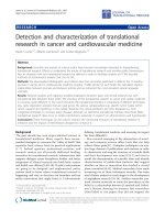
CAV DNA in embryos and egg shell membranes (ESM)
The nested PCR assay result indicated the presence of pos-
itive embryos ranging from 40–100% in different farms
from three states of Malaysia (Fig. 1).
Nucleotide sequence analysis
Nucleotide sequence analysis of a 498 bp region of CAV
genome from position 892 to 1389; numbering according
to Meehan et al. [14], encompassing the hypervariable
region of VP1 protein revealed total nucleotide variation
among the isolates ranging from 0.3 to 6.1% whilst the
overall maximum nucleotide variation is 6.5%. The nucle-
otide sequence alignment with the published isolates con-
sidered for comparison revealed 9 to 30 nucleotide
substitutions in the isolates from commercial broiler
chickens (Table 2). Based on comparisons of percentage
homologies, six isolates (MF1A, MF3C, M3B5, NF4A,
P12B and P24A) were found to have maximum homology
(97.1 to 99.1%) with SMSC-1 isolate, four isolates (M1B1,
NF3A, PYT4 and PPW4) were found to have maximum
homology (98.1 to 98.9%) with BL-5 isolate and the
remaining two (NF1D and NF2C) have similar maximum
homology (98.1%) both with isolates 3-1 and BL-5 (Table
1).
Compared to isolates from other geographical places
around the world, six out of 12 isolates (MF1A, MF3C,
M3B5, NF4A, P12B, and P24A) were found to have maxi-
mum homology with Australian isolate 704. The remain-
ing isolates showed maximum homology with isolates
from China (NF3A and PYT4), isolate A2 from Japan
(NF1D and NF2C) and CAV-B isolate from India (PPW4).
Only two of the isolates (M1B1 and NF3A) were found to
have maximum homology with isolates 26P4 and Del-
Ros from USA.
Amino acid sequence analysis
The amino acid sequence alignment with the published
isolates considered for comparison also showed 4 to 7
amino acid substitutions in the isolates from commercial
broiler breeder chickens (Table 2). The calculation of syn-
onymous and non-synonymous substitution rate demon-
strated omega values (K
a
/K
s
) of less than one suggesting
the occurrence of purifying (negative) selection in all the
12 isolates (Table 2). All the isolates have the omega value
ranging from 0.07 to 0.35 except for PPW4 with omega
value of 0.50. Eight variable amino acid positions were
detected in more than one isolate at amino acid positions
22, 75, 83, 97, 125, 139, 141, 144. Maximum variation
among the CAV isolates was observed at amino acid posi-
Virology Journal 2008, 5:128 />Page 3 of 11
(page number not for citation purposes)
tion 144. Proline (P) at position 22 for isolate NF4A and glycine (G) at position 48 for isolate PYT4 were unique
amino acid substitutions found only in the studied iso-
lates (Table 3).
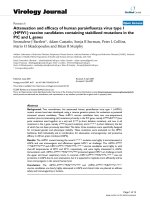
Phylogenetic analysis
Phylogenetic analysis based on 165 deduced amino acid
sequences of VP1 protein of the 12 isolates in comparison
to 20 previously identified isolates revealed the formation
of two clusters. All the 12 isolates were found both in clus-
ter I and II (Fig. 2). Seven of the isolates with amino acid
profile of 75-I, 97-L, 139-Q and 144-Q were clustered
together in cluster I while the remaining five isolates with
amino acid profile of 75-V, 97-M, 139-K and 144-E were
grouped under cluster II.
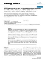
ELISA
The ELISA result shows that CAV infection is widespread
in these unvaccinated commercial broiler chicken farms
in Malaysia. Out of 52 chickens from which serum sample
was collected, 50 (96.15%) were positive for antibodies
against CAV (Fig. 3).
Out of the total number of hens sampled 26 hens (50%)
have anti-CAV antibody titers above 8600 (high protective
titers), 24 hens (46.15%) have the titer below 8600 but
Detection of CAV DNA in pooled embryonic tissues and ESM from eggs collected from commercial broiler breeder farmsFigure 1
Detection of CAV DNA in pooled embryonic tissues and ESM from eggs collected from commercial broiler
breeder farms. As it is shown on the graph, CAV DNA was detected in 40 to 100% of pooled embryonic tissue and ESM
samples tested for different commercial broiler breeder farms.
Table 1: Nucleotide percentage homologies of studied isolates
from the commercial broiler breeder hens in relation to
previously characterized Malaysian CAV field isolates.
Previously identified Malaysian CAV isolates
SMSC-1 BL-5 3-1
MF1A 98.9 94.9 94.5
MF3C 97.9 94.5 94.3
M3B5 97.1 95.1 94.9
NF4A 98.3 95.1 94.7
P12B 99.1 95.1 95.1
P24A 98.7 95.1 94.7
M1B1 94.9 98.9 98.1
NF3A 94.1 98.5 97.7
PYT4 94.1 98.5 97.7
PPW4 94.7 98.1 97.1
NF1D 94.7 98.1 98.1
NF2C 94.3 98.1 98.1
Accordingly, isolates MF1A, MF3C, M3B5, NF4A, P12B and P24A
were found to have maximum homology with SMSC-1 isolate, where
as four isolates (M1B1, NF3A, PYT4 and PPW4) were found to have
maximum homology with BL-5 isolate. However, isolates NF1D and
NF2C have similar maximum homology (98.1%) both with isolates 3-1
and BL-5.
Virology Journal 2008, 5:128 />Page 4 of 11
(page number not for citation purposes)
above 1000 (moderate protective titers) and 2 hens
(3.85%) have anti-CAV antibody titers below 1000 (neg-
ative for antibodies against CAV). The ELISA-based analy-
sis indicated that all farms had neutralizing antibodies.
Based on the correlation of ELISA titer to virus neutraliz-
ing antibody titer as recommended by the manufacturer
of the ELISA kit, 50% of hens found out to have high neu-
tralizing antibody titers that able to confer high level pro-
tection to the progeny whilst 46.15% of hens have low
neutralizing antibody titers affording low levels of protec-
tion to the progeny (Fig. 3).
Indirect immunoperoxidase staining
All the experimentally infected chickens from hyperim-
mune serum production produced the highest readable
antibody titer (>8661) which remained constant starting
from two weeks after the second inoculation. Sera from
the inoculated and control chickens were used as primary
antibodies for the indirect immunoperoxidase staining.
Specific positive staining was demonstrated in thymus
and bone marrow tissue sections from commercial broiler
breeder chickens that were detected positive for CAV
DNA. Positive staining was observed in lymphoblasts in
the cortex of the thymus (Fig. 4) and in hemocytoblasts in
the sinuses of the bone marrow (Fig. 5). Specific staining
was not demonstrated in tissue sections from spleen, liver,
duodenum, ovary and oviduct.
Discussion
In the present study, out of 420 organ samples tested, 75%
of spleen, 68.3% of bone marrow, 70% of thymus, 53.3%
of liver, 46.7% of duodenum, 66.7% of ovary and 48.3%
of oviduct tested were positive for CAV DNA. CAV repli-
cates mostly in lymphoid tissues of susceptible chickens
[15-17]. Cardona et al. [18] found out a few CAV positive
cells in spleen by in situ PCR and managed to detect CAV
in ovaries by in situ PCR even in the absence of splenic
virus. They also indicated that ovaries and to a lesser
degree infundibulum of the oviduct are sites for persist-
ence of CAV in hens. The finding of significantly higher
positive tissues in the spleen, bone marrow, thymus and
ovary as compared to duodenum, liver and oviduct in the
present study is similar to the aforementioned findings.
Therefore, we can suggest that spleen, thymus, and bone
marrow could serve as an excellent choice of organs for
the diagnosis of CAV infection while ovary representing
more favourable tissue for the persistence of CAV in the
reproductive organs in the broiler breeder hens.
Results from ELISA reading showed that 96.15% of blood
samples collected from the same farms were positive for
antibody against CAV indicating the widespread occur-
rence of CAV infection in these unvaccinated commercial
broiler breeder chicken farms. Testing pooled embryonic
tissue samples (thymus, bursa of Fabricius and spleen)
together with ESM showed positive embryos for CAV
DNA in the range of 40% to 100% for different commer-
cial broiler breeder chickens despite the presence of neu-
tralizing antibodies in majority of the hens (96.15%)
tested for CAV antibodies suggesting high level of occur-
rence of vertical transmission of viral DNA to the progeny.
The detection of CAV DNA in the ovary and oviduct of
commercial broiler breeder hens with virus neutralizing
antibodies and in their embryos supports the previous
evidence that CAV may remain in the gonads of antibody
positive chickens and can be vertically transmitted to their
progeny [18-21]. The finding by Schat et al. [2] indicated
low level of viral transcripts can be detected in the devel-
Table 2: Number of nucleotide and amino acid substitutions and Ka/Ks ratio of Malaysian CAV isolates
CAV isolates *No. of Nucleotide
Substitutions
*No. of amino acid
Substitutions
non-synonymous
substitution rate (K
A
)
Synonymous
substitution rate (K
S
)
Omega value (K
A
/K
S
)References
MF1A 28 6 0.017 0.199 0.08 This study
MF3C 30 6 0.017 0.225 0.07 This study
M3B5 27 6 0.017 0.191 0.09 This study
NF4A 27 7 0.020 0.179 0.11 This study
P12B 27 6 0.017 0.187 0.09 This study
P24A 18 6 0.017 0.191 0.09 This study
PPW4 12 7 0.020 0.039 0.50 This study
NF1D 13 6 0.017 0.053 0.33 This study
NF2C 13 6 0.017 0.055 0.31 This study
NF3A 11 4 0.012 0.047 0.25 This study
PYT4 11 5 0.015 0.042 0.35 This study
M1B1 9 4 0.012 0.036 0.32 This study
SMSC-1 26 5 0.017 0.187 0.09 [13]
3-1 8 4 0.011 0.060 0.19 [13]
BL-5 4 3 0.009 0.006 1.50 [12]
* compare to the consensus nucleotide and amino acid sequences. Pair wise comparison with the consensus nucleotide and amino acid sequence
indicated Ka/Ks ratio less than one for all the studied isolates which indicates the occurrence of negative selection.
Virology Journal 2008, 5:128 />Page 5 of 11
(page number not for citation purposes)
oping embryo during specific developmental periods sup-
porting the current hypothesis that, it may be possible
that a limited viral replication occurs in the embryos, but
if the embryos have VN antibodies, the VN antibodies pre-
vent the development of viremia in the embryos [19].
Viral antigens were identified only in individual lym-
phocytes in the cortex of the thymus and infected hemo-
cytoblasts in the bone marrow of tissues collected from
commercial broiler breeder chickens by the IPS. However,
consistent and observable differences on the intensity of
staining were not observed on those positive slides from
individual chickens with moderate and high protective
antibody titers at the same level of dilution of primary
antibodies. Specific staining could not be detected in
spleen, liver, duodenum, ovary and oviduct. Most of the
tissue sections obtained from commercial broiler breeder
chickens and tested positive by the nested PCR assay
turned out to be negative in IPS. This could be due to virus
replication in those tissues might be limited and below
the detection limit of the assay. It might also be related
with the age of commercial broiler breeder chickens or the
poor sensitivity of the technique compared to the other
two detection methods used in the present study namely
the nested PCR assay for detecting CAV DNA and ELISA
for detection of antibodies against CAV. Smyth et al. [17]
demonstrated viral antigens in the lymphoid tissues of
other organs including proventriculus, the ascending part
of duodenum, kidney and lung. However, they also con-
firmed that, infected cells in these tissues usually cannot
be detected for more than 22 days after infection at one
day of age.
Table 3: Amino acid substitutions in VP1 sequence of CAV isolates
Isolate Amino acid positions
22 48 75 83 97 125 139 141 144 157
Consensus H A V I M I K Q Q V
M1B1 . . E M
NF1D .L.L . E E .
NF2C .L.L . E E .
NF3A L . . E .
PYT4 . G* . . . L . . E .
NF4A P*.I.L.Q
PPW4 N.I.L.Q
P24A . . I . L . Q . . .
P12B . . I . L . Q . . .
M3B5 . . I . L . Q . . .
MF3C . . I . L . Q . . .
MF1A . . I . L . Q . . .
NIE/19.04/118/Nigeria . . I . L . Q . . .
BD-3/Bangladesh . . I . L . Q . . .
ISOLATE704/Australia . . I . L . Q . . .
130/Slovenia . . I L . Q . . .
CAV-B/India I.L.Q
SMSC-1/Malaysia . . I . L . Q . . .
BL5/Malaysia . . . . . . . . E .
BL5/P90/Malaysia . . . . . . . . K .
SMSC-1/P60/Malaysia . . . L . . . E E M
3-1/Malaysia . . . . . . . . E .
3-1/P60/Malaysia . . . . . . . E E M
CUX-1(M)/Germany . . . . . . . . D .
CUX-1(N)Germany . . . . . . . . D .
CIA-1/USA N . I . L . Q . . .
ConnB/USA N.
Del-Ros/USA . . . . . . . . E .
26P4/USA . . E M
CAV-A/India . . . . . L . . D .
A2/Japan . E E M
AF448446/China . . . . . . . . E .
Amino acids marked with an asterisk (*) are unique substitutions for isolates from commercial broiler breeder chickens. The isolates can be
grouped into two distinct groups based on their amino acid profile at positions 75, 97, 139 and 144. Five isolates (M1B1, NF1D, NF2C, NF3A and
PYT4) had amino acid profile of 75-V, 97-M, 139-K and 144-E.
Virology Journal 2008, 5:128 />Page 6 of 11
(page number not for citation purposes)
Comparisons of percentage homologies of the studied
CAV isolates with previously characterized local CAV iso-
lates showed diverse similarity among the local isolates.
The phylogenetic analysis of 165 deduced amino acid
sequences of the VP1 protein also revealed grouping of the
Malaysian CAV isolates into two major clusters (Fig. 2).
An overall similarity with CAV isolates circulating in
south and south-east Asia and Australia was also observed
while still having limited variation with isolates from dif-
ferent geographical parts of the world. Natesan et al. [22]
Phylogenetic relationship among 32 different CAV isolates based on partial VP1 amino acid sequencesFigure 2
Phylogenetic relationship among 32 different CAV isolates based on partial VP1 amino acid sequences. Note:
The boxes (n) indicate isolates identified in this study. The isolates were found both cluster I and II. Seven of the isolates with
amino acid profiles of 75-I, 97-L, 139-Q and 144-Q clustered together in cluster I. The remaining five studied isolates with
amino acid profiles of 75-V, 97-M, 139-K and 144-E were grouped under cluster II.
PPW4
CIA-1/USA
NF4A
Isolate704/Australia
M3B5
CAV-B/India
NIE/19.04/118-Nigeria
P24A
130/Slovenia
MF3C
BD-3/Banglade s h
P12B
MF1A
SMSC-1/Malays ia
BL5/P90/Malaysia
Cux-1(M)/Germany
Cux-1(N)/Germany
3-1/Malaysia
ConnB/USA
NF2C
NF1D
SMSC1/P60/M alaysia
A2-JP/Japan
3-1/P60/M alaysia
M1B1
CAV-A/India
PYT4
NF3A
De l-Ros /USA
AF448446/China
BL5-Malaysia
78
53
52
78
94
63
63
67
0.002
/
//
26P4/USA
Virology Journal 2008, 5:128 />Page 7 of 11
(page number not for citation purposes)
also found similar result in that Indian isolate (CAV-E)
having maximum similarity with Australian isolate iso-
late-704, Japanese isolate TR-20 and Malaysian SMSC-1
isolate. Unique amino acid residues observed in isolates
from commercial broiler breeder chickens include proline
(P) at amino acid position 22 and glutamine (G) at amino
acid position 48 in isolates NF4A and PYT4, respectively.
Islam et al. [8] identified amino acid residues at positions
75-I/T, 97-L, 139-Q and 144-Q can be used to group CAV
isolates into different groups. In the present study, two
distinct groups were observed in the current isolates based
on their amino acid profile at these positions. Seven of the
isolates from the commercial broiler breeder chickens
including previously characterized Malaysian isolate
SMSC-1, had 75-I, 97-L, 139-Q and 144-Q and clustered
together in cluster I of the deduced amino acid phyloge-
netic tree. Together in this group also included other iso-
lates from different geographical places. This includes,
CIA-1 from USA, CAV-B from India, BD-3 from Bangla-
desh, isolate 704 from Australia, isolate 130 from Slove-
nia and NIE/19.04/118 isolate from Nigeria. The
remaining five current isolates including previously char-
acterized Malaysian isolates BL-5 [12] and 3-1 [13] had
amino acid profile of 75-V, 97-M, 139-K and 144-E. These
isolates were found in cluster II of the deduced amino acid
phylogenetic tree with other isolates from around the
world. In addition, there was no evidence of recombina-
tion effect observed in the analysis of Malaysian CAV iso-
lates as reported by Van Santen et al. [23] on CAV isolates
from USA. In that study, they indicated that different CAV
isolates from Alabama can be divided into two groups
with one isolate showing an exceptionally different amino
acid profile of I-75, L-97, K-139 and E-144 suggesting of a
possible evidence of recombination event.
The analysis of the ratio of synonymous and non-synony-
mous substitution rate (omega value) indicated the pres-
ence of only purifying (negative) selection in the studied
isolates. This is similar to the result by Ducatez et al. [24]
where they indicated a very slow CAV virus evolution at
amino acid level corresponding to a strong negative selec-
tion (0.04 to 0.20) of VP1 gene in China and worldwide.
However, in this study, we found that one of the isolates
has omega value of 0.50 meanwhile five out of 12 isolates
have omega value between 0.25 to 0.35 and the rest with
omega value of 0.07 to 0.11 (Table 2). In our previous
study, we suggested that the BL-5 isolate was distantly
related to other Malaysian CAV isolates, SMSC-1 and 3-1
[12], has omega value of 1.50 suggesting of a positive
selection of VP1 protein in this isolate (Table 2).
The overall phylogenetic pattern and clustering of differ-
ent CAV isolates based on the partial VP1 gene in this
study was similar to previous one based on complete
sequence of VP1 gene [24] or the entire CAV genome [25]
for the common CAV isolates considered in all the three
studies. This suggests that relationships of different CAV
isolates can be determined on the basis of partial
sequence of VP1 gene due to the fact that most of the
amino acid substitutions in comparisons between isolates
lies in VP1 gene and more specifically on the N-terminal
half of VP1 gene.
Conclusion
Generally, from the present study we can conclude the
widespread occurrence of CAV infection in commercial
broiler breeder farms at least in the three states of Malay-
sia. Detection of significantly higher percentage of posi-
tive DNA from spleen, thymus and bone marrow make
these organs an excellent choice of organs in the screening
and diagnosis of flocks for CAV infection. The finding of
CAV DNA in embryos from broiler breeder chickens with
neutralizing antibodies supports the previous finding that
CAV may remain in the gonads of antibody positive chick-
ens and can be vertically transmitted to their progeny.
However, the importance of transmission of viral DNA
detected by nested PCR assay still needs further study and
explanation. The result also indicated the occurrence of
genetic variability in local CAV isolates that can be divided
at least into two groups based on characteristic amino acid
substitutions at positions 75, 97, 139 and 144 of the VP1
protein. However, the CAV isolates showed only negative
ELISA results of serum collected from commercial broiler breeder chickensFigure 3
ELISA results of serum collected from commercial
broiler breeder chickens. Fifty percent of the chickens
have ELISA S/N < 0.2 which indicates the presence of high
protective titers and able to afford high level protection to
the progeny, meanwhile 46.15% of the chickens have ELISA
S/N in the range of 0.2 to 0.8 affording low levels of protec-
tion to the progeny. Only 3.85% of the chickens have ELISA
S/N > 0.8 indicating negative result for antibody against CAV.
Virology Journal 2008, 5:128 />Page 8 of 11
(page number not for citation purposes)
selection based on the calculated omega value of the par-
tial sequences of the VP1 gene. The characterized CAV iso-
lates show overall similarity with CAV isolates circulating
in South East Asia and Australia while still having limited
variations with isolates from different geographical parts
of the world.
Methods
Broiler breeder farms
Tissue and blood samples were collected from 12 com-
mercial broiler breeder chicken farms located at three
states of Peninsular Malaysia. The farms were not vacci-
nated against CAV and the samples were collected from a
total of 60 broiler breeder hens that range in age from 25–
35 weeks.
Sample collection
A total of 420 organ samples were collected. Spleen, thy-
mus, liver, bone marrow, duodenum, ovaries and oviduct
were organs collected from each hen. Blood samples were
collected from 52 broiler breeder hens by veno-puncture
of the wing vein. Sera were separated and stored at -20°C
until used.
Ten eggs were also collected from each farm and incu-
bated for 18–20 days. Prior to hatching pooled embryonic
organ samples consisting of thymus, bursa of Fabricius
IPS performed on formalin fixed paraffin-embedded thymic tissuesFigure 4
IPS performed on formalin fixed paraffin-embedded thymic tissues. Thymic tissue slide from commercial broiler
breeder hen: a) infected lymphoblasts in the cortex demonstrated by brown staining (400×) b) IPS using CAV negative serum
as primary antibody and devoid of any specific brown staining (400×).
IPS performed on formalin fixed paraffin-embedded tissues from bone marrowFigure 5
IPS performed on formalin fixed paraffin-embedded tissues from bone marrow. Bone marrow tissue slide from
commercial broiler breeder hen: a) infected hemocytoblasts in the sinuses of the bone marrow demonstrated by brown stain-
ing (400×) b) IPS using CAV negative serum as primary antibody and devoid of any specific brown staining (200×).
Virology Journal 2008, 5:128 />Page 9 of 11
(page number not for citation purposes)
and spleen together with egg shell membrane (ESM) were
collected from individual embryos. Tissue samples from
the hens and embryos were stored at -20°C until DNA
extraction.
DNA Extraction from Samples
DNA was extracted from a total of 420 tissue sample from
hens and 52 pooled embryo samples. Briefly, tissue sam-
ples (1–5 mg) were homogenized in Phosphate Buffered
Saline (PBS) solution by grounding with a mortar and
pestle. Then the homogenate (~700 μl) was transferred
into 1.5 ml eppendorf tube and centrifuged at 13000 rpm
for 1 minute. The supernatant was then transferred into a
new microcentrifuge tube. DNA extraction was carried out
using MasterPure complete DNA and RNA purification kit
(Epicentre, Madison, WI), following the instructions of
the manufacturer with some modifications. The concen-
tration and purity of the extracted DNAs were determined
by a spectrophotometer (Beckman, USA) according to the
method described by Sambrook et al. [26].
Detection of CAV by nested PCR assay
The extracted DNA was first screened for CAV DNA using
a highly sensitive nested detection PCR as previously
described by Cardona et al. [18] with slight modifications.
The first-step PCR reaction was carried out using 20 pmol
each of the primers O3F and O3R amplifying a 386 bp
fragment of the VP3 gene [18]. The PCR reaction was car-
ried out in a total volume of 25 μl using the following
cycling parameters: initial denaturation of 94°C for 2 min
followed by 35 cycles of denaturation, annealing and
extension at 94°C for 2 min, 50°C for 1 min and 72°C for
1 min, respectively, and the final extension was carried
out at 72°C for 3 min. An aliquot of the first PCR reaction
(1 μl) was then added to 24 μl of a new mastermix (total
volume 25 μl) containing 20 pmol of the nested primers
N3 and primer N4 for amplification of a 209 bp nested
fragment of the VP3 gene as reported by Cardona et al.
[18]. The nested PCR assay was carried out in MyCycler
®
Thermal Cycler (Bio-Rad, Hercules, CA, USA). The PCR
products were analyzed by 1.8% agarose gel electrophore-
sis and the photographs were taken using Bio Imaging
System in GeneSnap program (SynGene, Cambridge,
UK).
Amplification of partial VP1 gene for sequencing
Spleen samples from each farm that were detected CAV
positive by VP3 nested PCR assay were used for amplifica-
tion of partial VP1 gene using primers VP1F and VP1R for
the first round amplification as described by Natesan et al.
[22]. Nested fragment of first round amplification were
amplified using primers O1F and PshA1R [18]. The first
round PCR condition was carried out using the following
cycling parameters: initial denaturation of 94°C for 4 min
followed by 35 cycles of denaturation, annealing and
extension at 94°C for 1 min, 57°C for 1 min and 72°C for
2 min, respectively, and the final extension was carried
out at 72°C for 8 min. The second synthesis was carried
out in a 50 μl reaction mixture with 1 μl of the first PCR
reaction product and cycling parameters similar to that
described for nested detection PCR. The PCR products
were run on 1.6% agarose gel electrophoresis and purified
from the gel by using GeneAll
®
kit (General Biosystem
Inc., Korea) following the supplied instructions.
Sequence and phylogenetic analysis
Using the gel purified PCR products, the partial nucle-
otide sequences of VP1 gene were determined by direct
sequencing in both direction using nested primers O1F
and PshA1R. Sequencing reactions were performed in MJ
Research PTC-225 Peltier Thermal Cycler using ABI
PRISM
®
BigDyeTM Terminator Cycle Sequencing Kits with
AmpliTaq DNA polymerase (FS enzyme) (Applied Biosys-
tems, CA, USA). Each sample was sequenced three times
to confirm consistency of the sequencing results.
DNA sequences of the 12 CAV isolates were aligned and
compared with 20 local and foreign CAV isolates retrieved
from the GenBank database. Sequences of VP1 gene for
the studied Malaysian isolates were submitted to Gen-
Bank under the following accession numbers: MF1A
[FJ167513
]; MF3C [FJ167514]; M1B1 [FJ167515]; M3B5
[FJ167516
]; NF1D [FJ167517]; NF2C [FJ167518]; NF3A
[FJ167519
]; NF4A [FJ167520]; P12B [FJ167521]; P24A
[FJ167522
]; PYT4 [FJ167523]; PPW4 [FJ167524]. The
retrieved CAV isolates sequence name, GenBank accession
numbers (in square brackets) and country are as follows:
Cux-1 [M-M55918
], Germany; Cux-1N [NC001427], Ger-
many; SMSC-1 [AF285882
], Malaysia; SMSC-1P60
[AF390102
], Malaysia; 3-1 [AF390038], Malaysia; 3-1P60
[AY040632
], Malaysia; BL-5 [AF527037], Malaysia; BL-5/
P90 [AY150576
], Malaysia; Isolate 704 [U65414], Aus-
tralia; CIA-1 [L14767
], USA; ConnB [U69548], USA; Del-
Ros [AF313470
], USA; 26P4 [D10068], USA; China
[AF448446
]; A2-[AB031296], Japan; BD-3 [AF395114],
Bangladesh; CAV-A [AY583755
], India; CAV-B
[AY583756
], India; NIE/19.04/118 [AJ888524], Nigeria;
130 [DQ016138
], Slovenia. Percentage of homology,
sequence identity matrix and translation from nucleotides
to amino acids were determined using BioEdit software
package version 7.01 [27]. Multiple sequence alignment
of nucleotide and translated amino acids were performed
using ClustalX software [28]. The phylogenic analysis of
165 deduced amino acids of the VP1 gene was performed
with the software MEGA4 for phylogenetic and molecular
evolutionary analyses using the Neighbor Joining Phylog-
eny reconstruction method with Poisson correction anal-
ysis and bootstrap consensus tree inferred from 1000
replicates [29]. Omega values [ratio of non-synonymous
(K
A
) to Synonymous (K
S
) substitution rates] was calcu-
Virology Journal 2008, 5:128 />Page 10 of 11
(page number not for citation purposes)
lated in comparison to consensus nucleotide sequences
using PAL2NAL program [30].
ELISA
The sera were tested using a commercial ELISA kit (Idexx
Lab, USA) at a 1:100 dilution and the results were
expressed as S/N ratios (sample to negative ratio) accord-
ing to manufacturer's instructions. Optical density value
was read at 650 nm wave length on an ELX 800™ micro-
plate reader (BIO-TEK Instruments, USA). The ELISA anti-
body titer has 78% correlation to virus neutralization
titers [19].
Experimental infection of chicks with CAV
Eight 5 days old specific-pathogen-free (SPF) chicks were
obtained from Veterinary Research Institute (VRI), Ipoh,
Perak, Malaysia. The chicks were divided into 2 groups.
Group 1 (n = 5) was inoculated intramuscularly with 1 ml
of SMSC-1 CAV isolate cell culture inoculum containing
10
5.5
TCID
50
/ml [13]. Group II (n = 3) was left uninocu-
lated as negative control chicks. Each group was reared
separately in different room and the chicks were observed
daily, and feed and water were provided ad libitum. The
chicks were sacrificed at 14 days p.i. (post inoculation) for
collection of organs. Tissue samples collected from
infected and uninoculated chicks were processed and con-
sidered as positive and negative control slides for immu-
noperoxidase staining (IPS), respectively. All
experimental research carried out on animals in this paper
(including chicken hyperimmune serum production) fol-
lowed internationally recognized guidelines and
approved by animal care and use committee at the Faculty
of Veterinary Medicine in University Putra Malaysia (Ref:
UPM/FPV/PS/3.2.1.1551/AUP-R4).
Specimen preparation for immunohistochemical staining
Tissue samples were fixed in 10% (v/v) neutral phos-
phate-buffered formalin for about 24 hrs and were then
trimmed to the thickness of 0.5 cm. The bone marrow
samples were decalcified by 5% nitric acid solution fol-
lowing the procedure of Luna [31]. Following tissue
processing, tissue blocks were sectioned at a thickness of 4
μm and collected on clean silanized microscope slides
[32,33].
Immunohistochemical staining
Prior to staining, the tissue slides were deparaffinized to
remove embedding media and rehydrated following the
supplied instructions. Antigen retrieval was achieved
using the microwave-based antigen retrieval technique
[33,34]. The staining procedure of the detection system
was carried out following the manufacturer's instruction
manual [DakoCytomation Envision
®
+ Dual Link System-
HRP (DAB+), Denmark]. A known negative and positive
antigen control and a negative serum control were
included in every procedure.
Chicken hyperimmune serum production
The hyperimmune serum production was carried out in
four 60 weeks old SPF broiler breeder roosters obtained
from VRI, Ipoh, Perak, Malaysia, with the following
immunization protocol: Briefly, at day 0, prior to inocula-
tion, blood samples were collected from all chickens to
test their freedom from CAV antibody. Then the roosters
were immunized orally with 2 ml of live CAV vaccine
AviPro
®
THYMOVAC (Lohmann Animal Health, Cux-
haven, Germany). The immunization regimen was
repeated at 14, 28, and 42 days after the first immuniza-
tion in combination with Freund adjuvant (Sigma, USA).
At day 56, blood was withdrawn from all the chickens
from the wing vein to separate the hyperimmune serum
produced. Commercial Idexx ELISA kit (Idexx Lab, West-
brook, Maine, USA) was used to evaluate the antibody
titer of the chicken hyperimmune serum at different levels
of immunization.
IgY purification from chicken hyperimmune serum
IgY purification was carried out using Pierce
®
Thiophilic
Adsorption Kit (Pierce, USA). The T-gel was initially used
according to the manufacturer's instruction for mamma-
lian immunoglobulin purification. For the purification of
IgY from chicken serum, the manufacturer's instruction
was followed together with optimized protocol of T-gel
chromatography for the purification of IgY from chicken
sera as described by Constantinoiu et al. [35].
Statistical methods
Data from the distribution of CAV DNA in organ samples
from commercial broiler breeder chickens were analyzed
by Kruskal-Wallis one-way ANOVA with significance
defined at P < 0.05. Groups with significance difference in
means from the rest were determined using Q-statistics
[36].
Competing interests
The isolated Malaysian CAV isolates are currently been
modified for development of live attenuated vaccines for
poultry.
Authors' contributions
Design and conception of the study (ARO, ZH, MHB,
TCG); samples detection by PCR assays (ZH, ARO, TCG),
Immunohistochemstry (ZH, MHB), sequence alignment
and phylogenetic tree analysis (ZH, ARO), manuscript
preparation (ZH, ARO, MHB); All the authors read and
approved the final manuscript.
Virology Journal 2008, 5:128 />Page 11 of 11
(page number not for citation purposes)
Additional material
Acknowledgements
This research was supported by grant number 09-02-04-0700-EA001 from
Ministry of Science, Technology and Innovation, Government of Malaysia.
Zerihun Hailemariam was supported by a grant from the Netherlands
Organization for International Cooperation in Higher Education (Nuffic).
References
1. Pringle CR: Virus taxonomy at the XIth International Con-
gress of Virology, Sydney, Australia. Arch Virol 1999,
144:2065-2069.
2. Schat KA, Circovirus infections: Chicken infectious anemia. In
Diseases of Poultry Edited by: Saif YM, Barnes HJ, Fadly AM, Glisson JR,
McDougald LR, Swayne DE. Iowa State University Press, Ames, IA;
2003:182-202.
3. Adair BM: Immunopathogenesis of chicken anemia virus
infection. Dev Comp Immunol 2000, 24:247-255.
4. Markowski-Grimsrud CJ, Schat KA: Infection with chicken anae-
mia virus impairs the generation of pathogen-specific cyto-
toxic T lymphocytes. Immunology 2003, 109:283-294.
5. McConnell CD, Adair BM, McNulty MS: Effects of chicken anemia
virus on cell-mediated immune function in chickens exposed
to the virus by a natural route. Avian Dis 1993, 37:366-374.
6. Yuasa N, Imai K, Watanabe K, Saito F, Abe M, Komi K: Aetiological
examination of an outbreak of haemorrhagic syndrome in a
broiler flock in Japan. Avian Pathol 1987, 16:521-526.
7. McNulty MS, Connor TJ, McNeilly F, McLoughlin MF, Kirkpatrick KS:
Preliminary characterisation of isolates of chicken anaemia
agent from the United Kingdom. Avian Pathol 1990, 19:67-73.
8. Islam MR, Johne R, Raue R, Todd D, Muller H: Sequence analysis
of the full-length cloned DNA of a chicken anaemia virus
(CAV) strain from Bangladesh: evidence for genetic group-
ing of CAV strains based on the deduced VP1 amino acid
sequences. J Vet Med B Infect Dis Vet Public Health 2002, 49:332-337.
9. Spackman E, Cloud SS, Pope CR, Rosenberger JK: Comparison of a
putative second serotype of chicken infectious anaemia virus
with a prototypical isolate I. Pathogenesis. Avian Dis 2002,
46:945-955.
10. Spackman E, Cloud SS, Pope CR, Rosenberger JK: Comparison of a
putative second serotype of chicken infectious anaemia virus
with a prototypical isolate II. Antigenicity. Avian Dis 2002,
46:955-960.
11. Rozanah AS, Aini I, Al-Ajeeli KS, Jalila A, Salim NB: Detection of
chicken anemia virus in flocks of commercial chicken in
Malaysia. Journal of Veterinary Malaysia 1995,
7:77-79.
12. Hasmah MS, Omar AR, Wan KF, Hair-Bejo M, Aini I: Genetic diver-
sity of chicken anemia virus following cell culture passaging
in MSB-1 cells. Acta Virologica 2004, 48:85-89.
13. Chowdhury S, Omar AR, Aini I, Hair-Bejo M, Jamaluddin AA, Md-Zain
BM, Kono Y: Pathogenicity, sequence and phylogenetic analy-
sis of Malaysian chicken anemia virus obtained after low and
high passages in MSB-1 cells. Archives of Virology 2003,
148:2437-2448.
14. Meehan BM, Todd D, Creelan JL, Earle JAP, Hoey EM, McNulty MS:
Characterization of viral DNAs from cells infected with
chicken anaemia agent: Sequence analysis of the cloned rep-
licative form and transfection capabilities of cloned genome
fragments. Arch Virol 1992, 124:301-319.
15. Adair BM, McNeilly F, McConnell CD, McNulty MS: Characteriza-
tion of surface markers present on cells infected by chicken
anemia virus in experimentally infected chickens. Avian Dis
1993, 37:943-950.
16. Hoop RK, Reece RL: The use of immunofluorescence and
immunoperoxidase staining in studying the pathogenesis of
chicken anemia agent in experimentally infected checkens.
Avian Pathol 1991, 20:349-355.
17. Smyth JA, Moffett DA, McNulty MS, Todd D, Mackie DP: A sequen-
tial histopathologic and immunocytochemical study of
chicken anemia virus infection at one day of age. Avian Dis
1993, 37:324-338.
18. Cardona CJ, Oswald WB, Schat KA: Distribution of chicken infec-
tious anemia virus in the reproductive tissues of specific
pathogen-free chickens. J Gen Virol 2000, 81:2067-2075.
19. Brentano L, Lazzarin S, Bassi SS, Klein TA, Schat KA: Detection of
chicken anemia virus in the gonads and in the progeny of
broiler breeder hens with high neutralizing antibody titers.
Veterinary Microbiology 2005, 105:65-72.
20. Cardona CJ, Lucio B, O'Connell P, Jagne J, Schat KA: Humoral
immune responses to chicken infectious anemia virus in
three strains of chickens in a closed flock. Avian Dis 2000,
44:661-667.
21. Miller MM, Katie AE, Oswald WB, Schat KA: Detection of chicken
anemia virus DNA in embryonal tissues and eggshell mem-
branes. Avian Dis
2003, 47:662-671.
22. Natesan S, Kataria JM, Dhama K, Rahul S, Baradhwaj N: Biological
and molecular characterization of chicken anemia virus iso-
lates of Indian origin. Virus Res 2006, 118:78-86.
23. van Santen VL, Li L, Hoerr FJ, Lauerman LH: Genetic characteriza-
tion of chicken anemia virus from commercial broiler chick-
ens in Alabama. Avian Dis 2001, 45:73-388.
24. Ducatez MF, Chen H, Guan Y, Muller CP: Molecular epidemiology
of chicken anemia virus (CAV) in Southeastern Chinese live
birds markets. Avian Diseases 2008, 52:68-73.
25. He C, Ding N, Fan W, Wu Y, Li J, Li Y: Identification of chicken
anemia virus putative intergenotype recombinants. Virology
2007, 366:1-7.
26. Sambrook J, Fitsch EF, Maniatis T: Molecular Cloning: A Laboratory Man-
ual Cold Spring Harbor, Cold Spring Harbor Press; 1989.
27. Hall TA: BioEdit: a user-friendly biological sequence align-
ment editor and analysis program for Windows 95/98/NT.
Nucl Acids Symp Ser 1999, 41:95-98.
28. Thompson JD, Gibson TJ, Plenwniak F, Jeanmoungin F, Higgins DG:
The Clustal_X windows interface: flexible strategies for mul-
tiple sequence alignment aided by quality analysis tools.
Nucleic Acids Res 1997, 25:4876-4882.
29. Tamura K, Dudley J, Nei M, Kumar S: MEGA4: Molecular Evolu-
tionary Genetics Analysis (MEGA) software version 4.0.
Molecular Biology and Evolution 2007, 24:1596-1599.
30. Suyama M, Torrents D, Bork P: PAL2NAL: robust conversion of
protein sequence alignments into the corresponding codon
alignments. Nucleic Acids Res 2006, 34:W609-W612.
31. Luna LG: Manual of Histologic Staining Methods of the Armed Forces Insti-
tute of Pathology The Blakiston Division, Mc Graw-Hill Book Company,
New York; 1968.
32. Bancroft JD, Stevens A, Stevens A: Theory and practice of histological
technique New York: Churchill Livingstone; 1996.
33. Key M, Boenisch T: Antigen retrieval. In Immunohistochemical stain-
ing methods Edited by: Key M. Dako corporation, Carpinteria;
2006:41-45.
34. Gown AM, de Wever N, Battifora H: Microwave-based antigenic
unmasking. A revolutionary new technique for routine
immunohistochemistry. Appl Immunohistochem
1993, 1:256-266.
35. Constantinoiu CC, Molley JB, Jorgensen WK, Coleman GT: Purifica-
tion of immunoglobulins from chicken Sera by thiophilic gel
chromatography. Poultry Science 2007, 86:1910-1914.
36. Petrie A, Watson P: Statistics for veterinary and animal science Blackwell
Science Ltd. UK; 1999.
Additional file 1
Tissue distribution of CAV DNA in various organs from commercial
broiler breeder hens. Values with the same lowercase superscript are not
significantly different (P < 0.05) by Kruskal-Wallis one-way ANOVA
analysis. The difference in the tissue distribution of CAV DNA between
spleen, bone marrow, thymus and ovary was found to be not significant.
However, the distribution of CAV DNA in liver, duodenum and oviduct
was found significantly less compared to spleen, thymus, bone marrow and
ovary.
Click here for file
[ />422X-5-128-S1.pdf]