Báo cáo hóa học: " Application of real-time PCR to quantify hepatitis B virus DNA in chronic carriers in The Gambia" pptx
Bạn đang xem bản rút gọn của tài liệu. Xem và tải ngay bản đầy đủ của tài liệu tại đây (278.16 KB, 7 trang )
BioMed Central
Page 1 of 7
(page number not for citation purposes)
Virology Journal
Open Access
Research
Application of real-time PCR to quantify hepatitis B virus DNA in
chronic carriers in The Gambia
Maimuna E Mendy*
1,2
, Steve Kaye
1,3
, Marianne van der Sande
1,4
, Pura Rayco-
Solon
1,5
, Pauline A Waight
1,6
, Deborah Shipton
1
, Dorka Awi
1
, Paul Snell
1
,
Hilton Whittle
1
and Samuel J McConkey
1,7
Address:
1
Medical Research Council, Atlantic Boulevard, Fajara, P O Box 273, Banjul, The Gambia,
2
Viral Disease programme, Medical Research
Council, Atlantic Boulevard, Fajara, P O Box 273, Banjul, The Gambia,
3
Imperial college, London, UK,
4
RIVM, Bithoven, The Netherlands,
5
Nutrition centre of the Philippines, Philippines,
6
National Protection Agency, Collindale, London, UK and
7
Royal College of Surgeons in Ireland,
Dublin, Ireland
Email: Maimuna E Mendy* - ; Steve Kaye - ; Marianne van der
Sande - ; Pura Rayco-Solon - ; Pauline A Waight - ;
Deborah Shipton - ; Dorka Awi - ; Paul Snell - ;
Hilton Whittle - ; Samuel J McConkey -
* Corresponding author
Abstract
Background/Aim: The study aimed at developing a real-time quantitative PCR assay to monitor
HBV serum virus load of chronic carriers enrolled in therapeutic trials.
Method: Quantitative real-time PCR assay was carried out using SYBR-Green signal detection and
primers specific to the S gene. Thermal cycling was performed in an ABi 5700 sequence detection
system. The assay was calibrated against an international HBV DNA standard and inter- and intra-
assay reproducibility determined. Levels of viral load were monitored for 1-year in lamivudine
treated carriers. Correlation between HBV DNA levels and HBeAg sero-status was determined in
untreated carriers.
Results: The qPCR assay showed good intra- and inter-assay reproducibility over a wide dynamic
range (1.5 × 10
3
to 1.5 × 10
8
copies/mL) and correlated well with those from a commercial assay
(r = 0.91, (p < 0.001). Viral load levels dropped dramatically but temporarily during and after a short
course of lamivudine therapy. HBV DNA was a more reliable indicator of the presence of virus
than HBe antigen and was detected in 77.0% (161/209) of HBeAg negative and in all HBeAg positive
carriers.
Conclusion: This method is reliable, accurate, and reproducible. HBV DNA Quantification by
qPCR can be used to monitor the efficacy of HBV therapy and useful in understanding the natural
history of HBV in an endemic area.
Published: 04 April 2006
Virology Journal 2006, 3:23 doi:10.1186/1743-422X-3-23
Received: 01 November 2005
Accepted: 04 April 2006
This article is available from: />© 2006 Mendy et al; licensee BioMed Central Ltd.
This is an Open Access article distributed under the terms of the Creative Commons Attribution License ( />),
which permits unrestricted use, distribution, and reproduction in any medium, provided the original work is properly cited.
Virology Journal 2006, 3:23 />Page 2 of 7
(page number not for citation purposes)
Introduction
Hepatitis B virus (HBV) is the leading cause of viral hepa-
titis in humans worldwide. Currently over two billion
people have evidence of previous HBV infection and 350
million have become chronic carriers of the virus, 60 mil-
lion of them residing in Africa [1]. In the Gambia, where
HBV is endemic, the prevalence of chronic infection is 10–
15% of the adult population [2,3]. Chronically infected
carriers have a high risk of developing liver damage and
hepatocellular carcinoma (HCC) and liver cancer is the
commonest cause of death in adult males in The Gambia
[4]. Detection of serological markers is the mainstay of
diagnosis of HBV infection and the most reliable marker
of HBV carriage is HBV surface antigen (HBsAg) in serum.
HBV e antigen (HBeAg) is generally used as secondary
marker to indicate high levels of virus in the blood. The
minority of chronic HBV carriers in whom HBeAg can be
detected have a particularly high risk of progressive liver
disease and end stage liver failure [5]. The monitoring of
hepatitis B virus DNA in serum is as important as serolog-
ical markers in predicting the clinical outcome of infec-
tion. More recently molecular diagnostic methods have
been used to quantify the levels of HBV DNA in serum as
a marker of viral replicative activity [6]. The detection and
quantification of HBV DNA is reported to have prognostic
value for the outcomes of acute and chronic HBV infec-
tions [7,8]. Quantification of HBV DNA may be a more
useful measure than HBeAg as genetic variants of HBV
may continue to replicate at high level without secreting
HBeAg. Quantification of HBV DNA can be useful to
assess the efficacy of antiviral therapy as a more direct
method of detecting viral replication than HBV serologic
markers [9,10]. The clinical management of HBV could be
improved by the use of accurate quantification of virus
load as a measure of replication of HBV in patients with
chronic liver disease. Prior to the development of the
polymerase chain reaction (PCR) a number of hybridisa-
tion methodologies were used to monitor HBV DNA lev-
els [11-14]. The introduction of PCR-based methods has
resulted in a large increase in the sensitivity of HBV DNA
detection and commercialisation of PCR-based methods
(e.g. HBV Monitor, Roche Diagnostic Systems) has lead to
widespread adoption of the methodology [15]. More
recently the development of real time PCR methodology
has further improved the ease with which HBV DNA levels
can be monitored and has increased the range over which
such levels can be accurately quantified [16,17].
We describe the development and validation of a quanti-
tative PCR (qPCR) method to measure the concentration
of HBV DNA in serum. The assay is based on the specific
amplification of HBV DNA using primers targeted to the
S-gene and detection in real-time with SYBR Green dye.
The specificity, reproducibility and detection limit of the
assay was examined. The assay was used to monitor HBV
DNA levels in patients on lamivudine therapy.
Viral load in HBeAg-positive and HBeAg-negative asymp-
tomatic HBV carriers was measured to assess the relation-
ship between serologic markers and levels of HBV DNA.
Material and methods
Study subjects
Two groups of asymptomatic HBV carriers were included
in the study. The first consisted of 22 male, asymptomatic
HBV carriers aged between 15 and 25 years recruited to a
therapeutic vaccine trial. As part of this trial some subjects
were randomised to receive the antiviral drug lamivudine
(GlaxoSmithKline). Fifteen subjects received a 98- day
course of lamivudine therapy alone and seven were mon-
itored as untreated controls. The volunteers were followed
for a period of 255 days; blood samples were collected at
baseline then on 28, 56, 77, 98, 161, 245 and 329 days
after the baseline visit. The Gambian Ethics Committee
reviewed and approved both studies.
The second group consisted of 318 HBV chronic carriers
aged between 1 and 73 years recruited during 1983 and
2003 community survey conducted in three rural villages
in The Gambia. An infant vaccination programme started
in these villages in 1984 so the majority of the HBV carri-
ers were over 20 years. Levels of viral load was compared
with HB sero-status.
Real-time quantitative PCR for HBV DNA
DNA was extracted from 200 µL of serum using the
QIAamp DNA Mini Kit (Qiagen, Hilden, Germany)
according to the manufacturer's instructions. DNA was
eluted into 100 µL nuclease-free water and 5 µl added to
a 25 µl PCR reaction mixture.
The reaction was carried out using a commercial SYBR-
Green reaction mix (Qiagen, Hilden, Germany). The kit
contains HotStarTaq polymerase which is included to
avoid false positives in the quantitative PCR. The primer
sequences were 5'-GTG TCT GCG GCG TTT TAT CA
(sense) and 5' GAC AAA CGG GCA ACA TAC CTT (anti-
sense) designed to amplify a 98 base pair product from
positions 379 to 476 of the HBV genome [18]. Thermal
cycling was performed in an ABi 5700 sequence detection
system (PE Applied Biosystems, Warrington, UK). Reac-
tion conditions were: 95°C for 15 minutes followed by 40
cycles of 94°C for 15 seconds, 55°C for 30 seconds and
72°C for 30 seconds. A four point standard curve (1.5 ×
10
8
copies per millilitre (cpm), 1.5 × 10
6
cpm, 1.5 ×
10
4
cpm, 1.5 × 10
2
cpm) was generated from a high titre
plasma donation quantified by end point dilution PCR.
The calibration of this standard was confirmed by com-
Virology Journal 2006, 3:23 />Page 3 of 7
(page number not for citation purposes)
parison with an International HBV DNA standard, (97/
746) (NIBSC, Potters Bar, UK). Test samples falling above
the top of the standard curve were re-assayed at a dilution
of 1:100. Each test run included positive and negative con-
trols. The performance of the assay was evaluated by com-
parison with a commercial assay (HBV Monitor, Roche
Molecular Systems, Inc., Branchburg, NJ 08876 USA) per-
formed according to the manufacturer's instructions.
Serology
Subjects were tested for HBV core antibody (anti-HBc)
and if indicated HBV surface antigen (HBsAg) and HBV e
antigen (HBeAg). Anti-HBc was measured by radio-
immunoassay (RIA) (Sorin Biochemica, Saluggia, Italy)
AB-COREK test kit. Samples, which were anti-HBc posi-
tive, were tested for HBsAg by reverse passive haemagglu-
tination assay (RPHA) (Wellcotest
®
, Murex Diagnostics,
Dartford, UK) and or by Determine™ HBsAg (Abbott Lab-
oratories), an immunochromatographic assay. HBsAg
positive subjects were tested for HBeAg using an enzyme
immunoassay (EIA) (Equipar Diagnostici, Saronno (Va),
Italy).
Data management
The data obtained in the ABi real time machine after the
PCR amplification and quantification of DNA was
exported as an Excel spreadsheet into an Access database
designed for the study. The viral results were merged with
HBV serological results prior to data analysis.
Results
Performance of the real-time qPCR
The performance of the new qPCR assay was examined by
determining the sensitivity, specificity, inter- and intra-
assay variability. HBV DNA standard was obtained by end
point dilution assay then calibrated with known standard
(97/746, NIBSC, Potters Bar, UK).
To determine the HBV DNA concentration of the top
standard serial half-log dilutions from 1:10 to 1:10
10
were
prepared from an HBeAg-positive plasma. The plasma was
diluted in HBV DNA-negative EDTA plasma. DNA was
extracted and amplified in quadruplicate reactions. The
end point, the dilution resulting in a mixture of positive
and negative reactions, was obtained at a dilution of
1:10
7.5
at which three of four reactions were positive. HBV
DNA concentration was calculated as 4.3 × 10
9.0
copies/
ml. This value was adjusted to 1.5 × 10
8
after direct com-
parison to the International HBV DNA standard.
The detection limit of the assay was 2.6 × 10
2
DNA copies
per mL when used to test a serial dilution of a 2.6 × 10
6
DNA standard. The assay was 100% specific when tested
against HBV seronegative sera from ten subjects. To evalu-
Table 1: Intra assay variability of the newly developed qPCR assay
DNA (copies DNA per mL) in experiment
Sample ID Experiment 1 Experiment 2 Experiment 3 Average SD CV (%)
MVA 300 2.6 × 10
6
2.5 × 10
6
1.6 × 10
6
2.3 × 10
6
0.104 1.60
MVA 307 1.9 × 10
9
4.5 × 10
9
1.7 × 10
9
2.7 × 10
9
0.191 2.00
MVA 313 1.2 × 10
8
1.2 × 10
8
1.2 × 10
8
1.2 × 10
8
0.004 0.50
MVA 315 2.5 × 10
6
2.2 × 10
6
1.9 × 10
6
2.2 × 10
6
0.046 0.70
T4040 4.7 × 10
3
4.9 × 10
3
6.4 × 10
3
5.4 × 10
3
0.060 1.60
Negative control not detected not detected not detected
The six samples represented in the table include five positive samples (MVA 300, MVA 307, MVA 313, MVA 315) and one negative sample (negative
for HBsAg). The samples were tested three times in the same assay.
Table 2: Inter assay variability of the newly developed qPCR assay
DNA (copies per mL) on different days
Sample ID Day 1 Day 2 Day 3 Average SD CV (%)
MVA300 2.7 × 10
6
1.4 × 10
6
2.7 × 10
6
2.3 × 10
6
0.19 3.0
MVA307 2.1 × 10
9
4.2 × 10
9
1.5 × 10
9
2.6 × 10
6
0.26 2.70
MVA313 1.4 x10
8
9.8 × 10
7
1.3 × 10
8
1.2 × 10
8
0.09 1.10
MVA315 1.3 × 10
6
3.3 × 10
6
2.0 x10
6
2.2 × 10
6
0.01 1.20
T4040 5.6 × 10
3
7.2 × 10
3
4.3 × 10
3
5.7 × 10
3
0.23 0.62
Negative control DNA not detected DNA not detected DNA not detected DNA not detected
The six samples represented in the table include five positive samples (MVA 300, MVA 307, MVA 313, MVA 315) and one negative sample (negative
for HBsAg). The samples were tested on three different days.
Virology Journal 2006, 3:23 />Page 4 of 7
(page number not for citation purposes)
ate the reproducibility of the assay five samples were
tested in triplicate on three different occasions. Variability
between the triplicate samples assayed in the same run
was 3.06% and variability between mean results of tripli-
cates samples in different runs was 2.35%. Overall varia-
bility = 3.03%. The coefficient of variation obtained from
intra-and inter assay was 1.08 and 1.72 respectively
(Tables 1 and 2).
To further validate the performance of the in-house
method it was compared to a commercial quantitative
PCR method (Roche Amplicor Monitor, Inc., Branchburg,
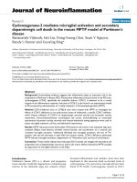
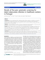
Comparison of real time PCR with the Roche Amplicor Monitor in 10 HBsAg positive samplesFigure 1
Comparison of real time PCR with the Roche Amplicor Monitor in 10 HBsAg positive samples. The log differ-
ence between the two assays was less than 1.0 over the dynamic range of 1.1 × 10
3
to 2.0 × 10
9
.
y = 1.0668x - 0.4338
R
2
= 0.8302
2.00
3.00
4.00
5.00
6.00
7.00
8.00
9.00
10.00
2.00 3.00 4.00 5.00 6.00 7.00 8.00 9.00 10.00
Roche Amplicor (DNA copies per mL)
Real time PCR (DNA copies per mL
)
Figure 1: Comparison of real time PCR with the Roche Amplicor Monitor in 10 HBsAg positive samples
Table 3: Viral load in relation to age group of carriers and HBeAg status
HBeAg Positive subjects HBeAg Negative carriers
Age group (Yrs) DNA positive (%) Viral load (GM) DNA positive (%) Viral load (GM) Total DNA positive in
all carriers
<5 50/50 (100%) 3.2 × 10
8
9/9 (100%) 4.3 × 10
4
59/69 (85.5%)
5–9 39/39 (100%) 3.8 × 10
8
15/21 (71.4%) 9.3 × 10
4
54/63(85.7%)
10–19 5/5 (100%) 1.7 × 10
9
11/13 (84.6%) 1.3 × 10
4
16/18 (88.9%)
20–29 11/11 (100%) 8.1 X10
7
71/91 (78.0%) 4.9 X10
3
82/102 (80%)
30–39 3/3 (100%) 5.5 X10
7
23/33 (69.6%) 2.6 X10
3
26/36 (72%)
>40 1/1 (100%) 4.6 X10
2
32/42 (76.1%) 6.02 X10
3
33/43 (77%)
Total 109/109 (100%) 161/209 (77.0%) 270
As it is most likely that HBV infection occurs in childhood in the Gambia, the age of the carriers is likely to represents the length of HBV carriage.
Virology Journal 2006, 3:23 />Page 5 of 7
(page number not for citation purposes)
NJ 08876 USA). Ten samples were assayed in the two
assays and the results are shown in Figure 1. Over a range
of 1.1 x10
3
to 2.0 × 10
9
DNA copies per mL the difference
between the two assays was less than 1.0 log. The correla-
tion between the log results of the two assays was high (r
= 0.91, p < 0.001).
Correlation of HBV DNA loads and HBeAg in
asymptomatic carriers
One hundred and eight (34%) of the 318 HBsAg positive
carriers had detectable HBeAg and the proportion
decreased with age (Chi squared test for trend p < 0.0001).
All HBeAg positive carriers tested positive for HBV DNA
compared to 164 (78%) of the HBeAg negative carriers
(Table 3.).
The geometric mean concentration of HBV DNA in
HBeAg-positive carriers was 4.6 log10 copies per ml
higher than in HBeAg-negative carriers (8.7 log10 copies
per ml vs. 4.1 log10 copies per ml, p < 0.0001). The viral
load was lower with increasing age in both HBeAg -posi-
tive and HBeAg negative carriers (p = 0.032, R = 0.207 for
HBeAg positive carriers and p <0.0001, R = 0.275 for
HBeAg negative carriers).
Monitoring of HBV DNA loads in subjects receiving
lamivudine therapy
The level of HBV DNA was measured in 16 asymptomatic
carriers (eight HBeAg positive and eight HBeAg negative)
on daily dose of 100 mg lamivudine for 98 days. HBV
DNA was measured at day 56, 98, 161, 245 and 329 after
the start of treatment with LMV. For comparison seven
asymptomatic untreated HBeAg negative carriers were
tested for HBV DNA. The baseline characteristics of the
two HBeAg negative groups (treated and untreated) were
similar (data not shown). None of the carriers had totally
cleared the virus as a result of LMV treatment however
HBV DNA decreased in patients on LMV and not in the
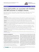
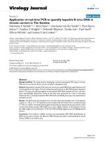
HBV DNA concentration in treated and untreated HBV carriersFigure 2
HBV DNA concentration in treated and untreated HBV carriers. The three groups consisted of HBeAg positive car-
riers treated with LMV (7), HBeAg negative carriers treated with LMV (8) and HBeAg positive carriers untreated and did not
receive any LMV (7). Patients were tested on day 56, 98, 161, 245 and 329 after LMV treatment. HBeAg positive carriers had
an additional test on day 77. The horizontal lines indicate error bars.
-4
-3
-2
-1
0
1
2
0 50 100 150 200 250 300 350 400
Time post treatment (Days)
log HBV DNA after base line subtraction
HBeAg pos treated HBeAg neg treated HBeAg neg untreated
Figure 2: HBV DNA concentration in treated and untreated HBV carriers
Virology Journal 2006, 3:23 />Page 6 of 7
(page number not for citation purposes)
untreated patients. Reduction in viral load was higher in
HBeAg positive carriers than in HBeAg negative carriers,
3.2 log compared to 1.5 respectively (Figure 2). Viral load
level returned to base line levels soon after the withdrawal
of LMV and this was more noticeable in the HBeAg posi-
tive group. There was subsequent rebound of viral load at
the withdrawal of LMV therapy. Fluctuation of level of
viral load was observed in the untreated group
Discussion
We have described the development and validation of a
real-time polymerase chain reaction (PCR) method based
on SYBR-Green for, measuring HBV DNA in serum and
plasma. We have demonstrated that the method had good
specificity, reproducibility and sensitivity and gave com-
parable results to those obtained with a commercial assay.
The primers used in this study have been shown to pro-
duce similar amplification/detection efficiency when used
to test samples of genotypes A and G [18]. The method
described had a lower threshold of detection than the
commercial assay with which it was compared [19]. In
general real-time PCR methodology is robust and easy to
perform and avoids many of the potential contamination
pitfalls that are associated with gel-based and hybridiza-
tion-based post-PCR detection methods.
The assay was used to assess the virological response to
short-term treatment with anti-viral medication. As for
HIV there is a large unmet need for treatment of chronic
HBV infection in sub-Saharan Africa. This is concurrent
with high morbidity and mortality from liver cancer and
cirrhosis. As antiviral therapies become more affordable
there will be requirement for virological assessment of the
successes of the treatment [20]. The assay described here
could have such a role. The lack of total clearance of HBV
DNA is most likely due to short-term treatment. Sustained
response after discontinuation of treatment was uncom-
mon in the carriers in this study and occurs in only 10–
15% of patients treated for years with LMV [21]. Drug
resistance in most cases after long-term therapy with LMV
has created the need for alternative form of treatment for
viral load reduction such as pegylated interferon [22,23].
Although LMV treatment can be effective in some cases,
low response rate to treatment is evident in HBeAg nega-
tive patients [24].
The new assay was used to describe the course of viraemia
in chronically infected people in The Gambia. The results
of the cross-sectional study suggest that the characteristics
of chronic HBV infection changes as people get older, or
have the infection for a longer time. Fewer older people
had HBeAg in the blood, suggesting that this is lost over
time in this group. The lower viral loads levels in those
who are HBeAg negative suggest that the host's control of
the virus replication is stronger and more efficient in those
who are older, or who have had the infection for longer.
This group, who have mostly been infected early in life,
are immunotolerant of the virus and often show little or
no clinical hepatitis [2]. There is evidence that infected
carriers are partly controlling HBV viral load, even before
they lose HBeAg from the blood. As shown in this study
there is a significant decline in viral load with age in both
HBeAg positive and HBeAg negative carriers.
The results of this study suggest that despite the immuno-
tolerance as they get older HBV carriers appear to clear
HBeAg and partially control viral replication. It is unclear
why the immune response may be more effective in those
who are older.
The cross-sectional component of this study shows that a
new rapid, robust, repeatable quantitative PCR assay for
HBV viral load can provide a useful tool to understand the
complex interactions between the HBV virus and the
infected host. This could be developed through its appli-
cation in a longitudinal study, ideally incorporating
simultaneous measures of the host immune response. The
longitudinal study of a group of chronic HBV carriers who
spontaneously loose HBeAg or seroconvert from HBsAg to
anti-HBs could be pivotal in elucidating the immunolog-
ical mechanisms, which affect the control of virus, which
these changes entail. This may be necessary prelude for the
development of effective immunomodulatory interven-
tions for the 350 millions of chronically infected individ-
uals.
Acknowledgements
We thank all the subjects of the study for volunteering to participate.
Thanks to Adam Jeng-Barry and Alasana Bah for laboratory assistance,
Joseph Bass, Yusupha Bah, Lamin Giana and Mansour Nyang for field assist-
ance. We would like to thank Adrian V. S. Hill for ideas and support for the
clinical trial. The lamivudine was a gift from Oxxon pharmaccines.
References
1. Lee WM: Hepatitis B virus infection. N Engl J Med 1997,
337:1733-1745.
2. Whittle HC, Inskip H, Bradley AK, McLaughlan K, Shenton F, Lamb
W, Eccles J, Baker BA, Hall AJ: The pattern of childhood hepatitis
B infection in two Gambian villages. J Infect Dis 1990,
161:1112-1115.
3. Viviani S, Jack A, Hall AJ, Maine N, Mendy M, Montesano R, Whittle
HC: Hepatitis B vaccination in infancy in The Gambia: pro-
tection against carriage at 9 years of age. Vaccine 1999,
17:2946-2950.
4. Bah E, Parkin DM, Hall AJ, Jack AD, Whittle H: Cancer in the Gam-
bia: 1988-97. Br J Cancer 2001, 84:1207-1214.
5. Fattovich G, Giustina G, Realdi G, Corrocher R, Schalm SW: Long-
term outcome of hepatitis B e antigen-positive patients with
compensated cirrhosis treated with interferon alfa. Euro-
pean Concerted Action on Viral Hepatitis (EUROHEP).
Hepatology 1997, 26:1338-1342.
6. Baker BL, Di Bisceglie AM, Kaneko S, Miller R, Feinstone SM, Wag-
goner JG, Hoofnagle JH: Determination of hepatitis B virus
DNA in serum using the polymerase chain reaction: clinical
significance and correlation with serological and biochemical
markers. Hepatology 1991, 13:632-636.
Publish with Bio Med Central and every
scientist can read your work free of charge
"BioMed Central will be the most significant development for
disseminating the results of biomedical research in our lifetime."
Sir Paul Nurse, Cancer Research UK
Your research papers will be:
available free of charge to the entire biomedical community
peer reviewed and published immediately upon acceptance
cited in PubMed and archived on PubMed Central
yours — you keep the copyright
Submit your manuscript here:
/>BioMedcentral
Virology Journal 2006, 3:23 />Page 7 of 7
(page number not for citation purposes)
7. Krogsgaard K, Kryger P, Aldershvile J, Andersson P, Brechot C: Hep-
atitis B virus DNA in serum from patients with acute hepati-
tis B. Hepatology 1985, 5:10-13.
8. Jardi R, Buti M, Rodriguez-Frias F, Cortina M, Esteban R, Guardia J,
Pascual C: The value of quantitative detection of HBV-DNA
amplified by PCR in the study of hepatitis B infection. J Hepa-
tol 1996, 24:680-685.
9. Brunetto MR, Oliveri F, Rocca G, Criscuolo D, Chiaberge E, Capalbo
M, David E, Verme G, Bonino F: Natural course and response to
interferon of chronic hepatitis B accompanied by antibody to
hepatitis B e antigen. Hepatology 1989, 10:198-202.
10. Perrillo RP, Schiff ER, Davis GL, Bodenheimer HCJ, Lindsay K, Payne
J, Dienstag JL, O'Brien C, Tamburro C, Jacobson IM: A randomized,
controlled trial of interferon alfa-2b alone and after pred-
nisone withdrawal for the treatment of chronic hepatitis B.
The Hepatitis Interventional Therapy Group. N Engl J Med
1990, 323:295-301.
11. Kuhns MC, McNamara AL, Perrillo RP, Cabal CM, Campbel CR:
Quantitation of hepatitis B viral DNA by solution hybridiza-
tion: comparison with DNA polymerase and hepatitis B e
antigen during antiviral therapy. J Med Virol 1989, 27:274-281.
12. Zarski JP, Kuhns M, Berck L, Degos F, Schalm SW, Tiollais P, Brechot
C: Comparison of a quantitative standardized HBV-DNA
assay and a classical spot hybridization test in chronic active
hepatitis B patients undergoing antiviral therapy. Res Virol
1989, 140:283-291.
13. Mendy ME, Fortuin M, Hall AJ, Jack AD, Whittle HC: Hepatitis B
virus DNA in relation to duration of hepatitis B surface anti-
gen carriage. Br J Biomed Sci 1999, 56:34-38.
14. Hendricks DA, Stowe BJ, Hoo BS, Kolberg J, Irvine BD, Neuwald PD,
Urdea MS, Perrillo RP: Quantitation of HBV DNA in human
serum using a branched DNA (bDNA) signal amplification
assay. Am J Clin Pathol 1995, 104:537-546.
15. Gerken G, Gomes J, Lampertico P, Colombo M, Rothaar T, Trippler
M, Colucci G: Clinical evaluation and applications of the
Amplicor HBV Monitor test, a quantitative HBV DNA PCR
assay. J Virol Methods 1998, 74:155-165.
16. Yeh SH, Tsai CY, Kao JH, Liu CJ, Kuo TJ, Lin MW, Huang WL, Lu SF,
Jih J, Chen DS, Chen PJ: Quantification and genotyping of hepa-
titis B virus in a single reaction by real-time PCR and melting
curve analysis. J Hepatol 2004, 41:659-666.
17. Kohmoto M, Enomoto M, Yano Y, Otani S, Minamitani S, Tamori A,
Habu D, Takeda T, Shiomi S, Seki S, Arakawa T, Nishiguchi S: Detec-
tion of serum hepatitis B virus DNA by real-time quantita-
tive polymerase chain reaction (TaqMan PCR) during
lamivudine treatment: comparison with three other assays.
Hepatol Res 2003, 26:125-133.
18. Garson JA, Grant PR, Ayliffe U, Ferns RB, Tedder RS: Real-time
PCR quantitation of hepatitis B virus DNA using automated
sample preparation and murine cytomegalovirus internal
control. J Virol Methods 2005, 126:207-213.
19. Mphahlele MJ, Lukhwareni A, Burnett RJ, Moropeng LM, Ngobeni JM:
High risk of occult hepatitis B virus infection in HIV-positive
patients from South Africa. J Clin Virol 2005, 35:14-20.
20. Janssen HL, van Zonneveld M, Senturk H, Zeuzem S, Akarca US,
Cakaloglu Y, Simon C, So TM, Gerken G, de Man RA, Niesters HG,
Zondervan P, Hansen B, Schalm SW: Pegylated interferon alfa-2b
alone or in combination with lamivudine for HBeAg-positive
chronic hepatitis B: a randomised trial. Lancet 2005,
365:123-129.
21. Barbaro G, Zechini F, Pellicelli AM, Francavilla R, Scotto G, Bacca D,
Bruno M, Babudieri S, Annese M, Matarazzo F, Di Stefano G, Barbarini
G: Long-term efficacy of interferon alpha-2b and lamivudine
in combination compared to lamivudine monotherapy in
patients with chronic hepatitis B. An Italian multicenter,
randomized trial. J Hepatol 2001, 35:406-411.
22. Lok AS, McMahon BJ: Chronic hepatitis B. Hepatology 2001,
34:1225-1241.
23. Honkoop P, Niesters HG, de Man RA, Osterhaus AD, Schalm SW:
Lamivudine resistance in immunocompetent chronic hepati-
tis B. Incidence and patterns. J Hepatol 1997, 26:1393-1395.
24. Abdel-Fattah S, el-Kholy MS, Abdel-Fattah SM, el-Shimi S, el-Rasad
MM, Mikhail TH, Wassef E: Delta virus and hepatitis B surface
antigen in chronic liver diseases. J Egypt Public Health Assoc 1991,
66:427-439.