Báo cáo hóa học: " Application of functional genomics to the chimeric mouse model of HCV infection: optimization of microarray protocols and genomics analysis" potx
Bạn đang xem bản rút gọn của tài liệu. Xem và tải ngay bản đầy đủ của tài liệu tại đây (317.33 KB, 8 trang )
BioMed Central
Page 1 of 8
(page number not for citation purposes)
Virology Journal
Open Access
Methodology
Application of functional genomics to the chimeric mouse model of
HCV infection: optimization of microarray protocols and genomics
analysis
Kathie-Anne Walters*
1
, Michael A Joyce
2
, Jill C Thompson
1
, Sean Proll
1
,
James Wallace
1
, Maria W Smith
1
, Jeff Furlong
1
, D Lorne Tyrrell
2
and
Michael G Katze
1
Address:
1
Department of Microbiology, University of Washington, Seattle, WA, USA and
2
Department of Medical Microbiology and Immunology,
University of Alberta, Edmonton, Alberta, Canada
Email: Kathie-Anne Walters* - ; Michael A Joyce - ; Jill C Thompson - ;
Sean Proll - ; James Wallace - ; Maria W Smith - ;
Jeff Furlong - ; D Lorne Tyrrell - ; Michael G Katze -
* Corresponding author
Abstract
Background: Many model systems of human viral disease involve human-mouse chimeric tissue.
One such system is the recently developed SCID-beige/Alb-uPA mouse model of hepatitis C virus
(HCV) infection which involves a human-mouse chimeric liver. The use of functional genomics to
study HCV infection in these chimeric tissues is complicated by the potential cross-hybridization
of mouse mRNA on human oligonucleotide microarrays. To identify genes affected by mouse liver
mRNA hybridization, mRNA from identical human liver samples labeled with either Cy3 or Cy5
was compared in the presence and absence of known amounts of mouse liver mRNA labeled in
only one dye.
Results: The results indicate that hybridization of mouse mRNA to the corresponding human gene
probe on Agilent Human 22 K oligonucleotide microarray does occur. The number of genes
affected by such cross-hybridization was subsequently reduced to approximately 300 genes both
by increasing the hybridization temperature and using liver samples which contain at least 80%
human tissue. In addition, Real Time quantitative RT-PCR using human specific probes was shown
to be a valid method to verify the expression level in human cells of known cross-hybridizing genes.
Conclusion: The identification of genes affected by cross-hybridization of mouse liver RNA on
human oligonucleotide microarrays makes it feasible to use functional genomics approaches to
study the chimeric SCID-beige/Alb-uPA mouse model of HCV infection. This approach used to
study cross-species hybridization on oligonucleotide microarrays can be adapted to other chimeric
systems of viral disease to facilitate selective analysis of human gene expression.
Published: 25 May 2006
Virology Journal 2006, 3:37 doi:10.1186/1743-422X-3-37
Received: 10 April 2006
Accepted: 25 May 2006
This article is available from: />© 2006 Walters et al; licensee BioMed Central Ltd.
This is an Open Access article distributed under the terms of the Creative Commons Attribution License ( />),
which permits unrestricted use, distribution, and reproduction in any medium, provided the original work is properly cited.
Virology Journal 2006, 3:37 />Page 2 of 8
(page number not for citation purposes)
Background
Hepatitis C virus (HCV), a blood-borne pathogen belong-
ing to the Flaviviridae family, is a major cause of chronic
hepatitis, progressive liver disease and hepatocellular car-
cinoma [1,2]. The HCV genome is positive-strand 9.6 kb
RNA which encodes a single open reading frame (ORF)
flanked by 5'and 3' highly structured un-translated
regions (UTR) [3]. Translation of the 3,011 amino acid
HCV polyprotein is initiated at an internal ribosome entry
site (IRES) located within the 5' UTR [3]. The HCV poly-
protein is co-and post-translationally cleaved into the
viral structural (core and envelope glycoproteins E1 and
E2) as well as nonstructural proteins (NS2, NS3, NS4A,
NS4B, NS5A and NS5B) by both host signal peptidases
and viral proteases.
Recently, an in vivo model of hepatitis C virus (HCV)
infection was developed involving a SCID mouse carrying
a urokinase plasminogen activator (uPA) transgene under
the control of the albumin promoter [4-7]. Expression of
the uPA transgene in the mouse liver causes a gradual
depletion of the mouse hepatocytes. Transplantation of
normal human hepatocytes into these SCID-beige/Alb-
uPA mice results in animals with chimeric human livers
which can then be infected with HCV. This model pro-
vides a unique opportunity to study virtually all aspects of
the HCV life cycle, including viral entry, replication, and
viral kinetics. The chimeric SCID/uPA mouse model also
provides a unique opportunity to study HCV-infected ani-
mals which have been transplanted with hepatocytes from
different donors, facilitating the analysis of host-specific
responses to HCV.
Global gene expression profiling, in which the expression
levels of thousands of genes are measured in a single
experiment, can provide a molecular portrait of cellular
events associated with a diseased state. Gene expression
studies have been particularly valuable in the study of
HCV-associated liver disease, including the identification
of specific patterns of gene expression associated with rap-
idly progressive fibrosis [8], predicting response to IFN
therapy [9], and identifying potential markers of HCV-
associated liver disease [10,11]. The use of functional
genomics to study chimeric systems, such as those used to
study HCV and HIV [12-14], is complicated by the poten-
tial cross-hybridization of mouse mRNA to the human
microarray. It is possible to selectively analyze the expres-
sion of human genes by RT-PCR using primers specific for
the human sequences but this restricts the analysis to a
limited number of genes that have already been estab-
lished as being interesting. Microarray experiments have
the advantage in that no prior knowledge about potential
genes/cellular pathways is required. Unfortunately, probe
design for oligonucleotide microarrays primarily focuses
on limiting cross-reactivity between different genes within
the human genome but does not necessarily take into
account cross-reactivity between species.
There is an interest in cross-species hybridization for a
variety of reasons, including the lack of microarrays for
mammalian systems other than human and rodent. Also,
evaluation of cross-species hybridization can potentially
be used to identify evolutionary conserved mechanisms
and pathways of expression control, metabolic pathways
or diseases. Cross-species hybridization on human micro-
arrays has already been demonstrated for a number of spe-
cies, including pig [15,16], bovine [17], feline [18],
Monodelphis domestica [19] and Macaca nemestrina [20].
However, the focus of the majority of these studies was to
take advantage of the cross-species hybridization for the
reasons outlined above.
The focus of the present study was to determine the feasi-
bility of applying functional genomics to a chimeric tis-
sue. The SCID-beige/Alb-uPA mouse contains a chimeric
liver and so tissue used for gene expression studies will
have a certain amount of mouse liver mRNA. Only the
gene expression in human hepatocytes is of interest as it is
only these cells, not the mouse hepatocytes, which are
infected with HCV. In addition, the percent of the liver
which contains human hepatocytes is variable both
between different regions of the same liver and between
individual mice. It is important to know not only which
probes on the microarray are affected by hybridization of
the mouse liver RNA but also to know the minimum per-
cent human which is required in the liver samples to limit
this cross-hybridization. Therefore, the goal was not sim-
ply to demonstrate cross-hybridization of mouse mRNA
on a human oligonucleotide microarray, but to assess the
level of this cross-hybridization, identify the genes
affected and determine how this affects the gene expres-
sion data from human tissue. The approach used to study
cross-species hybridization on oligonucleotide arrays can
be adapted to other chimeric human-mouse systems to
facilitate selective analysis of human gene expression.
Results and discussion
Isolation of liver samples with a high ratio of human to
mouse RNA
It has previously been established that the transplanted
human hepatocytes develop into red nodules within the
mouse liver, which is much paler in color [4]. To obtain
liver samples with a relatively high proportion of human
RNA, these red nodules were dissected from the chimeric
mouse livers. RNA and DNA were isolated from the same
sample by sequential Trizol extraction. The relative
amount of human and mouse DNA in each sample was
determined by semi-quantitative PCR of the single copy
gene for the α-subunit of succinyl-CoA synthetase (SCS-
α). The primers that were used for PCR were specific for
Virology Journal 2006, 3:37 />Page 3 of 8
(page number not for citation purposes)
conserved regions in the fourth and fifth exons of human
and mouse SCS-α. In mouse SCS-α the fourth intron is 68
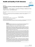
bp larger than in human. The results of PCR analysis of
two dissected (Figure 1, lanes 9–18) and two whole (lanes
19 and 20) mouse livers are shown in Figure 1. The extra
band that is amplified in the mouse samples was also
cloned, sequenced and identified as a gene of unknown
function found on the X chromosome. The dissection of
the liver F685 demonstrated the heterogeneous nature of
the chimeric livers, which varied between 4 and 80%
human. Typically, samples which contain between 70 and
88% human tissue could be obtained from each mouse.
Hybridization of mouse liver RNA on human
oligonucleotide microarrays in the presence of human liver
RNA
While samples containing relatively high percentages of
human tissue could be isolated from the chimeric livers,
there was still a variable level of contaminating mouse tis-
sue. Therefore, RNA isolated from these samples for use in
microarray experiments will contain RNA from mouse
hepatocytes. To identify genes affected by mouse liver
(ML) RNA hybridization, RNA from identical human liver
(HL) samples labeled with either Cy3 or Cy5 was com-
pared in the presence and absence of ML RNA.
A commercially available supply of normal HL RNA was
obtained from Ambion. The chimeric mouse model is uti-
lized primarily to study hepatotropic viruses and therefore
genes involved in the innate antiviral immune response
are of particular interest. To ensure that the cross-hybridi-
zation of these genes could be assessed, mouse liver RNA
from a transplanted HCV-infected SCID-uPA mouse was
used for the microarray experiments. This ML RNA was
100% mouse, based on the PCR assay, and RT-PCR anal-
ysis with mouse specific probes confirmed the expression
of interferon-regulated genes (data not shown). Table 1
shows the design of each microarray experiment. Genes
were selected as being differentially expressed based on 2
criteria, a greater than 95% probability of being differen-
tially expressed (P ≤ 0.05) and a fold change of at least 1.5-
fold or 2-fold. Typically, a 2-fold change (P ≤ 0.05) is used
to select differentially expressed genes in all microarray
experiments [11,21].
In the first experiment, HL labeled with either Cy3 or Cy5
fluorescent dye was compared in the absence of any ML
RNA. As the human liver samples are identical, the level of
differential gene expression between the Cy3 and Cy5
labeled samples should be minimal. As shown in Table 1,
this is indeed the case. In total, the expression ratios were
measured for 20,163 genes and no genes were selected as
Determination of the relative amount of human and mouse SCS-alpha genesFigure 1
Determination of the relative amount of human and mouse SCS-alpha genes. Genomic DNA isolated from dis-
sected chimeric livers (F685, F695, F552, F524) was amplified by PCR with primers specific for SCS-α as described in Materials
and Methods. A standard curve was generated using varying rations of human and mouse SCS-α containing plasmids (results
shown in lanes 1–8). The percentage of sample which is human is indicated below each lane number. The results of PCR of
pure mouse (ML) and human (HL) genomic liver DNA are shown in lanes 21 and 22, respectively.
Plasmid Ratio
Mouse:Human
1
:
8
2
:
7
3
:
6
4
:
5
5
:
4
6
:
3
7
:
2
8
:
1
F685
Nodules
F552
F524
ML HL
Mouse
452 bp
Human
384 bp
1 2 3 4 5 6 7 8 9 10 11 12 13 14 15 16 17 18 19 20 21 22 Lane
80 4 78 51 59 16 - - - - 59 46 % human
F695
Nodules
Virology Journal 2006, 3:37 />Page 4 of 8
(page number not for citation purposes)
being differentially expressed based on both 1.5 and 2-
fold changes.
Using this experiment as a base, various amounts of ML
RNA labeled with a single dye (either Cy3 or Cy5) was
added to the HL versus HL experiment. It was expected
that if the mouse RNA does not hybridize to the human
oligonucleotide probes, there should be no change in the
number of differentially expressed genes. However, if the
mouse liver RNA is hybridizing to the probes there should
be an increase in the number of differentially expressed
genes as the mouse RNA is present in only a single dye. In
this way, both the number and the identity of the genes
which are affected by ML RNA hybridization can be
assessed. Initially, a relatively high amount of mouse RNA
was spiked into the HL vs HL experiment, where 50% of
either Cy3 or Cy5-labeled RNA was ML RNA. This amount
of mouse RNA is substantially higher than what would be
considered acceptable for an experiment comparing the
gene expression in HCV-infected and naïve animals.
Therefore, it should give an indication of the highest level
of hybridization that can be expected. This experiment
was done in two slightly different ways. In one, the total
amount of HL RNA was kept constant: 1 µg HL-Cy3 and 1
µg HL-Cy5 + 1 µg ML RNA labeled with either Cy3 or Cy5.
In the other experiment, the amount of RNA for each
probe was kept constant: 1 µg HL-Cy3 versus 0.5 µg HL-
Cy5 + 0.5 µg ML-Cy5 (or flip dye experiment). This exper-
iment more closely represents how the actual array exper-
iments are done where equal amounts of total RNA are
used for each probe, rather than normalizing to the
amount of human RNA. However, both experiments were
done as it was unclear whether the amount of human
RNA labeled for each probe had to be equal to obtain the
very small number of differentially expressed genes
observed in the HL versus HL experiment. Again, genes
were selected as being differentially expressed based on
the criteria of at least a two-fold change and P value ≤
0.05. Unlike the experiment comparing only HL labeled
with Cy3 and Cy5, the addition of the ML RNA increased
the number of differentially expressed genes substantially.
The presence of the ML RNA increased the number of up-
regulated genes (2-fold) to either 1142 or 1132 (Table 1),
indicating that the mouse RNA was hybridizing to the
human oligonucleotide probes. However, even with 50%
mouse RNA, the number of genes affected was relatively
small, less than 5% of the total number of genes on the
slide. Comparison of the two experiments showed a very
high correlation coefficient on common signature genes,
0.99 (data not shown).
Reduction of mouse liver RNA hybridization to human
oligonucleotide microarrays
The addition of 50% mouse liver RNA increased the
number of up-regulated genes (2-fold) in a HL versus HL
experiment from 0 to approximately 1100, indicating that
the mouse RNA was likely hybridizing to the oligonucle-
otide probes. However, samples could be obtained from
HCV-infected mice that contain substantially lower per-
centages of mouse tissue, from 15–30%. To determine if
less cross-hybridization would occur in these samples, the
previous experiment was repeated with the exception that
20% ML RNA labeled in only a single dye was spiked into
the HL versus HL experiment. As shown in Table 1, a sub-
stantially smaller number of up-regulated genes (2-fold),
483, were observed in this experiment.
In an attempt to further reduce the amount of ML-RNA
hybridization, the specificity of the reaction was increased
by using more stringent hybridization conditions.
Increasing the hybridization temperature from 60°C to
65°C further reduced the number of up-regulated genes
(2-fold) to 328 (Table 1). The two experiments were com-
pared to ensure that the hybridization of the human liver
RNA was not significantly affected by the higher tempera-
ture. The correlation coefficient on common signature
genes is very high, 0.97, indicating that the effect on
human RNA hybridization was minimal (data not
shown). In addition, comparison of up-regulated genes
showed that the majority of the genes are common to
Table 1: Experimental Design for Microarray Experiments
Experiment Hybridization
Temperature (°C)
*Channel 1 *Channel 2 **Up-Regulated Genes
HL ML HL ML 2-fold 1.5-fold
1601-1-00
2 60 1 1 1 - 1142 2388
3 60 0.5 0.5 1 - 1132 2401
4 60 0.8 0.2 1 - 483 959
5 65 0.8 0.2 1 - 328 646
HL, human liver, ML, mouse liver,
* numbers indicate ug of fluorescent labeled RNA
** The expression ratio for up-regulated genes have a P value ≤ 0.05 based on n = 2 for each experiment and Rosetta Resolver error-model specific
for Agilent 22 K Human oligonucleotide microarray.
Virology Journal 2006, 3:37 />Page 5 of 8
(page number not for citation purposes)
both experiments (data not shown), further suggesting
that these up-regulated genes were caused by cross-hybrid-
ization of ML- RNA.
Validation of gene expression changes for genes known to
be affected by hybridization of mouse liver RNA
Real-time quantitative RT-PCR using species-specific
probes can differentiate whether the gene expression
change observed by microarrays is the result of viral-
induced changes in human hepatocytes or simply hybrid-
ization of mouse liver RNA. Three lipid metabolism
genes, CACH1, FABP1, and HMGCS2, shown to be
affected by hybridization of ML-RNA on the human arrays
were chosen to determine whether the gene expression
changes measured on the arrays was originating from the
human tissue or the mouse tissue. The species specificity
of each probe was first confirmed by performing real time
quantitative PCR analysis of commercially available
human and mouse liver RNA samples (data not shown).
These probes were then used to determine the expression
levels of these genes in HCV-infected chimeric samples
relative to donor-matched uninfected samples and the
results compared to the data obtained from the corre-
sponding microarray experiments. For both the microar-
ray experiments and quantitative RT-PCR, the expression
of these genes in HCV-infected animals was compared to
the respective uninfected controls. The microarray experi-
ments showed increased expression of these genes in
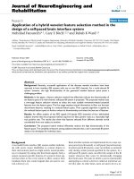
Mouse 1 and, to a lesser extent, Mouse 2 (Figure 2A).
Mouse 3 showed decreased expression of CACH1 and
HMGCS2 and essentially no change in FABP1 (Figure 2A).
Quantitative RT-PCR demonstrated a change in expres-
sion of these genes in both the mouse and human liver tis-
sue, although in general the change was more dramatic in
the human tissue (Figure 2B). It is possible that increased
the mouse hepatocytes are simply responding to increased
lipid/cholesterol released from the HCV-infected human
hepatocytes. Importantly, the results obtained using the
human-specific probes generally demonstrated a good
correlation with the gene expression data from the micro-
arrays, although the ratios calculated from quantitative
RT-PCR generally exceeded those obtained using microar-
rays. This indicates that the cross-hybridization of the ML-
RNA is not adversely effecting the gene expression data
generated using the microarray, at least with respect to the
three genes analyzed.
Bioinformatics analysis
To identify potential cross-hybridization candidates in sil-
ico, 20,143 Agilent Human 1A (V2) 60 mer oligonucle-
otide DNA sequences were searched using NCBI BLASTN
[26] for similar sequences within a 26,940 NCBI mouse
RefSeq [27] DNA sequence database, selecting the best
sequence match for each 60 mer with a BLAST expectation
value threshold of 10-3. 6,410 mouse RefSeq transcripts
representing 5,839 distinct genes showed significant iden-
tity to the 60 mer human probes. Average alignment
length for the 6,410 matches was 51/60 base pairs with
average identity of 92 percent. Of the 1124 genes showing
2-fold differential regulation at P value ≤ 0.05 with 50%
mouse RNA, 789 matched mouse RefSeq transcripts using
the above BLAST parameters with average alignment
length of 53/60 base pairs and average identity of 94 per-
cent. Of the 483 genes showing 2-fold differential regula-
tion at P ≤ 0.05 with 20% mouse RNA, 375 matched
mouse RefSeq transcripts with an average alignment
length of 53/60 base pairs and average identity of 94 per-
cent.
Conclusion
Chimeric systems are extremely valuable tools to study
human viral disease. They do, however, present a chal-
lenge for the use of genomics. While many studies have
examined cross-species hybridization on multiple array
platforms, including oligonucleotide microarrays [22-24],
none have investigated the effect of this cross-species
hybridization on gene expression studies using chimeric
tissue. The results of the current study demonstrate that
Expression of lipid metabolism genes in human and mouse liver tissueFigure 2
Expression of lipid metabolism genes in human and
mouse liver tissue. A. Comparison of gene expression lev-
els of lipid metabolism genes by either A. Microarray analysis
or B. Real-time PCR analysis using either human (solid bars)
or mouse (dotted bars)-specific probes. Data is shown as
log
10
ratio and reflects the difference in expression between
donor-matched HCV-infected and naïve liver tissue. Donor-
matched samples contained approximately the same percent-
age of human tissue. The three HCV-infected mice were
transplanted with hepatocytes from different donors.
-0.5
0
0.5
1
1.5
Mouse 1 Mouse 2 Mouse 3
HuCACH1
MmCACH1
HuFABP1
MmFABP1
HuHMGCS2
MmHMGCS2
Log
10
Ratio
-0.2
0
0.2
0.4
0.6
Mouse 1 Mouse 2 Mouse 3
CACH1
FABP1
HMGCS2
Log
10
Ratio
A
B
Virology Journal 2006, 3:37 />Page 6 of 8
(page number not for citation purposes)
functional genomics can be applied to the HCV-infected
chimeric SCID-beige/Alb-uPA mouse model and outline a
series of experiments that can be adapted to study the level
of cross-species hybridization in any chimeric tissue, thus
facilitating the use of genomics in these model systems.
While mouse liver mRNA from the SCID-beige/Alb-uPA
mouse did hybridize to the human oligonucleotide
microarray, the number of genes affected was relatively
small even when high percentages of mouse liver RNA was
present. This hybridization could be reduced by increas-
ing the stringency of the hybridization and also by using
liver samples that contained at least 80% human tissue.
Furthermore, species-specific quantitative RT-PCR dem-
onstrated that the hybridization of mouse RNA, in gen-
eral, did not significantly affect the human gene
expression measurement obtained from the microarray
analysis, at least with respect to the three genes analysed.
However, it is likely still important to use RT-PCR to vali-
date the expression level of known cross-hybridizing
genes as the extent of hybridization may vary between dif-
ferent probes.
Another way of examining cross-hybridization of the
mouse liver mRNA would be to apply the mouse mRNA
to the microarray in the absence of human mRNA. How-
ever, the purpose of this study was to determine the feasi-
bility of performing gene expression on chimeric tissue. It
was not simply to demonstrate that mouse liver RNA
would hybridize to human oligonucleotide microarray,
but rather to determine how extensive the hybridization
was in the presence of excess human RNA. In addition, the
platform of microarray used for these studies was a two
channel system, meaning that both the reference and
experimental samples are hybridized on the same micro-
array. As such, it generates ratio, rather than intensity,
gene expression data which makes it difficult to determine
simply the presence or absence of a transcript.
It is important to note that this gene set was derived exper-
imentally and so it is possible that additional genes which
are not expressed in the liver will also be affected by
hybridization of mouse RNA. Previous attempts, with
other array platforms, have been made to use bioinfor-
matics methods to "mask" or eliminate those probes from
further analysis which exceed a threshold of mismatching
base pairs between a probe and its corresponding tran-
script sequence [25]. Our analysis shows only a very small
difference in average alignment length and percent iden-
tity between genes showing significant differential regula-
tion with ML spike-ins and genes matching probes in
general. This emphasizes the importance of identifying
the genes affected by cross-hybridization experimentally
as opposed to using bioinformatics methods. While the
experiments outlined in this study were conducted specif-
ically to study virus-host interactions using the SCID-
beige/Alb-uPA mouse model of HCV infection, they can
be adapted to study the level of cross-species hybridiza-
tion in any chimeric tissue, thus facilitating the use of
genomics in these model systems.
Methods
Dissection of mouse livers and isolation of RNA and
genomic DNA
All mice were treated according to Canadian Council on
Animal Care guidelines. SCID-beige/Alb-uPA mice were
transplanted with human primary hepatocytes and then
infected with HCV as described previously [4,6]. Mice
were euthanized by cervical dislocation and the livers
were excised, dissected into small pieces and then snap
frozen in N
2(l)
. Total RNA was then isolated using a stand-
ard Trizol procedure. Genomic DNA was isolated from
the interphase and phenol/chloroform phase according to
the manufacture's specifications (Invitrogen). Both RNA
and genomic DNA were isolated from the same samples.
PCR of human and mouse genomic DNA
Genomic DNA from the liver of a SCID-beige/Alb-uPA
mouse containing human hepatocytes was amplified by
PCR with primers specific for the fourth and fifth exons of
the gene for the Succinyl-CoA synthetase alpha subunit
(SCS-α). The PCR conditions were: 0.01% (w/v) gelatin,
50 mM KCl, 1.5 mM MgCl
2
, 10 mMTris pH8.3, 0.2 mM
each of dATP, dGTP, dCTP, dTTP, 2.5 U of Taq polymer-
ase, and 0.25 µM of each of 5' ttgtgaatatggccaggcatg, (SCS-
ex5) and 5' caggagcaacggcttctgtc (SCS-ex4). The under-
lined nucleotide denotes the position of the nucleotide
that is present in the human sequence but not in the
mouse sequence. The PCR cycles were: 5 minutes at 95°C,
followed by 30 cycles of (30 seconds at 95°C, 30 seconds
at 45°C, 30 seconds at 72°C). The resulting PCR products
were separated by electrophoresis in a 1.5% agarose gel,
excised, purified using a Qiaquick gel extraction kit (Qia-
gen), cloned into pCR4 TOPO according to manufactures
specifications (Invitrogen) and sequenced. The cloned
fragments from mouse and human were 452 bp and 384
bp, respectively. For measurement of the relative amount
of human and mouse SCS-α the PCR conditions were as
follows: 1.2 µg of genomic DNA, 20 mM Tris pH 8.4, 50
mM KCl, 2 mM MgCl
2
, 0.2 mM each of dATP, dGTP,
dCTP, dTTP, 2.5 U of Taq polymerase, and 0.25 µM of
each of SCS-ex4 and SCS-ex5. PCR cycles were: 10 minutes
at 95°C followed by 40 cycles of (45 seconds at 95°C, 45
seconds at 68°C, 45 seconds at 72°C) and a final exten-
sion of 10 minutes at 72°C. The cloned plasmids were
used as standards, which were varied between 3.5 pg
human: 28 pg mouse and 28 pg human: 3.5 pg mouse per
reaction. To determine the relative amount of human
SCS-α gene in the sample, the PCR products were sepa-
rated by electrophoresis using a 2.5% agarose gel and then
stained with ethidium bromide. The bands were quanti-
Virology Journal 2006, 3:37 />Page 7 of 8
(page number not for citation purposes)
fied, a correction was made for the size difference, and the
results were plotted on a standard curve generated by the
plasmid controls.
Expression microarray format and data analysis
Microarray format, protocols for probe labeling, and array
hybridization are described at ro
slu.washington.edu. Human and mouse liver RNA were
labeled separately and then mixed prior to hybridization.
Human 1A Oligo Microarray (V2), which contains over
20,000 60-mer probes corresponding to over 18,000
human genes, were purchased from Agilent. Briefly, a sin-
gle experiment comparing two mRNA samples was done
with four replicate Human 1A (V2) 22 K oligonucleotide
expression arrays (Agilent Technologies) using the dye
label reverse technique. This allows for the calculation of
mean ratios between expression levels of each gene in the
analyzed sample pair, standard deviation and P values for
each experiment. Spot quantitation, normalization and
application of a platform-specific error model was per-
formed using Agilent's Feature Extractor software and all
data was then entered into a custom-designed database,
Expression Array Manager, and then uploaded into
Rosetta Resolver System 4.0.1.0.10 (Rosetta Biosoftware,
Kirkland, WA) and Spotfire Decision Suite 7.1.1 (Spotfire,
Somerville, MA).
Quantitative RT-PCR
Quantitative real-time PCR (rtPCR) was used to validate
the gene expression changes. Total RNA samples were
treated with RNAse-free DNase Treatment and Removal
Reagents (Ambion, Austin, TX). Reactions were per-
formed in quadruplicate on the ABI 7500 Real Time PCR
System (Applied Biosystems, Foster City, CA), using Taq-
Man chemistry with primer and probe sets selected from
the Assays-on-Demand product list (Applied Biosystems)
and two endogenous controls, GAPDH and 18S ribos-
omal RNA. Quantification of each gene, relative to the cal-
ibrator, was calculated by the instrument, using the
equation: 2-∆∆CT within the Applied Biosystems
Sequence Detections Software version 1.3.
Abbreviations
SCID, severe-combined immunodeficiency, PBMC,
peripheral blood mononuclear cells, HCV, Hepatitis C
virus, uPA, urokinase plasminogen activator, SCS-α, succi-
nyl-CoA synthetase, Cy, cyanine, CACH1, cytosolic acetyl-
CoA hydrolase, FABP1, fatty acid binding protein 1,
HMGCS2, HMG-CoA synthetase 2
Authors' contributions
KW participated in the design of the study, carried out
microarray experiments and drafted the manuscript. MJ
participated in sample preparation and characterization,
JC conducted the quantitative RT-PCR analysis, MS, SP
and JF participated in experimental design. JW performed
the sequence alignments and bioinformatics analysis. LT
and MK coordinated the study and helped to draft the
manuscript.
Acknowledgements
Financial Support: This work was supported by funding from the Cana-
dian Institute of Health Research and grants 5R01CA074131,
5R01A1049168, 5U19AI48214, 1P30DA01562501, 1R37DA004334,
R01DA12568, and R01DA16078 from the National Institutes of Health.
K.A.W. is supported by a Canadian Institute for Health Research Fellow-
ship.
References
1. Alter HJ, Seeff LB: Recovery, persistence, and sequelae in hep-
atitis C virus infection: a perspective on long-term outcome.
Semin Liver Dis 2000, 20:17-35.
2. Alter MJH, Margolis HS, Krawczynski K, Judson F, Mares A, Alexander
WJ, Hu PY, Miller JK, et.al.: The natural history of community-
acquired hepatitis C in the United States. N Engl J Med 1992,
321:1494-1500.
3. Lindenbach BD, Rice CM: Unravelling hepatitis C virus replica-
tion from genome to function. Nature 2005, 436:933-938.
4. Mercer DF, Schiller DE, Elliott JF, Douglas DN, Hao C, Rinfret A,
Addison WR, Fischer KP, Churchill TA, Lakey JR, Tyrrell DL, Knete-
man NM: Hepatitis C virus replication in mice with chimeric
human livers. Nat Med 2001, 7:927-933.
5. Meuleman P, Libbrecht L, De VR, de HB, Gevaert K, Vandekerckhove
J, Roskams T, Leroux-Roels G: Morphological and biochemical
characterization of a human liver in a uPA-SCID mouse chi-
mera. Hepatology 2005, 41:847-856.
6. Hsu EC, Hsi B, Hirota-Tsuchihara M, Ruland J, Iorio C, Sarangi F, Diao
J, Migliaccio G, Tyrrell DL, Kneteman N, Richardson CD: Modified
apoptotic molecule (BID) reduces hepatitis C virus infection
in mice with chimeric human livers. Nat Biotechnol 2003,
21:519-525.
7. Lindenbach BD, Meuleman P, Ploss A, Vanwolleghem T, Syder AJ,
McKeating JA, Lanford RE, Feinstone SM, Major ME, Leroux-Roels G,
Rice CM: Cell culture-grown hepatitis C virus is infectious in
vivo and can be recultured in vitro. Proc Natl Acad Sci U S A 2006,
103:3805-3809.
8. Smith MW, Walters KA, Korth MJ, Fitzgibbon M, Proll S, Thompson
JC, Yeh MM, Shuhart MC, Furlong JC, Cox PP, Thomas DL, Phillips JD,
Kushner JP, Fausto N, Carithers RLJ, Katze MG: Gene expression
patterns that correlate with hepatitis C and early progres-
sion to fibrosis in liver transplant recipients. Gastroenterology
2006, 130:179-187.
9. Chen L, Borozan I, Feld J, Sun J, Tannis LL, Coltescu C, Heathcote J,
Edwards AM, McGilvray ID: Hepatic gene expression discrimi-
nates responders and nonresponders in treatment of chronic
hepatitis C viral infection. Gastroenterology 2005, 128:1437-1444.
10. Smith MW, Yue ZN, Geiss GK, Sadovnikova NY, Carter VS, Boix L,
Lazaro CA, Rosenberg GB, Bumgarner RE, Fausto N, Bruix J, Katze
MG: Identification of novel tumor markers in hepatitis C
virus-associated hepatocellular carcinoma. Cancer Res 2003,
63:859-864.
11. Smith MW, Yue ZN, Korth MJ, Do HA, Boix L, Fausto N, Bruix J, Car-
ithers RLJ, Katze MG: Hepatitis C virus and liver disease: global
transcriptional profiling and identification of potential mark-
ers. Hepatology 2003, 38:1458-1467.
12. Camacho RE, Wnek R, Shah K, Zaller DM, O'Reilly RJ, Collins N, Fit-
zgerald-Bocarsly P, Koo GC: Intra-thymic/splenic engraftment
of human T cells in HLA-DR1 transgenic NOD/scid mice. Cell
Immunol 2004, 232:86-95.
Publish with BioMed Central and every
scientist can read your work free of charge
"BioMed Central will be the most significant development for
disseminating the results of biomedical research in our lifetime."
Sir Paul Nurse, Cancer Research UK
Your research papers will be:
available free of charge to the entire biomedical community
peer reviewed and published immediately upon acceptance
cited in PubMed and archived on PubMed Central
yours — you keep the copyright
Submit your manuscript here:
/>BioMedcentral
Virology Journal 2006, 3:37 />Page 8 of 8
(page number not for citation purposes)
13. yash-Rashkovsky M, Bentwich Z, Arditti F, Friedman S, Reisner Y,
Borkow G: A novel small animal model for HIV-1 infection.
FASEB J 2005, 19:1149-1151.
14. Zhang C, Cui Y, Houston S, Chang LJ: Protective immunity to
HIV-1 in SCID/beige mice reconstituted with peripheral
blood lymphocytes of exposed but uninfected individuals.
Proc Natl Acad Sci U S A 1996, 93:14720-14725.
15. Shah G, Azizian M, Bruch D, Mehta R, Kittur D: Cross-species com-
parison of gene expression between human and porcine tis-
sue, using single microarray platform preliminary results.
Clin Transplant 2004, 18 Suppl 12:76-80.:76-80.
16. Moody DE, Zou Z, McIntyre L: Cross-species hybridisation of pig
RNA to human nylon microarrays. BMC Genomics 2002, 3:27.
17. Adjaye J, Herwig R, Herrmann D, Wruck W, BenKahla A, Brink TC,
Nowak M, Carnwath JW, Hultschig C, Niemann H, Lehrach H:
Cross-species hybridisation of human and bovine ortholo-
gous genes on high density cDNA microarrays. BMC Genomics
2004, 5:83.
18. Dowling RJ, Bienzle D: Gene-expression changes induced by
Feline immunodeficiency virus infection differ in epithelial
cells and lymphocytes. J Gen Virol 2005, 86:2239-2248.
19. Wang Z, Dooley TP, Curto EV, Davis RL, VandeBerg JL: Cross-spe-
cies application of cDNA microarrays to profile gene expres-
sion using UV-induced melanoma in Monodelphis domestica
as the model system. Genomics 2004, 83:588-599.
20. Baskin CR, García-Sastre A, Tumpey TM, Bielefeldt-Ohmann H,
Carter VS, Nistal-Villán E, Katze MG: Integration of clinical data,
pathology, and cDNA microarrays in influenza virus-infected
pigtailed macaques (Macaca nemestrina). J Virol 2004,
78:10420-10432.
21. Smith MW, Walters KA, Korth MJ, Fitzgibbon M, Proll SC, Thompson
JC, Yeh MM, Shuhart MC, Furlong JC, Cox PP, Thomas DL, Phillips JD,
Kushner JP, Fausto N, Carithers RL, Katze MG: Gene expression
patterns that correlate with hepatitis C and early progres-
sion to fibrosis in liver transplant patients. Gastroenterol 2005:
In press
22. Ji W, Zhou W, Gregg K, Yu N, Davis S, Davis S: A method for
cross-species gene expression analysis with high-density oli-
gonucleotide arrays. Nucleic Acids Res 2004, 32:e93.
23. Gilad Y, Rifkin SA, Bertone P, Gerstein M, White KP: Multi-species
microarrays reveal the effect of sequence divergence on
gene expression profiles. Genome Res 2005, 15:674-680.
24. Sartor MA, Zorn AM, Schwanekamp JA, Halbleib D, Karyala S, Howell
ML, Dean GE, Medvedovic M, Tomlinson CR: A new method to
remove hybridization bias for interspecies comparison of
global gene expression profiles uncovers an association
between mRNA sequence divergence and differential gene
expression in Xenopus. Nucleic Acids Res 2006, 34:185-200.
25. Naef F, Huelsken J: Cell-type-specific transcriptomics in chi-
meric models using transcriptome-based masks. Nucleic Acids
Res 2005, %19;33:e111.
26. Altschul SF, Madden TL, Schaffer AA, Zhang J, Zhang Z, Miller W, Lip-
man DJ: Gapped BLAST and PSI-BLAST: a new generation of
protein database search programs. Nucleic Acids Res 1997,
25:3389-3402.
27. Pruitt KD, Tatusova T, Maglott DR: NCBI Reference Sequence
(RefSeq): a curated non-redundant sequence database of
genomes, transcripts and proteins. Nucleic Acids Res 2005,
33:D501-D504.