Báo cáo hóa học: " Members of the Hyposoter didymator Ichnovirus repeat element gene family are differentially expressed in Spodoptera frugiperda" docx
Bạn đang xem bản rút gọn của tài liệu. Xem và tải ngay bản đầy đủ của tài liệu tại đây (377.95 KB, 11 trang )
BioMed Central
Page 1 of 11
(page number not for citation purposes)
Virology Journal
Open Access
Research
Members of the Hyposoter didymator Ichnovirus repeat element gene
family are differentially expressed in Spodoptera frugiperda
L Galibert
1
, G Devauchelle
2
, F Cousserans
1
, J Rocher
1
, P Cérutti
2
, M Barat-
Houari
1
, P Fournier
1
and AN Volkoff*
1
Address:
1
UMR1231 INRA-UMII Biologie Intégrative et Virologie des Insectes (BIVI), Place Eugène Bataillon, Case Courrier 101, 34 095
Montpellier Cédex5, France and
2
UMR 5160 CNRS-UMI Baculovirus et Thérapie, 30 380 Saint Christol-lez-Alès, France
Email: L Galibert - ; G Devauchelle - ; F Cousserans - ;
J Rocher - ; P Cérutti - ; M Barat-Houari - ;
P Fournier - ; AN Volkoff* -
* Corresponding author
Abstract
Background: The abundance and the conservation of the repeated element (rep) genes in
Ichnoviruses genomes suggest that this gene family plays an important role in viral cycles. In the
Ichnovirus associated with the wasp Hyposoter didymator, named HdIV, 10 rep genes were identified
to date. In this work, we report a relative quantitative transcription study of these HdIV rep genes
in several tissues of the lepidopteran host Spodoptera frugiperda as well as in the H. didymator wasps.
Results: The data obtained in this work indicate that, in the early phases of infection (24 hours),
HdIV rep genes each display different levels of transcripts in parasitized 2
nd
instar or HdIV-injected
last instar S. frugiperda larvae. Only one, rep1, is significantly transcribed in female wasps. Transcript
levels of the HdIV rep genes were found as not correlated to their copy number in HdIV genome.
Our results also show that HdIV rep genes display different tissue specificity, and that they are
primarily transcribed in S. frugiperda fat body and cuticular epithelium.
Conclusion: This work is the first quantitative analysis of transcription of the ichnovirus rep gene
family, and the first investigation on a correlation between transcript levels and gene copy numbers
in Ichnoviruses. Our data indicate that, despite similar gene copy numbers, not all the members of
this gene family are significantly transcribed 24 hours after infection in lepidopteran larvae.
Additionally, our data show that, as opposed to other described HdIV genes, rep genes are little
transcribed in hemocytes, thus suggesting that they are not directly associated with the disruption
of the immune response but rather involved in other physiological alterations of the infected
lepidopteran larva.
Background
Polydnaviruses are obligatory endosymbionts of some
endoparasitic Hymenoptera from Ichneumonid and Bra-
conid families. They are integrated as provirus in wasp
chromosomes. Viral replication occurs in calyx cells of the
wasp ovary, and leads to the formation of multiple circu-
lar dsDNA encapsidated molecules. Viral particles accu-
mulate in the oviducts and are injected through
oviposition in the lepidopteran host larva.
Published: 19 June 2006
Virology Journal 2006, 3:48 doi:10.1186/1743-422X-3-48
Received: 07 February 2006
Accepted: 19 June 2006
This article is available from: />© 2006 Galibert et al; licensee BioMed Central Ltd.
This is an Open Access article distributed under the terms of the Creative Commons Attribution License ( />),
which permits unrestricted use, distribution, and reproduction in any medium, provided the original work is properly cited.
Virology Journal 2006, 3:48 />Page 2 of 11
(page number not for citation purposes)
Polydnaviruses do not replicate in the parasitized lepi-
dopteran host, but infect several host tissues, what leads
to viral gene expression in these tissues. Polydnaviruses
induce major physiological alterations in parasitized host
such as immune disruption, developmental arrest, hor-
monal alterations and a decrease in hemolymph storage
proteins [1-5].
Recent sequencing programs of the polydispersed polyd-
navirus genomes reveal that a large proportion of the
genes encoded by the circular DNA molecules are organ-
ized in gene families. This characteristic is common to the
two polydnavirus families, the Ichnoviruses (IV), associ-
ated with ichneumonid wasps, and the Bracoviruses (BV),
associated with braconid wasps [6,7]. We are studying the
Ichnovirus associated with the endoparasitoid wasp Hypo-
soter didymator (HdIV), where several gene families have
been identified so far [7,8]. Although only a fraction of
HdIV genome is presently known, 10 members of a gene
family named repeated element (rep) gene family have
already been identified in the genome. Originally
described by Theilmann & Summers [9] on the basis of
multiple cross-hybridization between several Campoletis
sonorensis IV (CsIV) genome segments, members of the rep
family possess a conserved 540-bp repeated element
motif, found singly or in multiple repeats [9-12]. The rep
gene family is the largest conserved ichnovirus gene fam-
ily identified to date [7,13]. Indeed, 30 members of the rep
gene family have been reported in the fully sequenced
CsIV genome, whereas 36 and ten rep genes are described
in Hyposoter fugitivus IV (HfIV) and Tranosema rostrale IV
(TrIV), respectively [7].
Although the function of the rep genes has not yet been
elucidated, their conservation among ichnoviruses and
their abundance in viral genomes both suggest that they
play an important role in viral cycles. To date, transcrip-
tion studies for ichnoviruses rep genes have been carried
out by Northern blot analysis [12,14] or by RT-PCR [11]
and have indicated that members of this gene family may
be transcribed in both wasp and caterpillar hosts [11,14]
and in different tissues of the parasitized lepidopteran
host [12,14]. Variations in the number of transcripts dur-
ing the first day after parasitism have also been suggested
for members of this gene family by Northern-blot analysis
[14]. Altogether, these results seem to indicate that rep
genes show a wide range of expression patterns, making it
difficult to identify any putative physiological function.
Based on the abundance of rep genes in ichnoviruses
genomes, one might expect that they have diverged in
their expression pattern, acquiring specificity for given tis-
sues, hosts or development stages.
In this work, we report the relative quantitative transcrip-
tion study of the 10 rep genes identified to date in HdIV.
The transcription studies were carried out on several tis-
sues of the lepidopteran host Spodoptera frugiperda as well
as in H. didymator adult wasps. Our data indicate that 24
hours after infection HdIV rep genes display different lev-
els of transcription in parasitized or HdIV-injected S. fru-
giperda. Surprisingly, one of the rep genes, rep1, is
significantly transcribed in female wasps. However, rep
genes remain preferentially transcribed in the lepidop-
teran host compared to the wasp host. Our data show that
transcription levels of the HdIV rep genes are not corre-
lated to their copy number in HdIV genome. In addition,
each HdIV transcribed rep gene displays tissue specificity,
and the primary targets are the lepidopteran host fat body
and cuticular epithelium.
Results and discussion
In HdIV, 10 rep genes are identified at present. Three have
been previously described in HdIV segment SH-E (rep1,
rep2 and rep3, [12]) and one in segment SH-G (named
rep12 in this work, [15]). More recently, six additional
sequences have been identified, which are available in the
GenBank database (accession numbers in Table 4).
Genome distribution and sequence analysis of HdIV rep
genes
Characterisation of the segments containing the six new
rep genes (rep4, rep5, rep6, rep7, rep8 and rep11) was
achieved by Southern-blot analysis and PCR amplifica-
tion of the corresponding circular molecules.
Southern-blot of HdIV segmented genome was performed
with oligonucleotide probes specific to each of the newly
identified rep genes, except for rep6. Indeed, rep6 and
rep11 share 98 % nucleotide identity in their coding
sequence and thus the rep6 probe was expected to cross-
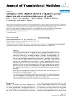
hybridize with rep11. As shown in Figure 1, the rep4
primer resulted in a hybridization band that co-localised
with the hybridization band obtained with the rep5
primer (Figure 1, compare rep4 and rep5 lanes). The rep6
probe hybridized with two HdIV segments, firstly with a
segment that co-localizes with the rep5 segment and sec-
ondly with another segment of lower size (Figure 1, rep6
lane). The faint hybridization band obtained with the rep7
probe had a size similar to that of the lower size segment
to which the rep6 probe hybridized (Figure 1, compare
rep7 and rep6 lanes). Lastly, the rep8 probe hybridized
with a segment of smaller size compared to the other rep
gene-containing HdIV segments. Thus, the Southern-blot
analysis indicated that the 6 new rep genes are encoded by
at least 3 different HdIV segments (Table 1).
The HdIV rep-encoding segments were further analysed by
PCR. Primers specific to the rep5 gene amplified a ~6 kbp
fragment, whereas those designed within the rep6 gene
amplified a ~5 kbp fragment. The HdIV super-helical (SH)
Virology Journal 2006, 3:48 />Page 3 of 11
(page number not for citation purposes)
segments are named alphabetically from the shortest to
the longest. Thus, based on their size, the segment con-
taining the rep5 gene was named SH-J and the one con-
taining rep6 was named SH-H. Presence of the rep5 and
rep6 genes was confirmed by partial sequencing of the two
PCR fragments (GenBank accession numbers DQ295920
and DQ295919, for the segments SH-J and SH-H respec-
tively). Sequencing revealed that SH-J also contained a
sequence corresponding to the rep11 gene, thus confirm-
ing Southern-blot analysis where the rep6 probe hybrid-
ized with SH-H and cross-hybridized with rep11 present
in SH-J (Figure 1, rep6 lane) whereas the rep5 probe
hybridized solely with SH-J (Figure 1, compare rep5 and
rep6 lanes). PCR using primers specific to the rep7 gene
resulted in a 3.1 kbp fragment. Sequencing of this PCR
fragment (GenBank accession number DQ295918
)
revealed a sequence identical to rep7. However, Southern-
blot analysis suggested that a larger segment encodes this
gene (Figure 1, rep7 lane). The discrepancy between the
two results could be explained by the existence of two seg-
Table 1: HdIV segments predicted to encode the rep genes analysed in this work. Segment names and putative sizes are indicated.
Segment names were given alphabetically from the shortest to the longest; however only segments for which a real (completely
sequenced; SH-E and SH-G) or estimated (PCR fragment; SH-J, SH-H and SH-A2 containing rep7 sequence) size could be given were
named. SH-x and SH-y stand for segments of unknown size (since molecular weigh marker represents linear DNA).Rep7 is underlined
because of discrepancy between PCR and Southern-blot results. For each segment, the rep gene(s) identified after sequencing of PCR
amplification fragments or by Southern-blot analysis are reported
segment size (kbp) sequencing/PCR Southern-blot
SH-J ~6 rep5, rep11 rep4
SH-y ~5 rep7?
SH-H ~5 rep6
SH-G 5.6 rep12
SH-E 4.6 rep1, rep2, rep3
SH-x ? rep8
SH-A2 3.1 rep7
Characterization of the HdIV genomic segments encoding the novel 6 rep genes by Southern-blot analysis with gene-specific oligonucleotide probesFigure 1
Characterization of the HdIV genomic segments encoding the novel 6 rep genes by Southern-blot analysis with gene-specific
oligonucleotide probes. The molecular weight marker corresponds to linear DNA (kb). Purified HdIV DNA was separated on
1% agarose gel and stained with BET (HdIV), then transferred to Nylon membrane for hybridization with oligonucleotide
probes specific to rep4 (rep4), rep5 (rep5), rep7 (rep7) and rep8 (rep8) genes. Due to high similarity between the rep6 and
rep11 coding sequences, the rep6 probe (rep6&11) should allow detection of both genes. SH-J, containing rep5 and rep11
genes, and SH-H, containing the rep6 gene, are indicated by vertical arrows.
10
8
6
5
4
3
2.5
rep7
rep8
rep5
HdIVkb
rep6
&11
rep4
SH-J
SH-H
Virology Journal 2006, 3:48 />Page 4 of 11
(page number not for citation purposes)
ments encoding this gene, similar to the rep1 gene, which
is encoded by both SH-E and SH-Evar [12]. However, this
result will need to be confirmed by identification and
sequencing of the rep7 hybridizing segment.
Therefore, based on both Southern-blot and PCR results,
we can conclude that the 10 HdIV rep genes are encoded
by at least 5 different HdIV molecules (Table 1).
Members of the rep family are characterized by a con-
served 540-bp repeated element motif, found singly or in
multiple repeats [9,11]. All the 10 rep genes identified to
date in HdIV encode proteins containing a single repeated
element motif (Figure 2). However, only part of the HdIV
genome is presently known and therefore we cannot
exclude existence of multiple-repeat containing genes in
this Ichnovirus.
All the rep genes described to date lack intron and encode
proteins with no predicted signal peptide [11,12,14]. The
HdIV deduced rep proteins analysed in this work follow
this rule. Moreover, immunofluorescence studies in cell
lines transfected with rep proteins coupled in their C-ter-
minal part to GFP confirm that the GFP-rep1, rep3 and
rep5 proteins are intracellular (Galibert et al., unpub.).
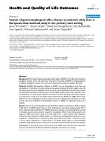
ClustalX alignment [16] of rep proteins from HdIV and
from other Ichnoviruses reveals a high degree of conserva-
tion in the repeated element motif (Figure 2). In contrast
to the repeat element motif, the N-terminal and C-termi-
nal sequences greatly diverge among the different rep
sequences. The close similarity between rep6 and rep11
(at both nucleotide and amino acid levels) suggests the
genes have diverged recently. Surprisingly, rep11 lacks the
C-terminal part of the repeated element motif, compared
to the other rep proteins.
Whole rep protein sequences of several Ichnoviruses con-
taining a single repeat and accessible on GenBank data-
base (10 HdIV, 2 HfIV, 1 TrIV and 25 CsIV proteins) were
aligned by ClustalX [16] to generate trees (data not
shown). Results did not indicate a clustering by virus spe-
cies, regardless of the method used, distance and parsi-
mony (PHYLIP package [17]), but rather a dispersion of
HdIV sequences among the other ichnovirus sequences
(data not shown). This distribution was different from
that seen in previous studies, comparing a lower number
of sequences, where rep proteins clustered by virus species
[12]. Phylogenetic analysis of this important and diversi-
fied gene family would require supplementary studies, in
order to understand if rep genes are derived from a single
ancestor gene or if several rep genes existed prior to the
association between an ichneumonid wasp and a polyd-
navirus ancestor.
Transcription in the parasitized lepidopteran host
Transcription of the 10 HdIV rep genes was analysed by
quantitative PCR during the first 24 hours following para-
sitism in S. frugiperda larvae parasitized at their 2
nd
instar.
Larvae parasitized by H. didymator rapidly exhibit reduced
food consumption and growth, and their development is
arrested at the end of the fourth larval instar, after the 8
days needed for completion of parasitoid larval develop-
ment.
Our data reveal that in the initial phases of parasitism
important differences are found between the transcript
levels of the different HdIV rep genes when considering
the overall expression of the genes (Figure 3A). The high-
est level of transcripts corresponds to the rep1 gene, fol-
lowed by the rep7, rep3 and rep2 genes. For example, 1
hour after parasitism, the ratios of rep1 transcripts (N0
value) to rep3, rep2 and rep7 transcripts are 8.4 ± 0.5, 7.0
± 0.2 and 5.4 ± 0.3, respectively; 24 hours after parasitism,
the ratios of rep1 compared to rep3, rep2 and rep7 are 8.2
± 0.3, 43.3 ± 0.3, and 14.2 ± 0.3, respectively. Because of
the high degree of identity between the rep6 and rep11
sequences, we were not able to design pairs of primers
specific for each of the genes. Therefore, the results
obtained in quantitative PCR include both genes. None-
theless, rep6 and rep11 transcript levels are generally simi-
lar to rep7 and significantly higher than other rep genes
such as rep4, rep8, rep5 or rep12. The rep5 and rep12 tran-
scripts are detected at very low levels in the parasitized lar-
vae (557.6 ± 0.3 fold and 1789.0 ± 0.3 fold respectively
less than rep1 at 24 h post-parasitism) suggesting that
transcription of these two genes in the lepidopteran host
may have no real biological significance.
As indicated in Figure 3A, transcript levels remain rela-
tively constant inside the whole parasitized S. frugiperda
larvae over the first 24 hours of parasitism, for each of the
HdIV rep genes with the exception of the rep3 gene, which
appears to have transcript levels that are 6-fold higher at
6–9 hours post-parasitism compared to other points in
the kinetic. These results are consistent with those
obtained by Theilmann & Summers [14] in CsIV who
observed through Northern blot experiments that some
rep genes were slightly more transcribed 2 h and 6 h after
parasitism than latter in parasitism (1d-8d). Nevertheless,
the biological significance of this peak of transcription for
the HdIV rep3 gene needs to be further investigated.
Transcript levels of the HdIV rep genes were also analysed
during the time course of parasitism (data not shown).
Preliminary results indicate that the differences in tran-
script levels between the HdIV genes are similar to those
observed in early phase of parasitism. Moreover, tran-
script levels remain constant over the duration of parasi-
toid development. Therefore, the 10 HdIV rep genes
Virology Journal 2006, 3:48 />Page 5 of 11
(page number not for citation purposes)
studied here do not show variations in the course of para-
sitism as it has been described for some bracovirus genes
[18].
In HdIV, the differences in transcript levels of the rep genes
inside the whole parasitized S. frugiperda larvae are not
related to their corresponding gene copy number in the
HdIV segmented genome. Indeed, rep1 is more tran-
scribed than both rep2 and rep3 although the 3 genes are
found on the same viral segments SH-E and SH-Evar [12]
and thus display the same gene copy numbers in the HdIV
genome. Different patterns and levels of transcripts in the
parasitized host for genes located on the same polydnavi-
rus segment have also been previously described for the
CsIV [19] and for the Bracovirus associated with Chelonus
inanitus [18]. The absence of a correlation between tran-
script level and gene copy number was further assessed by
estimating the relative copy numbers of each of the 10 rep
genes from purified HdIV DNA (Figure 3B). As expected,
our quantitative PCR assay revealed similar numbers of
ClutalX alignment of deduced amino acid sequences of HdIV and selected ichnoviruses rep genesFigure 2
ClutalX alignment of deduced amino acid sequences of HdIV and selected ichnoviruses rep genes. The first 2 letters indicate
ichnovirus species (Cs: Campoletis sonorensis; Hd: Hyposoter didymator; Hf: Hyposoter fugitivus; Tr: Tranosema rostrale), followed
by the name of the segment containing the corresponding gene (except for Hd ichnovirus) then the rep gene number. Arrow-
heads indicate beginning and ending of the conserved repeated element motif as defined by Theilmann & Summers [14]. Differ-
ent shades of grey indicate conserved residues. Consensus sequence represents conserved residues: in capital letters: residues
with >80% identity; p: polar residue; h: hydrophobic residue; l: aliphatic residue; +: positive residue; b: big residue; s: small res-
idue.
ź
Hdrep2 MNSSNNNVSRKTPAVPPLPASMNVSLSIKKIFQAAESLEFEEYQSFIQKLLHNMDMHPQIQAQLWRMSTHRFTA
Hdrep3 MTQYLSTPNQSTSEPVELPLRVIVYMARFLSVADYRSFVKSIWSDEIPSKTVRTKLWRMSTRKITT
Hdrep1 MESKENNVLPAFSVLQGAPLQREIVIPLDIILHLGDFLRFEDYRNFVKSIWPANDECDAVRNKLWQRSTHKIAI
Hdrep8 MSLFEE ESTSSRLSRAATRNGLHVPLDIILGMSEFLEFQDYHNFVRAFCPNGDEDEEVRAKLWQLSTHQVVT
Hdrep12 MSTPIPVVLRNKKASATRCSLLRRVAMKVLEYKTLQTHSPEN-VPGPLPSPFPYDIILHMSGYLKFEDYINFVRALWPHGDEDDSVRNKLWQLSTRSIIL
Hdrep5 MALQKD-EKSQRLTVDDFAKRGFRLPRGAPFNVNRYLNFD HEYRRFLLSTNKIEV
Hfc3rep MALRKE-KTTKKFSLDDFTKRGFRRPCGAPFNVSRYAMFD QEYRRFLLSTNKIEV
Hdrep6 MPFNEDHDRTHTYPVDTFTFRGPAMPNGAPFSFIEFIRFN VAYQQFLTSIRNLDL
Hdrep11 MPFHEDHDRTLTYPVDTFTSRGPAMPNGAPFNFAEFIRFN VAYQQFLTSIRNLDL
Hdrep4 MSSEESGVSQTVAVLEMG QHLEARTIDA
Hdrep7 MTMSPVERRMSQAVVYQEIS QDLKVSTIDA
Hfb15rep MGGRFRAFMTSLRKWISSSKTSQKASISPPCCDVVLYMSQFMPFEDFQNLVEAFWPNGGEDELIRQHLWKLSTRKYVT
CsBrep MESLETKESKVFTPVYFTSRQILLPLKMISYVSRFLKFEDFRKFIRAMWPNGEANVIFQELLERLSIRKFKA
TrFrep MRIIIGFESIHASSLLWPTEPSGRVRSFVQLRRIKRKRRTMMSPQ NEMSPPDVCLPNVLNVKNFEA
Consensus s hs h.h p.h b.bbhshpph.h
Hdrep2 TFLNGKPLVIRYNYDPSRLEEERVLFDVEYLFPVLGGVIPR ALSRFATASQIHSFVKKHVHLNRCADCEHAS-CPCDLG-HDQAQVRAFVQPAV D
Hdrep3 KFINGEPIEIEYNYDPGRIEEERVLINTKYLLPISGGIVAP VPKTFTSLTRINNFIQSAVELNVCSGHEYAC-CPCHLKNFNRHSATAFAKPSA T
Hdrep1 EFFNEEILNIEYNFDASRTKDQQFLFN VETLSPVFGGVVPP GTNQFLSASKLENFLRMHVHLNMCSRRQFAA-CSCHELKCGTYTGVKIVKPPK V
Hdrep8 EFLSGVRIPVIYNFNPWRREDEPLLIKVKSLSRIFGGIGAK LIDQFASVSTLHAFIEDHVHMDECSNLKYASSCLCHLGSHESSLGRTDPESPA G
Hdrep12 EFCNGKPLKVEYNYDPDRETEDRILINVENLLPTFGGAVP ARWEFVSVSQLRGFIKREIFFSKYP WARGALRNEENTSYTCKWSTH T
Hdrep5 TFFNGKSFQVLYNFDGTRTEEDRLLINWDTLTPLFGGVIPS GYRSFVRLPKIAKFVEKRIHLDQCEVGLHNS-CFCGRTPPDDLDIFW D
Hfc3rep TFFNNKSFQILYNFDAERPEEDRLLINWDTLTPLFGGVTPS GFRSFVRLAKIATFVEKRIHLDQCEVGLHNS-CFCERTPPDDLDIFW D
Hdrep6 TFLNGKACPVRYSFDATRPEEDRLLINWDWLMPLFWERPP GERDFVRLPVILQFVKN-IRFQKCKAVASDT-CSCEKKPNKVVSPGR E
Hdrep11 TFLNGKACPVRYSFDATRPEEDRLLINWDWLTPLFWERPP GERDFVRLPVILQFVKN-IRFQKCKAVASDT-CSCEKKPNKVVSPGL
Hdrep4 LFLNRKPLEVRYYFEDRGTEEPLIIIDVDSLRPIFKDVHRT TTKCFKPLDEISAFVRDNIQFNKCSNYQYAE-CVCHLLESRTDDPEFPELEGLPPN
Hdrep7 VFLNRKSLKIRYYCEDGGTEEPLIMLDVYSVKPILGN FLPR VAGSFVSLDNLCAFVKEEIHFDECWDYEYAD-CKCNLIGGRIVPPG-TKVQELPPS
Hfb15rep KFFNGKSLEVVYNYNTKRSKKDRILLNVKTLLPITGPIFPADTDVDELWMSPLELHDIVVTRFDMDKCWEYRYANCDCCERLHHTVEYPETFAEFCD I
CsBrep KFYNREEIEVEYKFDRERSGINWILINFKDLLPILGG IMLPD DEDKFQSIFTLEDFLKRNLKVHRCSGGIHTS CHNLGRDSDSDSE AKLD
TrFrep TFYNGRRLDIQYDFNPEKIVAERLRLNLESLLPLFGGIAPP GKAEFTTIDEISNFVNI-VNLDSCSSGIYAS-CSCHYNIPEKEQNLVYR
Consensus pFhN.c.h.lbYpass.+.pcpblllshc.L.Plhhshhs s pFhphspl Flpp.lphppC h ss.s.Cpb p s
ź
Hdrep2 ACHDRCFHHYCSQHVGYWLKLYLAPVVLLRERRASSADDRAAAESFLVFLSETVYFRGLNVQLRDSPLQSVPSWKRR
Hdrep3 ACASKHFHHYCSNHVNHWFNSFLNSVIRSQEGEEPFNED EAEIRLFLLDNMIYFRDNEIKLRSSHLYRVL
Hdrep1 ACRYGHFHHFCSQHVRDWVDIFLMSAVVKKEEGSPSDADMTKRLLAYMRDSVRLSGC
Hdrep8 SCPSGCFHHYCSQHLRYWLDVFLLPSIYS SP VFSSYR
Hdrep12 SKERSQSHPFRWKHVYWWVNEHLEPMITRRHQNSP
Hdrep5 SCSDQHFHHFCSLHVRSWLYLYLHPKILREESEQLFYETVWLGHSSNPDVLKYYATNNCKDTEILLDSARYLGYSSPCTRNRVKSL
Hfc3rep SCSDQHFHHFCSLHVRSWLWLYLHPKILSQESGPLFYNAVWNAHPSNLDVLQYYLMTGSKNTDILLDSARSLGHSSPSTRNRVKSLQMSF
Hdrep6 CKNELHWHHFCTAHVSAWLTKYMIPAILLKESKEMFTEIIANVHQNNPDVLEYYSMAERTDTQVLLDSARNSSGHGAA
Hdrep11
Hdrep4 DCPLGHFHHACSSCVNRWLNEYLRVLILLRESKPFFAKAAKEICSRVYQTDKFYEDHNQDCPEFYLRTAQRWT TLEYALDEMFVQ
Hdrep7 GCRR-HFHHACSSCVNSWLLEYLRMLILQRESEPAFAMAAEEICSRVLLSNDFGKCVHQVSSEFWLWIAQRWSLEGYVRYDVTNTWLRTTSASPITCKSL
Hfb15rep DCPYGHFHHYCVHHVSWWLMSYLHTSIQVQERRLAQPTPVPRSRRSFGYMLLRCWCIPAGAESRIVSDVFTSGSGVNDIGNLANL
CsBrep ICPFDHYHHFCPDHVIAWFKHYLLTAILLREGVYDELVKNANLPNADHLTSGRRRTEQYWLRVARRKKCRFSQ
TrFrep -CRDDHFHHYCASHVSAWFKLYLERAILLQDESKQFYYQLIADIHGPITAFEFTVGRYATAQYWLEYARTHVGRRRL
Consensus .p paHHhCs.pV Wh aL l pc
Virology Journal 2006, 3:48 />Page 6 of 11
(page number not for citation purposes)
gene copies for the genes encoded by the same segments,
rep1, rep2 and rep3. Furthermore, our results indicate that
rep1, for which a high level of transcripts was detected,
and rep12, a gene that is almost not transcribed, have sim-
ilar numbers of copies within the HdIV genome (Figure
3B, compare rep1 and rep12). Overall, our data indicate
that there are no significant differences within the HdIV
genome between the copy number for rep1, rep4, rep5,
rep7, rep8 and rep12. The rep6 and rep11 genes represent an
exception (Figure 3B, rep6&11). Since rep6 and rep11
genes are both amplified by rep6 primers, the N0 value
indicated in Figure 3B corresponds to the sum of rep6
(segment SH-H) and rep11 (segment SH-J) gene copies.
The proportion of the N0 value due to rep11 can be esti-
mated by the value obtained for rep5, since both genes are
on the same segment SH-H. This indicates that rep11 gene
(on SH-H segment) represents 15.5% of the total N0
value, whereas the rep6 gene (on SH-J segment) represents
84.5% of the N0 value. On the other side, quantification
of the signal intensity obtained on Southern-blot (Figure
1, column rep6&11) indicates that SH-J (containing rep6
gene) and SH-H (containing rep11 gene) represent 78%
and 15% hybridization signal, respectively. A third
hybridization signal with a high molecular weight seg-
ment, representing 7% of the total signal intensity, was
also detected in Southern-blot (Figure 1). Taken together,
our results indicate that SH-J, containing the rep6 gene, is
represented at least 5 times more than other rep-contain-
ing segments. Thus, although more abundant, rep6 is less
transcribed than rep1 in parasitized larvae, result that con-
firms absence of correlation between copy numbers and
transcription levels for the analysed HdIV rep genes.
To conclude, our results indicate that rep genes transcript
levels are variable inside the parasitized caterpillars and
are not linked to their relative copy numbers on HdIV
genome thus suggesting that transcript levels of the HdIV
rep genes are directly correlated to their promoter activi-
ties.
Transcription in the wasp host
Since some of the CsIV rep genes are transcribed in both
lepidopteran and hymenopteran hosts [11,14], we inves-
tigated transcription of HdIV rep genes in 2–3 days old H.
didymator female and male adult wasps. At this time, viral
replication is taking place in the calyx cells [20]. In the
female wasps, the rep genes are transcribed, but at a very
low level, with the exception of rep1, which was signifi-
cantly more transcribed compared to the other rep genes
(Figure 3C). In the male wasps, transcript level is more
than 200-fold lower than in females, suggesting that tran-
scription of HdIV rep genes is residual in male wasps. This
result differs from previous reports on C. inanitus bracov-
irus where 5 out of 6 analysed CiBV genes were tran-
scribed at similar levels in male and female wasps [18].
The finding that transcription of rep1 gene is restricted to
H. didymator females suggests an unexpected complex reg-
ulation of gene transcription, regardless transcripts are
generated from the integrated or from the excised viral
DNA. The remaining question is if rep1 transcription is
restricted or not to the replicative calyx cells and thus if it
may be related to HdIV viral particle production.
The HdIV rep1 gene is therefore the most transcribed rep
gene in both parasitized S. frugiperda and whole adult
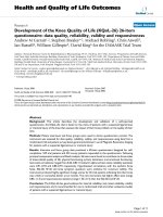
Expression profiles and gene copy number of the 10 rep genes identified in HdIV by relative quantitative PCRFigure 3
Expression profiles and gene copy number of the 10 rep
genes identified in HdIV by relative quantitative PCR. A.
Transcript levels in 2
nd
instar S. frugiperda parasitized larva,
over 1-h to 24-h time course study. B. Relative gene copy
numbers in HdIV genome. C. Transcript levels in H. didyma-
tor adult female and male wasps. D. Transcript levels in differ-
ent tissues of last instar S. frugiperda larvae 24 hours after
injection of HdIV (H: Hemocyte; FB: Fat Body; Ep: Cuticular
Epithelium; SN: Nervous System (Head); TD: Digestive
Track). Data are means ± SE of starting quantity of fluores-
cence (N0 value) for 6–9 measurements. For A, C and D,
data are normalized to housekeeping genes RNA polymerase
II and E2 ubiquitin ligase. For details, address to Methods
chapter.
0,00E+00
5,00E+00
1,00E+01
1,50E+01
2,00E+01
2,50E+01
3,00E+01
3,50E+01
4,00E+01
rep1 rep2 rep3 rep4 rep5 rep6&11 rep7 rep8 rep12
Gene Transcript Quantity Normalized to RNA
polymerase II & E2 ubiquitin ligase
H FB Ep SN TD
0,00E+00
5,00E-03
1,00E-02
1,50E-02
2,00E-02
2,50E-02
rep1 rep2 rep3 rep4 rep5 rep6&11 rep7 rep8 rep12
Gene Transcript Quantity Normalized to 18S
RNA
male wasps femal wasps
0,00E+00
2,00E-06
4,00E-06
6,00E-06
8,00E-06
1,00E-05
1,20E-05
1,40E-05
rep1 rep2 rep3 rep4 rep5 rep6&11 rep7 rep8 rep12
Gene Quantity (expressed in N0 number)
0,00E+00
2,00E-01
4,00E-01
6,00E-01
8,00E-01
1,00E+00
1,20E+00
1,40E+00
rep1 rep2 rep3 rep4 rep5 rep6&11 rep7 rep8 rep12
Gene Transcript Quantity Normalized to RNA
polymerase II & E2 ubiquitin ligase
1h 4h 6h 12h 24h
A
B
C
D
Virology Journal 2006, 3:48 />Page 7 of 11
(page number not for citation purposes)
female wasps. Whether transcription of rep1 gene is more
important in parasitized S. frugiperda larvae than in the
female wasp remains to be clearly established. By assum-
ing that reverse transcription and PCR efficiencies were
identical in the samples issued from both S. frugiperda lar-
vae and female wasps, we were able to compare the N0
values obtained. In both samples, non-normalized N0
values are around 4E-07, which indicates that rep1 tran-
script levels are similar in both insect hosts. We can there-
fore assume that transcription of the rep1 gene in female
wasps has a biological significance although it remains to
be clarified whether the related protein has a function in
the wasp and if this function is the same as that in the par-
asitized lepidopteran host.
Pattern of transcription in different tissues of HdIV-
infected S. frugiperda larvae
In order to assess if HdIV rep genes have tissue specific pat-
terns of transcription, quantitative analysis was performed
in different tissues of S. frugiperda last instar larvae. Our
results show that, 24 hours after HdIV injection, the HdIV
rep genes are preferentially transcribed in the fat body and
cuticular epithelium, and to a lower extent in the nervous
system of the infected host (Figure 3D). Finding of a pref-
erential transcription of the rep genes within these 3 tis-
sues is consistent with previous results obtained by
Northern-blot analysis for the HdIV rep1 gene [12].
In HdIV-injected last instar larvae, as in the parasitized 2
nd
instar larvae, rep4, rep5, rep8, rep12, but also rep7 show
very low transcript levels in all tissues examined, whereas
rep1, rep6, and to a lower extent, rep2 and rep3, are
detected at higher levels (Figure 3D). In this assay, where
tissues are analysed individually, rep1 transcripts are not
any longer the most abundant. Indeed, rep6 transcripts
level is similar to that of rep1 transcripts, in particular in
the fat body and cuticular epithelium (despite a high var-
iation between the biological samples for rep6 in cuticular
epithelium, as indicated by the standard error, Figure 3D).
This result has to be modulated by the fact that, in this
assay, both rep6 and rep11 transcripts were measured and
the proportion of each of the two genes is not known.
However, rep6 preferential transcription in HdIV-injected
last instar larvae fat body was corroborated by Northern
blot analysis using rep genes specific oligonucleotide
probes. Indeed, only one hybridization signal was
detected, which corresponded to the rep6 specific probe in
the fat body tissue (data not shown).
Our results indicate that the highest levels of rep gene tran-
scripts are detected in the fat body and the cuticular epi-
thelium (Figure 3D). Other ichnovirus genes of unknown
function, such as TrIV1, also target primarily the fat body
and the cuticular epithelium, with few transcripts detected
in hemocytes [21]. In these two tissues, the HdIV rep6 and
rep1 are the most represented transcripts, both at compa-
rable levels. The rep2 and rep3 transcripts are also detected
in fat body and cuticular epithelium, but at levels approx-
imately 10 to 15-fold lower than those of rep1 and rep6
genes. In the nervous system, we detected mainly rep2 and
rep1 transcripts, although at lower levels than in fat body
and cuticular epithelium. For example, rep2, the highest
transcribed gene in the nervous system, has 5-fold fewer
transcripts than the rep1 gene in fat body. Compared to
others tissues, transcripts in the digestive tract are almost
undetectable for all the genes considered, suggesting that
rep genes do not target this tissue. Whether this is due to
promoter activity or virus penetration in this tissue
remains to be determined.
Injection of purified viral HdIV particles inside S. fru-
giperda last instar larvae induces the inhibition of the cel-
lular immune response and results in reduction of larval
growth leading to abnormal or lack of pupation. Interest-
ingly, the rep genes are expressed at low levels in hemo-
cytes, as opposed to other HdIV genes [8,15,22] or genes
from other polydnaviruses, which frequently preferen-
tially target the blood cells [18,19,23,24]. The only rep
gene that is transcribed significantly in the hemocytes is
rep6 but transcript levels are still 6-fold less than in fat
body and cuticular epithelium.
Based on the nature of the tissues where HdIV rep genes
are preferentially transcribed and on the fact that rep pro-
teins remain intracellular, we can hypothesize that mem-
bers of this gene family play a small or an indirect role in
cellular immune-suppression. The rep genes may thus
mediate other physiological alterations of the parasitized
caterpillar such as developmental/growth arrest.
Conclusion
This study by relative quantitative PCR allowed us to dem-
onstrate that a number of HdIV rep genes are not tran-
scribed at the same levels in the parasitized lepidopteran
host. Even if transcript levels do not account for protein
activity and needs, we can make hypotheses to explain the
low transcript levels seen for some of the rep genes (rep4,
rep5, rep8, rep12). Firstly, rep genes could be involved in
host range for H. didymator wasp and those genes could be
more transcribed inside other hosts. Another possibility is
that these low transcribed rep genes have become pseudo-
genes, through genomic rearrangement in the wasp DNA.
For example HdIV SH-G contains rep12 and HdGorf1, but
the two open reading frames are on complementary
strands [15]. Differences in transcript level between
HdGorf1, which are similar to those of rep1 (data not
shown), and rep12 could be related to their orientation on
the viral segment. A third possibility would be that the rep
genes that were not detected in fat body, cuticular epithe-
Virology Journal 2006, 3:48 />Page 8 of 11
(page number not for citation purposes)
lium or nervous system are expressed in other, less abun-
dant tissues such as the endocrine glands.
HdIV rep genes seem to be specifically transcribed into the
Lepidoptera host rather than in the Hymenoptera host,
except maybe for the rep1 gene. In infected S. frugiperda
larvae, the rep genes transcripts are detected mostly in fat
body, cuticular epithelium and nervous system. Interest-
ingly rep3 gene transcripts are found at the same level than
rep1 transcripts in Sf9 cells infected with HdIV (data not
shown), showing that viral gene regulation can differ in in
vivo and in vitro systems.
The question whether rep genes have the same functions
in different tissues has yet to be answered. Based on their
transcription profiles, it is possible that rep genes do not
have a direct role in the disruption of the immune
response of the infected lepidopteran larva, but rather that
they contribute to the manipulation of lepidopteran host
larval growth and development.
Methods
Insect material
Rearing of Spodoptera frugiperda larvae and Hyposoter didy-
mator wasps, as well as HdIV virus and DNA purifications,
were conducted as described in [8].
For transcriptional studies in parasitized S. frugiperda lar-
vae, second instar larvae were placed in presence of H.
didymator female wasps for 3 hours. Negative controls cor-
responded to non-parasitized larvae.
To study the transcription of HdIV rep genes in HdIV-
injected S. frugiperda larvae tissues, purified virions were
injected into S. frugiperda last instar larvae (3 wasps equiv-
alent/larva, representing 28 μl). For negative controls, last
instar larvae were injected with an identical volume of
saline buffer (PBS).
Southern blot analysis for identification of HdIV rep genes
Identification of HdIV segments containing the new rep
genes was carried out by Southern blot analysis. 3 μg of
purified HdIV DNA and linear DNA molecular weight
marker (Eurogentec) were migrated on 1% agarose gel
and transferred on positively charged nylon membranes
(Boehringer). Gene specific oligonucleotide probes were
selected in the coding sequence of each rep gene
(sequences in Table 2). Specificity of the probe was ascer-
tained with Blastn at the NCBI
web.ensam.inra.fr/spodobase/. Membranes were pre-
hybridized for 3 hours at the same temperature than
hybridization (see below) in a solution containing 5X
Denhardt, 5X SSC, 0.1% SDS, and 100 μg/ml of salmon
sperm DNA. Hybridization was carried out for 20 hours
with oligonucleotide probe specific of each rep gene
(hybridization temperature is indicated next to the primer
in Table 2). The probes were labelled using γ-
32
P-ATP with
T4 polynucleotide kinase (Promega). A DNA weight
marker was hybridized with linear pUC-18 DNA labelled
with α-
32
P-dCTP in a random priming reaction in the
same conditions as described above (hybridization tem-
perature 42°C). Membranes were rinsed at room temper-
ature twice for 5 minutes in 2X SSC; 0.1% SDS solution,
and once for 10 minutes in 0.2X SSC; 0.1% SDS solution.
PhosphorImaging was performed on a STORM 840 appa-
ratus (Amersham). Quantification of bands intensity was
performed using ImageQuant 5.2 software from Amer-
sham.
PCR amplification of HdIV rep-containing segments
Characterization of HdIV segments containing the new rep
genes was conducted by PCR with primers specific for
each rep gene (Table 3). PCR was conducted with High
Fidelity Taq DNA polymerase (Invitrogen) in standard
conditions with 0.1 μg of DNA as a template and an
annealing temperature of 60°C.
Sequence analysis
Alignment of the deduced amino acid sequences encoded
by the 10 HdIV rep genes, the HfIV rep genes (Hfc3rep
(GenBank: AY597815
) and Hfb15 rep (GenBank:
AY570798
)), the TrIV TrFrep gene (GenBank: AF421353)
and the CsIV CsBrep gene (GenBank: AAA42923
) was car-
ried out with ClustalX [16] using default settings.
RNA isolation
To study the transcript levels of the rep genes in parasitized
S. frugiperda larvae, total RNA was isolated from second
instar larvae 1 h, 4 h, 6 h, 12 h and 24 h after parasitism.
For each time point, 15 larvae were collected and homog-
enized in 1 mL TRIzol reagent (Invitrogen). For tissue spe-
cific transcription analysis, tissues were collected from 10
last instar HdIV-injected S. frugiperda larvae, 24 h after
injection of HdIV or PBS. The tissues collected were hemo-
cytes, digestive track, head (for nervous system), fat body
and cuticular epithelium (including the muscles attached
to the cuticle). With the exception of hemocytes, which
were directly collected in TRIzol reagent, tissues samples
were rinsed in PBS prior to collection. Tissues were then
ground in 1 mL of TRIzol reagent. For the wasps' samples,
2 days old female and male wasps (20 of each) were
ground in 1 mL TRIzol reagent. For each assay, RNA was
collected from three independent sets of insects (biologi-
cal replicates).
Total RNAs were extracted following the manufacturer's
protocol. Total RNA samples were then incubated over-
night at -20°C in 2 M of LiCl, centrifuged 30 min at 7500
g, rinsed 2 times with ethanol 75% and re-suspended in
nuclease free water (Promega). RNA samples were quanti-
Virology Journal 2006, 3:48 />Page 9 of 11
(page number not for citation purposes)
fied through spectrometry. The quality of the extracted
RNA was confirmed on a 1% agarose gel.
To eliminate contaminating DNA, 8 μg of each RNA sam-
ple were treated with 8U of RQ1 DNAse (Promega) for 3
h at 37°C, following the manufacturer protocol. Samples
were then ethanol precipitated with sodium acetate,
rinsed twice in 75% ethanol and re-suspended in nuclease
free water. The RNA samples treated with RQ1 DNAse
were checked by PCR for the absence of contaminating
DNA before being submitted to RT-PCR. For the S. fru-
giperda RNA samples, the absence of genomic contaminat-
ing DNA was controlled with primers amplifying the actin
sequence (forward 5'-CAACTGGGACGACATGGAGAA-
GAT-3'; reverse 5'-CCACCGATCCATACGGAGTATTTC-
3'). The absence of viral DNA contamination was control-
led with primers amplifying the sequence of HdIV rep6
gene (forward 5'-ATGCCGTTCAACGAAGATCACGAC-3';
reverse 5'-GCTGCACCATGGCCGGAACTG-3'). For the H.
didymator RNA samples, we used the primers amplifying
the rep6 gene to control both absence of wasp and viral
genomic DNA. The following protocol was used: 0.5 μg of
each RNA sample served as matrix for RT-PCR using
SuperScript™ III One-Step RT-PCR System with Platinum
Taq DNA Polymerase (Invitrogen) and PCR using Platin-
ium Taq DNA polymerase with the same buffer as the RT-
PCR kit. The PCR program for both PCR and RT-PCR was
48°C 30 min; 94°C 5 min and 30 cycles 94°C 30 s; 55°C
30 sec; 1 min 68°C.
cDNA synthesis for relative quantitative PCR
Reverse transcription was carried out on 8 μg of total RNA
using SuperscriptII reverse transcriptase (Invitrogen), fol-
lowing the manufacturer's protocol. 1U of RNAsin plus
(Promega) was added in the reaction medium. Reverse
transcription was carried out for 3 h.
Relative quantitative PCR
For Relative Quantitative PCR, primers were designed
with the Primer Express software (version 2, Applied Bio-
system). The gene specificity of the primers was verified
using BLASTn (NCBI). The list of primers used is shown in
Table 4. The primers for rep1, rep2 and rep3 were designed
in such a way that the genes encoded by both SH-E and
SH-Evar [12] were amplified.
For transcription studies, each of the 3 biological replicate
samples was analysed in triplicate and non-template con-
trols were included in duplicate or triplicate in each assay.
Reactions were performed in 96-well PCR plates
(ABgene). For PCR using HdIV DNA as template, 0.16 ng
of DNA was used, and the experiment was conducted on
two sets of independently collected DNA samples (biolog-
ical replicates). For the PCR using cDNA as template, an
amount of cDNA corresponding to 100 ng reverse tran-
scribed total RNA was used. Each template was amplified
in a volume of 25 μl containing 1X PCR buffer (Invitro-
gen), 3 mM of MgCl2, 200 μM dNTP mix (Invitrogen), 0.2
μl of 1/2000 dilution stock solution of SYBR green I (Inv-
itrogen), 0.5 μM of ROX dye (Interchim), 0.4 μM of cou-
ples of primers and 0.1U of Platinium Taq DNA
polymerase (Invitrogen).
Relative Quantitative PCR were performed on an ABI
PRISM 7000 apparatus (Applied Biosystems) using the
following thermal profile: 95°C 2 min and 40 cycles:
95°C 15 sec, 60°C 1 min. The specificity of the amplicons
Table 3: List of primers used to amplify the rep-containing HdIV segments by Polymerase Chain Reaction
HdIV segment rep gene forward primer (5'-3') reverse primer (5'-3')
SH-A2 rep7 ATCTTAAAGTGAGCACTATTG
ACGC
CCTGCGAAATTTCTTGATACA
CCACAGCCT
SH-H rep6 AAGTGTTGCTTGACTCGGCT TCAAGTCCAGGTTTCGGATC
SH-J rep5 CTTGGTTACTCCAGCCCTTG ACTCCTCCGAATAAAGGCGT
Table 2: List of the gene-specific oligonucleotide probes used in Southern-blot. Hybridization temperature is indicated next to the
primer
Gene oligonucleotide (5'-3') Pre-hybridization & hybridization
temperature
rep4 GATGTTGCCCCATTTCTAGAACCGCAACAG 48°C
rep5 AGGGGCCCCACGCGGTAGACGAAACCCACG 54°C
rep6 &11 GCCCGCGGAACGTGAAGGTGTCCACCGGGT 50°C
rep7 CCTGCGAAATTTCTTGATACACCACAGCCT 47°C
rep8 TTCTCGTTGCAGCCCGTGACAGGCGCGAGC 53°C
Virology Journal 2006, 3:48 />Page 10 of 11
(page number not for citation purposes)
synthesised during the PCR was ascertained by perform-
ing a dissociation curve protocol from 60°C to 95°C.
Relative quantitative PCR results analysis
Analysis of Relative Quantitative PCR results was per-
formed with the program LinReg PCR developed by Ram-
akers et al. [25], using the Rn values (SYBR green I
fluorescence normalized to ROX passive dye fluorescence,
given by the Sequence Detection Software of Applied Bio-
system) as entries. This approach gives the initial number
of molecules presents in each sample (N0 value). The
mean of the 3 technical replicates N0 values was calcu-
lated.
Transcription results, obtained in S. frugiperda larvae, were
first normalized, according to Vandesompele et al. [26], to
the geometrical mean of 2 selected housekeeping genes:
the RNA polymerase II and the E2 ubiquitin-conjugating
enzyme. These two genes were chosen because their ratio
was constant regardless of the tissue studied. Transcrip-
tion results obtained in H. didymator wasps were first nor-
malized to the 18S RNA gene.
For comparison between biological replicates, we intro-
duced a second normalization step, aimed at reducing
variability due to possible different quantities of virus
inoculated or parasitism rates. Using the geNorm program
[26], we first controlled that, for a same tissue or for a
same time in the kinetic study, each rep gene behaves sim-
ilarly in the 3 biological samples. Then using the geNorm
program, a normalization factor was calculated for each
tissue or time point, taking the most stable genes identi-
fied by the previous control, with the M value (internal
gene-stability measure) set to 3. After normalisation, aver-
age values and standard errors were calculated for the 3
biological replicates.
Normalisation of the rep gene copy numbers on HdIV
genome between the 2 biological samples was carried out
using geNorm program as described above.
Abbreviations
HdIV: Hyposoter didymator IchnoVirus
PCR: Polymerase Chain Reaction.
rep: repeat element gene
Competing interests
The author(s) declare that they have no competing inter-
ests.
Authors' contributions
L. Galibert conducted the experiments, J. Rocher was in
charge of amplifying rep-containing HdIV segments, F.
Cousserans and M. Barat-Houari helped with qPCR exper-
iments, G. Devauchelle and P. Cerutti identified the novel
rep genes in HdIV genome, P. Fournier assisted in manu-
script composition with A N. Volkoff.
Aknowledgements
The authors are grateful to Bertrand Limier for providing the insects.
References
1. Balgopal MM, Dover BA, Goodman WG, Strand MR: Parasitism by
Microplitis demolitor induces alterations in the juvenile hor-
mone titers and juvenile hormone esterase activity of its
host, Pseudoplusia includens. Journal of Insect Physiology 1996,
42:337-345.
Table 4: List of the gene-specific primers used in relative quantitative PCR analysis. Gene names and accession numbers are indicated.
(*) Accession numbers for S. frugiperda correspond to Spodobase identifying numbers />gene name accession number qPCR forward primer (5'-3') qPCR reverse primer (5'-3')
HdIV
rep1 AF364055
AACGTGGAAACTTTGTCGCC CGTTCCTGGAGGGACTACCC
rep2 AF364055
TCGGTGTGCTGATTGTGAGC TCATGTCCCAAGTCACACGG
rep3 AF364055
GCCCCTGCCATTTGAAAAAT TCGCGAATGCAGTAGCACTG
rep4 AY499565
CGGCGTGTCACAAACTGTTG GCTTCAAGATGTTGCCCCATT
rep5 AY499566
GGAAGACCGCCTGCTTATCA CCTCCGAATAAAGGCGTCAGT
rep6 AY499567
AAGGCCAGAAGAAGATCGCC AGAGGCATGAGCCAGTCCC
rep7 AY499568
TCGTATCGTTCCACCGGGTA CAGCCAGATGGTGGAAGCTC
rep8 AY499569
GTTTTGCCCCAATGGTGATG TGCCACAGTTTTGCTCGAAC
rep11 AY501383
same as rep6 gene same as rep6 gene
rep12 AF479654
GGGTCGCAATGAAGGTGCTA CTGGCGAGTGTGTTTGCAAT
H. didymator
18S RNA AY433942
CATCGTGGTGCTCTTCATTGA CAAAGTAAACGTACCGGCCC
S. frugiperda
E2 ubiquitin ligase SF9L03548
(*) ACTTGTGGCCCGCATACACT GGATCGGCACAATAAATGGG
RNA polymerase II SF9L00930
(*) TGCCATCGGGAAAATGAAAT TTCTCTGCACCTTATTGGGTCTC
Publish with BioMed Central and every
scientist can read your work free of charge
"BioMed Central will be the most significant development for
disseminating the results of biomedical research in our lifetime."
Sir Paul Nurse, Cancer Research UK
Your research papers will be:
available free of charge to the entire biomedical community
peer reviewed and published immediately upon acceptance
cited in PubMed and archived on PubMed Central
yours — you keep the copyright
Submit your manuscript here:
/>BioMedcentral
Virology Journal 2006, 3:48 />Page 11 of 11
(page number not for citation purposes)
2. Malva C, Varricchio P, Falabella P, La Scaleia R, Graziani F, Pennacchio
F: Physiological and molecular interaction in the host-parasi-
toid system Heliothis virescens-Toxoneuron nigriceps: cur-
rent status and future perspectives. Insect Biochem Mol Biol 2004,
34:177-183.
3. Shelby KS, Adeyeye OA, Okot-Kotber BM, Webb BA: Parasitism-
linked block of host plasma melanization. J Invertebr Pathol
2000, 75:218-225.
4. Shelby KS, Webb BA: Polydnavirus infection inhibits synthesis
ofan insect plasma protein, arylphorin. J Gen Virol 1994, 75(Pt
9):2285-2292.
5. Webb BA: Polydnavirus biology, genome structure, and evo-
lution. In The Insect Viruses Edited by: Miller LK, Ball LA. Plenum Pub-
lishing Corporation, New York; 1998:105-139.
6. Espagne E, Dupuy C, Huguet E, Cattolico L, Provost B, Martins N,
Poirie M, Periquet G, Drezen JM: Genome sequence of a polyd-
navirus: insights into symbiotic virus evolution. Science 2004,
306:286-289.
7. Webb BA, Strand MR, Dickey SE, Beck MH, Hilgarth RS, Barney WE,
Kadash K, Kroemer JA, Lindstrom KG, Rattanadechakul W, et al.:
Polydnavirus genomes reflect their dual roles as mutualists
and pathogens. Virology 2006, 347:160-174.
8. Volkoff AN, Cerutti P, Rocher J, Ohresser MC, Devauchelle G,
Duonor-Cerutti M: Related RNAs in lepidopteran cells after in
vitro infection with Hyposoter didymator virus define a new
polydnavirus gene family. Virology 1999, 263:349-363.
9. Theilmann DA, Summers MD: Physical analysis of the Campo-
letis sonorensis virus multipartite genome and identification
of a family of tandemly repeated elements. Journal of Virology
1987, 61:2589-2598.
10. Fleming JGW, Krell P: Polydnavirus genome organization. In
Parasites and Pathogens of Insects Volume I. Parasites. Edited by: Beckage
NE. Academic Press, Inc; 1993:189-225.
11. Hilgarth RS, Webb BA: Characterization of Campoletis sono-
rensis ichnovirus segment I genes as members of the repeat
element gene family. J Gen Virol 2002, 83:2393-2402.
12. Volkoff AN, Beliveau C, Rocher J, Hilgarth R, Levasseur A, Duonor-
Cerutti M, Cusson M, Webb BA:
Evidence for a conserved polyd-
navirus gene family: ichnovirus homologs of the CsIV repeat
element genes. Virology 2002, 300:316-331.
13. Kroemer JA, Webb BA: Polydnavirus genes and genomes:
emerging gene families and new insights into polydnavirus
replication. AnnuRev Entomol 2004, 49:431-456.
14. Theilmann DA, Summers MD: Identification and comparison of
Campoletis sonorensis virus transcripts expressed from four
genomic segments in the insect hosts Campoletis sonorensis
and Heliothis virescens. Virology 1988, 167:329-341.
15. Galibert L, Rocher J, Ravallec M, Duonor-Cerutti M, Webb BA,
Volkoff AN: Two Hyposoter didymator ichnovirus genes
expressed in the lepidopteran host encode secreted or
membrane-associated serine and threonine rich proteins in
segments that may be nested. J Insect Physiol 2003, 49:441-451.
16. Thompson JD, Gibson TJ, Plewniak F, Jeanmougin F, Higgins DG: The
CLUSTAL_X windows interface: flexible strategies for mul-
tiple sequence alignment aided by quality analysis tools.
Nucleic Acids Res 1997, 25:4876-4882.
17. Felsenstein J: PHYLIP (Phylogeny Inference Package). 1993.
18. Bonvin M, Kojic D, Blank F, Annaheim M, Wehrle I, Wyder S, Kaeslin
M, Lanzrein B: Stage-dependent expression of Chelonus inani-
tus polydnavirus genes in the host and the parasitoid. J Insect-
Physiol 2004, 50:1015-1026.
19. Kroemer JA, Webb BA: Ikappabeta-related vankyrin genes in
the Campoletis sonorensis ichnovirus: temporal and tissue-
specific patterns of expression in parasitized Heliothis vires-
cens lepidopteran hosts. J Virol 2005, 79:7617-7628.
20. Volkoff AN, Ravallec M, Bossy JP, Cerutti P, Rocher J, Cerutti M,
Devauchelle G: The replication of Hyposoter didymator polyd-
navirus:Cytopathology of the calyx cells in the parasitoid.
Biology of theCell 1995, 83:1-13.
21. Beliveau C, Levasseur A, Stoltz D, Cusson M: Three related TrIV-
genes: comparative sequence analysis and expression in host
larvae and Cf-124T cells. J Insect Physiol 2003, 49:501-511.
22. Volkoff AN, Rocher J, Cerutti P, Ohresser MC, d'Aubenton-Carafa Y,
Devauchelle G, Duonor-Cerutti M: Persistent expression of a
newly characterized Hyposoter didymator polydnavirus
gene in long-term infected lepidopteran cell lines. J Gen Virol
2001, 82:963-969.
23. Glatz RV, Asgari S, Schmidt O: Evolution of polydnaviruses as
insect immune suppressors. Trends Microbiol 2004, 12:545-554.
24. Turnbull MW, Volkoff AN, Webb BA, Phelan P: Functional gap
junction genes are encoded by insect viruses. Curr Biol 2005,
15:R491-492.
25. Ramakers C, Ruijter JM, Deprez RH, Moorman AF: Assumption-
free analysis of quantitative real-time polymerase chain
reaction (PCR) data. Neurosci Lett 2003, 339:62-66.
26. Vandesompele J, De Preter K, Pattyn F, Poppe B, Van Roy N, De
Paepe A, Speleman F: Accurate normalization of real-time
quantitative RT-PCR data by geometric averaging of multi-
ple internal control genes. Genome Biol 2002, 3:. RESEARCH0034