Báo cáo hóa học: " Natural products that reduce rotavirus infectivity identified by a cell-based moderate-throughput screening assay" pot
Bạn đang xem bản rút gọn của tài liệu. Xem và tải ngay bản đầy đủ của tài liệu tại đây (395.77 KB, 11 trang )
BioMed Central
Page 1 of 11
(page number not for citation purposes)
Virology Journal
Open Access
Methodology
Natural products that reduce rotavirus infectivity identified by a
cell-based moderate-throughput screening assay
Mark E Shaneyfelt, Anna D Burke, Joel W Graff, Mark A Jutila and
Michele E Hardy*
Address: Veterinary Molecular Biology, Montana State University, Bozeman, MT 59715, USA
Email: Mark E Shaneyfelt - ; Anna D Burke - ; Joel W Graff - ;
Mark A Jutila - ; Michele E Hardy* -
* Corresponding author
Abstract
Background: There is widespread interest in the use of innate immune modulators as a defense
strategy against infectious pathogens. Using rotavirus as a model system, we developed a cell-based,
moderate-throughput screening (MTS) assay to identify compounds that reduce rotavirus
infectivity in vitro, toward a long-term goal of discovering immunomodulatory agents that enhance
innate responses to viral infection.
Results: A natural product library consisting of 280 compounds was screened in the assay and 15
compounds that significantly reduced infectivity without cytotoxicity were identified. Time course
analysis of four compounds with previously characterized effects on inflammatory gene expression
inhibited replication with pre-treatment times as minimal as 2 hours. Two of these four
compounds, α-mangostin and 18-β-glycyrrhetinic acid, activated NFκB and induced IL-8 secretion.
The assay is adaptable to other virus systems, and amenable to full automation and adaptation to a
high-throughput format.
Conclusion: Identification of several compounds with known effects on inflammatory and antiviral
gene expression that confer resistance to rotavirus infection in vitro suggests the assay is an
appropriate platform for discovery of compounds with potential to amplify innate antiviral
responses.
Background
Interest in the use of innate immune modulating agents
recently has increased in the context of developing effec-
tive biodefense strategies. Increasing natural disease resist-
ance by administration of agonists that stimulate
pathogen recognition receptors and gene expression path-
ways is an approach that would provide broad protection
from infection without need for pathogen-specific vac-
cines. Stimulating broadly reactive immune responses to
viral, as well as bacterial and protozoan pathogens, has
shown efficacy in animal models and is the subject of
recent reviews that address the utility of immune potenti-
ators in developing infectious disease defense strategies
[1,2]. The number of such compounds currently in clini-
cal development lends support for this approach [3].
To be seriously considered as a candidate antiviral drug or
innate immune agonist, rapid and quantitative assess-
Published: 01 September 2006
Virology Journal 2006, 3:68 doi:10.1186/1743-422X-3-68
Received: 22 June 2006
Accepted: 01 September 2006
This article is available from: />© 2006 Shaneyfelt et al; licensee BioMed Central Ltd.
This is an Open Access article distributed under the terms of the Creative Commons Attribution License ( />),
which permits unrestricted use, distribution, and reproduction in any medium, provided the original work is properly cited.
Virology Journal 2006, 3:68 />Page 2 of 11
(page number not for citation purposes)
ment of activity and toxicity in cell culture are prerequisite
to lead compound development [4]. Therefore, develop-
ment of screening assays in cell lines that can simultane-
ously support virus replication and be responsive to
measures of inhibition of virus replication are a priority.
Cell-based high-throughput screening (HTS) assays that
test for compounds active against hepatitis C virus (HCV),
HIV and SARS coronavirus utilize recombinant viruses
and changes in reporter gene expression in engineered cell
lines to measure antiviral activity [5-8]. For example, a
dual replicon assay system that combined reporter gene
assays and FRET was used to screen for drugs effective
against HCV [5]. Advantages of these cited assays include
rapid readout and an ability to perform screens with
viruses that must be handled under elevated biosafety
conditions.
The intent of the studies reported here was to establish a
cell-based screening assay that could identify compounds
that inhibit virus replication by inducing antiviral gene
expression pathways. Although the assay could identify
compounds that target distinct steps of the virus replica-
tion cycle, we are interested in those that stimulate cellular
responses necessary to confer initial resistance to rotavirus
infection. The theory behind this approach is that ampli-
fication of the antiviral response will override virus-
encoded immune evasion strategies and restrict replica-
tion to subclinical levels.
Rotaviruses are responsible for the majority of childhood
morbidity and mortality from viral gastroenteritis [9]. Sev-
eral rotavirus vaccines in clinical trials show promising
efficacy, suggesting that a long-term goal of rotavirus gas-
troenteritis becoming a vaccine preventable disease is
attainable [10]. However, the significant mortality associ-
ated with rotavirus illness in the developing world sug-
gests approaches to enhance the antiviral immune
response and consequent natural resistance to rotavirus
infection need to be explored. The importance of this
issue and the significant amount of data available on rota-
virus replication led us to use this virus as a model system.
We report development of an assay that uses unmodified
adherent epithelial cells to measure reductions in rotavi-
rus infectivity in response to treatment of cells with a vari-
ety of compounds. A natural product library consisting of
280 compounds derived from plant extracts was screened,
and several compounds that inhibited rotavirus infectivity
in a dose-dependent manner were identified. All of the
compounds that passed the designated criteria of a hit
have been reported to have direct effects on inflammatory
or antiviral gene expression or on virus replication. Devel-
opment of such a platform to screen compounds for the
ability to diminish virus replication easily can be applied
to other virus systems where direct measurement of activ-
ity in epithelial cells is desirable.
Results
Screening assay development
An ELISA-based assay that successfully measured neutral-
ization of rotavirus infectivity has been reported [11].
Similarly, we adapted an immunofluorescent (IF) infec-
tivity assay to a moderate-throughput screening (MTS)
format to measure changes in rotavirus infectivity follow-
ing treatment of cells with compounds from a natural
product library. In the standard IF assay, MA104 cells are
cultured to confluence in 96-well microtiter plates, then
infected with rotavirus in triplicate wells and incubated
for 18–20 hours. Virus replication then is detected by
indirect IF and replication is quantified by counting fluo-
rescent focus forming units (FFU) in a dilution series by
fluorescence microscopy. To adapt the assay to an MTS
format that would necessarily eliminate manual counting
of FFU, an HRP-conjugated secondary antibody was used
in place of the FITC-conjugated antibody, and the assay
was developed with chemiluminescent substrate. Virus
replication then was quantified in a microplate fluorome-
ter with readout of relative light units (rlu). Use of an
enzymatic signal to measure infectivity was validated by
comparing changes in chemiluminescent signal versus
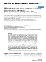
virus dose measured by IF. The data shown in figure 1
illustrate the range of increase in chemiluminescent signal
that corresponds with increasing infectious units in a 10
-
1/3
virus dilution series. These data demonstrate that
changes in the magnitude of the enzymatic signal accu-
rately reflect changes in the number of infectious units
measured by IF.
Luminescent signal magnitude corresponds with infectious unitsFigure 1
Luminescent signal magnitude corresponds with
infectious units. Cells were infected with 10
-1/3
serial dilu-
tions of rotavirus for 18 hours. Plates were fixed and probed
with anti-VP6 mAb A6M followed by peroxidase-conjugated
goat anti-mouse IgG. Signals were developed with chemilumi-
nescent substrate and measured on a ThermoElectron
Fluroskan and scored as relative light units (rlu). Error bars
are standard errors of the means (n = 5).
-6 -5 -4 -3 -2 -1
0
10000
20000
30000
log [Virus Dilution]
rlu
Virology Journal 2006, 3:68 />Page 3 of 11
(page number not for citation purposes)
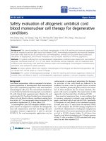
In order to establish internal inhibition controls for the
assay, we measured reductions in infectivity in response to
cytokines known to induce antiviral gene expression.
Cells were pre-treated with IFNα, IFNγ, or a combination
of both cytokines. IFNα and IFNγ both reduced infectivity,
and the magnitude of reduction increased to ~80% when
cells were treated with the combination of both cytokines
(Figure 2). The combination of IFNα and IFNγ consist-
ently yielded the greatest reduction in virus replication.
Therefore, this mixture of cytokines, and IL-2 which is not
known to have direct antiviral activity, were included as
internal positive and negative controls, respectively, in
each plate of every experiment.
Assay validation
Plate uniformity assessments were performed according
to the recommendations of the NIH Chemical Genomics
Center in order to determine whether our assay was suita-
ble for eventual adaptation to a high-throughput format.
The Z' coefficient was established with mock infected cells
representing the minimum signal value, virus infected
cells representing the maximum signal value, and the
IFNα/IFNγ mixture as the midrange signal. Experiments
consisting of three plates each run on three consecutive
days were performed and an example of the data from one
day is shown in Figure 3. No evidence of significant drift
or edge effect was observed. The Z' coefficient measures
the quality of an HTS assay by comparing data variation
and the signal dynamic range, and a value of > 0.5 estab-
lishes the assay as excellent for screening [12,13]. The Z'
values ranged from 0.69 – 0.82 for individual plates, and
the aggregate Z' value from all plates and all days was
0.64. A signal-to-noise value of 4.96 was calculated with
the aggregate data from all plates. All of the statistical cri-
teria for intra-plate assessment were met [14].
The assay results show some inter-plate variability. For
example, although all within-day fold-shifts in signal were
less than 2, all average (between)-day fold-shifts were not.
We expect some degree of variability due simply to the
variations in the growth status of cells from day to day.
Importantly, we established sufficient intra-plate controls
to account for minor variances associated with cell status.
Together the validation and statistical data support the
assay as a viable platform for screening compound librar-
ies.
Compounds from a natural product library reduce
rotavirus infectivity
A 280 compound natural product library was screened to
identify compounds that reduced rotavirus infectivity.
Forty-seven (17%) compounds were selected for a second
round of screening and were tested both for dose-depend-
ency and for effects on cell viability. A representative data-
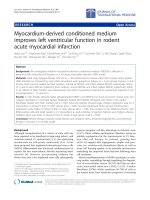
set is shown in Figure 4. Figure 4A shows data obtained
from the initial screen, and the data in figure 4B illustrate
dose-dependent reductions in rotavirus infectivity in
response to treatment of cells with each of the four repre-
sentative compounds. The observed reduction in infectiv-
ity was not a result of generalized cytotoxicity because cell
viability did not decrease upon treatment beyond the
DMSO only controls (Figure 4B). The one exception was
that mangostin was toxic at 10 μg/ml. Dose-dependency
and cell viability assays were performed in parallel with
cells seeded at the same densities from the same cell sus-
pension.
Forty-seven compounds from the primary screen were
subjected to second round screening and 32 were elimi-
nated from consideration for follow-up studies. Ten com-
pounds did not reduce infectivity above the established
threshold in the second round, generating a true false pos-
itive hit rate of ~20%. Twenty compounds proved cyto-
toxic as measured by the cell viability assay, and two
compounds are known toxins with gross effects on cell
metabolism (e.g. protein synthesis inhibitors). The two
toxins were eliminated from further consideration
because we are interested in identifying compounds that
stimulate or enhance antiviral signaling pathways. The
remaining 15 compounds were designated true hits (5 %)
and are listed in Table 1.
Establishment of internal controls: IFNα and IFNγ reduce rotavirus infectivity in the assayFigure 2
Establishment of internal controls: IFNα and IFNγ
reduce rotavirus infectivity in the assay. Cells were
treated with indicated amounts of each cytokine or a combi-
nation of both prior to infection. Infections were allowed to
proceed for 18 hours and chemiluminescent measurement of
reduction in rotavirus infectivity was performed as described
in the text and in the legend to Figure 1. Error bars are
standard errors of the means.
Virus Only
D
1
0
0
U
/m
l
I
FN
-
J
100 U/ml IFN-
J
,
I
FN-
D
100 U/ml I
F
N-
0
100
200
300
400
500
n = 15
n = 12
n =12
n = 12
rlu
Virology Journal 2006, 3:68 />Page 4 of 11
(page number not for citation purposes)
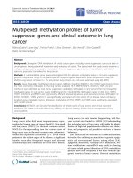
Assay validationFigure 3
Assay validation. Cells were treated according the provided template [14]. Three individual experiments, each containing
three plates, were conducted on three separate days. A) The left panels show a representative set of data from day two of the
plate uniformity assay with the data graphed in a well-row orientation where the wells are labeled horizontally. The panels on
the right contain the same data in a well-column orientation. B) Aggregate data of all nine plates over three days in a well-row
orientation.
0
5000
10000
15000
20000
0 1224364860728496
Max
Mid
Min
0
5000
10000
15000
20000
0 8 16 24 32 40 48 56 64 72 80 88 96
Ma x
Mid
Min
0
5000
10000
15000
20000
0 122436486072 8496
Max
Mid
Min
0
5000
10000
15000
20000
0 8 16 24 32 40 48 56 64 72 80 88 96
Max
Mid
Min
0
5000
10000
15000
20000
0 1224364860728496
Ma x
Mid
Min
0
5000
10000
15000
20000
0 8 16 24 32 40 48 56 64 72 80 88 96
Ma x
Mid
Min
1
2
3
A
0
5000
10000
15000
20000
0 32 64 96 128 160 192 224 256 288
max
mid
min
B
Natural products identified in the MTS assay reduce rotavirus infectivity in a dose-dependent mannerFigure 4
Natural products identified in the MTS assay reduce rotavirus infectivity in a dose-dependent manner. Repre-
sentative data are shown for four compounds: 18-β-glycyrrhetinic acid, abietic acid, mangostin, and all-trans retinoic acid. A)
Data from initial screen. The y-axis is relative light units (n = 3). B) Secondary screen that includes a dose-response (red) and
cell viability (blue) data. The left y-axis is relative light units and the right y-axis is viability (n = 3). Error bars are standard errors
of the means. The highest concentration of mangostin was toxic under the conditions of this assay.
18-E-Glycyrrhetinic Acid
V
i
r
u
s
1% D
MSO
0.5% DMSO
0
.
1%
DM
S
O
I
L-
2
J,
IFN-
DIFN-
g
/ml
P
1
0
g/ml
P
5
g
/ml
P1
0
1000
2000
3000
4000
5000
6000
7000
8000
9000
rlu
Abietic Acid
V
i
rus
1%
D
MSO
0.5% D
M
S
O
0.1% DMSO
I
L
-
2
JI
FN
-D
I
F
N
-
g/ml
P
1
0
g
/ml
P5
g
/
ml
P
1
0
1000
2000
3000
4000
5000
6000
7000
8000
9000
10000
11000
12000
13000
14000
rlu
All-trans-Retinoic Acid
Virus
1% D
M
S
O
0
.
5%
DM
S
O
0.1% DMSO
I
L-2
J
I
FN
-
D
IFN
-
g/
m
l
P
10
g
/ml
P5
g/mlP
1
0
1000
2000
3000
4000
5000
6000
7000
8000
9000
10000
rlu
D-Mangostin
Virus
1% D
MSO
0.5% DMSO
0.1% D
M
S
O
IL-
2
J
IFN-
DI
F
N-
g
/ml
P
10
g/mlP
5
g/
ml
P
1
0
1000
2000
3000
4000
5000
6000
7000
8000
9000
10000
11000
12000
13000
rlu
AB
All-trans-Retinoic Acid
Mo
c
k
V
i
r
u
s
0.04% DMSO
0.02% D
MSO
0
.
004% D
MSO
IL
-2
J
/I
F
N
-
D
I
F
N-
g
/
m
l
P
1
0
g
/
m
l
P
5
g/
m
l
P
1
0
5000
10000
15000
20000
0
1000
2000
3000
4000
5000
6000
D -Mangostin
Mo
c
k
V
i
r
u
s
0.04% DMSO
0.02% D
MSO
0
.
004% D
MSO
IL
-2
J
/I
F
N
-
D
I
F
N-
g
/
m
l
P
1
0
g
/
m
l
P
5
g/
m
l
P
1
0
5000
10000
15000
20000
0
1000
2000
3000
4000
5000
6000
18-E-Glycyrrhetinic Acid
Mo
c
k
V
i
r
u
s
0.04% DMSO
0.02% D
MSO
0
.
004% D
MSO
IL
-2
J
/I
F
N
-
D
I
F
N-
g
/
m
l
P
1
0
g
/
m
l
P
5
g/
m
l
P
1
0
5000
10000
15000
20000
0
1000
2000
3000
4000
5000
6000
Abietic Acid
Mock
V
irus
0.0
4% D
M
SO
0.02% DMSO
0.
0
04% DMS
O
I
L
-2
J
/I
F
N-
D
IFN
-
g/ml
P
10
g
/
ml
P
5
g/ml
P
1
0
5000
10000
15000
20000
0
1000
2000
3000
4000
5000
6000
Virology Journal 2006, 3:68 />Page 5 of 11
(page number not for citation purposes)
Table 1: Compounds that reduce rotavirus infectivity without cytotoxicity
Manufacturer's ID Number Chemical Name FW Chemical Family Known Functions (Select references)
Group A
TNP00130 18-β-Glycyrrhetinic Acid 470.68 Triterpene -Antiviral activity against a number of DNA and RNA
viruses [25]
-Inhibits gap junction communication
TNP00088 Abietic Acid 302.45 Diterpene -Inhibits acute inflammation after topical or oral
administration [26]
-Reduces neutrophil infiltration [26]
-Reduces COX-2 and TNF-α expression [16]
-Activates PPAR-γ in macrophages [16]
TNP00194 All-trans-Retinoic Acid 300.44 Retinoid -Increases expression of type I interferon receptors [27]
TNP00140 α-Mangostin 410.46 Xanthone -Used in treatment of skin infections, wounds and
diarrhea in Southeast Asia [15]
-γ-Mangostin inhibits NF-κB activation and COX-2
expression [15]
-α-Mangostin preferentially inhibits growth of HL60 cells
[28]
-Induces caspase-9 and -3 activation in HL60 cells [28]
Group B
TNP00307 Kinetin-9-Riboside 347.33 Phytohormone -Cytokinin
-Antiviral activity against Tobacco Mosaic Virus [29]
-Causes a decrease in (poly rI) (poly rC) stimulated
interferon response in VSV challenged mice [30]
-Reverses the effect of endotoxin-enhanced host
resistance [31]
TNP00064 7,3'-Dihydroxyflavone 254.24 Flavone
TNP00050 6,7-Dimethoxyflavone 282.29 Flavone
TNP00044 8-Hydroxy-7-Methoxyflavone 268.27 Flavone
TNP00151 Genistein 270.24 Isoflavone -Tyrosine kinase inhibitor [32]
-Down-regulates iNOS [33]
-Inhibits NF-κB activation [33]
-Inhibits COX-2 induction [33]
-Inhibits LPS induced IL-1β, IL-6, and TNF-α production
in monocytes
TNP00227 Capsaicin 305.42 Phenylalkyl-
Amine Alkaloid
-Inhibits NF-κB activation [34]
-Activates transient receptor potential vanilloid-1 (TRPV-
1) [35]
TNP00256 Securinine 217.26 Pseudoalkaloid -GABA receptor antagonist
-Antibacterial activity against E. coli, Staph. aureus and
Myc. smegmatis [36]
-Antimalarial activity [37]
TNP00231 Isopimaric Acid 302.46 Diterpene -Inhibitory activity against multidrug-resistant strains of
Staphylococcus aureus [38]
-activates large-conductance Ca
2+
-activated K
+
channel
α-subunit [39]
TNP00292 Parthenolide 248.32 Sesquiterpene
Lactone
-inhibits NFκB activation by preventing induction of IκB
kinase [40]
TNP00006 Unknown 358.48
TNP00014 Unknown 388.50
Virology Journal 2006, 3:68 />Page 6 of 11
(page number not for citation purposes)
Time course of inhibitory effects
The four compounds shown as representative data in Fig-
ure 4 were analyzed further because of prior reports of
their effects on components of innate immune signaling
pathways [15-17]. A time-course and dose-response of
each compound were established in the immunofluores-
cent focus reduction assay. Cells were treated with each
compound for various times ranging from 12 hours pre-
infection to 2 hours post-infection (Figure 5). Significant
levels of inhibition of virus replication were observed with
each compound when added as early as 2 hours prior to
infection. The level of inhibition did not change dramati-
cally as pre-treatment times increased up to 12 hours. All
compounds showed some ability to reduce infectivity
when added at the time of infection, and 18-β glycyrrhe-
tinic acid, all-trans retinoic acid and mangostin were
somewhat effective when added at 1 or 2 hours post-infec-
tion. Interestingly, addition of abietic acid post-infection
showed a small, but significant increase in infectivity that
also was dose-dependent.
α
-mangostin and 18
β
-glycyrrhetinic acid activate NF
κ
B
and induce IL-8 secretion
The design of our assay measures inhibition of rotavirus
replication but does not distinguish whether antiviral sig-
naling pathways are activated or whether the compounds
block specific steps of the virus replication cycle. To test
the hypothesis that virus replication was reduced because
cell signaling pathways involved in antiviral and inflam-
matory gene expression were induced by selected com-
pound treatment, NFκB activation and IL-8 secretion was
measured following each treatment. α-mangostin and 18
β-glycyrrhetinic acid induced NFκB activation, whereas
all-trans retinoic acid and abietic acid did not beyond the
levels of the control (Figure 6). The levels of IL-8 expres-
Time course of inhibition of rotavirus replicationFigure 5
Time course of inhibition of rotavirus replication. Cells were treated with indicated compounds for various intervals
ranging from 12 hr pre-infection to 2 hr post-infection. Each time point was assayed in triplicate and virus was quantified by
counting FFU. Open squares are IFN control. Red, 7.5 μg/ml, blue, 5 μg/ml, and green, 2.5 μg/ml The y-axis represents percent
inhibition and the x-axis is time of infection. Error bars are standard errors of the means (n = 3).
18-
E
-Glycyrrhetinic Acid
-12 -10 -8 -6 -4 -2 0 2 4
-40
-20
0
20
40
60
80
100
Abietic Acid
-12 -10 -8 -6 -4 -2 0 2 4
-40
-20
0
20
40
60
80
100
All-trans-Retinoic Acid
-12 -10 -8 -6 -4 -2 0 2 4
-40
-20
0
20
40
60
80
100
D
-Mangostin
-12 -10 -8 -6 -4 -2 0 2 4
-40
-20
0
20
40
60
80
100
Virology Journal 2006, 3:68 />Page 7 of 11
(page number not for citation purposes)
sion measured for each compound were consistent with
levels of activation of NFκB (Figure 7).
Discussion
We designed a MTS assay capable of identifying com-
pounds that reduce rotavirus infectivity, toward a long-
term goal of discovering compounds that activate innate
immune signaling pathways to reduce the disease impact
of acute viral infections. We screened a library consisting
of 280 natural products purified from plant extracts, and
several compounds were identified that reproducibly
inhibited rotavirus replication without cytotoxicity. The
assay has been validated statistically, as well as by the
observation that compounds selected for further study
were purchased from different sources and showed repro-
ducible inhibition of virus replication. In addition, fol-
low-up assays measured reductions in infectivity in
response to compound treatment in the standard rotavi-
rus IF assay and the degrees of replication inhibition and
dose-dependence correlated with the values obtained in
NFκB activation in infected and uninfected cells in the presence of selected compoundsFigure 6
NFκB activation in infected and uninfected cells in the presence of selected compounds. Infected or mock infected
MA104 cells were treated with indicated compounds at a final concentration of 7.5 μg/ml. Nuclear extracts were prepared 6
hours post-infection and activation of NFκB was assessed and quantified by commercial ELISA. Squares indicate DMSO only
treated cultures and triangles indicate compound treated cultures. The y-axis is OD
450
nm. The data were analyzed by two-way
ANOVA (n = 3).
18-
E
-Glycyrrhetinic Acid
+ Viru
s
- Viru
s
0.0
0.1
0.2
0.3
0.4
0.5
0.6
0.7
0.8
Abietic Acid
+
Viru
s
- Viru
s
0.0
0.1
0.2
0.3
0.4
0.5
0.6
0.7
0.8
All-trans-Retinoic Acid
+ Viru
s
- Viru
s
0.0
0.1
0.2
0.3
0.4
0.5
0.6
0.7
0.8
D
-Mangostin
+
Viru
s
- Viru
s
0.0
0.1
0.2
0.3
0.4
0.5
0.6
0.7
0.8
Virology Journal 2006, 3:68 />Page 8 of 11
(page number not for citation purposes)
the MTS assay. We calculated a true hit rate for the natural
product library of 5% and a false positive rate of ~20%.
We performed numerous optimization assays to reduce
variability presented by variation in growth properties of
our cell line. We also have initiated a screen of a 10,000
compound synthetic chemical library, and after screening
~2500 compounds, our true hit rate is 1.8% (data not
shown). These true hit percentages yield a large, but not
prohibitive, number of compounds for follow-up studies
both in vitro and in animal models. Together the data sug-
gest our assay is an effective platform for screening candi-
date compounds for antiviral activity in adherent
epithelial cells.
In this study, we sought to identify compounds with
innate immune modulating effects that resulted in cellu-
lar resistance to rotavirus infection. The format of the
assay, that is, readout of reduction in infectivity, does not
distinguish between compounds that act on antiviral sig-
naling pathways and those that may target specific steps in
the virus replication cycle, such as entry or replicase activ-
ity. However, two of the four compounds selected for fol-
low-up study activated NFκB and induced IL-8 secretion.
Moreover, the majority of compounds called a hit in the screen
for which data is available has been previously described to
affect inflammatory or anti-inflammatory gene expression or
pathogen growth in vitro (Table 1). These observations
strongly support the assertion that our assay is appropri-
ate, but not exclusive, for discovery of immune potentia-
tors, as we intended.
There has been a resurgence of interest in natural products
as drug candidates for a variety of reasons including
increased interest in infectious disease prevention and
therapy, a lower percentage of chemical properties that
negatively affect permeation and absorption, and the pro-
pensity of natural products to act by affecting protein-pro-
tein interactions [18]. Important for the studies described
here, natural products are known to modulate immune
responses and cell signaling pathways. Table 1 lists com-
pounds designated hits in this study, along with some of
the reported functions. Compounds were grouped into
those chosen for further study (Group A), and those
known to stimulate or repress inflammatory responses or
innate immune signaling pathways and two with
unknown functions (Group B). Also included in Group B
are compounds with reported antiviral, antibacterial, or
anti-protozoan activity. We have not yet deciphered
mechanisms by which these compounds inhibit rotavirus
replication in vitro. However, the fact that several of the
compounds, for example, abietic acid, genistein, and cap-
saicin, interfere with NFκB activation and cyclo-oxygenase
2 (COX2) expression is noteworthy (see Table 1 for refer-
ences). NFκB activation is an important regulator of
COX2 expression; COX2 activity and COX-mediated pros-
taglandin synthesis is necessary for rotavirus infectivity in
CaCo-2 intestinal cells [19]. Interestingly, the addition of
prostaglandin E
2
(PGE
2
) restored infectivity reduced by
the COX inhibitor [19] and three of the compounds we
chose for follow-up studies all have an inhibitory effect on
either synthesis or release of PGE
2
(see Table 1 references).
The ability of α-mangostin and 18 β-glycyrrhetinic acid to
inhibit rotavirus replication when both compounds acti-
vate NFκB is most likely because antiviral states are estab-
lished upon treatment of the cells with each compound
prior to infection. The definitive mechanisms by which
the compounds identified in this natural product library
screen warrant further investigation.
We intend this MTS assay to serve as a platform for discov-
ery of candidate adjuvants that will be effective against
acute viral infections at mucosal surfaces. Rotavirus is an
ideal model system for these purposes for several reasons.
First, rotaviruses cause gastrointestinal illness in most
mammalian species and so their relevance as mucosal
pathogens is clear. Second, these viruses are well charac-
terized with respect to structure, antigenicity, and mecha-
nisms of virus replication, and thus an excellent resource
for mechanistic follow-up studies is available. Third, rota-
virus is promiscuous in its tropism for cultured cell lines,
and multiple cell lines of different types and species of ori-
gin, including primary cell lines [20], support productive
IL-8 expression cells treated with selected compoundsFigure 7
IL-8 expression cells treated with selected com-
pounds. MA104 cells were treated with indicated com-
pounds for 24 hours and IL-8 in the supernatants was
measured with a commercial ELISA. ** = p < 0.01, *** = p <
0.001, n = 4.
Vi
r
u
s
Mock
-G
lycyrr
hetin
ic A
cid
E
18-
Abietic Ac
i
d
All-tr
ans-R
e
t
ino
i
c Acid
-Mangostin
D
0.00
0.05
0.10
0.15
0.20
0.25
0.30
0.35
0.40
0.45
***
**
***
***
O.D.
450 nm
Virology Journal 2006, 3:68 />Page 9 of 11
(page number not for citation purposes)
virus replication. The ability to propagate rotavirus in a
variety of cell types supports high throughput applica-
tions that study general as well as cell-type specific innate
immune responses. Fourth, both small and large animal
models of natural infection allow relatively rapid evalua-
tion of the efficacy of candidate compounds in vivo.
Finally, the fact that rotavirus employs a mechanism to
down-regulate antiviral gene expression allows considera-
tion of possible evasion strategies when selecting and test-
ing candidate immune potentiators [21,22]. Current
development efforts include expansion of screening stud-
ies to other lytic RNA viruses such as influenza virus, and
adaptation of the assay to a fully automated format.
Methods
Cells, virus, and rotavirus monoclonal antibodies
MA104 monkey kidney epithelial cells were maintained
in M199 medium (MediaTech Cell Grow) supplemented
with 5% fetal bovine serum (FBS; Atlanta Biologicals).
Isolation and cultivation of G serotype 6 (G6) bovine
rotavirus strain NCDV has been described [23]. Mono-
clonal antibody (MAb) E4 reacts with major structural
protein VP6 of most group A rotavirus strains [24]. MAb
A6M recognizes VP6 and was generated by immunizing
mice with G6 bovine rotavirus strain B641 and screening
hybridoma supernatants for antibodies that react with
rotavirus specific proteins.
Reagents and chemicals
A library containing 0.5 mg each of 280 natural products
purified from plant extracts was purchased from TimTec
(TimTec Corporation). Compounds were reconstituted in
500 μl of dimethyl sulfoxide (DMSO) and the library was
stored at -80°C. Individual compounds α-mangostin
(TimTec or Indofine), 18-β-glycyrrhetinic acid (18-BGA;
Fluka), abietic acid (AA; Sigma), and all-trans-retinoic acid
(ATRA; Sigma) were reconstituted in DMSO to final stock
concentrations of 25 mg/ml.
Recombinant human interferon-α (IFN-α; BioSource
International, Inc) was diluted to a concentration of 1 ×
10
5
U/ml in phosphate buffered saline (PBS) containing
0.1% bovine serum albumin (BSA). Recombinant human
interferon-γ (IFN-γ) and recombinant human interleukin-
2 (IL-2; Peprotech Inc) were diluted to 2 × 10
6
U/ml and
10 μg/ml, respectively, in PBS.
Horseradish peroxidase (HRP)-conjugated goat anti-
mouse IgG (H+L), HRP-conjugated goat anti-mouse IgG
(H+L) F(ab')
2
fragments, and FITC-conjugated goat anti-
mouse IgG were purchased from Jackson ImmunoRe-
search Laboratories.
Screening assay
Compound treatments and virus infections
Working stock solutions of each compound from the
library were prepared to twice the desired final concentra-
tions of 20 μg/ml, 10 μg/ml, and 2 μg/ml in serum-free
M199. Working stock solutions of assay controls con-
sisted of 2%, 1% DMSO, and 0.2% DMSO, a mixture of
200 U/ml each of IFN-α and IFN-γ, and 2 ng/ml of IL-2.
MA104 cells were cultured to confluence in 96-well black-
walled plates (Costar). The culture media was decanted
and replaced with 50 μl of M199. 50 μl of 2X control and
experimental stock solutions were added to respective
wells, in triplicate, and plates were incubated for 4 hours
at 37°C. Following the 4 hour incubation, the contents of
each plate were removed and 8.9 × 10
5
ffu/well of trypsin-
activated NCDV in 0% M199 was added to appropriate
wells. Mock infected wells received 50 μl of 0% M199. 50
μl of fresh 2X control and experimental compounds were
added and at 18 hours post-infection, the cells were fixed
for 10 minutes with 80% acetone.
Cell-based ELISA
The wells were blocked for one hour at room temperature
with 100 μl of PBS containing 3% bovine serum albumin
(w/v) and 0.05% Tween-20. The plates were washed one
time with 400 μl of wash buffer consisting of PBS and
0.05% Tween 20. 50 μl of 20 μg/ml A6M or E4 (1:25
hybridoma supernatant) in PBS containing 0.05% Tween
20 and 0.5% dry milk was added to the wells and incu-
bated for one hour at room temperature. The plates were
washed four times with wash buffer, then 50 μl of a 1:500
dilution of HRP-conjugated F(ab')
2
in 0.5% Blotto was
added and the plates were incubated for one hour at room
temperature. Following a final wash, 100 μl of BM Chemi-
luminescence ELISA Substrate (Roche Diagnostics) was
added and reactions were allowed to proceed for 4 min-
utes to reach a steady state of enzymatic activity. Signals
were measured on a ThermoElectron Fluroskan (Thermo-
Electron Cooperation) with an integration time of 1,000
ms.
Cell viability assay
Cell viability assays for compound toxicity were set up
similar to compound screening except cells were not
infected over the course of the experiments. Cell viability
was measured with the CellTiter-Glo Luminescent Cell
Viability Assay (Promega) according to instructions pro-
vided by the manufacturer.
Statistics and criteria for "hit" designation
Compounds that showed a greater than 60% decrease in
signal at 5 μg/ml when compared to the 0.5% DMSO con-
trol, and p < 0.05 as determined by a one-tailed student's
t test, were selected for secondary screening. Additionally,
Virology Journal 2006, 3:68 />Page 10 of 11
(page number not for citation purposes)
compounds that showed a greater than 90% decrease in
signal when compared to the 1% DMSO control and p <
0.05 also were selected for further screening. Compounds
that showed a greater than 10% decrease in signal and a p
< 0.05 in the cell viability assay were determined to be
toxic.
Plate uniformity assessment
The recommendations of the National Institutes of Health
Chemical Genomics Center's Assay Guidance Manual Ver-
sion 4.1 for plate uniformity assessments were followed.
Three separate experiments consisting of three plates each
were performed on three different days. Confluent
MA104 cells were pretreated for 4 hours with 100 μl of
either media or a mixture of 100 U/ml each of IFN-α and
IFN-γ. All media was serum free and contained 1%
DMSO.
The pre-treatment media was decanted and the cells were
infected with 50 μl of 8.9 × 10
5
pfu/ml of trypsin-activated
NCDV. Mock infected wells were treated with 50 μl of
serum free media. 50 μl of fresh 2X treatment media con-
taining 2% DMSO was added to the respective wells and
the plates were incubated for 18 hours. The plates were
fixed, labeled and quantified according to the assay proce-
dure described above. All calculations were performed
using the Assay Guidance Manual's spreadsheet [14].
Immunofluorescent focus assay
Immunofluorescent assays (IF) for rotavirus infectivity
were performed as previously described [24]. Cells were
cultured in 96-well plates and were mock infected or
infected with NCDV at approximately 150 ffu/well. Pre-
or post-treatment with compounds was performed for the
indicated times as described above. 18 hours post-infec-
tion, the cells were fixed for 10 minutes with 80% acetone.
Incubations with primary and secondary antibody were as
described above, except the secondary antibody was FITC-
conjugated goat anti-mouse IgG. Fluorescent foci were
counted by microscopy (Nikon Eclipse TE300).
NF
κ
B and IL-8 assays
MA104 cells in 100 × 20 mm culture dishes were treated
with 7.5 μg/ml of selected compounds in serum-free
M199 containing 0.03% DMSO. The effects of the pres-
ence of virus on NFκB activation and IL-8 secretion in the
assays was tested by infecting cells with trypsin-activated
NCDV at an moi of 10 pfu/cell at the time of compound
treatment. In all cases, incubation periods were 6 hours.
NF-κB activation was quantified with TransAM NF-κB p50
Transcription Factor Assay Kit (Active Motif). Nuclear
extracts were prepared with CelLytic NuCLEAR Extraction
Kit (Sigma) following the manufacturer's protocol.
Nuclear protein concentration was determined with the
D
c
Protein Assay (Bio-Rad Laboratories), and 20 μg of
nuclear protein was used in the assay. Statistical analysis
of the data was performed by two-factor ANOVA.
IL-8 secretion was measured with the Quantikine Human
IL-8 system (R & D Systems) following the manufacturer's
protocol. Compound treatments and infections were per-
formed as described above, except that the incubation
periods were extended to 24 hours. The data were ana-
lyzed by student's t test with a threshold of significance set
to p < 0.001.
Competing interests
The author(s) declare that they have no competing inter-
ests.
Authors' contributions
MES established the assay, performed most of the experi-
ments and assisted in preparation of the manuscript. ADB
performed a significant number of screening assays and
data analysis, and participated in manuscript preparation.
JWG purified and characterized virus stocks, assisted in
assay development and in manuscript preparation. MAJ
contributed to study design, assay development, data
analysis and manuscript preparation. MEH conceived of
the study, participated in design and is responsible for
study oversight.
Acknowledgements
This work was supported by DoD contract W9113M-04-1-0010, USDA
NRI/CGP 02657 to MEH, the Montana Agriculture Experiment Station, and
PHS grant P20 RR020185. ADB gratefully acknowledges financial support
from the Arnold and Mabel Beckman Foundation administered through the
MSU Beckman Scholars Program.
References
1. Hackett CJ: Innate immune activation as a broad-spectrum
biodefense strategy: prospects and research challenges. J
Allergy Clin Immunol 2003, 112:686-694.
2. Pashine A, Valiante NM, Ulmer JB: Targeting the innate immune
response with improved vaccine adjuvants. Nat Med 2005,
11:S63-S68.
3. Amlie-Lefond C, Paz DA, Connelly MP, Huffnagle GB, Whelan NT,
Whelan HT: Innate immunity for biodefense: a strategy whose
time has come. J Allergy Clin Immunol 2005, 116:1334-1342.
4. Johnston PA, Johnston PA: Cellular platforms for HTS: three
case studies. Drug Discov Today 2002, 7:353-363.
5. O'Boyle DR, Nower PT, Lemm JA, Valera L, Sun JH, Rigat K, Colonno
R, Gao M: Development of a cell-based high-throughput spe-
cificity screen using a hepatitis C virus-bovine viral diarrhea
virus dual replicon assay. Antimicrob Agents Chemother 2005,
49:1346-1353.
6. Blair WS, Isaacson J, Li X, Cao J, Peng Q, Kong GF, Patick AK: A
novel HIV-1 antiviral high throughput screening approach
for the discovery of HIV-1 inhibitors. Antiviral Res 2005,
65:107-116.
7. Adelson ME, Pacchia AL, Kaul M, Rando RF, Ron Y, Peltz SW, Dough-
erty JP: Toward the development of a virus-cell-based assay
for the discovery of novel compounds against human immu-
nodeficiency virus type 1. Antimicrob Agents Chemother 2003,
47:501-508.
8. Ivens T, Eynde CV, Acker KV, Nijs E, Dams G, Bettens E, Ohagen A,
Pauwels R, Hertogs K: Development of a homogeneous screen-
ing assay for automated detection of antiviral agents active
Publish with BioMed Central and every
scientist can read your work free of charge
"BioMed Central will be the most significant development for
disseminating the results of biomedical research in our lifetime."
Sir Paul Nurse, Cancer Research UK
Your research papers will be:
available free of charge to the entire biomedical community
peer reviewed and published immediately upon acceptance
cited in PubMed and archived on PubMed Central
yours — you keep the copyright
Submit your manuscript here:
/>BioMedcentral
Virology Journal 2006, 3:68 />Page 11 of 11
(page number not for citation purposes)
against severe acute respiratory syndrome-associated coro-
navirus. J Virol Methods 2005, 129:56-63.
9. Parashar UD, Hummelman EG, Bresee JS, Miller MA, Glass RI: Global
illness and deaths caused by rotavirus disease in children.
Emerg Infect Dis 2003, 9:565-572.
10. Ward RL: Rotavirus vaccines: is the second time the charm?
Curr Opin Investig Drugs 2005, 6:798-803.
11. Knowlton DR, Spector DM, Ward RL: Development of an
improved method for measuring neutralizing antibody to
rotavirus. J Virol Methods 1991, 33:127-134.
12. Iversen PW, Eastwood BJ, Sittampalam GS, Cox KL: A comparison
of assay performance measures in screening assays: signal
window, Z' factor, and assay variability ratio. J Biomol Screen
2006, 11:247-252.
13. Zhang JH, Chung TD, Oldenburg KR: A Simple Statistical Param-
eter for Use in Evaluation and Validation of High Through-
put Screening Assays. J Biomol Screen 1999, 4:67-73.
14. [ />].
15. Nakatani K, Yamakuni T, Kondo N, Arakawa T, Oosawa K, Shimura
S, Inoue H, Ohizumi Y: gamma-Mangostin inhibits inhibitor-
kappaB kinase activity and decreases lipopolysaccharide-
induced cyclooxygenase-2 gene expression in C6 rat glioma
cells. Mol Pharmacol 2004, 66:667-674.
16. Takahashi N, Kawada T, Goto T, Kim CS, Taimatsu A, Egawa K,
Yamamoto T, Jisaka M, Nishimura K, Yokota K, Yu R, Fushiki T: Abi-
etic acid activates peroxisome proliferator-activated recep-
tor-gamma (PPARgamma) in RAW264.7 macrophages and
3T3-L1 adipocytes to regulate gene expression involved in
inflammation and lipid metabolism. FEBS Lett 2003,
550:190-194.
17. Kolla V, Lindner DJ, Xiao W, Borden EC, Kalvakolanu DV: Modula-
tion of interferon (IFN)-inducible gene expression by retin-
oic acid. Up-regulation of STAT1 protein in IFN-
unresponsive cells. J Biol Chem 1996, 271:10508-10514.
18. Koehn FE, Carter GT: The evolving role of natural products in
drug discovery. Nat Rev Drug Discov 2005, 4:206-220.
19. Rossen JW, Bouma J, Raatgeep RH, Buller HA, Einerhand AW: Inhi-
bition of cyclooxygenase activity reduces rotavirus infection
at a postbinding step. J Virol 2004, 78:9721-9730.
20. Buckner D, Wilson S, Kurk S, Hardy M, Miessner N, Jutila MA: Use
of Early Passage Fetal Intestinal Epithelial Cells in Semi-
high-throughput Screening Assays: An Approach to Identify
New Innate Immune System Adjuvants. J Biomol Screen 2006.
21. Graff JW, Mitzel DN, Weisend CM, Flenniken ML, Hardy ME: Inter-
feron regulatory factor 3 is a cellular partner of rotavirus
NSP1. J Virol 2002, 76:9545-9550.
22. Barro M, Patton JT: Rotavirus nonstructural protein 1 subverts
innate immune response by inducing degradation of IFN reg-
ulatory factor 3. Proc Natl Acad Sci USA 2005, 102:4114-4119.
23. Woode GN, Kelso NE, Simpson TF, Gaul SK, Evans LE, Babiuk L:
Antigenic relationships among some bovine rotaviruses:
serum neutralization and cross-protection in gnotobiotic
calves. J Clin Microbiol 1983, 18:358-364.
24. Zheng SL, Woode GN, Melendy DR, Ramig RF: Comparative stud-
ies of the antigenic polypeptide species VP4, VP6, and VP7 of
three strains of bovine rotavirus. J Clin Microbiol
1989,
27:1939-1945.
25. Pompei R, Flore O, Marccialis MA, Pani A, Loddo B: Glycyrrhizic
acid inhibits virus growth and inactivates virus particles.
Nature 1979, 281:689-690.
26. Fernandez MA, Tornos MP, Garcia MD, de las HB, Villar AM, Saenz
MT: Anti-inflammatory activity of abietic acid, a diterpene
isolated from Pimenta racemosa var. grissea. J Pharm Pharma-
col 2001, 53:867-872.
27. Hamamoto S, Fukuda R, Ishimura N, Rumi MA, Kazumori H, Uchida
Y, Kadowaki Y, Ishihara S, Kinoshita Y: 9-cis retinoic acid
enhances the antiviral effect of interferon on hepatitis C
virus replication through increased expression of type I
interferon receptor. J Lab Clin Med 2003, 141:58-66.
28. Matsumoto K, Akao Y, Yi H, Ohguchi K, Ito T, Tanaka T, Kobayashi
E, Iinuma M, Nozawa Y: Preferential target is mitochondria in
alpha-mangostin-induced apoptosis in human leukemia
HL60 cells. Bioorg Med Chem 2004, 12:5799-5806.
29. Milo GE Jr, Srivastava BI: Effect of cytokinins on tobacco mosaic
virus production in tobacco pith tissue cultures. Virology 1969,
39:621-623.
30. De Clercq E, Nuwer MR, Merigan TC: The role of interferon in
the protective effect of a synthetic double-stranded polyri-
bonucleotide against intranasal vesicular stomatitis virus
challenge in mice. J Clin Invest 1970, 49:1565-1577.
31. Kessel RW, Braun W: Endotoxin-enhanced host resistance:
reversal by kinetin riboside. Nature 1966, 211:1001-1002.
32. Akiyama T, Ishida J, Nakagawa S, Ogawara H, Watanabe S, Itoh N,
Shibuya M, Fukami Y: Genistein, a specific inhibitor of tyrosine-
specific protein kinases. J Biol Chem 1987, 262:5592-5595.
33. Kim HP, Son KH, Chang HW, Kang SS: Anti-inflammatory plant
flavonoids and cellular action mechanisms. J Pharmacol Sci
2004, 96:229-245.
34. Singh S, Natarajan K, Aggarwal BB: Capsaicin (8-methyl-N-vanil-
lyl-6-nonenamide) is a potent inhibitor of nuclear transcrip-
tion factor-kappa B activation by diverse agents. J Immunol
1996, 157:
4412-4420.
35. Geppetti P, Trevisani M: Activation and sensitisation of the
vanilloid receptor: role in gastrointestinal inflammation and
function. Br J Pharmacol 2004, 141:1313-1320.
36. Mensah JL, Lagarde I, Ceschin C, Michel G, Gleye J, Fouraste I: Anti-
bacterial activity of the leaves of Phyllanthus discoideus. J
Ethnopharmacol 1990, 28:129-133.
37. Weenen H, Nkunya MH, Bray DH, Mwasumbi LB, Kinabo LS, Kilimali
VA, Wijnberg JB: Antimalarial compounds containing an alpha,
beta-unsaturated carbonyl moiety from Tanzanian medici-
nal plants. Planta Med 1990, 56:371-373.
38. Smith E, Williamson E, Zloh M, Gibbons S: Isopimaric acid from
Pinus nigra shows activity against multidrug-resistant and
EMRSA strains of Staphylococcus aureus. Phytother Res 2005,
19:538-542.
39. Imaizumi Y, Sakamoto K, Yamada A, Hotta A, Ohya S, Muraki K, Uch-
iyama M, Ohwada T: Molecular basis of pimarane compounds
as novel activators of large-conductance Ca(2+)-activated
K(+) channel alpha-subunit. Mol Pharmacol 2002, 62:836-846.
40. Hehner SP, Hofmann TG, Droge W, Schmitz ML: The antiinflam-
matory sesquiterpene lactone parthenolide inhibits NF-
kappa B by targeting the I kappa B kinase complex. J Immunol
1999, 163:5617-5623.