Báo cáo hóa học: " The effects of N-terminal insertion into VSV-G of an scFv peptide" pot
Bạn đang xem bản rút gọn của tài liệu. Xem và tải ngay bản đầy đủ của tài liệu tại đây (310.23 KB, 8 trang )
BioMed Central
Page 1 of 8
(page number not for citation purposes)
Virology Journal
Open Access
Research
The effects of N-terminal insertion into VSV-G of an scFv peptide
Hanna Dreja* and Marc Piechaczyk
Address: Institut de Génétique Moléculaire de Montpellier, UMR 5535, IFR122, CNRS, France
Email: Hanna Dreja* - ; Marc Piechaczyk -
* Corresponding author
Abstract
Recombinant retroviruses, including lentiviruses, are the most widely used vectors for both in vitro
and in vivo stable gene transfer. However, the inability to selectively deliver transgenes into cells of
interest limits the use of this technology. Due to its wide tropism, stability and ability to pseudotype
a range of viral vectors, vesicular stomatitis virus G protein (VSV-G) is the most commonly used
pseudotyping protein. Here, we attempted to engineer this protein for targeting purposes.
Chimaeric VSV-G proteins were constructed by linking a cell-directing single-chain antibody (scFv)
to its N-terminal. We show that the chimaeric VSV-G molecules can integrate into retroviral and
lentiviral particles. HIV-1 particles pseudotyped with VSV-G linked to an scFv against human Major
Histocompatibility Complex class I (MHC-I) bind strongly and specifically to human cells. Also, this
novel molecule preferentially drives lentiviral transduction of human cells, although the titre is
considerably lower that viruses pseudotyped with VSV-G. This is likely due to the inefficient fusion
activity of the modified protein. To our knowledge, this is the first report where VSV-G was
successfully engineered to include a large (253 amino acids) exogenous peptide and where attempts
were made to change the infection profile of VSV-G pseudotyped vectors.
Background
Retroviruses, including lentiviruses, integrate into the
genome of host cells, and the expression of the transduced
genes can persist throughout cell divisions. Hence,
murine leukemia virus (MLV)- and lentivirus-based vec-
tors are among the most commonly used tools for gene
transfer in eukaryotic cells in the laboratory, and may one
day become clinically important. Lentiviral vectors have
also the additional advantage of transducing non-dividing
cells, which broadens their application to both resting and
terminally differentiated cells.
Despite continuous improvement of retroviral and lenti-
viral gene transfer over the past years [1-3], the current
inability to target infection to cells of interest remains a
severe limitation, preventing the development of efficient,
safe and cost-effective clinical application. A number of
reports have already been published to this end (for
review, see [4-6]). The majority of these studies were
attempts to redirect the tropism of the ecotropic envelope
glycoprotein (GP) of MLVs by the addition of ligand
motifs, which bind to specific molecules associated with
the cell membrane. However, these approaches generally
met with limited success. Although the engineered viruses
usually did bind to the new receptors, infection titres were
low. Inefficient transduction was mostly due to dimin-
ished fusion activity of the engineered GP, which conse-
quently prevented infectious translocation of the viral
capsids into cells [7-9].
Retroviral and lentiviral GPs are made of two parts, pro-
duced from the same precursor following proteolytic mat-
Published: 02 September 2006
Virology Journal 2006, 3:69 doi:10.1186/1743-422X-3-69
Received: 21 June 2006
Accepted: 02 September 2006
This article is available from: />© 2006 Dreja and Piechaczyk; licensee BioMed Central Ltd.
This is an Open Access article distributed under the terms of the Creative Commons Attribution License ( />),
which permits unrestricted use, distribution, and reproduction in any medium, provided the original work is properly cited.
Virology Journal 2006, 3:69 />Page 2 of 8
(page number not for citation purposes)
uration. SU, or surface protein, recognises the viral
receptor, and TM, the transmembrane protein, carries the
fusion activity and tethers the GP to virions [4-6]. How-
ever, retroviruses and lentiviruses can be pseudotyped by
a number of GPs from other viruses, such as the hemag-
glutinin (HA) of influenza virus, the envelope proteins
(E1 and E2) of Sindbis virus and the G protein of vesicular
stomatitis virus (VSV-G). These have all higher fusion
activity than the native GPs and remain tightly attached to
virions. HA has already been engineered for targeting pur-
poses through N-terminal addition of various ligands, of
which one successfully redirected MLV tropism towards
human melanoma cells [10]. E2 has also been genetically
modified to display the immunoglobulin-binding
domain of Staphylococcus aureus protein A [11]. After addi-
tion of antibodies specific for certain cell membrane
markers, a relatively efficient retargeted infection of pseu-
dotyped MLV- and HIV based vectors was observed in vitro
[11], as well as in vivo [12]. Recently, E2 was engineered to
include a scFv against CCR5, which specifically directed
lentiviral vectors to CCR5-expressing cells [13].
These findings are promising for future vector modifica-
tions, although HA and the Sindbis proteins are seldom
used for gene transfer protocols. Due to its broad tropism
and stability, VSV-G, on the other hand, is the most widely
used protein for pseudotyping retroviral and lentiviral
vectors [14,15]. VSV-G is a trimerised transmembrane
molecule, although its exact structure is not fully known.
Moreover, its ligand has not been identified [16], which
hampers rational design of targeting strategies. Addition-
ally, only a few permissible sites for short (2–10 amino
acids) peptide insertions have been isolated [17-20]. Nev-
ertheless, these studies all confirmed that VSV-G might be
amenable to genetic engineering for targeting purposes.
Guibinga et al inserted a 10 amino acid collagen-binding
peptide close to the N-terminal of VSV-G, and could show
specific attachment of MLV- and HIV-1-based vectors to
collagen matrix [17]. To date, however, no redirected cell
transduction has been reported. We therefore attempted
to target infection by attaching a large ligand binding
domain, an scFv against MHC-I, directly in the N-terminal
of the protein, a site that Yu and Schaffer confirmed per-
missive. We show that the novel GP, with its large exoge-
nous peptide, (i) is processed and transported to the cell
surface, (ii) provides a new binding specificity but (iv)
transduces target cells very inefficiently, although better
than control scFv/VSV-G. We speculate that this is due to
an inefficient fusion activity, and discuss potential
improvements.
Results and discussion
As a model system, we decided to target MHC-I molecules
on human cells, as these membrane receptors can mediate
cell infection by retroviral and lentiviral vectors [11,21-
23]. As already described [23], a scFv against MHC-I
(αMHC) consists of the heavy and light chain variable
regions of a mouse monoclonal antibody (B9.12.1) [24],
coupled by flexible spacer. This peptide was fused to the
N-terminal of the mature coding sequence of VSV-G
(αMHC/VSV-G). Although certain anti-MHC-I mono-
clonal antibodies are known to inhibit HIV production,
B9.12.1 appears to have a minor effect on the viral life
cycle [25]. As a control, we used a similar construct, con-
taining an anti-hen egg lysozyme scFv (αHEL) [26], which
does not recognise any surface markers on human cells.
For immunodetection purposes, the C-terminal of the
VSV-G cDNA was fused to an HA sequence. The two chi-
maeras were obtained by inserting the HA-containing
VSV-G cDNA downstream of scFv sequences in vectors
originating from Moloney MLV constructs [27]. Conse-
quently, the leader sequence from Moloney MLV GP is
used, and 6 aminoacids from the original GP are retained
between the scFv and VSV-G (Figure 1).
VSV-G is glycosylated, folded and trimerised in the endo-
plasmatic reticulum prior to export to the Golgi [28].
Changes in the protein structure often results in inappro-
priate processing [18] (our own unpublished observa-
tions). We therefore assessed the intracellular distribution
of the scFv/VSV-G molecules in transfected HeLa cells,
revealed by a rat anti-HA antibody. HA-tagged VSV-G and
scFv/VSV-G proteins were all found scattered throughout
the cells and a fraction of the protein were detected in, or
very close to the cellular membrane (data not shown).
With the conformation-specific anti-VSV-G antibody
8G5F11 (a generous gift by Dr D. Lyles), VSV-G and scFv/
VSV-G molecules were also detected by flow cytometry on
the surface of transfected HeLa and 293T cells, implying
that the engineered VSV-G proteins retain conformational
resemblance to the native molecule. Therefore, we have
succeeded in generating correctly processed hybrid pro-
teins, which is in accord with a recent report that showed
that the N-terminal of VSV-G is permissive for short pep-
tide insertion [20]. We next assessed the incorporation of
the chimaeras into lentiviral particles. To this aim, expres-
sion vectors for VSV-G, αMHC/VSV-G and αHEL/VSV-G
were co-transfected with the pCMV᭝R8.2 helper plasmid
expressing Gag, Pol and accessory HIV-1 proteins together
with pHRCMV-EGFP HIV-1-based lentiviral vector [29].
Viral particles were prepared from culture supernatants
and analysed by immunoblotting for the presence of VSV-
G proteins. As shown in Figure 1B, the αMHC/VSV-G and
αHEL/VSV-G chimaeras were incorporated in HIV-1
recombinant particles at levels reflecting those in the
transfected cells. There was, however, slightly lower
amounts of the chimaeric proteins versus the parental ver-
sion in transfected cells, which may be a result of
decreased synthesis (different expression plasmids) or
reduced stability of the new molecules.
Virology Journal 2006, 3:69 />Page 3 of 8
(page number not for citation purposes)
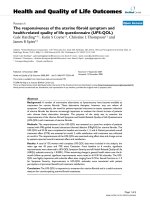
scFv-VSV-G expression plasmids and incorporation in HIV-1 derived particlesFigure 1
scFv-VSV-G expression plasmids and incorporation in HIV-1 derived particles. a) Expression plasmids: (I) VSV-G expression
plasmid (PM 730). The 1.6 kB HindIII-BamHI VSV-G fragment (serotype Indiana) was transferred from pFB.VSVG (J.M. Heard,
Paris, France) into pcDNA3 (InVitrogen) by PCR cloning according to standard procedures. A haemagglutinin (HA) sequence
was added at the C terminus of VSV-G for immunodetection. (II and III) scFv/VSV-G expression plasmids.
The chimaeric con-
structs were generated by PCR-based cloning. Mature VSV-G (from amino acid 17) was amplified from PM 730 and introduced
into the PM 441 and PM 442 plasmids [23]. These constructs originate from an MLV-derived plasmid (FBMOSALF [31]), modi-
fied to contain a scFv (αMHC and αHEL, respectively [27]), upstream of the GP gene. Consequently, the resulting constructs
(αMHC/VSV-G and αHEL/VSV-G) express the genes from the MLV LTR, with a MLV leader sequence (L
mlv
) and 6 additional
amino acids from the virus. (IV and V) Vectors for production of HIV-1 derived viral particles.
A HIV-1-based lentiviral vector
(V) (CD 416; pHRCMV) [29], into which EGFP gene had been inserted, together with a helper plasmid (CD 417; pCMV᭝R8.2)
[29] expressing Gag, Pol and accessory HIV-1 proteins (IV), were used for production of HIV-1 particles. Expression vectors,
physical maps and primer sequences are available upon request. b. VSV-G- and scFv/VSV-G pseudotyped HIV-1 particles, pro-
duced in 293T cells. 2 × 10
6
293T cells in 10 cm diameter tissue culture dishes were transiently transfected with 5 μg of an
LTR-driven EGFP vector (pHRCMV-EGFP) and 4 μg of a helper plasmid (pCMV᭝R8.2) [29], using the calcium phosphate co-
precipitation procedure [36]. 5 μg PM 730 or 30 μg scFv/VSV-G plasmid (PM 981 or PM 983) were also included. DNA precip-
itates were removed after 16 hours, and the viral supernatants were collected 24–48 hours later and pelleted by ultracentrifu-
gation (BeckmanCoulter) at 25 kRPM, 4°C for 2 hours and resuspended in 1% of the original volume. Cell lysates and 2 μl
concentrated scFv/VSV-G virus or 10 μl of non-concentrated VSV-G virus were separated on a 12 % SDS polyacrylamide gel
and transferred onto Protran nitrocellulose membranes (Schleicher and Schuell). HA-tagged VSV-G and scFv/VSV-G were
detected using a rat anti-HA antibody (Sigma), followed by horse radish peroxidase (HPO) conjugated anti-RatIgG (Dako).
p24Gag was detected using SF2 rabbit monoclonal antibody (NIH AIDS Research and Reference Reagent Program) followed by
anti-rabbit IgG/HPO (Santa Cruz), and was used as an internal reference to normalise for the virion protein quantities. The
membranes were developed with Renaissance chemoluminescence kit (NEN Life Science Products), as recommended by the
supplier.
a.
I) PM 730 (VSV-G-HA)
II) PM 981 (αMHC/VSV-G)
III) PM 983 (αHEL/VSV-G)
IV) CD 416 (pHRCMV-EGFP)
V) CD 417 (pCMVR8.2)
Cell extracts
HIV particles
Anti-HA
Anti-HIV Gag
VSV-G
p24Gag
Pr55Gag
scFv/VSV-G
b.
-ve ctrl
αMHC/VSV-G
VSV-G
αHEL/VSV -G
-ve ctrl
αHEL/VSV -G
αMHC/VSV -G
gag pol
pCMV
pA
tat rev
Ψ
RRE
LTR
EGFP
Ψ
LTR
gag
RRE
cPPT
pCMV
pCMV
pA
VSV-G HA
LTR
LTR
VSV-G HA
L
mlv
6aa αHEL-scFv
LTR
LTR
VSV-G HA
L
mlv
6aa αMHC-scFv
Virology Journal 2006, 3:69 />Page 4 of 8
(page number not for citation purposes)
Next, we investigated whether αMHC/VSV-G could medi-
ate specific viral binding to human cells. HIV-1-derived
particles were pseudotyped with either VSV-G or the scFv/
VSV-G molecules and placed in the presence of murine
Balb/C fibroblasts or of human 293T cells, which were
then analysed by flow cytometry. No viral binding to
mouse cells was seen with any of the pseudotyped vectors
(Figure 2). It is possible that the scFvs had masked/inacti-
vated the natural receptor-binding site of VSV-G. How-
ever, the lack of VSV-G binding is puzzling, as the protein
efficiently drives infection of most cell types. Although
not quantified precisely, the affinity of VSV-G for its recep-
tor is presumably low, as maximal binding of radiola-
belled VSV to Vero cells was shown to require 12 hours of
incubation at 4°C [30]. Hence, we suggest that VSV-G
pseudotyped viral particles bind to cells by low-affinity
attachment which does not resist thorough washing steps.
As for human cells, VSV-G and αHEL/VSV-G gave both
poor binding signals, reminiscent of what was observed
with mouse fibroblasts, whereas αMHC/VSV-G bound
well to target cells. This is in agreement with the binding
of natural HA, which attached to cells less strongly than its
ligand-modified variants [10]. Taken together, our data
suggests that αMHC/VSV-G can mediate specific and
robust attachment to human cells via MHC class I. How-
ever, it does not exclude that scFV/VSV-G chimaeras can-
not bind to the VSV-G receptor, as binding of native VSV-
G could not be visualised.
Having found the selective binding properties of αMHC/
VSV-G molecules, we assessed if the virus would discrimi-
natingly infect human cells. αMHC/VSV-G and αHEL/
VSV-G pseudotyped EGFP expressing HIV-1 derived parti-
cles were used to infect human or mouse cells. However,
we observed a dramatic drop in infectivity with the mod-
ified VSV-G molecules as compared to the native VSV-G.
To distinguish between reduced fusion activity and bind-
ing is difficult, as we were not been able to quantify the
binding of VSV-G to cells. However, αMHC/VSV-G
attaches to human cells, but the fusogenicity is very poor,
as shown by analysis of syncytium formation in trans-
fected HeLa cells (data not shown). This is suggestive of
partly dysfunctional fusion machinery, although some
activity remains, as these proteins still mediate infection
significantly better than bald viral particles. To properly
titre the αMHC/VSV-G and αHEL/VSV-G pseudotyped
viruses, the particles were concentrated by ultracentrifuga-
tion (x100). αHEL/VSV-G pseudotyped particles retain
some infective activity (Table 1), as these vector prepara-
tions are still more infective (x10) than bald (VSV-G neg-
ative) viruses. αMHC/VSV-G pseudotyped HIV-1
transduces 293T cells more efficiently than αHEL/VSV-G,
but is significantly lower than VSV-G. However, the two
VSV-G chimaeras were similarly unsuccessful in infecting
murine cells. Although inefficient, we have engineered a
molecule that satisfies the selective criteria as it can medi-
ate preferential infection of a certain cell type.
To confirm that the selective infection of human cells by
the αMHC/VSV-G virus was attributed to the targeting
scFv, we blocked the MHC-1 molecules on the human
cells with the monoclonal antibody B9.12.1 prior to infec-
tion. A 50% loss in infectivity by the αMHC/VSV-G HIV-1
particles was observed with the highest concentration of
MAb (1 μg/ml) (Figure 3). The αHEL/VSV-G control virus,
already with a very low titre, was not affected by the pres-
ence of the antibody (Figure 3). Also, the infectivity of the
VSV-G pseudotyped virions remained unchanged when
the target cells were pre-treated with the antibody (data
not shown). That we did not succeed in preventing all
Binding of VSV-G- or scFv/VSV pseudotyped HIV-1 particles to target cellsFigure 2
Binding of VSV-G- or scFv/VSV pseudotyped HIV-1 particles
to target cells. 5 × 10
5
293T or Balb/C cells were incubated
with 1 ml (non-concentrated) pseudotyped HIV-1 particles
from transiently transfected 293T cells for 30 minutes on ice.
Cells were washed twice with phosphate buffered saline
(PBS, pH 7.0) and incubated in block buffer (BB: 10% bovines
serum albumine, 0.1 M Glycine in PBS (pH 7.0)) for 30 min-
utes on ice, which was then replaced by 200 μl of 5G8F11
hybridoma supernatant, kindly donated by Dr Douglas Lyles
(Winston-Salem NC, US). After 1 hour on ice, the cells were
washed twice with BB and resuspended in 100 μl fluorescein
isothiocyanate-conjugated anti-mouse IgG antibody (FITC-
Ab) (Sigma), diluted 100 times in BB. The cells were rinsed
again after 1 hour, fixed with 0.2 % formaldehyde and ana-
lysed using a FACScalibur fluorescence-activated cell sorter
(Becton Dickinson).
293T cells
α
H
E
L
/
V
S
V
-
G
α
M
H
C
/
V
S
V
-
G
c
o
n
t
r
o
l
Balb/C cells
αMHC/VSV -G
VSV-G
αHEL/VSV-G
VSV-G
control
a.
b.
Virology Journal 2006, 3:69 />Page 5 of 8
(page number not for citation purposes)
infection events with an excess of antibody is difficult to
explain. It is possible that the natural turnover of MHC-I
allows recycled molecules to appear on the surface, avail-
able for viral binding. Similarly, Marin et al could not
completely inhibit the infection of MHC-I targeted MLV
virus with the same antibody [23]. Although not com-
plete, we show that by blocking the targeted molecule, the
titre of αMHC/VSV-G virus can be reduced, suggesting
that the infectivity is dependent on the exogenous cell
directing peptide.
This is the first demonstration of a directed, albeit still
inefficient, VSV-G based transduction system. Improve-
ment of titres may be achieved by including flexible [13]
or cleavable linkers between the subunits in the chimaeric
molecule. Also, replacing the αMHCI scFv with other cell
targeting peptides will be important to validate the
potency of this targeting model. If successfully improved,
this prototype may bear fruit in future gene therapy stud-
ies.
Conclusion
To selectively deliver transgenes into target cells could be
of interest when utilising gene transfer vectors. GPs of dif-
ferent viruses have been modified to meet this end. How-
ever, VSV-G, the most commonly used pseudotyping
protein for retro- and lentiviral vectors, has not yet been
successfully adapted for directed gene delivery. Recently,
reports have shown that this protein is indeed amenable
to small peptide insertions. Here, we expend this by link-
ing a large (253 aa) cell-directing scFv directly to its N-ter-
minal. These hybrid proteins are processed and get
transported to the surfaces of transfected cells. They are
also capable of pseudotyping lentiviral particles, which
are shown to specifically attach to target cells. However,
the fusogenicity of the novel proteins are diminished and
the resulting titres of the viral particles are reduced. Nev-
ertheless, on specific target cells, the infectivity is still
higher than with the control vector. This is the first dem-
onstration of a directed, albeit still inefficient, VSV-G
based transduction system. Importantly, we show that
VSV-G can accept large peptic additions in its N-terminal,
which should encourage further improvements. Hence,
this prototype may bear fruit in future gene therapy stud-
ies.
Inhibition of infection with an anti-MHC antibodyFigure 3
Inhibition of infection with an anti-MHC antibody. Superna-
tant from HIV-1-producing 293T cells were passed through a
0.45 μm filter (Sarstedt). αMHC/VSV-G and αHEL/VSV-G
HIV-1 samples were concentrated 100 times by centrifuga-
tion (25 kRPM at 4°C for 2 hours in a BeckmanCoulter ultra-
centrifuge) and were resuspended in 1% bovine serum
albumine. 70% confluent target cells (293T cells) were
treated with 1.0 μg/ml B9.12.2 mAb for 30 minutes prior to
infection with concentrated αMHC/VSV-G or αHEL/VSV-G
pseudotyped virions for 16 hours in the presence of 5 μg/ml
polybrene. 48 hours later, EGFP positive clones (colony
forming units (cfu)/ml) were counted. 100% transduction
corresponds to the cfu obtained by the αMHC/VSV-G parti-
cles after pre-treatment of an isotype-matched antibody con-
trol. The results are representative of four independent
experiments, and error bars indicate the standard error of
the mean.
Infection %
0
10
20
30
40
50
60
70
80
90
100
0 µg/ml MAb 1 µg/ml MAb
αMHC/VSV-G
αHEL/VSV-G
Table 1: Infection assay on human cells.
Virus Exp 1 Exp 2 Exp 3 Exp 4 Exp 5 Exp 6 Exp 7
αMHC/VSV-G 7200 3020 3620 2880 1400 1720 1800
αHEL/VSV-G 1120 620 760 380 260 360 420
Supernatant from HIV-1-producing 293T cells were passed through a 0.45 μm filter (Sarstedt). αMHC/VSV-G and αHEL/VSV-G HIV-1 samples
were concentrated 100 times by centrifugation (25 kRPM at 4°C for 2 hours in a BeckmanCoulter ultracentrifuge) and were resuspended in 1%
bovine serum albumine. VSV-G pseudotyped HIV-1-derived viral particles were directly used without prior concentration to infect target cells.
When required, the virus was stored at -80°C. 50% confluent target cells, either human 293T (depicted) and HeLa cells, mouse Mus Dunni cells or
monkey Cos-7 cells (not shown), were cultured with dilutions of virus for 16 hours in the presence of 5 μg/ml polybrene. 48 hours later, green
fluorescent colonies were counted. Titres (colony forming units (cfu)/ml) on infected 293T cells of αMHC/VSV-G and αHEL/VSV-G particles from
7 independent experiments are shown. VSV-G pseudotyped HIV-1 was used as control for successful virus production and infection, and generally
gave titers of 10
7
-10
6
cfu/ml (data not shown).
Virology Journal 2006, 3:69 />Page 6 of 8
(page number not for citation purposes)
Materials and methods
Engineering of VSV-G and scFv/VSV-G expression plasmids
The 1.6 kB HindIII-BamHI VSV-G fragment (serotype
Indiana) was transferred from pFB. VSV-G into pcDNA3
(InVitrogen). To introduce a HA tag in the C terminal of
VSV-G, the cDNA was amplified with a T7-specific sense
primer (5' TAATACGATCACTTTAGGG) and an antisense
oligo, including the HA sequence (in miniscule), a stop-
codon and an Xho site (5' CCCCTCGAGTTA agcgtaatcag-
gaacatcataaggata CTTTCCAAGTCGGTTCATCTC). The
product was digested with HindIII and XhoI, and rein-
serted into pcDNA3.
To generate scFv/VSV-G molecules, the sequence for
mature VSV-G (from amino acid 17) was amplified with a
sense primer, also containing a NotI site and an additional
nucleotide to retain the reading frame (5' CCCGCG-
GCCGCA
AAGTTCACCATAGTTTTTCCACAC). The anti-
sense primer hybridises to the HA sequence, contains a
stopcodon
and carries a Cla I site (5' CCCATCGAT
TTA
AGCGTAATCAGGAACATCATA). The NotI/ClaI
restricted PCR product was ligated into NotI/ClaI-cleaved
PM441 and PM442 plasmids [23]. These constructs origi-
nate from an MLV-derived plasmid (FBMOSALF [31]),
modified to contain an scFv (αMHC and αHEL, [27]),
upstream of the GP gene. Consequently, the resulting con-
structs (αMHC/VSV-G and αHEL/VSV-G) express the gene
from the MLV LTR, with a MLV leader sequence and 6
additional amino acids from the virus (see Fig 1).
Restriction enzymes were purchase from Roche or Invitro-
gene and all oligonucleotides were obtained from Sigma.
Cells and culture conditions
HeLa [32], 293T [33], TelCeb6 [31], Cos-7 [34] and Mus
Dunni cells [35] were grown at 37°C in Dulbecco's modi-
fied Eagle's medium (Sigma), supplemented with 10%
heat inactivated foetal calf serum (Gibco), 100 units/ml
streptomycin, 100 units/ml penicillin and 2 mM L-
glutamine in a humified 5% CO
2
incubator.
Transient expression of
α
MHC/VSV-G and
α
HEL/VSV-G
HeLa or 293T cells were seeded on 6-well plate at 60%
confluency. The following day, cells were transiently
transfected using the classic CaPO
4
co-precipitation
method [36] with 5 μg DNA/well. The precipitate was
removed and gene expression was confirmed 24–48 hours
later by Western Blot, immunofluorescence or flow
cytometry.
Production of VSV-G and scFv/VSV-G pseudotyped
lentiviral particles
To express HIV-1 particles, 293T cells (75% density) in a
10-cm culture plate were transiently transfected with 5 μg
of an LTR-driven EGFP vector (pHRCMV-EGFP) and 4 μg
of a helper plasmid (pCMV᭝8.2) [29]. 5 μg VSV-G or 30
μg scFv/VSV-G plasmids were also included. DNA precip-
itate was removed after 16 hours, and the viral superna-
tants were collected 24–48 hours later.
Immunoblotting assays of VSV-G and scFv/VSV-G
For virion protein preparation, 1 ml of culture superna-
tant from virus producing cells were adjusted to 10 mM
CaCl
2
and left at room temperature for 30 minutes. Pre-
cipitated viruses were spun down at 13 k rpm at 4°C for 1
minute and resuspended in 50 μl of electrophoresis load-
ing buffer. Cells were resuspended in triplex lysis buffer
(50 mM Tris-HCl pH8.0, 150 mM NaCl, 0.2% NaN
3
,
0.1% SDS, 1% NP40, 0.5% Na-deoxycholate, 2 mg/ml
leupeptin, 1 mM phenylmethyl sulfone fluoride) and left
on ice for 30 minutes. Cell debris and nuclei were
removed by centrifugation (13 k rpm at 4°C for 10 min-
utes). The samples were fractionated through SDS poly-
acrylamide (10%) gels (SDS-PAGE) and transferred to
Protran nitrocellulose membranes (Schleicher and
Schuell). VSV-G and scFv/VSV-G carry an HA tag, and were
detected by a rat anti-HA antibody (Sigma), followed by a
horseradish peroxidase (HPO) conjugated anti-RatIgG
(Dako). p24Gag, detected by SF2 rabbit monoclonal anti-
body (NIH AIDS Research and Reference Reagent Pro-
gram) and an anti-rabbit IgG/HPO (Santa Cruz), was used
as an internal reference to normalise the virion proteins.
The membranes were developed with Renaissance chem-
oluminescence kit (NEN Life Science Products), as recom-
mended by the supplier.
Detection of scFv/VSV-G by immunofluorescence assay
Transfected HeLa or 293T cells were incubated with a con-
formation specific anti-VSV-G antibody (5G8F11, a gener-
ous gift by Dr Douglas Lyles, Winston-Salem) for 30
minutes, washed and revealed by a fluorescein isothiocy-
anate conjugated anti-mouse IgG antibody (FITC-anti-
MuIg; Sigma). VSV-G expressing cells were detected under
a fluorescence microscope (Zeiss).
Distribution of intracellular, HA-tagged VSV-G was
assessed in paraformaldehyde-fixed, Triton-X permeabi-
lised transfected HeLa cells, grown on cover slips. The pro-
teins were visualised with a rat anti-HA antibody together
with a FITC labelled anti-Rat IgG (both Sigma), and ana-
lysed with a confocal microscope (Leica).
Detection of scFv/VSV-G by flow cytometry
2 × 10
5
transfected 293T cells were collected in phosphate
buffered saline (PBS), incubated in block buffer (BB: 10%
bovines serum albumin, 0.1 M Glycine in PBS) for 30
minutes on ice, which was replaced by 200 μl of 5G8F11
hybridoma supernatant. After 1 hour on ice, the cells were
washed twice with BB and resuspended in 100 μl FITC-
anti-MuIg. The cells were rinsed again after 1 hour, fixed
Virology Journal 2006, 3:69 />Page 7 of 8
(page number not for citation purposes)
with 0.2 % formaldehyde and analysed on a FACScalibur
fluorescence-activated cell sorter (Becton Dickinson).
VSV-G binding assays
5 × 10
5
human 293T and HeLa cells, and Mus Dunni cells
were incubated with 1 ml pseudotyped HIV-1 particles
from transiently transfected 293T cells for 30 minutes on
ice. Cells were washed two times with PBS. scFv/VSV-Gs or
VSV-G, attached to the cell surfaces, were detected as pre-
viously described.
Infection assays
Supernatant from HIV-1-producing 293T cells were
passed through a 0.45 μm filter (Sarstedt). Some samples
were concentrated 100 times by centrifugation (25 k rpm
at 4°C for 2 hours in a BeckmanCoulter ultracentrifuge)
and were carefully resuspended in 1% BSA. When
required, the virus was stored at -80°C.
50% confluent target cells, either human 293T and HeLa
cells, mouse Mus Dunni cells or monkey Cos-7 cells, were
cultured with dilutions of virus for 16 hours in the pres-
ence of 5 mg/ml polybrene. 48 hours later, green fluores-
cent colonies were counted or cells were analysed by flow
cytometry.
To block αMHC/VSV-G driven infection, target cells were
preincubated with the B9.12.1 (< 1 μg/ml, Beckman Coul-
ters) for 30 minutes before addition of the virus.
Competing interests
The author(s) declare that they have no competing inter-
ests.
Authors' contributions
HD participated in the design of the project, carried out
the practical work and drafted the manuscript. MP con-
ceived and managed the project.
Acknowledgements
HD was funded by a Marie Curie Individual Training Fellowship. Dr Douglas
Lyles, Winston-Salem NC, US, kindly provided the 5G811 hybridoma. Dr
Gordon Daly helped proof reading of the manuscript.
References
1. Lundstrom K: Latest development in viral vectors for gene
therapy. Trends Biotechnol 2003, 21:117-122.
2. Gould DJ, Favorov P: Vectors for the treatment of autoim-
mune disease. Gene Ther 2003, 10:912-927.
3. Sinn PL, Sauter SL, McCray PBJ: Gene therapy progress and pros-
pects: development of improved lentiviral and retroviral
vectors design, biosafety, and production. Gene Ther 2005,
12:1089-1098.
4. Sandrin V, Russell SJ, Cosset FL: Targeting retroviral and lentivi-
ral vectors. Curr Top Microbiol Immunol 2003, 281:137-178.
5. Lavillette D, Russell SJ, Cosset FL: Retargeting gene delivery
using surface-engineered retroviral vector particles. Curr
Opin Biotechnol 2001, 12:461-466.
6. Karavanas G, Marin M, Salmons B, Gunzburg WH, Piechaczyk M: Cell
targeting by murine retroviral vectors. Crit Rev Oncol Hematol
1998, 28:7-30.
7. Zhao Y, Zhu L, Lee S, Li L, Chang E, Soong NW, Douer D, Anderson
WF: Identification of the block in targeted retroviral-medi-
ated gene transfer. Proc Natl Acad Sci U S A 1999, 96:4005-4010.
8. Benedict CA, Tun RY, Rubinstein DB, Guillaume T, Cannon PM,
Anderson WF: Targeting retroviral vectors to CD34-express-
ing cells: binding to CD34 does not catalyze virus-cell fusion.
Hum Gene Ther 1999, 10:545-557.
9. Karavanas G, Marin M, Bachrach E, Papavassiliou AG, Piechaczyk M:
The insertion of an anti-MHC I ScFv into the N-terminus of
an ecotropic MLV glycoprotein does not alter its fusiogenic
potential on murine cells. Virus Res 2002, 83:57-69.
10. Hatziioannou T, Delahaye E, Martin F, Russell SJ, Cosset FL: Retro-
viral display of functional binding domains fused to the
amino terminus of influenza hemagglutinin. Hum Gene Ther
1999, 10:1533-1544.
11. Morizono K, Bristol G, Xie YM, Kung SK, Chen IS: Antibody-
directed targeting of retroviral vectors via cell surface anti-
gens.
J Virol 2001, 75:8016-8020.
12. Morizono K, Xie Y, Ringpis GE, Johnson M, Nassanian H, Lee B, Wu
L, Chen IS: Lentiviral vector retargeting to P-glycoprotein on
metastatic melanoma through intravenous injection. Nat
Med 2005, 11:346-352.
13. Aires da Silva F, Costa MJ, Corte-Real S, Goncalves J: Cell type-spe-
cific targeting with sindbis pseudotyped lentiviral vectors
displaying anti-CCR5 single-chain antibodies. Hum Gene Ther
2005, 16:223-234.
14. Lu X, Humeau L, Slepushkin V, Binder G, Yu Q, Slepushkina T, Chen
Z, Merling R, Davis B, Chang YN, Dropulic B: Safe two-plasmid
production for the first clinical lentivirus vector that
achieves >99% transduction in primary cells using a one-step
protocol. J Gene Med 2004, 6:963-973.
15. Quinonez R, Sutton RE: Lentiviral vectors for gene delivery into
cells. DNA Cell Biol 2002, 21:937-951.
16. Coil DA, Miller AD: Phosphatidylserine is not the cell surface
receptor for vesicular stomatitis virus. J Virol 2004,
78:10920-10926.
17. Guibinga GH, Hall FL, Gordon EM, Ruoslahti E, Friedmann T: Ligand-
modified vesicular stomatitis virus glycoprotein displays a
temperature-sensitive intracellular trafficking and virus
assembly phenotype. Mol Ther 2004, 9:76-84.
18. Li Y, Drone C, Sat E, Ghosh HP: Mutational analysis of the vesic-
ular stomatitis virus glycoprotein G for membrane fusion
domains. J Virol 1993, 67:4070-4077.
19. Schlehuber LD, Rose JK: Prediction and identification of a per-
missive epitope insertion site in the vesicular stomatitis virus
glycoprotein. J Virol 2004, 78:5079-5087.
20. Yu JH, Schaffer DV: Selection of novel vesicular stomatitis virus
glycoprotein variants from a peptide insertion library for
enhanced purification of retroviral and lentiviral vectors. J
Virol 2006, 80:3285-3292.
21. Roux P, Jeanteur P, Piechaczyk M: A versatile and potentially gen-
eral approach to the targeting of specific cell types by retro-
viruses: application to the infection of human cells by means
of major histocompatibility complex class I and class II anti-
gens by mouse ecotropic murine leukemia virus-derived
viruses. Proc Natl Acad Sci U S A 1989, 86:9079-9083.
22. Etienne-Julan M, Roux P, Carillo S, Jeanteur P, Piechaczyk M: The effi-
ciency of cell targeting by recombinant retroviruses depends
on the nature of the receptor and the composition of the
artificial cell-virus linker. J Gen Virol 1992, 73 ( Pt 12):3251-3255.
23. Marin M, Noel D, Valsesia-Wittman S, Brockly F, Etienne-Julan M,
Russell S, Cosset FL, Piechaczyk M: Targeted infection of human
cells via major histocompatibility complex class I molecules
by Moloney murine leukemia virus-derived viruses displaying
single-chain antibody fragment-envelope fusion proteins. J
Virol 1996, 70:2957-2962.
24. Rebai N, Malissen B: Structural and genetic analyses of HLA
class I molecules using monoclonal xenoantibodies. Tissue
Antigens 1983, 22:107-117.
25. Briant L, Benkirane M, Girard M, Hirn M, Iosef C, Devaux C: Inhibi-
tion of human immunodeficiency virus type 1 production in
infected peripheral blood mononuclear cells by human leu-
Publish with BioMed Central and every
scientist can read your work free of charge
"BioMed Central will be the most significant development for
disseminating the results of biomedical research in our lifetime."
Sir Paul Nurse, Cancer Research UK
Your research papers will be:
available free of charge to the entire biomedical community
peer reviewed and published immediately upon acceptance
cited in PubMed and archived on PubMed Central
yours — you keep the copyright
Submit your manuscript here:
/>BioMedcentral
Virology Journal 2006, 3:69 />Page 8 of 8
(page number not for citation purposes)
kocyte antigen class I-specific antibodies: evidence for a
novel antiviral mechanism. J Virol 1996, 70:5213-5220.
26. Ward ES, Gussow D, Griffiths AD, Jones PT, Winter G: Binding
activities of a repertoire of single immunoglobulin variable
domains secreted from Escherichia coli. Nature 1989,
341:544-546.
27. Russell SJ, Hawkins RE, Winter G: Retroviral vectors displaying
functional antibody fragments. Nucleic Acids Res 1993,
21:1081-1085.
28. Coll JM: The glycoprotein G of rhabdoviruses. Arch Virol 1995,
140:827-851.
29. Naldini L, Blomer U, Gallay P, Ory D, Mulligan R, Gage FH, Verma IM,
D. T: In vivo gene delivery and stable transduction of nondi-
viding cells by a lentiviral vector. Science 1996, 272:263-267.
30. Schlegel R, Sue TT, Willingham MC, Pastan I: Inhibition of VSV
binding and infectivity by phosphatidylserine: is phosphati-
dylserine a VSV-bidning site? Cell 1983, 32:639-646.
31. Cosset FL, Takeuchi Y, Battini JL, Weiss RA, Collins MK: High-titer
packaging cells producing recombinant retroviruses resist-
ant to human serum. J Virol 1995, 69:7430-7436.
32. Scherer WF, Syverton JT, Gey GO: Studies on the propagation in
vitro of poliomyelitis viruses. IV. Viral multiplication in a sta-
ble strain of human malignant epithelial cells (strain HeLa)
derived from an epidermoid carcinoma of the cervix. J Exp
Med 1953, 97:695-710.
33. Graham FL, Smiley J, Russell WC, Nairn R: Characteristics of a
human cell line transformed by DNA from human adenovi-
rus type 5. J Gen Virol 1977, 36:59-74.
34. Gluzman Y: SV40-transformed simian cells support the repli-
cation of early SV40 mutants. Cell 1981, 23:175-182.
35. Lander MR, Chattopadhyay SK: A Mus dunni cell line that lacks
sequences closely related to endogenous murine leukemia
viruses and can be infected by ectropic, amphotropic, xeno-
tropic, and mink cell focus-forming viruses. J Virol 1984,
52:
695-698.
36. Sambrook J, Fritsch E, Maniatis T: Molecular cloning: a laboratory
manual. In Cold Spring Harbor Laboratory Press 2nd edition. Cold
Spring Harbor, Cold Spring Harbor Laboratory Press; 1989.