báo cáo hóa học:" Analysis of machine perfusion benefits in kidney grafts: a preclinical study" pdf
Bạn đang xem bản rút gọn của tài liệu. Xem và tải ngay bản đầy đủ của tài liệu tại đây (1022.92 KB, 13 trang )
RESEA R C H Open Access
Analysis of machine perfusion benefits in kidney
grafts: a preclinical study
Nader Vaziri
1,2
, Raphaël Thuillier
1,3,7
, Frederic D Favreau
1,3,7
, Michel Eugene
1,4,7
, Serge Milin
1
, Nicolas P Chatauret
1,4,7
,
Thierry Hauet
1,3,4,7*
, Benoit Barrou
1,4,5,6,7
Abstract
Background: Machine perfusion (MP) has potential benefits for marginal organs such as from deceased from
cardiac death donors (DCD). However, there is still no consensus on MP benefits. We aimed to determine machine
perfusion benefits on kidney grafts.
Methods: We evaluated kidney grafts preserved in ViaspanUW or KPS solutions either by CS or MP, in a DCD pig
model (60 min warm ischemia + 24 h hypothermic preservation). Endpoints were: function recovery, quality of
function during follow up (3 month), inflammation, fibrosis, animal survival.
Results: ViaspanUW-CS animals did not recover function, while in other groups early follow up showed similar
values for kidney function. Alanine peptidase and b-NAG activities in the urine were higher in CS than in MP
groups. Oxydative stress was lower in KPS-MP animals. Histology was improved by MP over CS. Survival was 0% in
ViaspanUW-CS and 60% in other groups. Chronic inflammation, epithelial-to-mesenchymal transition and fibrosis
were lowest in KPS-MP, followed by KPS-CS and ViaspanUW-MP.
Conclusions: With ViaspanUW, effects of MP are obvious as only MP kidney recovered function and allowed
survival. With KPS, the benefits of MP over CS are not directly obvious in the early follow up period and only
histological analysis, urinary tubular enzymes and red/ox status was discriminating. Chronic follow-up was more
conclusive, with a clear superiority of MP over CS, independently of the solution used. KPS was proven superior to
ViaspanUW in each preservation method in terms of function and outcome. In our pre-clinical animal model of
DCD transplantation, MP offers critical benefits.
Introduction
Stati c cold storage (CS) using the University of Wiscon-
sinsolution(Viaspan
®
)(UW)isthegoldstandardof
preservation of kidneys obtained from dece ased donors
[1]. Its introduction in the late nineteen eighties has
reduced the incidence of delayed graft function (DGF)
and improved graft survival of kidneys obtained from
donations after brain death [2]. Nevertheless, the grow-
ing use of expanded criteria donors (ECD), donors with
acute renal failure [3,4] and deceased after cardiac death
donors (DCD) has increased t he DGF incidence of graft
preserved by UW [5] or by CS in general [6,7].
UseofDCDgraftsintheclinicislimitedbyahigh
rate of primary non function and DGF [7-9], in
correlation with the length of the warm ischemia period
[6]. However, as they represent a significant increase in
the pool of donors (30%), which is of particular impor-
tance in the current shortage (only one out of three
patients on the waiting list receives a kidney), finding
the optimal way to preserve these organs and improve
their quality as become a first order issue.
Hypothermic machine perfusion (MP) preservation is
increasingly being used as an alte rnative preservation
method to CS. Studies have reported a reduction of
DGF after MP compared to CS [10-18], however th e
solutions u sed were different, and some studies lacked
proper randomization. These early clinical data were
supported by experimental studies, conducted in large
animal models of DCD using different preservation solu-
tions, reporting improvements of kidney function after
MP [19-22]. Nevertheless, not all animal studies support
the superiority of MP over CS in DCD models. Indeed,
* Correspondence:
1
Inserm U927, Poitiers, Poitiers F-86021, France; Univ Poitiers; Faculté de
Médecine et de Pharmacie, Poitiers, F-86034, France
Full list of author information is available at the end of the article
Vaziri et al. Journal of Translational Medicine 2011, 9:15
/>© 2011 Vaziri et al; licensee BioMed Central Ltd. This is an Open Access article distributed under the terms of the Creative Commons
Attribution License ( which permits unres tricted use, distribution, and reproduction in
any medium, provided the original wor k is properly cited.
MP of pig DCD kidneys using a combination of Belzer
machine perfusion solution (MPS) and Viaspan
®
did not
reveal any superior effect to ViaspanUW-CS [20] and
when the same preservation solution was used in both
the CS- and MP- groups, no significant difference
between MP and CS preservation could be observed in
dogs [21] or pigs [23] for WI times of up to 60 min. A
better performance of ViaspanUW-MP was, however,
reported for longer WI times in dogs [21]. These experi-
mental data question the necessity of MP for DCD kid-
neys. Clinical evidence on the use of MP and its benefits
can be conflicting [24-26], however recent clinical trials
show small but significant benefits of MP over CS [27]
in terms of DGF rate and one year survival of grafts
from all categories of donors and furthe r studies
demonstrated some benefits from MP in terms of DGF
and function in a DCD subset [28].
Hence, clinical evidence for the superiority of MP over
CS in DCD kidney transplantation is accumulating and
interest in MP is still growing [29-32] as new machines
[33,34] and preservation c oncepts [35] ar e being developed.
Nevertheless there is also a need for preclinical stu dies in a
standardized transplantation model to investigate t he bene-
fits of MP on both acute and chronic kidney injury.
The present study uses a recently developed porcine
model mimicking conditions o f DCD class I and II
[36,37], by 60 minutes of WI before organ collection
andstorage.Weproposeafour-waycomparisonusing
preservation with Viaspan
®
(ViaspanUW), the gold stan-
dard in CS, either by CS or MP, and preservat ion with
Kidney preservation solution-1
®
(KPS), recommended
for MP, either by CS or MP.
We will measure function recovery, quality of func-
tion, chr onic immune r esponse de velopment, chronic
fibrosis development and animal survival. This will allow
us to determine a ‘machine effect’ inde pendent ly of the
solution used, as well as measure benefits of clinical MP
(KPS-MP) versus clinical CS (ViaspanUW-CS).
Methods
Surgical procedures and Experimental groups
The DCD model was performed in large white male pigs
(INR A, GEPA, Surgères, France) (30-35 kg) accord ing to
the guidelines of the French Ministry of Agriculture for
the use and care of laboratories animals as previously
described [37]. Briefly, WI was induced by right renal
pedicl e clamping for 60 min, condition s inducing consis-
tent damages [37]. The right kidney was removed, cold
flushed with the same solution used for either MP or CS,
and preserved for 24 hours at 4°C either by static storage
(CS), o r by MP using the Lifeport
®
machine (Organ
Recovery System, USA) with either ViaspanUW (Via-
span
®
, Bristol-Myers Squibb, France) or KPS (KPS-1
®
,
Organ Recovery Systems, Brussels). Solution composition
is detailed in Table 1. At the end of the preservation per-
iod, the kidney is transplanted in the same animal, and the
left kidney is removed to reproduce the nephron mass in
transplanted patients. Average anastomosis time was 30 ±
5 min and no complications were observed between the 2
surgical procedures.
4 groups were studied: 1)ViaspanUW-CS:kidneys
preserved in Viaspan
®
solution by CS (n = 6); 2)Viaspa-
nUW-MP: kidney preserved in Viaspan
®
by MP (n = 8);
3)KPS-CS: kidneys preserved in CS (n = 7); 4)KPS-MP:
Kidneys pr eserved in KPS-1
®
solution by MP ( n = 7).
Results between experimental groups were compared to
a group of normal animals (Control; Sham Operated
sex-, age- and weight-matched, n = 7).
Primary non-function (PNF) of the graft was defined
as a total absence of urine output for 7 consecutive days
after transplantation and since dialysis is not available in
our animal facility, animals with PNF were sacrificed.
Organ perfusion parameters
The Lifeport
®
kidney transporter operated in pulsatile
mode, with a maximum systolic pressure set at 4 0
Table 1 Solutions Composition
Componants Blood ViaspanUW KPS
Ions (mM)
Na
+
140 30 80
K
+
5 125 25
Mg
2+
0.8 5 5
Ca
2+
2.5 0.5
Cl
-
104 0.5
SO4
2-
1.4 5
H
2
PO4
2-
3.2 25 25
HCO3- 25
HEPES 10
Additives
Glucose 7 10
Raffinose 30
Ribose 5
lactobionate 100
adenosine 5 5
glutathion 4 4
allopurinol 1
Mannitol 30
Colloids (g/L)
HES 50 50
Physico-chimie
pH 7.4 7.3 7.4
Viscosité (cSt) 1.6 2.4 3.15
Osmolarité (mOsm) 308 320 320
Vaziri et al. Journal of Translational Medicine 2011, 9:15
/>Page 2 of 13
mmHg and frequency at 60 min
-1
. The initial perfusion
pressure w as set at 35 mmHg. This setting was cor-
rected hourly, according to the clinical protocol recom-
mended by the “ Agence de Biomédecine” (France),
based on the organ’s value of perfusion resistance (mm
Hg/(mL/min) displayed in real time on the machine
screen, represen ting t he quotient of pressure divi ded by
flow. The perfusion pressure was corr ected accordi ng to
the 3 following criteria of resistance value: 1) inferior or
equal to 0.3 mm Hg/(mL/min), the perfusio n pressure
setting was decreased at a rate of 5 mmHg/h with a
minimal perfusion pressure of 20 mmHg; 2) ranging
from 0.3 to 0.6 mm Hg/(mL/min), the perfusion pres-
sure setting was maintained at 35 mmHg; 3) equal or
over 0.6 mm Hg/(mL/min), the perfusion pressure set-
ting was increase d at a rate of 5 mmHg/h with a maxi-
mal perfusion pressure of 45 mmHg. Overall mean
pressure was 31.5 ± 2.5 mmHg in the KPS-MP group
and 33.4 ± 1.5 mmHg in the ViaspanUW-MP group.
Functional parameters
Animals were placed in individual metabolic cages for
blood and urine collection. Functional parameters were
measured using an automatic analyzer (Modular auto-
matic analyzer, Roche Diagnostic, Meylan, France).
Activities of brush border enzyme alanine aminopepti-
dase and lysosomal enzyme N-acetyl -b-D-glucosamini-
dase (NAG) were determined in urine as previously
described [38], briefly, NAG activity was determined on
a Roche Modular P system (Roche Di agnostics, Meylan,
France) and AAP determination was mea sured using
storage method and colorimetric assay. NAG and AAP
activity (U/L) was expressed as a ratio with urinary crea-
tinine (mmol/L) so as to adjust for differences in urinary
flow of the sample.
Histopathological studies
Serial ultrason-guided percutaneous biopsies were per-
formed at day 7 and M1 and larger tissue samples were
collected at 3 month after sacrifice. Samples were e ither
frozen at -80°C or fixed in formalin then embedded in
paraffin. All sections were examined and photographed
under blind conditions by a pathologist and a nephrolo-
gist. A standard procedure was used to estimate the level
of tubulointerstitial fibrosis using the Picro Sirius red
staining, as described previously [39]. ED1+ and CD3+
cell invasion was measured on frozen sections from the
graft at 3 months, stained with specific antibodies (South-
ernBiotech, USA). 10 high powered fields (400X) were
randomly selected and the number of positive cells deter-
mined in a blinded fashion. Immunostaining was per-
formed for Vimentin (Dako, Sweden). The per centage of
staining was determined by computerized image analysis
in 10 randomly selected fields (×200) of each slide.
Statistical methods
Results are shown as mean ± SEM. For the statistical
analysis among groups, we used NCSS software (NCSS
LLC, USA) an one-way ANOVA analysis with Tukey-
Kramer test for multiple comparisons in case of normal-
ity (Skewness, Kurtosis and Omnibus tests) and equality
of variance (Modifie d-Levene Equal-Variance Test) and
Kruskal-Wallis Multiple-Comparison Z- Value Test
(Dunn’s Test) in case these parameters were not met.
Correlation were evaluated with Pearson and S pearman
tests and a 2 way ANOVA test was per formed to check
influence of preservation t echniques and solutions. Sta-
tistical significance was accepted for P < 0.05.
Results
Organ characteristics
Kidney’ s w eights before preservation did not differ
between the experimental groups (166.9 ± 7.4 g). After
preservation, kidneys from ViaspanUW-CS group had
lost the most weight (115.0 ± 7.7 g) while KPS-CS kid-
neys did not change significantly (155.3 ± 13.8 g, p <
0.05 to UW-CS). ViaspanUW-MP organs seemed to
gain weight (191.8 ± 16.3 g) while KPS-MP had signifi-
cantly gained we ight (208.6 ± 13.2 g, p < 0.05 to all).
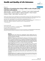
Organ resistance was significantly higher and flow rate
significantly lower at the start of perfusion for Viaspa-
nUW-MP grafts compar ed to KPS-M P organs (p < 0.05,
Figure 1A and 1B).
Function recovery (Figure 1C to 1G)
Animals from the ViaspanUW-CS group never recovered
diuresis, their serum creatinine increased steadily until
day 7 when the obvious lack of function recovery and
generally poor state of the animal lead us to euthanize
them. ViaspanUW-MP and KPS-CS groups recovered
diuresis by day 4 p ost reperfusion, functional recovery
was similar except for a lower creatinine peak at day 5 (p
< 0.05) and a higher osmolarity ratio from D5 to D11 for
KPS-CS (p < 0.05). KPS-MP demonstrat ed better func-
tion recovery with diuresis resuming at D3, lower s erum
creatinine levels and a similar osmolarity ratio to Viaspa-
nUW-MP. MP groups also demonstrated controlled gly-
cosuria by D11 (p < 0.05 versus KPS-CS), while glycemia
was normal in all groups (data not shown).
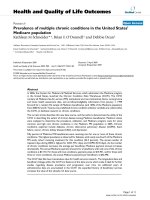
Urinary enzymes (Figure 2 A and 2B)
Measurement of urinary levels of p roximal tubule
enzymes alanine peroxydase and N-acetyl-b-D-glucosa-
minidase (b-NAG) showed e arly high level s followed by
a progressive reduction with time, sign of tubular
damage and s low recovery of structure. KPS-MP grafts
showed fastest and most effective recovery, with Viaspa-
nUW-MP and KPS-CS showing consistently higher
levels (p < 0.05).
Vaziri et al. Journal of Translational Medicine 2011, 9:15
/>Page 3 of 13
Figure 1 Perfusion Parameters and Kidney function following reperfusion. A: Flow rate and B: Resistance of machine perfused kidneys. C:
Diuresis before and after transplantation. D: Serum creatinine before and after transplantation. E: Sodium excretion fraction. F: Glycosuria. G:
Osmolarity ratio between blood and urine. Shown are mean ± SEM, statistics: † : p < 0.05 to ViaspanUW CS; * : p < 0.05 to ViaspanUW MP; ° : p
< 0.05 to KPS CS; ¶ : p < 0.05 to KPS-MP.
Vaziri et al. Journal of Translational Medicine 2011, 9:15
/>Page 4 of 13
Oxydative Stress (Figure 2, C)
Measure in peripheral blood of the ratio of oxidized glu-
tathione over total glutathione, reflecting the oxidative
stress state of the animal, showed lowest levels at all
time points for KPS-MP group (p < 0.05). ViaspanUW-
MP group showed equal or lower levels than KPS-CS.
ViaspanUW-CS showed the highest levels for the dura-
tion of the follow up. Statistical analysis showed that use
of MP was correlated with lower oxidized glutathione
levels at Day3 (R
2
= 0.76, p < 0.0001) and 2 way
ANOVA showed an influence of solution (p < 0.05) and
perfusion technique (p < 0.001) while no additive influ-
ence was determined. At day 7, MP was also correlated
with lower levels (R
2
= 0.5 4, p < 0.01 ) and 2 wa y
ANOVA showed additive effect of solution and perfu-
sion technique (p < 0.01). Use of KPS was not correlated
with lower levels at day 3 while it was slightly correlated
with levels at day 7 (R
2
= 0.41, p < 0.01)
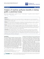
Tissue histology (Figure 3, Table 2)
Evaluation of tissue histology at D7 showed intense tis-
sue damage and necrosis for ViaspanUW-CS grafts.
There was significantly reduced damage in the Viaspa-
nUW-MP group (p < 0.05) compared to ViaspanUW-
CS. KPS grafts tended to show lower amount of damage
compared to ViaspanUW kidneys. At D14 and M1,
ViaspanUW-MP consistently showed more tissue
damage (p < 0.05 at M1) and tubulo-interstitial invasion
compared to KPS-CS, and further reduction was
observed in KPS-MP kidneys (p < 0.05 to both at M1).
Immune response development (Figure 4)
Immunostaining for monocyte/macrophages (ED1+)
showed consistently lower invasion level in KPS-MP
group (p < 0.05), while KPS-CS and ViaspanUW-MP
demonstrated similar cell n umber until M1. After 3
month, invasion in KPS-CS was lower than in Viaspa-
nUW-MP (p < 0.05). Staining for CD3+ showed lower
levels in KPS groups co mpared to ViaspanUW gro ups
throughout the duration of the follow up (p < 0.05).
KPS-MP grafts had lower invasion levels compared to
KPS-CS starting from M1 u ntil M3 (p < 0.05). U se of
KPS was correlated with lower invasion lovels for both
ED1+ (R
2
= 0.75, p < 0.0001) and CD3+ (R
2
=0.78,p<
0.0001). Within the KPS groups, MP was correlated
with lower invasion (ED1+: R
2
= 0.96, p < 0.0001; CD3
+: R
2
= 0.98, p < 0.0001)
Epithelial to Mesenchymal Transition (Figure 5)
Evaluation of Vimentin sta ining at 3 mon th revealed
high levels of Vimentin expression in ViaspanUW-MP
kidneys. Expression was halved in KPS-CS kidney (p <
0.05) and further diminished in KPS-MP grafts (p < 0.05
to both KPS-CS and ViaspanUW-MP).
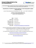
Renal survival, Function and Interstitial Fibrosis/Tubular
Atrophy (Figure 6)
No animal of the ViaspanUW-CS group survived
beyond D7. Three months after transplantation, survival
was lowest in KPS-CS group, f ollowed by KPS-MP with
ViaspanUW-MP showing the highest survival rate,
although the differences were no t significant. Morpholo-
gical analysis (Additiona l file 1) reveale d extens ive
necrosis and tubule loss at week 1 for cases of primary
non function (PNF), graft loss at weeks 2 and 4 was due
Figure 2 Tubular integrity and Red/Ox Status following
reperfusion. A: Alanine aminopeptidase activity in urine. B: b-N-
acetylglucosaminidase activity in urine. C: Blood reduced gutathion
over total glutathion ratio. Shown are mean ± SEM, statistics: † :p<
0.05 to ViaspanUW CS; * : p < 0.05 to ViaspanUW MP; ° : p < 0.05 to
KPS CS; ¶ : p < 0.05 to KPS-MP.
Vaziri et al. Journal of Translational Medicine 2011, 9:15
/>Page 5 of 13
Figure 3 Graft Hist ology. Representative PAS staining of kidney biopsies at day 7 and Month 1 post transplantation. LBB: loss of brush border;
CD: Endoluminal cell detachment; Ti: tubulo-interstitial inflammation.
Vaziri et al. Journal of Translational Medicine 2011, 9:15
/>Page 6 of 13
to high rate of inflammation and tubulitis. Serum creati-
nine was highest in ViaspanUW-MP group, followed by
KPS-CS (p < 0.05) and KPS-MP (p < 0.05 to both).
This order was also found when evaluating fibrosis
development: ViaspanUW-MP kidneys showed mo re
than 30% fibrosis, while KPS-CS neared 20% (p < 0.05
to ViaspanUW-MP). Fibrosis development in KPS-MP
was negligible and did not differ from control. Here
also, use of KP S correlated with lower fibro sis (R
2
=
0.65, p < 0.01). Within the KPS grou ps, MP was corre-
lated with lower fibrosis (R
2
= 0.87, p < 0.01).
Discussion
Herein, we demonstrate in a preclinical study using a
highly reproducible swine model of transplantation the
benefits of MP over CS, particularly in regards to
chronic outcome.
We performed static preservation with both Viaspa-
nUW and KPS, demonstrating the superior ity of KPS in
terms of function recovery, histology at D7 and survival.
Comparisons of these two groups offers a perspective
on studies generally performed on machine perfusion:
when two different solution s are used for static and
machine preservatio n, the obse rved effect is not solely
due on perfusion but also depends significantly on the
solution used. Our 4 groups/2 variables approach cir-
cumvents this bias, highlighting the importance of ani-
mal studies in large animals to assess the benefits of
novel therapies, as indeed such setting is impossible in
the clinic.
Weight variations of kidney grafts are classically
observed during preservation. Our observation of weight
loss for CS and weight gain for MP are consistent with
a similar experimental design in pigs [23]. In addition,
increases in kidney weight after MP have been pre-
viously reported to have no significant impact on the
graft outcome [40].
Comparing ViaspanUW-CS to ViaspanUW-MP allows
us to determine the benefits of machine perfusion with
the current high-K
+
gold standard in static preservation.
Although ViaspanUW is not used for MP in clinical set-
tings, using identical preservation solutio n focuses the
analysis solely on the effect of perfusion. Early follow up
with classical tools such as serum cre atinine do not
allow to determine differences between the two meth-
ods. In our setting, pigs were not dialyzed thus analysis
of diuresis was pertinent, but this would not be the case
in the clinic. Interestingly, measure of peripheral blood
Table 2 Histological Evaluation
ViaspanUW-CS ViaspanUW-MP KPS-CS KPS-MP
Brush Border loss
D7 5.0 ± 0.0 4.1 ± 0.3 † 3.6 ± 0.4 † 3.6 ± 0.5 †
D14 n/a 3.2 ± 0.7 3.0 ± 0.6 2.0 ± 0.4 *
M1 n/a 3.6 ± 0.8 2.0 ± 0.5 * 1.2 ± 0.3 * °
Endoluminal Detachment
D7 5.0 ± 0.0 4.3 ± 0.2 † 3.3 ± 0.3 † 3.0 ± 0.6 †
D14 n/a 3.6 ± 0.8 2.8 ± 0.6 2.0 ± 0.4 *
M1 n/a 2.8 ± 0.6 2.0 ± 0.5 * 1.0 ± 0.2 * °
Tubulo-interstitial Inflammation
D7 necrosis 3.0 ± 0.1 3.0 ± 0.1 2.0 ± 0.1
D14 n/a 3.2 ± 0.2 3.0 ± 0.1 2.0 ± 0.1 *
M1 n/a 2.6 ± 0.3 2.0 ± 0.1 1.0 ± 0.1 * °
Statistics: †:p < 0.05 to UW-CS, *:p < 0.05 to UW-MP, °: p < 0.05 to KPS-CS
Figure 4 Inflammation. A: Representative images of typical ED1+
(top) and CD3+ (bottom) staining of kidneys from each group. B:
graphical representation of the number of ED1 positive cells at each
time point for each group. C: graphical representation of the
number of CD3 positive cells at each time. Shown are mean ± SEM,
statistics: † : p < 0.05 to ViaspanUW CS; * : p < 0.05 to ViaspanUW
MP; ° : p < 0.05 to KPS CS; ¶ : p < 0.05 to KPS-MP.
Vaziri et al. Journal of Translational Medicine 2011, 9:15
/>Page 7 of 13
gluthathion red/ox status provided discriminating infor-
mation between the groups, which was enhanced by
analysis of histology at day 7. Use of UW d emonstrates
in the clearest f ashion the benefits of MP: while high
concentration of potassium induce vasoconstriction, as
seen in the resistance index at beginning of perfusion,
themachineisabletorescuethisnegativeeffectand
regulate flow, allowing the organ to better face the stress
Figure 5 EMT development. A, B, C and D: Representative staining for Vimentin at 3 months. E: quantification of staining in each group.
Shown are mean ± SEM, statistics: † : p < 0.05 to ViaspanUW CS; * : p < 0.05 to ViaspanUW MP; ° : p < 0.05 to KPS CS; ¶ : p < 0.05 to KPS-MP.
Vaziri et al. Journal of Translational Medicine 2011, 9:15
/>Page 8 of 13
of reperfusion, with dramatic benefits on outcome parti-
cular ly su rvival, such as found in the clinic [27,28]. This
model thus offers a unique opportunity for further clari-
fication of the e xact mechanisms through which MP
provides this protection.
Benefits of machine perfusion were also not immedi-
ately obvious between KPS-CS and KPS-MP groups:
diuresis and creatinine levels were close, as were other
functional parameters usually available in the clinic.
Here also, discrimination was possible with measure of
Glutathion red/ox. Moreover, since both groups pro-
duced urine, proximal tubule enzymes activity assay in
the urine was invaluable. Alanine aminopeptidase and
b-N-acetylglucosaminidase are found in kidney tubular
cell s br ush border and their presence in urine is a com-
monly accepted sign of tubular damage [41], their activ-
ity level in the urine revealed a superiority of MP in
maintaining tissue integrity at all time point, which was
confirmed by histological analysis of the grafts
parenchyma.
Early follow up of ViaspanUW-M P and KPS-CS
showed similar values on t he tests we performed, high-
lighting the existence of a solution bias when comparing
preservation strategies. Altogether, results from the early
follow up do not permit a clear discrimination between
CS and MP, unless we consider less orthodox tests such
as gluta thione red/ox or urinary tubular enzyme activity
assays. Excretion of Na
+
and glycosuria, in a context of
normoglycemia, also offered a degree of discrimination
between experimental groups for tubular necrosis and
tubular dysfunction.
In the case of glutathione red/ox, a clear correlation
was drawn between the use of M P and lower oxidative
stress, and both solution and perfusion technique
demonstrated an effect on this paramete r. However,
addition of effects was not found until day 7. We thus
0
5
10
15
20
25
30
35
40
UW-CS
UW-MP
% Sirius Red Staining
D
D: Control
E: UW-MP
G: KPS MP
F: KPS CS
KPS-CS
KPS-MP
Control
*
°
*
0.00
0.25
0.50
0.75
1.00
1.25
1.50
1.75
0
100
200
300
Proteinuria (g/24h)
B
UW-CS
UW-MP
Creatinemia (μM)
C
KPS-CS KPS-MP
*
°
*
H
*
UW-CS
UW-MP
KPS-CS KPS-MP
A
0 2 4 6 8 10 12
0
25
50
75
100
Time
Percent survival
ViaspanUW CS
ViaspanUW MP
KPS CS KPS MP
Figure 6 3 month Outcome. Survival was measured and represented by a Kaplan-Meier plot (A). Function was determined: Creatinemia (B) and
proteinuria (C) Representative images of Sirius Red staining of sections obtained from Control (D), ViaspanUW-MP (E), KPS-CS (F) KPS-MP (G)
kidneys. Original magnification x100. H: Quantification of fibrosis in kidneys from each group studied. Shown are mean ± SEM, statistics: † :p<
0.05 to ViaspanUW CS; * : p < 0.05 to ViaspanUW MP; ° : p < 0.05 to KPS CS; ¶ : p < 0.05 to KPS-MP.
Vaziri et al. Journal of Translational Medicine 2011, 9:15
/>Page 9 of 13
identify an independent machine effect, however the
relatively small differences observed herein would l ikely
not be present in the clinic due to disparities in patients
population , while in iden tical pigs statistical significance
is obtainable. Grafts histology analysis confirmed the
superiority of MP over CS, however these tests may not
be standard in clinical practice. Thus, measurement of
the benefits of MP is difficult in short follow up studies,
particularly if the preservation solution bias is not
circumvented.
We followed animals for 3 month post reperfusion. In
this large animal model, this length permit s us to follow
the development of chronic lesions such as immune
response and interstitial fibrosis and tubular atrophy
(IFTA). The summated effects of damage sustained by
organ preservation and reperfusion [42] lead to loss of
graft function, and ultimately loss of the grafts itself,
often due to the development of IFTA [43]. This pathol-
ogy is also strongly correlated with immune r esponse
[42,44-46]. Herein, KPS-MP showed less innate and
adaptative invasion compared to KPS-CS, which showed
lower levels that ViaspanUW-MP. Use of KPS correlated
with lower invasion, and w ithin the KPS groups we
showed that the use of MP correlated with better out-
come. Unfortunately, absence of data from the UW-CS
group did not allow us to perform further statistical
analysis. This confirms the benefits of the machine on
chronic immune response development. The Viaspa-
nUW-MP fared poorly compared to KPS groups, how-
ever its superiority to ViaspanUW-CS is demonstrated
in terms of animal sur viva l. These results are in contra-
diction to a study conducted on dogs [21], however the
setting of the study and the anatomy of the dog kidney
render the comparison of data difficult.
Epithelial to mesenchymal transition (EMT), a process
through which polarized tubular cells are driven to de-
differentiate and al ter their phe notype towards that of a
mobile and fast proliferating mesenchymal cell [47], is
shown to be a repair mechanism that can be deregulated
during injury and promote interstitial fibrosis [48-50].
Our results show that Vimentin s taining, a marker o f
EMT, is high in ViaspanUW-MP, lower in KPS-CS and
close to control levels in KPS-MP. Thus, the machine
effect is also fo und in a major pathway leading to fibro-
sis and graft loss. We measured the extend of fibrosis
using Sirius red and showed a similar order in the grade
of lesion: ViaspanUW-MP was highest and KPS-CS
showed half the degree of fibrosis of ViaspanUW-MP.
KPS-MP group did not show a degree of fibrosis higher
than control.
Considering no ViaspanUW-CS animal survived to the
end of the follow up, no comparison is possible in
regards to chronic lesions such as immune response or
fibrosis, however previous studies using the same
protocol as ViaspanUW-CS showed a 27% survival rate
with im portant immune respo nse and IFTA (47%)
[51,52]. ViaspanUW-MP showed better survival,
strengthening the results of a similar study inv estigating
the short-term eff ects (7 days) of ViaspanUW-MP in a
pig model [19], also reporting trends towards a better
early kidney function after MP [19,23]. Our results
demonstrate superiority of KPS over ViaspanUW solu-
tion in our animal model, independently of the preserva-
tion strategy. UW is a high K
+
and low Na
+
solution
[53], proposed to maintain intracellular ionic balance.
However high potassium has been shown to induce cel-
lular depolarization, decrease cellular ATP content and
activates voltage-dependent channels, such as calcium
channels [54,55]. Influx of calcium can result in vaso-
constriction impairing organ perfusion during washout
and reperfusion, participating in the ‘ no ref low’ phe-
nomenon [56-58]. Recently, studies have shown equal or
improved results of low potassium/high s odium ratio
such as KPS [1], consistent with our findings. Use of
Mannitol instead of lactobionate in KPS may also
account for the better performance, as this compound
has reactive oxygen species scavenging properties [1].
The present study uses large white pigs, an animal well
suited for preclinical studies as it is close to humans, par-
ticularly in regards to the multipapillar multilobular orga-
nization of its kidney, only found in higher mammals,
implying a complex vascular bed making these organs
particularly sensitive to IRI [59]. In this setting, we deter-
mined that the benefits of machine perfusion, with a
machine currently used in the clinic, are most evident on
chronic graft outcome. Indeed, discrimination between
the groups in the early time points was only possible
through assays rarely performed in transplant centers
and thus could explain the relatively small benefits found
in clinical studies investigating the machine effect [27].
However, our results suggest that chronic follow up of
these patients will uncover a wider rift between MP and
CS, as chronic lesions start to develop.
The exact mechanisms by which MP minimizes the
activation of lesional pathways in our study remain to
be elucidated. MP actions may include a complete per-
fusion of the organ promoting a thorough washout of
blood and subseq uent tissue equilibration with the pre-
servation solution. This more efficient washout has been
previously reported to limit the aggregation of erythro-
cytes [60]. Finally, t he maintenance of a flow may pro-
tect against depolarization of the endothelial cell
membrane which is linked to generation of ROS,
increased intracellular Ca
2+
concentration, and activa-
tion of NO synthases [61]. Hence, more mechanistic
studies are necessary to unravel the exact mechanism of
action in MP, in order to focus on improvement and
optimal application of this technique.
Vaziri et al. Journal of Translational Medicine 2011, 9:15
/>Page 10 of 13
The present study appears limited by the use of an iso-
graft model, devoid of the influence of immunosuppres-
sants. However, machine perfusion has been developed to
optimize graft preservation, hence address ischemia reper-
fusion injury. Thus, we felt that an allograft model, with
the addition of immunosuppressors and their own set of
deleterious side effects, would dilute the impact of our
results. We thus sacrificed relevance to the clinic by the
use of i sograft in order to obtain clarity of our results in
regards to the benefits of machine perfusion. Another lim-
itation is the fact that our model does not follow exactly
the setting of classes I and II of the Maastricht criteria.
Indeed, it normally includes no more than 30 min arrest
before starting the CPR procedure, which is then contin-
ued during the transport to the hospital (generally with a
machine); then as failure to resuscitate is pronounced
there is a 5 min no touch period. All these steps should
not exceed 150 min. The patient is then either cold per-
fused or a extracorporeal circuit is put in place, giving
enough time to secure consent from the family and collect
the organs, which are then machine perfused. It is obvious
that a correct modelling of this situation should include all
these steps, and we are actually in the process of adapting
such procedures on the pig. However in the meantime we
are using 60 min WI as it reproduces as closely as possible
the conditions of DCD.
Conclusion
In a study using a preclinical model of DCD kidney
transplantation, we demonstrate the superiority of MP
over CS independently of the solution used for perfu-
sion. Our results suggest significant benefits on graft
outcome, particularly evident on the chronic effects of
IRI with a protection against chronic immune response,
EMT and IFTA.
Additional material
Additional file 1: Representative graft morphology for kidney lost
during follow up. Morphological analysis of grafts lost during follow up
revealed extensive necrosis and tubule loss at week 1 for cases of
primary non function (PNF). Graft loss at weeks 2 and 4 was due to high
rate of inflammation and tubulitis.
Acknowledgements
We deeply thank Sandrine Joffrion, Dominique Lochon and William Hebrard
for their excellent technical assistance, Dr Jérome Cau for his surgical advices
and Pr Jean-Michel Goujon for his commentaries on the histological analysis.
We extend these thanks to our funding sources, the Conseil Général de la
Vienne, Région Poitou Charentes, the Banque Tarneaud, Poitiers, CHU de
Poitiers and Inserm, the Société Francophone de Transplantation, the French
Foundation of Transplantation.
Author details
1
Inserm U927, Poitiers, Poitiers F-86021, France; Univ Poitiers; Faculté de
Médecine et de Pharmacie, Poitiers, F-86034, France.
2
Service d’Urologie et
chirurgie de la transplantation, Pavillon V - Hôpital Edouard Herriot - 5, place
d’Arsonval, 69437 Lyon, France.
3
CHU Poitiers, Pole UBM, Service de
Biochimie, Poitiers, F-86021, France.
4
IBISA, Domaine expérimental du
Magneraud, Surgères, F17700, France.
5
Service d’Urologie et Transplantation,
Hôpital Pitié Salpétrière, Groupe Hospitalier Universitaire Est, 75651 Paris
cedex 13, Paris, France.
6
Université Pierre et Marie Curie, 75005 Paris cedex,
Paris France.
7
FLIRT : Fédération pour L’étude de l’Ischémie Reperfusion en
Transplantation, Poitiers, F-86034, France.
Authors’ contributions
NV carried out the animal experiment design, surgery, data gathering. RT
carried out the data management and processing, writing the paper. FF
carried out experimental design, running experiments, data gathering. NC
carried out the experiments, data gathering. SM carried out the experimental
design, running experiments, data gathering. ME carried out the study
design. TH carried out the study design, writing the paper. BB carried out
the animal experimental design, study design. All authors read and
approved the final manuscript.
Competing interests
The authors declare that they have no competing interests.
Received: 28 September 2010 Accepted: 25 January 2011
Published: 25 January 2011
References
1. Maathuis MH, Leuvenink HG, Ploeg RJ: Perspectives in organ preservation.
Transplantation 2007, 83:1289-1298.
2. Ploeg RJ, van Bockel JH, Langendijk PT, Groenewegen M, van der
Woude FJ, Persijn GG, Thorogood J, Hermans J: Effect of preservation
solution on results of cadaveric kidney transplantation. The European
Multicentre Study Group. Lancet 1992, 340:129-137.
3. Deroure B, Kamar N, Depreneuf H, Jacquet A, Francois H, Charpentier B,
Rostaing L, Durrbach A: Expanding the criteria of renal kidneys for
transplantation: use of donors with acute renal failure. Nephrol Dial
Transplant 25:1980-1986.
4. Zuckerman JM, Singh RP, Farney AC, Rogers J, Stratta RJ: Single center
experience transplanting kidneys from deceased donors with terminal
acute renal failure. Surgery 2009, 146:686-694, discussion 694-685.
5. Cooper JT, Chin LT, Krieger NR, Fernandez LA, Foley DP, Becker YT,
Odorico JS, Knechtle SJ, Kalayoglu M, Sollinger HW, D’Alessandro AM:
Donation after cardiac death: the university of wisconsin experience
with renal transplantation. Am J Transplant 2004, 4:1490-1494.
6. Keizer KM, de Fijter JW, Haase-Kromwijk BJ, Weimar W: Non-heart-beating
donor kidneys in the Netherlands: allocation and outcome of
transplantation. Transplantation 2005, 79:1195-1199.
7. Rudich SM, Kaplan B, Magee JC, Arenas JD, Punch JD, Kayler LK, Merion RM,
Meier-Kriesche HU: Renal transplantations performed using non-heart-
beating organ donors: going back to the future? Transplantation 2002,
74:1715-1720.
8. Kootstra G: The asystolic, or non-heartbeating, donor. Transplantation
1997, 63:917-921.
9. Asher J, Wilson C, Gok M, Balupuri S, Bhatti AA, Soomro N, Rix D, Jaques B,
Manas D, Shenton B, Talbot D: Factors predicting duration of delayed
graft function in non-heart-beating donor kidney transplantation.
Transplantation proceedings 2005, 37:348-349.
10. Alijani MR, Cutler JA, DelValle CJ, Morres DN, Fawzy A, Pechan BW,
Helfrich GB: Single-donor cold storage versus machine perfusion in
cadaver kidney preservation. Transplantation 1985, 40:659-661.
11. Balupuri S, Buckley P, Snowden C, Mustafa M, Sen B, Griffiths P, Hannon M,
Manas D, Kirby J, Talbot D: The trouble with kidneys derived from the
non heart-beating donor: a single center 10-year experience.
Transplantation 2000, 69:842-846.
12. Daemen JH, de Vries B, Kootstra G: The effect of machine perfusion
preservation on early function of non-heart-beating donor kidneys.
Transplantation proceedings 1997, 29:3489.
13. Kwiatkowski A, Wszola M, Kosieradzki M, Danielewicz R, Ostrowski K,
Domagala P, Lisik W, Fesolowicz S, Michalak G, Trzebicki J, et al: The early
and long term function and survival of kidney allografts stored before
transplantation by hypothermic pulsatile perfusion. A prospective
randomized study. Ann Transplant 2009, 14:14-17.
Vaziri et al. Journal of Translational Medicine 2011, 9:15
/>Page 11 of 13
14. Kwiatkowski A, Wszola M, Kosieradzki M, Danielewicz R, Ostrowski K,
Domagala P, Lisik W, Nosek R, Fesolowicz S, Trzebicki J, et al: Machine
perfusion preservation improves renal allograft survival. Am J Transplant
2007, 7:1942-1947.
15. Moustafellos P, Hadjianastassiou V, Roy D, Muktadir A, Contractor H,
Vaidya A, Friend PJ: The influence of pulsatile preservation in kidney
transplantation from non-heart-beating donors. Transplantation
proceedings 2007, 39:1323-1325.
16. Reznik ON, Bagnenko SF, Loginov IV, Iljina VA, Ananyev AN, Eremich SV,
Moysyuk YG: Machine perfusion as a tool to select kidneys recovered
from uncontrolled donors after cardiac death. Transplantation proceedings
2008, 40:1023-1026.
17. van der Vliet JA, Kievit JK, Hene RJ, Hilbrands LB, Kootstra G: Preservation
of non-heart-beating donor kidneys: a clinical prospective randomised
case-control study of machine perfusion versus cold storage.
Transplantation proceedings 2001, 33:847.
18. Wight JP, Chilcott JB, Holmes MW, Brewer N: Pulsatile machine perfusion
vs. cold storage of kidneys for transplantation: a rapid and systematic
review. Clinical transplantation 2003, 17:293-307.
19. Treckmann J, Nagelschmidt M, Minor T, Saner F, Saad S, Paul A: Function
and quality of kidneys after cold storage, machine perfusion, or
retrograde oxygen persufflation: results from a porcine
autotransplantation model. Cryobiology 2009, 59:19-23.
20. Nicholson ML, Hosgood SA, Metcalfe MS, Waller JR, Brook NR: A
comparison of renal preservation by cold storage and machine
perfusion using a porcine autotransplant model. Transplantation 2004,
78:333-337.
21. Lindell SL, Compagnon P, Mangino MJ, Southard JH: UW solution for
hypothermic machine perfusion of warm ischemic kidneys.
Transplantation 2005, 79:1358-1361.
22. Hosgood SA, Yang B, Bagul A, Mohamed IH, Nicholson ML: A comparison
of hypothermic machine perfusion versus static cold storage in an
experimental model of renal ischemia reperfusion injury. Transplantation
89:830-837.
23. La Manna G, Conte D, Cappuccilli ML, Nardo B, D’Addio F, Puviani L,
Comai G, Bianchi F, Bertelli R, Lanci N, et al: An in vivo autotransplant
model of renal preservation: cold storage versus machine perfusion in
the prevention of ischemia/reperfusion injury. Artificial organs 2009,
33:565-570.
24. Irish WD, Katz E: Cold machine perfusion or static cold storage of
kidneys: why the debate continues. Am J Transplant 10:1955-1956.
25. Watson CJ, Wells AC, Roberts RJ, Akoh JA, Friend PJ, Akyol M, Calder FR,
Allen JE, Jones MN, Collett D, Bradley JA: Cold machine perfusion versus
static cold storage of kidneys donated after cardiac death: a UK
multicenter randomized controlled trial. Am J Transplant 10:1991-1999.
26. Reich DJ, Mulligan DC, Abt PL, Pruett TL, Abecassis MM, D’Alessandro A,
Pomfret EA, Freeman RB, Markmann JF, Hanto DW, et al:
ASTS
recommended practice guidelines for controlled donation after cardiac
death organ procurement and transplantation. Am J Transplant 2009,
9:2004-2011.
27. Moers C, Smits JM, Maathuis MH, Treckmann J, van Gelder F, Napieralski BP,
van Kasterop-Kutz M, van der Heide JJ, Squifflet JP, van Heurn E, et al:
Machine perfusion or cold storage in deceased-donor kidney
transplantation. The New England journal of medicine 2009, 360:7-19.
28. Jochmans I, Moers C, Smits JM, Leuvenink HG, Treckmann J, Paul A,
Rahmel A, Squifflet JP, van Heurn E, Monbaliu D, et al: Machine perfusion
versus cold storage for the preservation of kidneys donated after
cardiac death: a multicenter, randomized, controlled trial. Annals of
surgery 252:756-764.
29. Yuan X, Theruvath AJ, Ge X, Floerchinger B, Jurisch A, Garcia-Cardena G,
Tullius SG: Machine perfusion or cold storage in organ transplantation:
indication, mechanisms, and future perspectives. Transpl Int 2010,
23:561-570.
30. Taylor MJ, Baicu SC: Current state of hypothermic machine perfusion
preservation of organs: The clinical perspective. Cryobiology 2010, 60:
S20-35.
31. Cooper JT, Freeman RB: The value of machine perfusion in deceased
donor kidney transplantation. Am J Kidney Dis 2009, 54:410-412.
32. Tullius SG, Garcia-Cardena G: Organ procurement and perfusion before
transplantation. The New England journal of medicine 2009, 360:78-80.
33. Schreinemachers MC, Doorschodt BM, Florquin S, van den Bergh
Weerman MA, Zernecke A, Idu MM, Tolba RH, van Gulik TM: Pulsatile
perfusion preservation of warm ischaemia-damaged experimental
kidney grafts. The British journal of surgery 97:349-358.
34. Doorschodt BM, Schreinemachers MC, Florquin S, Lai W, Sitzia M,
Zernecke A, Tolba RH: Evaluation of a novel system for hypothermic
oxygenated pulsatile perfusion preservation. The International journal of
artificial organs 2009, 32:728-738.
35. Hosgood SA, Barlow AD, Yates PJ, Snoeijs MG, van Heurn EL, Nicholson ML:
A Pilot Study Assessing the Feasibility of a Short Period of
Normothermic Preservation in an Experimental Model of Non Heart
Beating Donor Kidneys. The Journal of surgical research .
36. Favreau F, Rossard L, Zhang K, Desurmont T, Manguy E, Belliard A, Fabre S,
Liu J, Han Z, Thuillier R, et al: Expression and modulation of translocator
protein and its partners by hypoxia reoxygenation or ischemia and
reperfusion in porcine renal models. American journal of physiology 2009,
297:F177-190.
37. Jayle C, Favreau F, Zhang K, Doucet C, Goujon JM, Hebrard W, Carretier M,
Eugene M, Mauco G, Tillement JP, Hauet T: Comparison of protective
effects of trimetazidine against experimental warm ischemia of different
durations: early and long-term effects in a pig kidney model. American
journal of physiology 2007, 292:F1082-1093.
38. Mueller PW, MacNeil ML, Steinberg KK: Stabilization of alanine
aminopeptidase, gamma glutamyltranspeptidase, and N-acetyl-beta-D-
glucosaminidase activity in normal urines. Archives of environmental
contamination and toxicology 1986, 15:343-347.
39. Grimm PC, Nickerson P, Gough J, McKenna R, Stern E, Jeffery J, Rush DN:
Computerized image analysis of Sirius Red-stained renal allograft
biopsies as a surrogate marker to predict long-term allograft function. J
Am Soc Nephrol 2003, 14:1662-1668.
40. Wilson CH, Gok MA, Shenton BK, Balupuri S, Gupta AJ, Asher J, Talbot D:
Weight increase during machine perfusion may be an indicator of organ
and in particular, vascular damage. Ann Transplant 2004, 9:31-32.
41. Nicot GS, Merle LJ, Charmes JP, Valette JP, Nouaille YD, Lachatre GF, Leroux-
Robert C: Transient glomerular proteinuria, enzymuria, and nephrotoxic
reaction induced by radiocontrast media. Jama 1984, 252:2432-2434.
42. Nankivell BJ, Chapman JR: Chronic allograft nephropathy: current
concepts and future directions. Transplantation 2006, 81:643-654.
43. El-Zoghby ZM, Stegall MD, Lager DJ, Kremers WK, Amer H, Gloor JM,
Cosio FG: Identifying specific causes of kidney allograft loss. Am J
Transplant 2009, 9:527-535.
44. Ferenbach D, Kluth DC, Hughes J: Inflammatory cells in renal injury and
repair. Seminars in nephrology 2007, 27:250-259.
45. Fletcher JT, Nankivell BJ, Alexander SI: Chronic allograft nephropathy.
Pediatric nephrology (Berlin, Germany) 2009, 24:1465-1471.
46. Thuillier RMR: The Immunology of Chronic Allograft Injury. In Chronic
Allograft Failure: Natural History, Pathogenesis, Diagnosis and Management
Edited by: Ahsan N: Landes Bioscience 2007.
47. Kalluri R, Neilson EG: Epithelial-mesenchymal transition and its
implications for fibrosis. The Journal of clinical investigation 2003,
112:1776-1784.
48. Djamali A, Reese S, Yracheta J, Oberley T, Hullett D, Becker B: Epithelial-to-
mesenchymal transition and oxidative stress in chronic allograft
nephropathy. Am J Transplant 2005, 5:500-509.
49. Vongwiwatana A, Tasanarong A, Rayner DC, Melk A, Halloran PF: Epithelial
to mesenchymal transition during late deterioration of human kidney
transplants: the role of tubular cells in fibrogenesis. Am J Transplant 2005,
5:1367-1374.
50. Bedi S, Vidyasagar A, Djamali A: Epithelial-to-mesenchymal transition and
chronic allograft tubulointerstitial fibrosis. Transplantation reviews
(Orlando, Fla 2008, 22:1-5.
51. Favreau F, Thuillier R, Cau J, Milin S, Manguy E, Mauco G, Zhu X,
Lerman LO, Hauet T: Anti-thrombin therapy during warm ischemia and
cold preservation prevents chronic kidney graft fibrosis in a DCD model.
Am J Transplant 2009, 10:30-39.
52. Thuillier R, Favreau F, Celhay O, Macchi L, Milin S, Hauet T: Thrombin
inhibition during kidney ischemia-reperfusion reduces chronic graft
inflammation and tubular atrophy. Transplantation 2010,
90:612-621.
53. Belzer FO, Southard JH: Principles of solid-organ preservation by cold
storage. Transplantation 1988, 45:673-676.
Vaziri et al. Journal of Translational Medicine 2011, 9:15
/>Page 12 of 13
54. Rauen U, de Groot H: New insights into the cellular and molecular
mechanisms of cold storage injury. J Investig Med 2004, 52:299-309.
55. Salahudeen AK: Cold ischemic injury of transplanted kidneys: new
insights from experimental studies. American journal of physiology 2004,
287:F181-187.
56. Brodsky SV, Yamamoto T, Tada T, Kim B, Chen J, Kajiya F, Goligorsky MS:
Endothelial dysfunction in ischemic acute renal failure: rescue by
transplanted endothelial cells. American journal of physiology 2002, 282:
F1140-1149.
57. Mazzoni MC, Borgstrom P, Intaglietta M, Arfors KE: Lumenal narrowing and
endothelial cell swelling in skeletal muscle capillaries during
hemorrhagic shock. Circulatory shock 1989, 29:27-39.
58. Mazzoni MC, Intaglietta M, Cragoe EJ Jr, Arfors KE: Amiloride-sensitive Na+
pathways in capillary endothelial cell swelling during hemorrhagic
shock. J Appl Physiol 1992, 73:1467-1473.
59. Simmons MN, Schreiber MJ, Gill IS: Surgical renal ischemia: a
contemporary overview. The Journal of urology 2008, 180:19-30.
60. Morariu AM, Vd Plaats A, W VO, NA TH, Leuvenink HG, Graaff R, Ploeg RJ,
Rakhorst G: Hyperaggregating effect of hydroxyethyl starch components
and University of Wisconsin solution on human red blood cells: a risk of
impaired graft perfusion in organ procurement? Transplantation 2003,
76:37-43.
61. Chatterjee S, Chapman KE, Fisher AB: Lung ischemia: a model for
endothelial mechanotransduction. Cell biochemistry and biophysics 2008,
52:125-138.
doi:10.1186/1479-5876-9-15
Cite this article as: Vaziri et al.: Analysis of machine perfusion benefits in
kidney grafts: a preclinical study. Journal of Translational Medicine 2011
9:15.
Submit your next manuscript to BioMed Central
and take full advantage of:
• Convenient online submission
• Thorough peer review
• No space constraints or color figure charges
• Immediate publication on acceptance
• Inclusion in PubMed, CAS, Scopus and Google Scholar
• Research which is freely available for redistribution
Submit your manuscript at
www.biomedcentral.com/submit
Vaziri et al. Journal of Translational Medicine 2011, 9:15
/>Page 13 of 13