báo cáo hóa học:" Elevated expression of CDK4 in lung cancer" ppt
Bạn đang xem bản rút gọn của tài liệu. Xem và tải ngay bản đầy đủ của tài liệu tại đây (1.3 MB, 9 trang )
RESEARCH Open Access
Elevated expression of CDK4 in lung cancer
Aibing Wu
1†
, Bin Wu
2†
, Jinsong Guo
6†
, Weiren Luo
1
, Dong Wu
2
, Huiling Yang
4
, Yan Zhen
1
, Xiaoli Yu
1
, Hao Wang
1
,
Ying Zhou
1
, Zhen Liu
3*
, Weiyi Fang
1*
and Zhixiong Yang
5*
Background: The aim of the present study was to analyze the expression of Cyclin-dependent kinase 4 (CDK4)in
lung cancer and its correlation with clinicopathologic features. Furthermore, the involvement of CDK4-mediated cell
cycle progression and its molecular basis were investig ated in the pathogenesis of lung cancer.
Methods: Using immunohistochemistry analysis, we analyzed CDK4 protein expression in 89 clinicopathologically
characterized lung cancer patients (59 males and 30 females) with ages ranging from 36 to 78 years and
compared them to 23 normal lung tissues. Cases with cytoplasmic and nuclear CDK4 immunostaining score values
greater than or equal to 7 were regarded as high expression while scores less than 7 were considered low
expression. The correlation between the expression level of CDK4 and clinical features was analyzed. Furthermore,
we used lentiviral-mediated shRNA to suppress the expression of CDK4 and investigate its function and molecular
mechanism for mediating cell cycle prog ression.
Results: The expression level of CDK4 protein was significantly increased in lung cancer tissues compared to
normal tissues (P < 0.001). In addition, high levels of CDK4 protein were positively correlated with the status of
pathology classification (P = 0.047), lymph node metastasis (P = 0.007), and clinical stage (P = 0.004) of lung cancer
patients. Patients with higher CDK4 expr ession had a markedly shorter overall survival time than patients with low
CDK4 expression. Multivariate analysis suggested the level of CDK4 expression was an independent prognostic
indicator (P < 0.001) for the survival of patients with lung cancer. Use of lentiviral-mediated shRNA to inhibit the
expression of CDK4 in lung cancer cell line A549 not only inhibited cell cycle progression, but also dramatically
suppressed cell proliferation, colony formation, and migration. Furthermore, suppressing CDK4 expression also
significantly elevated the expression of cell cycle regulator p21
Conclusion: Overexpressed CDK4 is a potential unfavorable prognostic factor and mediates cell cycle progression
by regulating the expression of p21 in lung cancer
Background
Lung cancer is the world’s most prevalent cancer accord-
ing to the World H ealth Organization, with 1.2 million
new cases every year. Nearly all lung cancer s arise due to
smoking and men are more frequently diagnosed than
women. However, a rise in female smoking worldwide
has started reversing the trend.
In China, about 300,000 lung cancer patients (23/
100,000) are diagnosed each year [1]. Unfortunately, most
lung cancer patients tend to present with an advanced
stage of disease due to its deep location within the lungs
and lack of symptoms during early stages. This may con-
tribute to the overall poor prognosis of most lung cancer
patients. Therefore, it is of great interest to identify factors
which provide early diagnosis, more accurate prognosis
prediction, and allow development of novel therapeutic
strategies.
Genetic abnormalities found in lung cancer typically
affect two general classes of genes: oncogenes and tumor
suppressors. Cancer-promoting oncogenes are typically
activated in cancer cells, giving those cells new properties,
such as hyperactive growth and division, protection against
programmed cell death, or loss of respect for normal tissue
boundaries. CDK4 is part of the cyclin-dependent kinase
family. The protein encoded by this gene is a member of
* Correspondence: ; ;
† Contributed equally
1
Cancer Research Institute of Southern Medical University, 510515,
Guangzhou, PR China
3
Department of Pathology, Medical College of Guangzhou, 510450,
Guangzhou, PR China
Full list of author information is available at the end of the article
Wu et al. Journal of Translational Medicine 2011, 9:38
/>© 2011 Wu et al; licensee BioMed Central Ltd. This is an Open Access article distributed under the terms of the Creative Com mons
Attribution License ( which perm its unrestricted use, distribution, and reproduction in
any medium, provided the original work is properly cited.
the Ser/Thr protein kinase family and is highly similar to
the gene products of S. cerevisiae cdc28 and S. pombe
cdc2. It is a catalytic subunit of the protein kinase complex
important for G1 cell cycle progression. Transition
through G1-S phases, is controlled by the regulatory sub-
units D-type cyclins(CDK4 and CDK6) and CDK inhibitor
p16(INK4a). Marval et al. found that CDK4 has higher
oncogenic activity than cyclin D1(CCND1) and it markedly
enhanced malignant skin tumorigenesis in CDK4 trans-
genic mice [2]. Furthermore, overexpression of CDK4 has
been showed in many tumor types, including oral squa-
mous cell carcinoma [3], pancreatic endocrine tumors [4],
lung cancer [5,6], and nasopharyngeal carcinoma [7],
suggesting that CDK4 is a key factor in promoting the
initiation and development of tumors.
In order to clarify the role of CDK4 in the pathogen-
esis of lung cancer, we explored the correlation of its
protein expression with clinicopathologic features of
lung cancer patients. We found that the expression
levels of CDK4 were higher in lung cancer tumors com-
pared to those in normal lung tissues. This increased
CDK4 expression was associated with the progression
and poor prognosis of lung cancer patients. Further-
more, suppressing the expression of CDK4 elevated
tumor suppressor p21 expression, which may function
to reduce cell proliferation and migration.
Materials and methods
Sample collection
Eighty-nine (89) paraffin-embedded lung cancer and 23
normal lung samples were obtained from the First
Affiliated Hospital of Guangdong Medical School,
Zhanjiang City, China. In the 89 lung canc er cases,
there were 59 ma les and 30 females with ages ranging
from 36 to 78 years. The clinical follow-up time of
patientsrangedfrom6to55months.Foruseofthese
clinical materials for research purposes, prior consent
from the patients and approval from the Ethics Com-
mittees of this hospital was obtained. Histological clas-
sification and clinicopatholo gic staging of th e samples
were performed according to the rules of according to
the WHO histologic classification.
Immunohistochemistry
Paraffin sections (4 μm) from samples were deparaffinized
in 100% xylene and re-hydrated in descending ethanol
series and water according to standard protocols. Heat-
induced antigen retrieval was performed in 10 mM citrate
buffer for 2 min at 100°C. Endogenous peroxidase activity
and non-specific antigen were blocked with peroxidase
blocking reagent containing 3% hydrogen peroxide and
serum, followed by incubation with goat anti-human
CDK4 antibody (1:100) (Santa, MA, USA) for overnight at
4°C. After w ashing, the sections were incubated with
biotin-labeled rabbit anti-goat antibody for 10 min at
room temperature, and subsequently were incubated with
streptavidin-conjugated horseradish peroxidase (HRP)
(Maixin Inc, China). The peroxidase reaction was devel-
oped using 3, 3-diaminobenzidine chromogen solution in
DAB buffer substrate. Sections were visualized with DAB
and counterstained with hematoxylin, mounted in neutral
gum, and analyzed using a bright field microscope.
Evaluation of staining
The immunohistochemically stained tissue sections were
reviewed and scored separately by two pathologists
blinded to t he clinical parameters. Expression of CDK4
in the nucleus and in the cytoplasm was independently
evaluated. For cytoplasmic staining, the score was evalu-
ated according to the sum of cytoplasm staining inte n-
sity and the percentage of positive staining areas of
cells. The staining intensity was scored as previously
described(0-3) [8,9] and the percentage of positive stai n-
ing areas of cells was defined as a scale of 0 to 3 where
0 represents <10%, 1 is 10-25%, 2 is 26-75%, and 3 is
≥76%. For nuclear staining, the staining score was
defined based on the sum of nuclear staining intensity
and the number of positive nuclear staining. Nuclear
staining intensity score was consistent with cytoplasm
and positive nuclear staining scores were defined as fol-
lows: 0 represents <10%, 1 is 10-50%, 2 is 51-80%, and 3
is ≥80%. The sum of the cytoplasm and nuclear staining
scores was used as the final staining score for CDK4
(0-12). For statistical analysis, a final staining score of 0-
6or7-12wasrespectivelyconsideredtobeloworhigh
expression.
Establishment of lung cancer A549 cell line with stably
expressing shRNA-CDK4
We selected two sequences(CDK4 509: Sense:5’
CGCGTCCCCGCATGTAGACC AGGACCTAAGTT-
CAAGAGACTTAGGTCCTGGTCTACATGCTTTTTG-
GAAAT 3’ Antisense:5’CGATTTCCAAAAAGCATG
TAGACCAGGACCTAAGTCTCTTGAACTTAGGTCCT
GGTCTACATGCGGGGA 3’) CDK4 1097 Sense:5’CGCG
TCCCCGCAGCACTCTTATCTACATAATTCAAGA-
GATTATGTAGATAAGAGTGCTGCTTTTTGGAAAT
3’ ;Antisense:5’ CGATTTCCAAAAAGCAGCACTCT-
TATCTACATAATCTCTTGAATTATGTAGATAAG
AGTGCTGCGGGGA 3’ )fortargetingtheCDK4 gene
using the BLOCK-It RNAi Designer (Invitrogen, Carlsbad,
CA). The preparation of lentiviral vectors expressing
human CDK4 short hairpin RNA (shRNA) was performed
using the pLVTHM-GFP Lentiviral RNAi Expression Sys-
tem. Replication-incompetent lentivirus was produced by
cotransfection of the pLVTHM/CDK4-shRNA expression
vector and ViraPower packaging mix containing an opti-
mized mixture of two packaging plasmids: psPAX2 and
Wu et al. Journal of Translational Medicine 2011, 9:38
/>Page 2 of 9
pMD2.G into 293FT cells. Lung cancer A549 cells were
infected with lentiviral particles containing specific or
negative control vectors and the single colony with strong
GFP expression was selected to establish stable silencing
cell lines. The total RNA of these cell clones was isolated,
and the levels of CDK4 mRNA were measured using real-
time PCR examination.
Western blot Analysis
Cells were lysed in RIPA Buffer (50 mM Tris-HCl pH
8.0, 1 mM EDTA p H 8.0, 5 mM DTT, 2% SDS), and
protein concentration was determined using BCA assay
(Beyotime Inc, China). Total protein (30 μg) was
resolved using a 10% SDS-PAGE gel and electro-trans-
ferred to polyvinylidene fluoride membranes (Invitrogen,
Carlsbad, CA), and blocked with 5% nonfat dry milk in
Tris-buffered saline, pH 7.5. Membranes were immuno-
blotted overnight at 4°C with rabbit polyclonal anti-
CDK4 antibody(1:500), anti-ACTB antibody(1:400) and
p21(1:200)(Santa Cruz Biotechnology, CA, USA). An
HRP-conjugated anti-rabbit IgG antibody w as used as
the secondary antibody (Zhongshan Inc, China).
Cell Proliferation
Cell proliferation was analyzed using MTT assay (Sigma,
St. Louis, USA). Briefly, 1 × 10
3
cells were seeded into a
96-well plate with quadruplicate repeat for each condi-
tion. After 24 h of incubation, MTT reagent was added
to each well and incubated for 4 h. The formazan crys-
tals formed by viable cells were then solubilized in
DMSO and measured at 490 nm for the absorbance (A)
values. Each experiment was performed in triplicate.
Colony Formation Assay
About 100 cells were added to each well of a 6-well cul-
ture plate, and each cell group contained 2 wells. After
2 weeks of incubation, cells were washed twice with PBS
and stained with Giemsa solution. The number of colo-
nies containing ≥ 50 cells was counted under a micro-
scope. The colony formation efficiency was calculated
as: efficiency = (number of colonies/number of cells
inoculated) × 100%. Each experiment was performed in
triplicates.
Cell Cycle
To evaluate cell cycle distribution, cells were seeded on
10 cm-diameter plates in RPMI 1640 culture medium
containing 10% NBCS. After 48 h of incubation, a total
of1×10
6
cells were harvested, rinsed with cold PBS,
and fixed with 70% ice-cold ethanol for 48 h at 4°C.
Fixed cells were rinsed with cold PBS followed by incu-
bation with PBS containing 10 μg/mL propidium iodide
and 0.5 mg/mL RNase A for 15 min at 37°C. The DNA
content of labeled cells was acquired using FACS
Caliber cytometry (BD Biosciences). Each experiment
was performed in triplicates.
In Vitro Cell Migration Assay
Cells growing in the log phase were t reated with trypsin
and re-suspended as single-cell solution. A total of 1 × 10
5
cells were seeded on a fibronectin-coated polycarbonate
membrane insert in a transwell apparatus (Corning Inc.,
Corning, NY). In the lower chamber, 600 μlofRPMI1640
with 10% NBCS was added as chemoattractant. After the
cells were incubated for 12 h, the insert was washed with
PBS, and cells on the top surface of the insert were
removed by a cotton swab. Cells adhering to the lower
surface were fixed with methanol, stained with Giemsa,
and counted under a microscope in five predetermined
fields (× 200). All assays were independently repeated at
least three times.
Expression examination of Cell cycle factors
Changes in expression of cell cycle regulators CDK1,
CDK2, CDK6, CCND1, p15, p16, p21, and p27 were first
detected by real-time PCR in pLVTHM/ CDK4-shRNA
and contro l expression vector. Subsequently, genes with
markedly differential expression were further validated
by western blot. Real-time PCR and western blot were
carried out as described above.
Statistical analysis
All data were analyzed for statistical significance using
SPSS 13.0 software. The Mann-Whitney U test was
applied to the examination of relationship between
CDK4 exp ression levels and clinicopa thologic character-
istics. Survival analysis was performed using Kaplan-
Meier method. Multivariate Cox proportional hazards
method was used for analyzing the relationship between
the variables and patient’ s sur vival tim e. One-way
ANOVA was used to determine the differences between
groups for a ll in vitro analyses. A P value of less tha n
0.05 was considered statistically significant.
Results
Immunohistochemical analysis of CDK4 protein
expression in lung cancer and normal lung tissues
We measured the expression levels and subcellular locali-
zation of CDK4 protein in 89 archived paraffin-embedded
lung cancer samples and 23 normal lung tissues using
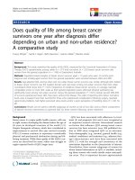
immunohistochemical staining (Figure 1A-E). Specific
CDK4 protein staining was found in the cytoplasm and
nucleus of normal and malignant lung tissues. Further-
more, we observed that in 50.6% (45/89) of lung cancer
samples, CDK4 protein was highly expressed. In compari-
son, only 8.7%(2/23) of normal lung samples had highly
expressed CDK4 protein, significantly lower than that in
the lung cancer samples (P < 0.001) (Table 1).
Wu et al. Journal of Translational Medicine 2011, 9:38
/>Page 3 of 9
Relationship between clinicopathologic characteristics
and CDK4 expression in lung cancer patients
The relationship between c linicopathologic characteris-
tics and CDK4 expression levels in individuals with lung
cancer are summarized in Table 2. We did not find a
significant association of CDK4 expression levels with
patient’ sage,sex,smoking,degreeofdifferentiation,
tumor size (T classification), or status of distant metas-
tases (M classification) in 89 lung cases. However, we
observed that the expression level of CDK4 was posi-
tively correlated with the status of pathology classifica-
tion(P = 0.047) lymph node metastasis (N classi fication)
(N0-N1 vs.N2-N3)(P = 0.007), and clinical stage (I-II
vs. III-IV) (P = 0.004) in lung cancer patients (Table 2).
Survival analysis
To investiga te the prognostic value of CDK4 expression
for lung cancer, we assessed the association between the
expression levels and patient survival using Kaplan-
Meier analysis with the log-rank test. In 89 lung cancer
cases with prognosis information, we observed that the
level of CDK4 protein expression was significantly corre-
lated with the overall survival of lung cancer patients
(Figure 1F). Patients with higher level s of CDK4 expres-
sion had poorer survival rates than those with lower
levels of CDK4 expression (P < 0.001). In addition,
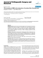
Figure 1 Expression of CDK4 protein predicts lung cancer patients’ survival time. AandB: Strong expression of CDK4 in lung cancer
samples; C and D: Weak expression of CDK4 in lung cancer sample; E:Weak expression of CDK4 in normal lung tissue. F. Kaplan-Meier survival
analysis of overall survival duration in 89 lung cancer patients according to CDK4 protein expression. The log-rank test was used to calculate
p values.
Table 1 Protein expression of CDK4 between lung cancer
and normal lung tissues
Group Protein expression P value
Cases High expression Low expression
Cancer 89 45(50.6%) 44 (49.4%)
Normal 23 21(8.7%) 2 (91.3%) 0.000
Wu et al. Journal of Translational Medicine 2011, 9:38
/>Page 4 of 9
smoking, degree of tumor differentiation, T/N/M classi-
fications and clinical stages were also significantly corre-
lated with patients’ survival (P = 0.05, P = 0.004, P =
0.018, P = 0.003, P =0.039,andP < 0.001 respectively).
To determine whether CDK4 is an independent prog-
nostic factor for lung cancer, we performed multivariate
analysis of CDK4 expression adjusted for the same para-
meters. The results indicated that the level of CDK4
expression was an independent prognostic facto r for
lung cancer (P < 0.001) (Table 3).
Reduced CDK4 Expression Suppressed the Proliferation of
lung cancer cells in vitro
To study the biological function of CDK4,weuseda
lentiviral vector containing shRNA to specifically target
and stably knock down the expression of CDK4 in A549
cells, a lung cancer cell line with high endogenous
levels. Eight stably trans fected cell clones were obtained
(C1, C2, C3, C4, D1, D2, D3, D4) (Figure 2A). Real-time
PCR analysis showed that CDK4 mRNA expression in
C1, C2, and D1 cells was markedly reduced compared
to empty vector control clone A549 cells(PLV-Ctr).
Further, decreased expression of CDK4 protein was con-
firmed by w estern blotting in these three clones com-
pared to PLV-Ctr and A549 cells(Figure 2B). C1 and D1
clones with significantly reduced CDK4 protein expres-
sion were finally chosen for further experiments.
We examined the effect of decreased CDK4 expression
on lung cancer cell growth in vivo. Using an MTT assay,
we found that the parental lung cancer A549 cells had a
similar growth rate as PLV-Ctr cells over a seven-day per-
iod, the growth of shRNA-CDK4 cells was significantly
slower than the former two lines from day 3 (P < 0.05)
(Figure 2C). Interestingly, this result was also consistent in
the plate clone formation test. Both the parental A549
cells and the PLV-Ctr cells formed a similar number of
colonies on plate over a two-week peri od [(68 ± 8.54) vs.
(65 ± 8.00)]. In contrast, knocking down endogenous
Table 2 Correlation between the clinicopathologic characteristics and expression of CDK4 protein in lung cancer
CDK4 (%)
Characteristics n High expression Low expression P
Gender
Male 59 30(50.8%) 29 (49.2%)
Female 30 15(50%) 15 (50%) 1.000
Age(y)
≥65 39 21 (53.8%) 18 (46.2%)
<65 50 24 (48%) 26(52%) 0.671
Smoking
Yes 38 23 (60.5%) 15 (39.5%)
No 51 22 (43.1) 29 (56.9) 0.135
Pathology classification
squamous cell carcinoma 39 15(38.5%) 24(61.5%)
adenocarcinoma 46 17(40%) 29(60%)
small cell undifferentiated carcinoma 4 3(75%) 1(25%) 0.047*
Differentiated degree
High 25 9(36%) 16(64%)
middle 34 21(61.8%) 13(38.2%)
Low or undifferentiated 30 15(50%) 15(50%) 0.150*
T classification
T1+T2 71 32(45.1%) 39(54.9%)
T3+T4 18 13(72.2%) 5(27.8%) 0.063
N classification
N0+N1 58 23 (39.7%) 35 (60.3%)
N2+N3 31 22 (71%) 9 (29%) 0.007
Distant metastasis
Negative 3 3 (100%) 0 (0%)
Positive 86 42 (48.8%) 44 (51.2%) 0.242
Clinical stage
I~II 55 21(38.2%) 34(61.8%)
III~IV 34 24 (70.6%) 10(29.4%) 0.004
*Kruskal Wallis Test.
Wu et al. Journal of Translational Medicine 2011, 9:38
/>Page 5 of 9
CDK4 could dramatically reduce the number of colonies
in C1 cells(40 ± 8.0) and D1 cells(24.33 ± 5.13) (P < 0.05)
(Figure 2D).
Knock-down of CDK4 Inhibited Migration and Cell Cycle
Progression
Cell migration is a key step during tumor development
and metastasis. We tested the ability of A549 cells to
migrate through t he 8 μm pores on the polycarbonate
membrane, and fo und that the knock-down of endogen-
ous CDK4 expression could significantly decrease cell
migration of C1 cells(114 ± 26.75) and D1 cells(80 ± 7.31)
compared to the parental cells(288.2 ± 41.78) or PLV-Ctr
cells (254 ± 34.28) (P < 0.05) (Figures 3A).
We measured the alteration of cell cycle progression
after CDK4 knock-down. Using flow cytom etry analysis,
we found that CDK4-deficient cells showed a significant
increase in G1 phase population cells and a decrease in
S phase cells compared to the PLV-Ctr and the parental
A549 cells (P < 0.05) (Figure 3B).
CDK4 Inhibited the Expression of p21 in A549 cells
The above results indicated that over-expression CDK4
may play an important role in promoting the development
of lung cancer. We further examined the effect of CDK4
on the expression of key regulators of G1-S cell cycle tran-
sition including CDK1, CDK2, CDK6, CCND1, p15, p16,
p21,andp27. Real-time PCR indicated that reducing the
levels of CDK4 significantly activates the expression of
tumor suppressor p21 by 3.12-fold(Figure 4A). Further, we
measured the protein levels of p21 in cells deficient of
CDK4 by western blot. CDK4-deficient cells had increased
levels of p21 protein compared to the parental A549 cells
and cells expressing the control vector (Figure 4B). Our
results suggest that CDK4 may be involved in the develop-
ment of lung cancer by antagonizing the effect of p21.
Discussion
Lung cancer is a disease which con sists of uncontrolled
cell growth in tissues of the lung that may lead to metas-
tases. These growths may ultimately contribute to the
majority of the lung cancer deaths. However, the molecu-
lar mechanisms linking the initiation and development of
lung cancer are not completely understood.
CDK4 has gained prominence as a significant cancer-
related gene, as its function is to drive cell-cycle progres-
sion by phosphorylating the retinoblastoma protein. Over-
expression of CDK4 has been described in many tumors,
including lung cancer.
In this investigation, we analyzed the e xpression of CDK4
protein in lung cancer and normal lung tissues by immu-
nohistochemistry. We found that CDK4 was mainly coex-
pressed in nucleus and cytoplasm in lung cancer tissues
and predominantly expressed in cytoplasm in normal lung
tissues. Furthermore, we presented evidence that CDK4 in
nucleus and total protein levels was overexpressed in lung
cancer tissues compared to normal lung tissues. Our
reports were analogous to Wikman [5], Dobashi [6], and
Table 3 Summary of univariate and multivariate Cox regression analysis of overall survival duration
Univariate analysis Multivariate analysis
Parameter P HR 95%CI P HR 95%CI
Age
≥65vs. <65 years 0.573 1.160 0.692-1.946
Gender
Male vs. female 0.061 0.574 0.322-1.025
Smoking
Yes vs. No 0.05 0.586 0.344-0.999 0.145 0.656 0.372-1.156
Pathology classification
Squamous vs. Adenocarcinoma vs. Small cell undifferentiated 0.883 1.036 0.648-1.656
Differentiation degree
High vs. Middle vs.Low 0.004 1.660 1.176-2.343 0.001 2.076 1.370-3.144
T classification
T
1
-T
2
vs. T
3
-T
4
0.018 2.020 1.130-3.612 0.609 0.819 0.381-1.759
N classification
N
0
-N1 vs. N
2–
N
3
0.003 2.259 1.323-3.860 0.996 1.003 0.273-3.692
M classification
M
0
vs. M
1
0.039 3.436 1.066-11.078 0.088 3.666 0.825-16.293
Clinical stage
Ⅰ-Ⅱ vs. Ⅲ-Ⅳ 0.000 2.586 1.515-4.412 0.470 1.605 0.445-5.787
CDK4 expression
High vs. Low * 0.000 6.420 3.473-11.867 0.000 6.714 3.329-13.451
Wu et al. Journal of Translational Medicine 2011, 9:38
/>Page 6 of 9
Lingfei [10] et al’s results, suggesting that CDK4 partici-
pates in the pathogenesis of lung cancer.
CDK4 is a protein kinase of the CDK family that is
important for cell cycle G1 phase progression, and its
expression pattern is associated with clinical pathology
parameters of lung cancer patients. Yoshida et al. found
that CDK was predominantly expressed in low-grade
osteosarcomas compared to benign histological mimics,
which suggested that CDK4 can be a marker distinguish-
ing low-grade osteosarcoma from benign mimics [11].
Zhang et al. reported that overexpression of CDK4 wa s
positively correlated with Duke’s stage of colorectal can-
cer [12]. In our study, we found that CDK4 overexpres-
sion was significantly correlated with the status of
pathology classification, lymph node metastasis, and clin-
ical stage of lung cancer patients. CDK4 appears to be
more highly expressed in adenocarcinomas compared to
the other two histologic subtypes. Similar to the report
from Dobashi et al., we found that overexpression of
CDK4 was correlated with lymph node metast asis and
statistically higher in the N2/N3 group compared to the
N0/N1 group [6]. In addition, overexpression of CDK4
was positively related to advanced disease status of lung
cancer patients. Our results suggested CDK4 overexpres-
sion in lung cancer may accelerate tumor progression by
promoting cell growth.
Further, we presented the evidence that CDK4 protein
expression in lung cancer was inversely correlated with
patient’s overall survival. Patients with higher expression
of CDK4 protein had an overall shorter survival time.
According to univariate analysis, patient ’soverallsurvi-
val is also inversely proportional to smoking, tumor
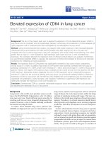
Figure 2 Down-regulation of CDK4 inhibited cell growth in vitro. A. Markedly reduced mRNA expression of CDK4 after shRNA-CDK4: 8 single
clone cells(C1-C4,D1-D4) compared with PLV-Ctr by real-time PCR. B. Significantly decreased protein expression of CDK4 was found in shRNA-
CDK4 cells(C1,C2,D2) compared with PLV-Ctr and A549 cells by western blot. ACTB was used as internal control. C. The cell growth of parental
A549 cells and their stable derivatives, PLV-Ctr and shRNA-CDK4, was examined by MTT assay over a seven-day period. *P < 0.05, as compared to
A549 and PLV-Ctr cells. D. The anchorage-dependent growth of parental A549 cells and their stable derivatives, PLV-Ctr and shRNA-CDK4, was
examined by plate colony formation assay. *P < 0.05, as compared to A549 and PLV-Ctr cells.
Wu et al. Journal of Translational Medicine 2011, 9:38
/>Page 7 of 9
differentiated degree, and T/N/M classification. Multi-
variate analyses showed that i ncreased expression of
CDK4 protein was a significant predictor o f poor prog-
nosis for lung cancer patients. Our reports were not
consistent with Dobashi [6] and Ghazizadeh’sresults
[13]. The discrepancy is most likely due to the different
sample source, sample number, and evaluation method
used. However, our results suggest CDK4 is a clinical
significant biomarker for NPC prognosis.
In previous studies, overexpression of CDK4 had been
shown to promote cell proliferation by driving cell cycle
progression [14-16]. To understand the biological func-
tions of CDK4 in lung cancer, we employed a loss-of-
function approach by knocking down the expression
level of endogenous CD K4.Tothatend,wechoseto
use lung cancer A549 cell line which express high levels
of endogenous CDK4 for our study. Similar to results
publishedbyRetzer-Lidl,An,andRodriguez-Puebla
et al. [14-16], we found that CDK4 plays a role in pro-
moting cell proliferation and migration in vitro. Further-
more, we also found that inhibition of CDK4 could
significantly retard the cell cycle transition from G1 to S
phase. These results strongly support an oncogenic role
for CDK4 in the development of lung cancer.
Based on the increased population of G1-S arrested
cells after inhibiting CDK4 expression, we examin ed
mRNA expression levels of relevant cell cycle factors.
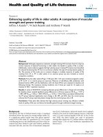
Figure 3 Reduced CDK4 expression inhibited cell migration and cell cycle progression in vi tro. A: The migrating capability of parental
A549 cells and their stable derivatives, PLV-Ctr and shRNA-CDK4, was examined by transwell and boyden chamber assay. B: Cell cycle profile was
determined by FACS Caliber cytometry. Data were presented as mean ± SD for three independent experiments. *P < 0.05, as compared to PLV-
Ctr and A549 cells.
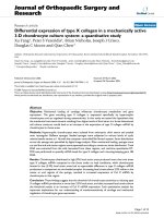
Figure 4 Down-regulation of CDK4 elevated the expr ession of
p21 protein. A.mRNA expression of p21 was inhibited in shRNA-
CDK4 cells compared to PLV-Ctr cells and parental A549 cells. B: p21
protein expression was suppressed in shRNA-CDK4 cells compared
to PLV-Ctr cells and parental A549 cells. Data were presented as
mean ± SD for three independent experiments. *P < 0.05.
Wu et al. Journal of Translational Medicine 2011, 9:38
/>Page 8 of 9
CDK1, CDK2, CDK6, CCND1, p15, p16, p21,andp27
[17-21] were first examined in shRNA-CDK4 and con-
trol cells by real-time PCR. The results indicated that
the reduction of endogenous CDK4 expression markedly
elevated the expression level of tumor suppressor p21
(≥2 folds). Further, we confirmed the upregulated pro-
tein expression of p21 in CDK4-inhibited cells.
In summary, our results provide evidence that CDK4
may be involved in the development of lung cancer.
Furthermore, we also demonstrated that CDK4 could
serve as a potential independent prognostic factor for
lung cancer patients. Due to the limited sample size of
patients in our investigation, further studies would be
needed to verify these findings and establish the role of
CDK4 as a reliable clinical predictor for lung cancer
outcome. Finally, o ur work is the first to present that
CDK4 mediates cell cycle progression by regulating the
expression of p21 expression in lung cancer.
Acknowledgements
Grants support: National 863 High Technology Research and Development
program of China(No.2006AA02A404); Natural science fund of Guangdong
Province (NO.8151051501000058)
Author details
1
Cancer Research Institute of Southern Medical University, 510515,
Guangzhou, PR China.
2
Department of Respiratory Medicine, Affiliated
Hospital of Guangdong Medical College, 524000, Zhanjiang, PR China.
3
Department of Pathology, Medical College of Guangzhou, 510450,
Guangzhou, PR China.
4
School of Pharmacy, Guangdong Medical College,
523808, Dongguan, PR China.
5
Cancer Center, Affiliated Hospital of
Guangdong Medical College, 524000, Zhanjiang, PR China.
6
Department of
Bioinformatics, Southern Medical University, 510515, Guangzhou, PR China.
Authors’ contributions
AW, DW, JG, WL, HY, YZ, XL, HW, and YZ performed this research. WF, ZL
and ZY collected, analyzed, and interpreted data and wrote the manuscript.
WF, ZL, and ZY supervised all the work. All authors have read and approved
the final manuscript.
Competing interests
The authors declare that they have no competing interests.
Received: 27 December 2010 Accepted: 11 April 2011
Published: 11 April 2011
References
1. Houwen L: State of the art: lung cancer in China. Ann Thorac Cardiovasc
Surg 2003, 9(3):147-148.
2. Miliani de Marval PL, Macias E, Conti CJ, Rodriguez-Puebla ML: Enhanced
malignant tumorigenesis in Cdk4 transgenic mice. Oncogene 2004,
23(10):1863-1873.
3. Poomsawat S, Buajeeb W, Khovidhunkit SO, Punyasingh J: Alteration in the
expression of cdk4 and cdk6 proteins in oral cancer and premalignant
lesions. J Oral Pathol Med 2010, 39(10):793-799.
4. Lindberg D, Hessman O, Akerström G, Westin G: Cyclin-dependent kinase
4 (CDK4) expression in pancreatic endocrine tumors. Neuroendocrinology
2007, 86(2):112-118.
5. Wikman H, Nymark P, Väyrynen A, Jarmalaite S, Kallioniemi A, Salmenkivi K,
Vainio-Siukola K, Husgafvel-Pursiainen K, Knuutila S, Wolf M, Anttila S: CDK4
is a probable target gene in a novel amplicon at 12q13.3-q14.1 in lung
cancer. Genes Chromosomes Cancer 2005, 42(2):193-199.
6. Dobashi Y, Goto A, Fukayama M, Abe A, Ooi A: Overexpression of cdk4/
cyclin D1, a possible mediator of apoptosis and an indicator of
prognosis in human primary lung carcinoma. Int J Cancer 2004,
110(4):532-541.
7. Fang W, Li X, Jiang Q, Liu Z, Yang H, Wang S, Xie S, Liu Q, Liu T, Huang J,
Xie W, Li Z, Zhao Y, Wang E, Marincola FM, Yao K: Transcriptional patterns,
biomarkers and pathways characterizing nasopharyngeal carcinoma of
Southern China. J Transl Med 2008, 6:32.
8. Masunaga R, Kohno H, Dhar DK, Ohno S, Shibakita M, Kinugasa S,
Yoshimura H, Tachibana M, Kubota H, Nagasue N: Cyclooxygenase-2
expression correlates with tumor neovascularization and prognosis in
human colorectal carcinoma patients. Clin Cancer Res 2000,
6(10):4064-4068.
9. Liu Z, Li X, He X, Jiang Q, Xie S, Yu X, Zhen Y, Xiao G, Yao K, Fang W:
Decreased expression of updated NESG1 in nasopharyngeal carcinoma:
Its potential role and preliminarily functional mechanism. Int J Cancer
2010.
10. Lingfei K, Pingzhang Y, Zhengguo L, Jianhua G, Yaowu Z: A study on p16,
pRb, cdk4 and cyclinD1 expression in non-small cell lung cancers. Cancer
Lett 1998, 130(1-2):93-101.
11. Yoshida A, Ushiku T, Motoi T, Shibata T, Beppu Y, Fukayama M, Tsuda H:
Immunohistochemical analysis of MDM2 and CDK4 distinguishes low-
grade osteosarcoma from benign mimics. Mod Pathol 2010,
23(9):1279-1288.
12. Zhang LL, Zheng cq: Expression of p16 and CDK4 in colonic carcinoma
and pericancerous mucose. Journal of Shenyang Medical College 2009,
11(2):74-76.
13. Ghazizadeh M, Jin E, Shimizu H, Fujiwara M, Arai S, Ohaki Y, Takemura T,
Kawanami O: Role of cdk4, p16INK4, and Rb expression in the prognosis
of bronchioloalveolar carcinomas. Respiration 2005, 72(1):68-73.
14. Retzer-Lidl M, Schmid RM, Schneider G: Inhibition of CDK4 impairs
proliferation of pancreatic cancer cells and sensitizes towards TRAIL-
induced apoptosis via downregulation of survivin. Int J Cancer 2007,
121(1):66-75.
15. Rodriguez-Puebla ML, Miliani de Marval PL, LaCava M, Moons DS,
Kiyokawa H, Conti CJ: Cdk4 deficiency inhibits skin tumor development
but does not affect normal keratinocyte proliferation. Am J Pathol 2002,
161(2):405-411.
16. An HX, Beckmann MW, Reifenberger G, Bender HG, Niederacher D: Gene
amplification and overexpression of CDK4 in sporadic breast carcinomas
is associated with high tumor cell proliferation. Am J Pathol 1999,
154(1):113-118.
17. Cole AM, Myant K, Reed KR, Ridgway RA, Athineos D, Van den Brink GR,
Muncan V, Clevers H, Clarke AR, Sicinski P, Sansom OJ: Cyclin D2-cyclin-
dependent kinase 4/6 is required for efficient proliferation and
tumorigenesis following Apc loss. Cancer Res 2010, 70(20):8149-8158.
18. Larrea MD, Liang J, Da Silva T, Hong F, Shao SH, Han K, Dumont D,
Slingerland JM: Phosphorylation of p27Kip1 regulates assembly and
activation of cyclin D1-Cdk4. Mol Cell Biol 2008, 28(20):6462-6472.
19. Yu X, Luo Y, Zhou Y, Zhang Q, Wang J, Wei N, Mi M, Zhu J, Wang B,
Chang H, Tang Y: BRCA1 overexpression sensitizes cancer cells to
lovastatin via regulation of cyclin D1-CDK4-p21WAF1/CIP1 pathway:
analyses using a breast cancer cell line and tumoral xenograft model. Int
J Oncol 2008, 33(3):555-563.
20. Frizelle SP, Kratzke MG, Carreon RR, Engel SC, Youngquist L, Klein MA,
Fourre L, Shekels LL, Kratzke RA: Inhibition of both mesothelioma cell
growth and Cdk4 activity following treatment with a TATp16INK4a
peptide. Anticancer Res 2008, 28(1A):1-7.
21. Braden WA, McClendon AK, Knudsen ES: Cyclin-dependent kinase 4/6
activity is a critical determinant of pre-replication complex assembly.
Oncogene 2008, 27(56):7083-7093, 27.
doi:10.1186/1479-5876-9-38
Cite this article as: Wu et al.: Elevated expression of CDK4 in lung
cancer. Journal of Translational Medicine 2011 9:38.
Wu et al. Journal of Translational Medicine 2011, 9:38
/>Page 9 of 9