báo cáo hóa học:" Value of large scale expansion of tumor infiltrating lymphocytes in a compartmentalised gas-permeable bag: interests for adoptive immunotherapy" pptx
Bạn đang xem bản rút gọn của tài liệu. Xem và tải ngay bản đầy đủ của tài liệu tại đây (519.71 KB, 9 trang )
RESEARCH Open Access
Value of large scale expansion of tumor
infiltrating lymphocytes in a compartmentalised
gas-permeable bag: interests for adoptive
immunotherapy
Thomas Zuliani
1,3†
, Julien David
3†
, Sylvain Bercegeay
1
, Marie-Christine Pandolfino
1
, Isabelle Rodde-Astier
4
,
Amir Khammari
2
, Cécile Coissac
4
, Bruno Delorme
4
, Soraya Saïagh
1
and Brigitte Dréno
1,2,3*
Abstract
Background: Adoptive cell therapy (ACT) has emerged as an effective treatment for patients with metastatic
melanoma. However, there are several logistical and safety concerns associated with large-scale ex vivo expansion
of tumour-specific T lymphocytes for widespread availability of ACT for cancer patients. To address these problems
we developed a specific compartmentalised bag allowing efficient expansion of tumour-specific T lymphocytes in
an easy handling, closed system.
Methods: Starting from lymph nodes from eight melanoma patients, we performed a side-by-side comparison of
Tumour-Infiltrating Lymphocytes (TIL) produced after expansion in the compartmentalised bag versus TIL produced
using the standard process in plates. Proliferation yield, viability, phenotype and IFNg secretion were comparatively
studied.
Results: We found no differences in proliferation yield and c ell viability between both TIL production systems.
Moreover, each of the cell products complied with our defined release criteria before being administered to the
patient. The phenotype analysis indicated that the compartmentalised bag favours the expansion of CD8+ cells.
Finally, we found that TIL stimulated in bags were enriched in reactive CD8+ T cells when co-cultured with the
autologous melanoma cell line.
Conclusions: The stimulation of TIL with feeder cells in the specifically designed compartmentalised bag can
advantageously replace the conventional protocol using plates. In particular, the higher expansion rate of reactive
CD8+ T cells could have a significant impact for ACT.
Background
Adoptive cell therapy (ACT) has been successfully
implemented as a modality for the treatment of cancers
for almost twenty years. The applications of this therapy
for cancer have included treatment of hematopoietic
cancers through the targeting of viral antigens [1], renal
cancer carcinogenesis [2] an d metastatic melanoma
[3-5], with good evidence of efficacy. Important progress
has been achieved in the field of melanoma and in
recent clinical trials, objective response rates of between
50% and 70% have been obtained when combined with
immunodepletion [6]. More recently, in a phase II study
it was shown that at the metastatic stage, 43% of the
patients with stage III and IV melanoma experienced an
objective clinical response after treatment with autolo-
gous melanA/Mart-1-specific T lymphocyte clones [7].
These encouraging results emphasize the need to further
develop this cancer therapy strategy.
ACT is an immunotherapy technique in which autolo-
gous tumour-infiltrating lymphocytes are isolated from
resected metastatic lesions, expanded in culture, usually
along with vaccines or growth factors, and re-
* Correspondence:
† Contributed equally
1
Cell and Gene Therapy Unit (UTCG): CIC biotherapy INSERM 0503 Hôtel-
Dieu University Hospital 44093 Nantes cedex 01 France
Full list of author information is available at the end of the article
Zuliani et al. Journal of Translational Medicine 2011, 9:63
/>© 2011 Zuliani et al; licensee BioMed Central Ltd. This is an Open Access article distribute d under the terms of the Creative Co mmons
Attribution License ( which permits unrestricted use, distribution, and reproduction in
any medium, provided the original work is properly cited.
administered to patients. In this approach very large
numbers of TIL need to be generated and administer ed.
The common procedure for lymphocyte expansion starts
in multi-well plates or in T-flasks. TIL are initially sti-
mulated with recombinant human IL-2, irradiated allo-
genic peripheral blood mononuclear cells and
sometimes B-EBV cells as feeder cells. Expansion is then
completed by transferring cells to gas-permeable bags in
the presence of recombinant human IL-2. Although this
procedure has proved its efficacy in generating large
numbers of viable activated TIL, it presents some limita-
tions in the context of GMP, especially during the
expansion phase in multi-well plates/flasks. The main
limitation is tha t during this period, cells ne ed to be fed
ever y 3-4 days and plates and flasks constitute an “open
system” , allowing potential contamination of the cell
therapy product during handling. In addition, the large
quantities of cells needed for each infusion require the
use of multiple containers, which has two drawbacks;
first, it can introduce variability in cell preparation and
second, the handling procedures are labour-intensive
and time-consuming. In order to make this stage of TIL
production more standardised, safer and easier, we
developed a specifically dedicated bag prototype for TIL
expansion on feeder cells. The main aim was to facilitate
cell-to-cell contact between TIL and feeder cells. This
was achi eved by dividing the bags into two asymmetric
compartments: one small compartment into which TIL
and feeder cells were injected, above a larger compart-
ment containing the medium, separated by a discontinu-
ous welding.
By comp arative analysis of TIL produced using a stan-
dard multi-well plate method and in the specifically
developed bags, we report here that bags can advanta-
geously replace plates. First, bags have the advantage of
being a safe, closed system which is much easier to han-
dle than plates. Second, TIL produced in bags were
comparable to those produced in plates in terms of
quantity and viability. Third, we found that the pro por-
tion of CD8+ T cells at the end of the production pro-
cess was higher in bags. Finally, this higher proportion
of CD8+ cells produced correlated with a higher num-
ber of CD8+ T cells producing IFNg when TIL were
placed in contact with the autologous melanoma cell
line.
Methods
Tumour samples and cell line
Tumour samples were o btained from 8 patients with
melanoma-invaded lymph nodes (LN). All patients
signed an informed consent form approved by the Ethics
Committee (Pays de La Loir e) for the use of surgical
samples for research. All samples were immediately
transferred to the cell and gene therapy unit following
surgical resection. LAZ 388 cell line, an Epstein Barr
virus-transformed B-cell line, was kindly provided by
Thierry Hercend.
Establishment of autologous melanoma cell lines
Melanoma cell lines were established as previously
described [8]. Briefly, fresh l ymph nodes with metastasis
were minced into small tumour explants (approximately
1-2 mm
3
) with scissors and a biopsy punch. The result-
ing fragment suspension was centrifuged, and then
pieces were inoculated (at a rate of 2 or 3 per well) into
the wells of a 24-well plate (NUNC) and 1.5 ml per well
of RPMI/FCS (10%) was added. The plates were placed
at 37°C in a humidified incubator with 5% CO
2
.The
plates were observed under a light mi croscope every
week and sub-cultured if necessary.
TIL generation
TIL w ere produced according to a procedure d escribed
previously [9] (See Figure 1 for an illustration of the TIL
production process for ACT). Briefly, TIL were isolated
from 8 tumour samples by culturing cryopreserved frag-
ments of stage III tumour-invaded lymph nodes in two
12-well tissue culture plates with X-vivo 15 serum-free
medium (Lonza) containing 150 U/ml IL-2 (Novartis),
for 10 to 14 days. To perform large-scale e xpansion,
0.39 × 10
6
TIL from these short-term culture were pla-
ted i n thirteen U bottom 96-well microplates at a den-
sity of 300 viable lymphocytes/well with irradiated
feeder cells in 150 μlofIL-2medium.For0.39×10
6
TIL, 26 × 10
6
irradiated LAZ cells and 52 × 10
6
allo-
genicPBMCwereusedasfeedercells.PHA-L(Sigma-
Aldrich) was added on day 0 (15 μg/ml). After three
days, most of the remaining PHA-L was removed by
replacing the culture medium. Cells were cultured for a
further 10 days and fed every 3-4 days by removing 1/3
of the medium and by replacing it with fresh X-vivo15
+ 150 UI/ml IL-2. This is the protocol that is currently
run for a clinical phase III study (NCT00200577), except
that in order to obtain sufficient cells for patient infu-
sion (>10
9
cells), 1.8 × 10
6
cells are plated with the fee-
der cells, the equivalent of plating sixty 96-well plates.
In this study, we compare in parallel the stimulation/
expansionof0.39×10
6
TIL grown with feeder cells in
thirteen 96-well plates versus the same quantity of TIL
grown with the same quantity of feeder cells in one spe-
cially manufactured bag (MacoPharma, french patent
07/00252). This polyolefin bag consists of a small com-
partment (bottom part) and a larger one ( upper part),
separated by a discontinuous welding allowing medium
exchange betwee n both compartments. Firstly, TIL and
feeder cells are inje cted into the bottom compartment
of the bag, diluted in 10 ml X-vivo 15 med ium contain-
ing 150 UI/ml IL-2. Then, 185 ml medium containing
Zuliani et al. Journal of Translational Medicine 2011, 9:63
/>Page 2 of 9
Ly mph no des
excision
Initiation of TILs culture
from fragments
TILs + irr CMN
+ irr LAZ
VS
D0
D13
Amplification
in culture bags
Tranfer in
infusion bags
IV infusion
D20
60 X
Cryopreservation
of lymph nodes
fragments
D-10
Ly mph no des
sectioning
Ly mph no des
excision
Initiation of TILs culture
from fragments
TILs + irr CMN
+ irr LAZ
VS
D0
D13
Amplification
in culture bags
Tranfer in
infusion bags
IV infusion
D20
60 X
Cryopreservation
of lymph nodes
fragments
D-10
Ly mph no des
sectioning
A
B
Medium
injection site
Identification and attach zone
Cells injection
site
Thickening
Discontinous
welding
Lower compartment
(V=15 ml)
Upper compartment
(V=195ml)
135 mm
17 mm
93 mm
5 mm
10 mm 2 mm
15 mm
Medium
injection site
Identification and attach zone
Cells injection
site
Thickening
Discontinous
welding
Lower compartment
(V=15 ml)
Upper compartment
(V=195ml)
135 mm
17 mm
93 mm
5 mm
10 mm 2 mm
15 mm
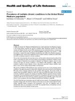
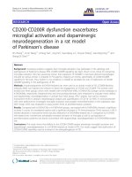
Figure 1 TIL amplification after stimulation in multiple 96-wells plates or in bags. A, Illustration of the TIL production process for ACT.
Briefly, lymph nodes are excised from patients and sectioned for cryopreservation. If the patient is included in the protocol, node fragments are
thawed and TIL cultured in 12-well plates for 10 to 14 days. Then, TIL together with feeder cells (irradiated PBMC and irradiated LAZ cells) are
plated in sixty 96-well plates for stimulation. Ten days later, TIL are pooled and expanded in culture bags before being administered to patients
(see “Materials and Methods” for details). Even though 96-well plates have the advantage of enhancing TIL stimulation through close contact
between TIL and feeders cells, they represents a time- and labour-intensive step in the TIL production process. Furthermore, as it requires
multiple containers (i.e. sixty 96-well plates), this method is a source of potential contamination and variability of the cell therapy product. In
order to make TIL stimulation with feeder cells safer and more straightforward, we developed a specific compartmentalised bag. B, Schematic
representation of the compartmentalized bag. This bag is composed of a size-reduced lower compartment (L, 135 mm × H, 17 mm; Vmax 15
ml) that allows close contact between cells. This compartment has been thermoformed to avoid plastic sticking and facilitate cell injection. TIL
and feeder cells are injected into this compartment through the bottom injection site. Then culture medium is added in the larger upper
compartment (L, 135 mm × H, 93 mm; Vmax 195 ml) via the upper injection site. The two compartments are separated by a discontinuous
welding (forteen 2 mm weldings separated by 5 mm) that allows exchanges between both compartments but prevents the passage of cells
from the lower to the upper compartment during culture medium renewal.
Zuliani et al. Journal of Translational Medicine 2011, 9:63
/>Page 3 of 9
PHA-L are carefully added to the upper compartment of
the bag to avoid cell transfer from bottom to top. For
both the p late and b ag conditions, thirteen days after
plating with feeder cells TIL were recovered, adjusted to
1×10
6
cells/ml in 150 UI/ml IL-2 X-vivo 15 medium
and transferred into standard Lifecell culture bags (Bax-
terfenwall)forexpansionforafurther7days(Figure
1). It has to be no ted that the Baxter expansion bags
whose production has recently been stopped could effi-
ciently be replaced by uncompartmentalized Miltenyi
bags or Mabio Clinicell system. Three days after transfer
to the expansion bags, cells were fed with fresh X-
vivo15 medium containing 150 UI/ml IL-2, readjusted
to 1 × 10
6
cells/ml and grown until day 20. TIL in bags
and in plates were then recovered at day 20 for immu-
nophenotypic characterization and to assess IFNg pro-
ductionbyTILinthepresenceoftheautologous
melanoma cell line. Immunophenot ypic characterization
was a lso performed at days 13 an d 17 of the expansion
process.
Flow cytometry analysis of CD3, CD4, CD8 and CD19 cell
populations
At several stages of TIL generation (days 13, 17 and 20),
we performed flow cytometry analy sis to monitor CD3,
CD4, CD8 and CD19 expression. Briefly, 0.2 × 10
6
cells
were rinsed twice in PBS. Cells were then stained simul-
taneously with anti-CD3-PC5 mAb and either anti-CD4
FITC mAb, anti-CD8 FITC mAb or a nti-CD19 FITC
mAb for 30 minutes at 4°C protected from light. Isotype
matched controls were performed under the same con-
ditions. All the Abs were purchased from BD Bio science
(France) except the anti-CD3-PC5 mAb, which was pur-
chased from Beckman/Coulter (Marseille, France).
Finally, cells were rinsed twice in PBS and resuspended
in PBS/PFA (1%) until the flow cytometry analysis. A
minimum of 10
4
viable cell gated events were analysed
on a FACScalibur flow cytometer using Cell Quest soft-
ware (Becton Dickinson, Grenoble, France). Data were
reanalysed with winMDI software (developed by JC
Trotter).
IFNg production by TIL in response to autologous
melanoma cell lines
Approximately 10
5
lymphocytes were stimulated by 3 ×
10
5
autologous melanoma cells in 200 μLofX-vivo15
medium in the presence o f brefeldin A, 10 μg/ml
(Sigma, St Louis MO, USA) in round-bottom 96-well
plates. The culture was incubated for 6 h at 37°C in a
5% CO
2
humidified atmosphere. Cells were stained for
surface markers with fluorochrome-labelled monocl onal
antibodies (anti-human CD4-APC, anti-human CD8-
FITC, BD Biosciences, France). For intracytoplasmic
IFNg staining , cells were fixed for 10 min at room
temperature in a 4% paraformaldehyde solution in PBS
(Sigma), then washed and stored at 4°C until labelling.
Fixed stimulated lymphocytes were stained for IFNg
production according to the previously described
method [10]. Anti-IFN-g specific antibody was pur-
chased fr om BD Biosciences, France. After staining, cells
were resuspended in PBS until the flow cytometry analy-
sis. A minimum of 10
4
cells were analysed on a FACS-
calibur flow cytometer using Cell Quest software
(Becton Dickinson, Grenoble, France). Data were reana-
lysed with winMDI software.
Statistical analyses
Results are expressed as mean ± SEM. The statistical
differences between values were determined by means
of the Wilcoxon matched pairs test. A difference
between values was considered statistically significant if
p-value < 0.05.
Results
Cell recovery and viability are similar when TIL are
initially co-cultured with feeder cells in plates or in bags
Currently , TIL produced for clinical use shou ld comply
with specific set criteria which chara cterize the final
product before it is administered to the patient. Among
them, cell viability must be ≥70% and the infus ion dose
higher than 10
9
viable TIL. This dose corresponds to a
cell production initiated with 1.8 × 10
6
TIL obtained
from lymph node fragments and co-cultured with feeder
cells in sixty 96-well plates. At the end of the process, it
corresponds to a 555-fold amplification. The prototype
of the bag developed to replace the expansion phase in
plates could be loaded with a maximum of 0.39 × 10
6
TIL, corresponding to an equivalent of thirteen 96-well
plates (see materials and methods). Starting with 0.39 ×
10
6
TIL in one bag compared to 13 plates, we obtained
a similar quantity of cells at the end o f the process.
Hence, at day 20 we recovered a mean of 1021 × 10
6
±
296 cells produced in plates versus 922 × 10
6
±282
produced in b ags for all the donors (Figure 2A). Extra-
polated t o standard production conditions (starting
from 1.8 × 10
6
TIL),thiscorrespondedtoayieldof
approximately 4706 × 10
6
cells in plates versus 4250 ×
10
6
cells in bags and a 2618-fold and 2361-fold expan-
sion respectively (Figure 2B). As regards cell viability, it
wasalwaysover70%forallthedonors,withameanof
84.9% ± 4.3 in bags versus 79.6% ± 7 .2 in plates at day
20 (Figure 2C).
Selective expansion of CD8+ cells in bags
We examined the phenotype of cells produced in both
bag and plate test containers. CD3, CD4, CD8 and
CD19 expression were ana lysed at day 13 (end of feeder
cell stimulation), at day 17 and at the end of the
Zuliani et al. Journal of Translational Medicine 2011, 9:63
/>Page 4 of 9
expansion period at day 20. For all the samples, the per-
centage of CD3+ cells was always ≥98%. This high per-
centage of CD3+ cells was confirme d by the fact that
fewer than 1% of CD19+ cells were found during cell
expansion for all the samples (data not shown), confirm-
ing that LAZ cells were no longer proliferating after
irradiation. CD3+ cells were further examined by double
staining with either CD4 or CD8. The results for CD4
and CD8 expression of cells produced in plates an d in
bags are presented in Table 1 (day 20). Percentages of
CD4 and CD8 at day 13 and day 17 were not signifi-
cantly different th an at day 20 (data not shown).
0
200
400
600
800
1000
1200
1400
0 5 10 15 20 25
0
10
20
30
40
50
60
70
80
90
100
Viability (%)
Plates Bags
NS, p=0.11
Cell Amplification
0
500
1000
1500
2000
2500
3000
3500
4000
NS, p=0.67
Da
y
s of culture
Tota l number of cells (X10
6
)
*, p=0.008
*, p=0.004
NS, p=0.56
Plates
Bags
A
C
B
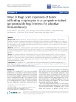
Figure 2 Comparison of TIL proliferation in plates and bags. A, Total quantity of TIL produced in plates and bags during the culture period.
B, Cell amplification of TIL produced in bags and plates between day 0 and day 20. C, Viability of TIL produced in bags and in plates at the end
of the process (day 20). The data are the means of experiments carried out on triplicate from 8 donors. p-values are shown.
Table 1 Proliferation yield and phenotype analysis of lymphocytes expanded from metastatic lymph nodes of eight
patients with melanoma
Patients Initial cell number Final cell number Fold expansion CD4
a
CD8
a
CD8+/IFNg+ TIL
(×10
6
) (×10
6
) % Nb (10
6
) % Nb (10
6
) % Nb (10
6
)
01 (plates) 0.39 794 (±501) 2035 (±1287) 56 (±8) 439 (±257) 43 (±9) 342 (±257) 2 (±1) 5 (±2)
01 (bag) 0.39 1128 (±445) 2892 (±1141) 11 (±2) 123 (±35) 85 (±2) 953 (±366) 14 (±1) 129 (±45)
02 (plates) 0.39 982 (±50) 2518 (±128) 80 797 (±49) 19 (±4) 191 (±54) 2 (±1) 3 (±1)
02 (bag) 0.39 1215 (±153) 3115 (±393) 17 (±6) 207 (±50) 80 (±3) 981 (±166) 5 (±1) 45 (±14)
03 (plates) 0.39 1698 (±182) 4352 (±466) 5 (±1) 85 (±19) 95 (±1) 1612 (±172) 15 (±8) 241 (±106)
03 (bag) 0.39 773 (±366) 1984 (±938) 1 (±0) 6 (±4) 99 (±0) 766 (±362) 15 (±5) 107 (±31)
04 (plates) 0.39 895 (±217) 2296 (±557) 76 (±2) 676 (±158) 21 (±2) 192 (±55) 1 (±1) 2 (±0)
04 (bag) 0.39 600 (±240) 1539 (±615) 61 (±18) 387 (±257) 33 (±16) 180 (±94) 2 (±1) 3 (±2)
05 (plates) 0.39 989 (±180) 2537 (±463) 24 (±4) 236 (±66) 75 (±4) 741 (±132) 6 (±1) 46 (±9)
05 (bag) 0.39 969 (±88) 2484 (±225) 8 (±3) 76 (±17) 90 (±2) 870 (±100) 12 (±4) 107 (±44)
06 (plates) 0.39 910 (±199) 2334 (±509) 42 (±4) 387 (±119) 60 (±7) 533 (±60) 5 (±1) 26 (±3)
06 (bag) 0.39 1323 (±152) 3393 (±389) 10 (±5) 132 (±71) 91 (±5) 1205 (±162) 23 (±2) 281 (±58)
07 (plates) 0.39 1126 (±199) 2889 (±509) 24 (±5) 274 (±73) 75 (±2) 851 (±174) 16 (±4) 132 (±27)
07 (bag) (plates)) 0.39 795 (±174) 2039 (±446) 6 (±1) 46 (±18) 93 (±1) 737 (±162) 26 (±8) 184 (±29)
08 (plates) 0.39 772 (±123) 1982 (±316) 71 (±12) 552 (±142) 31 (±13) 234 (±81) 2 (±1) 4 (±1)
08 (bag) 0.39 571 (±163) 1465 (±417) 46 (±17) 281 (±182) 54 (±14) 296 (±24) 5 (±3) 14 (±9)
For each patients, 3 or 4 independent cell expansions were conducted in parallel in bags and in plates and analysed at day 20. Values are the means obtained
from these independent cell amplifications. Bracketed values show the standard deviation.
a
, gated on lived CD3 + cells.
Zuliani et al. Journal of Translational Medicine 2011, 9:63
/>Page 5 of 9
Figure 3A illustrates the phenotypes of recovered TIL at
day 20 for the patient 06 after expansion in plates versus
in bags. Figure 3B illustrates the percentages of CD4+
and CD8+ cells produce d in plates and in bags for each
donor. For all the donors, the mean percentage of CD8+
cells in bags is 78% ± 23 versus 52.4% ± 28 in plates
and the mean percentage of CD4+ cells is 20.1 ± 21.6 in
bags versus 47.2 ± 28 in plates.
In response to autologous melanoma cell line stimulation,
TIL in bags strongly express IFNg compared to TIL in
plates
Following 20 days of expansion, TIL grown after feeder
cell stimulation in plates and in bags were tested for
their ability to produce IFNg in response to autologous
melanoma cell line stimulation. Figure 4A illustrates the
density plots of CD8 and IFNg expression following sti-
mulation by the autologous melanoma cell line of TIL
expanded in plates and in bags from the patient 06. No
IFNg production was seen when TIL were cultured in
the absence of the autologous melanoma cell line. Six
out of eight donors (donors 01, 02, 05, 06, 07 a nd 08),
showed a higher percentage of specific TIL expressing
IFNg when cells were stimulated in bags co mpared to
stimulation in plates. For the other two donors (03 and
04), these percentages were equal (see Table 1 and Fig-
ure 4B). For all the patients, the mean percentage of
CD8+/IFNg+TILwas12.5±8.7inbagsversus6.12±
6.1 in plates. Since the percentage of CD8+ cells is
higher when cells are stimulated in bags, it results in a
higher quantity of t otal CD8+/IFNg+ cells produced in
bags versus standard plates, 109 × 10
6
±93and57×
10
6
± 86 respectively (Figure 4C).
Discussion
We have undertaken the improvement of the ex vivo
TIL expansion process by initiating the stimulation/
0
10
20
30
40
50
60
70
80
90
100
Ba
g
sPlates
CD4+ cells (%)
BagsPlates
CD8+ cells (%)
0
10
20
30
40
50
60
70
80
90
100
110
CD8CD4
Plates
Bags
A
46 %
53 %
11 %
89 %
B
P=0.0015
P=0.0028
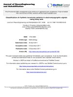
Figure 3 CD4 and CD8 expression by TIL produced in plates vs
bags. A, representative cytometry histograms of TIL produced in
plates (upper panels) and bags (lower panels) stained for CD4 (left
panels) and CD8 (right panels) from patient 06. White histograms
correspond to isotypic controls. B, graphical representation of the
percentage of CD8 (left) and CD4 (right) positive cells produced in
plates vs bags. Circles correspond to the mean of the percentage of
CD8 and CD4 positive cells obtained for each donors. For both
graphics, left and right circles correspond to TIL expanded in plates
and bags respectively. For each line the horizontal bar corresponds
to the mean of the 8 donors. The difference between means is
indicated by the p-value.
A
CD8
IFN
J
4.5 %
21.5 %
+ auto
l
ogous
melanoma cell line
- autologous
melanoma cell line
Plates
Bags
0
5
10
15
20
25
30
35
40
B
C
D8+
/
IFNgamma+
cells (%)
01 02 03 04 05 06 07 08
0
50
100
150
200
250
300
Ba
g
sPlates
CD8+/ IFNgamma+
cells (X10
6
)
P=0.026
Figure 4 IFNg expression by TIL produced in plates vs bags. A,
representative cytometry dot plots of cells produced in plates
(upper panel) and bags (lower panel) double stained for CD8 and
IFNg (see “Materials and Methods”) after 6 hours in contact (right
panel) or not (left panel) with the autologous melanoma cell line.
Representative data obtained from patient 06. Percentages of CD8
+/IFNg+ cells are shown. B, graphical representation of the
percentage of CD8+ cells expressing IFNg in plates (black
histograms) and in bags (white histograms) for all donors. C, Total
number of CD8+/IFNg expressing cells produced in plates versus
bags. Circles correspond to the mean of CD8+/IFNg expressing cells
obtained for each donors. Left and right circle lines correspond to
TIL expanded in plates and bags, respectively. For each line the
horizontal bar corresponds to the mean of the 8 donors. The
difference between these means is indicated by the p-value.
Zuliani et al. Journal of Translational Medicine 2011, 9:63
/>Page 6 of 9
expansion phase with feeder cells in a specifically
designed gas-permeable compartmentalized bags. We
demonstrated that these bags can efficiently replace the
standard protocol using plates for TIL stimulation/
expansion in term of safety, yield and specificity of the
TIL produced.
The potential antineoplastic efficacy of A CT is now
well established and recent clinical trials have demo n-
strated the possibility of inducing tumor regression in
patients with melanoma. However, ACT has some lim-
itations for the widespread development of this thera-
peu tic approach due to cost and safety concerns related
to ex vivo cell amplification and any improvement of
existing protocols for TIL expansion would participate
to the development of availability of ACT. Recently
some bioreactors have proven useful for some clinical
appli cations with lymphocytes. More specifically, a bior-
eactor developed at Aastrom Biosciences has been tested
to replace the second step of TIL expansion process in
T-flasks/bags with OKT3, feeder cells and high concen-
tratio n IL-2. This system has been reported to be suita-
ble to expand TIL by safety and efficacy assays [11].
However,itsusewasnotrecommendedbytheauthors
for TIL infusion in ACT clinical trials to treat patients
with advanced melanoma; the major negative aspects
was the reduced cell yiel d compared to th e current pro-
tocols and also the fixed and non-scalable design of the
system that limits opportunity for in-process monitoring
of the cell product and impedes cell concentration
adjustments during the amplification.
Currently, TIL expansion relies on a first “open” step
which consists of a micro culture initiated from tumor
fragments or a single-cell digestion that are perf ormed
on multiwell plates. During this phase, TIL can be
selected for their reactivity against autologous melanoma
cell line before expansion. The second step, which con-
sists in the rapid amplififcation of TIL obtained from
tumor, is also initiated in an “ open” system. In our
group, TIL are first stimulated and expanded in multiple
96 well plates with low IL-2 concentration, PHA-L, with
pooled CMN and LAZ as feeder cells before being
transferred in gas permeable bags (see material and
methods). In an other routinely used protocol, TIL are
expanded first in multiple T-175 flasks with pooled
CMN as feeder cells in presence of OKT3 and high IL-2
concentration. Although, the use of multiple 96-well
plates has the disadvantage of being labour intensive
compared to T-175 flasks, we observed that T-175 flasks
were not suitable for optimal expansion of TIL with o ur
amplification system using PHA-L, LAZ and low IL-2
concentration. Nevertheless, the use of either multiple
T-175 flasks or 96-well plates represent another “open”
step in the TIL amplification process. Thus, in order to
both secure and also facilitate stimulation and expansion
of TIL in an easy handling reproducible system, we
developed a specific vessel.
Chemical and physical properties of containers in
which cells are grown, could have significant impact on
cell proliferation as well as differentiation. I n the first
step of development of an optimised device for TIL
amplification on f eeder cells, w e designed two similar
uncompartmentalized bags, one in EVA (ethyl vinyl
acetate) and one in polyolefin, both materials being
commonly used for cell culture bags. Results indicated
that polyolefin bags allowed greater TIL amplification
than EVA ones. However, when they were grown in
uncompartmentalized polyolefin bags, TIL represented
only 40% of the amount of TIL obtained according to
our standard process in 96-well plates (data not shown).
We finally designe d another prototype in which a size-
reduced compartment was created to locally increase
cell concentration in the bag and enhance cell to cell
contact between TIL and feeder cells.
In this work, we demonstrate that TIL c an be effi-
ciently expanded after stimulation with feeder cells in
the specifically designed compartmentalised bag (Nantes
university hospital patent 07/00238). In all cases, starting
the production from eight lymph nodes from patients
with advanced melanoma, we we re able with this bag to
produce the same quantity of cells at the end of the TIL
expansion process as with our conventional method
with plates. Moreover, proliferation yield and viability
were compatible with the release criteria for patient
infusion stated in our current TIL clinical protocol.
Interestingly, we also showed that the ratio of CD8
+/CD4+ cells is different betwe en TIL produced in bags
or in plates. Bags favour the proliferation of CD8+ cells
as compared to plates. In our study, the mean percen-
tage of CD8+ cells from the 8 patients was 78%, versus
52.4% in plates. The reason why the compartmentalised
bag repeatedly induces more CD8+ TIL proliferation
than plates remains at the moment unknown. However,
we can hypothesized that it is, a least in part, related to
the polyolefin material component of the bag. Indeed,
when TIL were first amplified in the uncompartmenta-
lized polyolefin bag, we already observed that the per-
centage of CD8+ cells in bag was higher than in plates.
Moreover, the fact that it was not observed with the
similar uncompartmentalised EVA bag, argues for a role
of the polyolefin in this phenomenon suggesting a cru-
cial role of the container. By s tudying the reactivity of
TIL to the autologous melanoma cell lines via IFNg pro-
duction, we also found that bags expand a significantly
greater quantity of reactive CD8+ T lymphocytes than
plates. These results may have a significant impact on
the efficacy of adoptive cell therapy. Indeed, the anti-
tumoral activity of CD8 T cells has been widely demon-
strated in mice and humans [12,13]. The evidence
Zuliani et al. Journal of Translational Medicine 2011, 9:63
/>Page 7 of 9
indicates that the presence of infiltrated CD8 T cells
within tumours is positively correlated with better p rog-
nosis in cutaneous melanoma [14] as well as in several
other types of cancer [15,16]. Moreover, in our group, a
direct correlation was demonstrated between the clinical
outcome of patients with one invaded lymph node trea-
ted with TIL and the presence of CD 8+ cells producing
IFNg that were injected into the patients [17].
It has been proposed that the extensive in vitro stimu-
lation and expansion required to obtain high quantities
of cells may impair the activity and proliferative poten-
tial of these cells in vivo once adoptively transferred
[18,19]. The invitroexpansion of T cells for ACT has
been shown to induce progressive CD8+ T cell differen-
tiation into a late effector state that makes T cells less
effective in mediating anti-tumour responses in vivo.
Hence, it is proposed that culturing tumour-infiltrating
lymphocytes for a limited period of time in vitro would
increase the lymphocyte population capable of mediat-
ing tumour regression in vivo. T his was recently con-
firmed by the in vitro and in vivo demonstrations that
minimally cultured TIL display optimal characteristics
for ACT [20-22]. In this s tudy, in o rder to pe rform a
side-by-side analysis of TIL proliferation, cells were
injected into the l ower compartment of the bag with
identical TIL and feeder cell ratios and total cell con-
centration to those u sed in the standard protocol with
plates. Preliminary data indicate that by increa sing the
cell concentration in the lower compartment of the bag
and reducing the feeder cell stimulation period, it is
possible to increase cell r ecovery (data not shown). In
addition, for technical and logistical reasons, the TIL
expansion process in plates is initiated with 1.8 × 10
6
TIL, while far more TIL are usually obtained from mela-
noma lymph node explant cultures. The initial limited
numbers of TIL used for amplification is primarily due
to the fact that 1.8 × 10
6
TIL correspond to the plating
of sixty 96-well plates, which is time-consuming and
labour-intensive e ven for highly qualified staff, particu-
larly in a GMP environment. Because bags are easier to
handle it should be possible to start TIL expansion with
far more than 1.8 × 10
6
cells. Moreover, in order to
further increase cell yield we are also developing a larger
compartmentalized bag based on this prototype. This
could result in a shortening of the cell production pro-
cess, which would be beneficial for the patient i n terms
of cell therapy product availability but also efficacy.
This compartmentalised bag could be useful in other
situations in addition to a TIL expansion protocol for
ACT. Other immunotherapy strategies rely on the gen-
eration/selection of antigen-specific T reactive lympho-
cytes. These cells may be obtained by peptide
immunomagnetic sorting [23], cloning [7], or generated
after activation by tumour antigen- or apoptotic body-
pulsed dendritic cells [24,25]. Whatever the method for
selecting or generating antigen specific T cells, they
need to undergo large-scale expansion, which is primar-
ily performed by feeder cell stimulation. This empha-
sises the usefulness of this type of compartmentalised
bag.
Conclusions
We report here the processing of a specific compart-
mentalised bag into which TIL and feeder cells are
inoculated in a restricted volume and that can accu-
rately replace the standard system using multiple 96-
well plates, thus allowing exogenous expansion of T IL
in a quicker, more easy-handling and transposable, safer
and cost-effective way. Moreover we show that in addi-
tion to producing the same quantity of cells as the stan-
dard procedu re using plates, the bag allows an improved
expansion of specific CD8+ cells regarding IFNg produc-
tion of TIL co-cultured with the autologous melanoma
cell line. Taken together our data represent a ma jor
improvement of the exogenous TIL expansion process
and may contribute to the development and availability
of ACT for the treatment of patients with cancers that
can be treated by immunotherapy.
List of abbreviations
TIL: tumor infiltrated lymphocytes; ACT: adoptive cell therapy; IFNγ:
Interferon gamma; EVA: ethyl vinyl acetate; IL-2: interleukin-2; PHA-L:
phytoheamagglutinin-L; PBMC: peripheral blood mononuclear cells.
Acknowledgements
This work was supported by the Association Française contre les Myopathies
(AFM) and the European Consortium “Cancer immunology and
immunotherapy” related to the “Life sciences, Genomics and Biotechnology
for Health” priority of the 6
th
Framework Program (FP6) FP6-2004-
LIFESCIHEALTH-5.
Author details
1
Cell and Gene Therapy Unit (UTCG): CIC biotherapy INSERM 0503 Hôtel-
Dieu University Hospital 44093 Nantes cedex 01 France.
2
Dermatological
Oncology Department: CIC biotherapy INSERM 0503 Hôtel-Dieu University
Hospital 44093 Nantes cedex 01, France.
3
Immunodermatology Laboratory:
CIC biotherapy INSERM 0503 Hôtel-Dieu University Hospital 44093 Nantes
cedex 01, France.
4
MacoPharma, 59200 Tourcoing, France.
Authors’ contributions
TZ and JD carried out the cell culture, immunoassays and analysis and
interpretation of the data. They drafted the manuscript. SB have made
substantial contribution in the study design. MCP have produced the
autologous melanoma cell lines and have been involved in IFNγ production
assays. AK contributed to patient inclusion. IR-A, CC and B Delorme have
developed, designed and produced the bags for cell culture. SS and B
Dréno supervised and participated in the study design, result interpretation
and in the writing. B Dréno participated in the recruitment and clinical
follow-up of the patients. All authors read and approved the final
manuscript.
Competing interests
The authors declare that they have no competing interests.
Received: 25 January 2011 Accepted: 16 May 2011
Published: 16 May 2011
Zuliani et al. Journal of Translational Medicine 2011, 9:63
/>Page 8 of 9
References
1. Marijt WA, Heemskerk MH, Kloosterboer FM, Goulmy E, Kester MG, van der
Hoorn MA, et al: Hematopoiesis-restricted minor histocompatibility
antigens HA-1- or HA-2-specific T cells can induce complete remissions
of relapsed leukemia. Proc Natl Acad Sci 2003, 100:2742-2747.
2. Figlin RA, Thompson JA, Bukowski RM, Vogelzang NJ, Novick AC, Lange P,
Steinberg GD, Belldegrun AS: Multicenter, randomized, phase III trial of
CD8(+) tumor-infiltrating lymphocytes in combination with recombinant
interleukin-2 in metastatic renal cell carcinoma. J Clin Oncol 1999,
17:2521-2529.
3. Dreno B, Nguyen JM, Khammari A, Pandolfino MC, Tessier MH, Bercegeay S,
Cassidanius A, Lemarre P, Billaudel S, Labarriere N, Jotereau F: Randomized
trial of adoptive transfer of melanoma tumor-infiltrating lymphocytes as
adjuvant therapy for stage III melanoma. Cancer Immunol Immunother
2002, 51:539-546.
4. Khammari A, Nguyen JM, Pandolfino MC, Quereux G, Brocard A,
Bercegeay S, Cassidanius A, Lemarre P, Volteau C, Labarriere N, et al: Long-
term follow-up of patients treated by adoptive transfer of melanoma
tumor-infiltrating lymphocytes as adjuvant therapy for stage III
melanoma. Cancer Immunol Immunother 2007, 56:1853-1860.
5. Fang L, Lonsdorf AS, Hwang ST: Immunotherapy for advanced melanoma.
J Invest Dermatol 2008, 128:2596-2605.
6. Rosenberg SA, Dudley ME: Cancer regression in patients with metastatic
melanoma after the transfer of autologous antitumor lymphocytes. Proc
Natl Acad Sci USA 2004, 101(Suppl 2):14639-14645.
7. Khammari A, Labarriere N, Vignard V, Nguyen JM, Pandolfino MC, Knol AC,
Quereux G, Saiagh S, Brocard A, Jotereau F, Dreno B: Treatment of
metastatic melanoma with autologous Melan-A/MART-1-specific
cytotoxic T lymphocyte clones. J Invest Dermatol 2009, 129:2835-2842.
8. Jotereau F, Pandolfino MC, Boudart D, Diez E, Dreno B, Douillard JY,
Muller JY, LeMevel B: High-fold expansion of human cytotoxic T-
lymphocytes specific for autologous melanoma cells for use in
immunotherapy. J Immunother (1991) 1991, 10:405-411.
9. Tessier MH, Pandolfino MC, Jotereau F, Boudart D, Litoux P, Dreno B: Home
therapy with autologous tumour-infiltrating lymphocytes and
subcutaneous interleukin-2 in metastatic melanoma. Eur J Cancer 1996,
32A:735-736.
10. Jung T, Schauer U, Heusser C, Neumann C, Rieger C: Detection of
intracellular cytokines by flow cytometry. J Immunol Methods 1993,
159:197-207.
11. Klapper JA, Thomasian AA, Smith DM, Gorgas GC, Wunderlich JR, Smith FO,
Hampson BS, Rosenberg SA, Dudley ME: Single-pass, closed-system rapid
expansion of lymphocyte cultures for adoptive cell therapy. J Immunol
Methods 2009, 30:90-99.
12. Shankaran V, Ikeda H, Bruce AT, White JM, Swanson PE, Old LJ,
Schreiber RD: IFNgamma and lymphocytes prevent primary tumour
development and shape tumour immunogenicity. Nature 2001,
410:1107-1111.
13. Chiao EY, Krown SE: Update on non-acquired immunodeficiency
syndrome-defining malignancies.
Curr Opin Oncol 2003, 15:389-397.
14. Ladanyi A, Kiss J, Somlai B, Gilde K, Fejos Z, Mohos A, Gaudi I, Tímár J:
Density of DC-LAMP(+) mature dendritic cells in combination with
activated T lymphocytes infiltrating primary cutaneous melanoma is a
strong independent prognostic factor. Cancer Immunol Immunother 2007,
56:1459-1469.
15. Zhuang X, Xia X, Wang C, Gao F, Shan N, Zhang L: A high number of CD8
+ T cells infiltrated in NSCLC tissues is associated with a favorable
prognosis. Appl Immunohistochem Mol Morphol 2010, 18:24-28.
16. Zhang D, Shankar P, Xu Z, Harnisch B, Chen G, Lange C, Lee SJ, Valdez H,
Lederman MM, Lieberman J: Most antiviral CD8 T cells during chronic
viral infection do not express high levels of perforin and are not directly
cytotoxic. Blood 2003, 101:226-235.
17. Labarriere N, Pandolfino MC, Gervois N, Khammari A, Tessier MH, Dreno B,
Jotereau F: Therapeutic efficacy of melanoma-reactive TIL injected in
stage III melanoma patients. Cancer Immunol Immunother 2002,
51:532-538.
18. Gattinoni L, Klebanoff CA, Palmer DC, Wrzesinski C, Kerstann K, Yu Z,
Finkelstein SE, Theoret MR, Rosenberg SA, Restifo NP: Acquisition of full
effector function in vitro paradoxically impairs the in vivo antitumor
efficacy of adoptively transferred CD8+ T cells. J Clin Invest 2005,
115:1616-1626.
19. Gattinoni L, Powell DJ Jr, Rosenberg SA, Restifo NP: Adoptive
immunotherapy for cancer: building on success. Nat Rev Immunol 2006,
6:383-393.
20. Besser MJ, Shapira-Frommer R, Treves AJ, Zippel D, Itzhaki O, Hershkovitz L,
Levy D, Kubi A, Hovav E, Chermoshniuk N, Shalmon B, Hardan I, Catane R,
Markel G, Apter S, Ben-Nun A, Kuchuk I, Shimoni A, Nagler A, Schachter J:
Clinical responses in a phase II study using adoptive transfer of short-
term cultured tumor infiltration lymphocytes in metastatic melanoma
patients. Clin Cancer Res 2010, 16:2646-2655.
21. Itzhaki O, Hovav E, Ziporen Y, Levy D, Kubi A, Zikich D, Hershkovitz L,
Treves AJ, Shalmon B, Zippel D, Markel G, Shapira-Frommer R, Schachter J,
Besser MJ: Establishment and large-scale expansion of minimally
cultured “young” tumor infiltrating lymphocytes for adoptive transfer
therapy. J Immunother 2011, 34:212-220.
22. Tran KQ, Zhou J, Durflinger KH, Langhan MM, Shelton TE, Wunderlich JR,
Robbins PF, Rosenberg SA, Dudley ME: Minimally cultured tumor-
infiltrating lymphocytes display optimal characteristics for adoptive cell
therapy. J Immunother 2008, 31:742-751.
23. Labarriere N, Gervois N, Bonnin A, Bouquie R, Jotereau F, Lang F: PBMC are
as good a source of tumor-reactive T lymphocytes as TIL after selection
by Melan-A/A2 multimer immunomagnetic sorting. Cancer Immunol
Immunother 2008, 57:185-195.
24. Melief CJ: Cancer immunotherapy by dendritic cells. Immunity 2008,
29:372-383.
25. Huarte E, Fisher J, Turk MJ, Mellinger D, Foster C, Wolf B, Meehan KR,
Fadul CE, Ernstoff MS: Ex vivo expansion of tumor specific lymphocytes
with IL-15 and IL-21 for adoptive immunotherapy in melanoma. Cancer
Lett
2009, 285:80-88.
doi:10.1186/1479-5876-9-63
Cite this article as: Zuliani et al.: Value of large scale expansion of
tumor infiltrating lymphocytes in a compartmentalised gas-permeable
bag: interests for adoptive immunotherapy. Journal of Translational
Medicine 2011 9:63.
Submit your next manuscript to BioMed Central
and take full advantage of:
• Convenient online submission
• Thorough peer review
• No space constraints or color figure charges
• Immediate publication on acceptance
• Inclusion in PubMed, CAS, Scopus and Google Scholar
• Research which is freely available for redistribution
Submit your manuscript at
www.biomedcentral.com/submit
Zuliani et al. Journal of Translational Medicine 2011, 9:63
/>Page 9 of 9