báo cáo hóa học:" No relationship between the distribution of mast cells and the survival of stage IIIB colon cancer patients" potx
Bạn đang xem bản rút gọn của tài liệu. Xem và tải ngay bản đầy đủ của tài liệu tại đây (796.83 KB, 6 trang )
RESEARCH Open Access
No relationship between the distribution of
mast cells and the survival of stage IIIB colon
cancer patients
Qing Xia
1,2
, Xiao-Jun Wu
1,3
, Qiang Zhou
1,2,5
, Jing-Zeng
1,4
, Jing-Hui Hou
1,4
, Zhi-Zhong Pan
1,3
and
Xiao-Shi Zhang
1,2*
Abstract
Background: Mast cells promote the progression of exper imental tumors and might be a valuable therapeutic
target. However, the relevant clinical evidence is still controversial. This study analyzed the relationship between
the distribution of mast cells and the survival of patients with colon cancer to study whether mast cells contribute
to tumor progression.
Materials and methods: Ninety-three cases of pathologically confirmed primary cancer tissues matched with
adjacent normal mucosa, metastases of regional-draining lymph nodes and regional-draining lymph nodes without
metastases were collected from stage IIIB colon carcinoma patients between January 1997 and July 2004 at the
Cancer Center of Sun Yat-Sen University. Tryp tase-positive mast cells were counted. The relationships of the
distribution of mast cells with clinicopathologic parameters and 5-year survival were analyzed.
Results: Although the mast cell count in the mucosa adjacent to the primary colon cancer was significantly higher
than that in the stroma of the primary colon cancer, no difference in mast cell counts was observed betw een the
stroma in lymph node metastasis and the lymph tissue adjacent to the metastasis. Additionally, the mast cell count
in the regional-draining lymph node without the invasion of cancer cells was significantly higher than that in the
stroma of lymph node metastasis and adjacen t lymph tissue. However, none of those mast cell counts was related
to 5-year survival.
Conclusion: Although mast cell count varied with location, none of the mast cell counts was related to 5-year
survival, suggesting that mast cells do not contribute to the progression of stage IIIB colon cancer.
Keywords: Mast cells, Colon cancer, Survival, Progression
Background
In addition to the genetic alterations of cancer cells, it
is believed that the inf iltration of immune cells, such
as dendritic cells, T cells, macrophage s, and mast cells,
are involved in the progression of colon cancer [1-6].
For example, mast cells might impact tumor progres-
sion by induction of angiog enesis, tissue remodeling,
immune cell recruitment and direct cytotoxicity
against cancer cells [7-9]. Because c-kit i nhibitors such
as imatinib and sunitinib have been approved in
clinical practice and mast cells depend on c-kit, mast
cells might be a new target for cancer t herapy [10]. In
animal models, p olyps are infiltrated by pro-inflamma-
tory mast cells and their precursors. Depletion of mast
cells, either pharmacologically or through the genera-
tion of chimeric mice with genetic lesions in mast cell
development, leads to a p rofound remission of existing
polyps [11]. The i nteraction between mast c ells and
Tregcellsshiftsthelocalbalanceofimmunesurveil-
lance in favor of tumor p rogression [12]. However, the
relevant clinical evidence is controversial. For example,
although Yodavudh and Nielsen reported that mast
cell count was an independe nt prognostic factor for
* Correspondence:
1
State Key Laboratory of Oncology in South China, Sun Yat-sen University
Cancer Center, Guangzhou 510060, China
Full list of author information is available at the end of the article
Xia et al. Journal of Translational Medicine 2011, 9:88
/>© 2011 Xia et al; license e BioMed Central Ltd. This is an Open Access ar ticle distributed under t he terms of the Creative Commons
Attribution License ( http://c reativecommons.org/licenses/by/2.0), which permits unrestricted use, distribution, and reproduction in
any medium, provided the original work is properly cited.
patients with colorectal cancer, this result was not con-
firmed by other groups [13-18].
Because these previous studies focused on the infiltra-
tion of mast cells into primary colorectal cancers and
the function of mast cells might vary with their location
in cancer tissue, it is reasonable to examine the distribu-
tion of mast cells a nd its relationship with the progres-
sion of colon cancer to identify the role of mast cells in
this process. Therefore, the current study examined the
mast cell counts in primary and metastatic tumors, as
well as regional-draining lymph nodes without metas-
tases, to study whether mast cells contribute to the pro-
gression of colon cancer.
Materials and methods
Materials
Ninety-three cases of pathologically confirmed primary
tumor tissues matched with adjacent normal colon
mucosa, metastases of regional-draining lymph nodes
and regional-draining lymph nodes without metastases
were collected from stage IIIB colon cancer patients
between January 1997 and July 2004 at the Cancer C en-
ter of Sun Yat-Sen University. All the patients under-
went radical surgery, and none of them had undergone
either chemotherapy or radiotherapy before the collec-
tion of the samples. The histopathologic characteristics
of the colon carcinoma tissue specimens were confirmed
by blinded review of t he original pathology slides. The
TNM classification system of the American Joint Com-
mittee on Cancer (edition 7) was used for clinical sta-
ging, and the World Health Organization classification
(2000 version) was used for pathologic grading. The
study was conducted in accordance with the Helsinki
Declaration and approved by the Ethic s Committee of
our institution. Patients were informed of the investiga-
tional nature of the study and provided their written
informed consent.
Follow-up of patients
Follow-up was provided to all of the patients. All
patients were observed at 3-month intervals during the
first year, once every 6 months in the second year, and
by telephone or mail communication once every year
thereafter for a total of 5 years. If recurrence or metasta-
sis occurred, 5-Fu-based chemotherapy was adminis-
tered according to the NCCN guidelines. Overall
survival (OS) was defined as the time from surgery to
death or was censored at the last known living date.
Immunohistochemistry
The specimens were fixed in formaldehyde and
embedded in paraffin. Tissue sections of 5 μm thickness
were cut, dried, deparaffinized, and rehydrated in a ser-
ies of alcohols and xylene before antigen retrieval by
pressure cooker treatment in citrate buffer (pH 6.0) for
3 minutes. Then endogenous peroxidase was blocked
with 3% hydrogen peroxide incubation. Mouse anti-
human mast cell tryptase monoclonal antibody (at 1:160
000 dilution, Sero tec, Oxfo rd, UK) was used. Immunos-
taining was performed using an EnVision+ Dual Link
Kit (DakoCytomation, Denmark) according to the man-
ufacturer’ s instructions. The samples were developed
with a substrate-chromogen solution [3,3’-diaminobenzi-
dine dihydrochloride (DAB)] for 3-5 minutes. Sections
were then counterstained with hematoxylin and
mounted in non-aqueous mounting medium.
Mast cell evaluation
The count of tryptase-positive mast cells in the cancer
stroma of a primary tumor is denoted as MCC
stroma
.
The stained sections were first screened under lower
power (×100) to identify the areas with the most mast
cells in the tumor stroma. MCC
stroma
was then counted
under ×400 magnification (1 mm² per HP) in five fields
of vision with an ocular micrometer. The number of
mast cells in every field is expressed as MC/HP. Mean
MCC
stroma
= total number of mast cells in the five fields
divided by five. Additionally, the mast cell counts in the
normal mucosa adjacent to the colon ca ncer (MCC
adja-
cent
), in the stroma of matched lymph node metastasis
(MCC
slnm
), in the normal lymph tissue adjacent to the
lymph node metastasis (MCC
alnm
) and in the regional-
draining lymph node without metastasis (MCC
lnwm
)
were evaluated as MCC
stroma
. All evaluated section were
obtained from areas far from the area of necrosis and H.
E. staining was review ed in uncertain cases. T he mast
cell count in each sec tion was scored independently by
two pathologists with no prior knowledge of clinico-
pathologic parameters. The inter-observer agreement for
the MCC was 81%. Disagreements were re-evaluated
until a consensus was reached.
Statistical analysis
Statistical analyses were p erformed using SPSS 13. 0
software for Windows (SPSS Inc, Chicago, IL, USA).
Descriptive statistical tests, including the mean, stan-
dard deviation, and media n, were calculated according
to standard methods. The relationships between the
various clinicopathologic characteristics and the MCC
parameters were compared and analyzed using chi-
square tests, likelihood ratio, and linear-by-linear asso-
ciation, as appropriate. The non-parametric Wilcoxon
signed ranks test and Kruskal-Wallis test were used to
evaluate the significance of the differences of the
mean ranks. Univariate and multivariate analyses were
based on the Cox proportional hazards regression
model. A two-tailed P < 0.05 was considered statisti-
cally significant.
Xia et al. Journal of Translational Medicine 2011, 9:88
/>Page 2 of 6
Results
The distribution of mast cells
The cytoplasm of mast cells stained brown. In primary
tumor tissue, the mast cell count in normal mucosa
adjacent to colon cancer (MCC
adjacent
) w as significantly
higher than that in the stroma of the primary colon can-
cer ( MCC
stroma
) (P = 0.000). However, no difference in
mast cell count was observed between the stroma in
lymph node metastasis (MCC
slnm
) and the adjacent
lymph tissue (MCC
alnm
) (P = 0.752). Additionally, the
mast cell count in the regional-draining lymph node
without metastasis (MCC
lnwm
) was significantly higher
than that in the lymph tissue adjacent to lymph node
metastasis (MCC
alnm
) (P = 0.000) (Figure 1 andTable 1).
Relationships between the distribution of mast cells and
clinicopathologic characteristics
We used the chi-square test to assess the relationships
between the distribution of mast cells and clinicopathologic
characteristics. The results show that MCC
alnm
(the mast
cell count in the normal lymph tissue adjacent to metasta-
sis) was correlated w ith pathologic classifications and
pathologic grades. MCC
alnm
was higher in papillary and
tubular adenomas than th at in mucoid and signet ring ade-
nomas. Addi tionally, higher MCC
alnm
also occurred in
lower-grade colon cancers, while higher MCC
lnwm
occurred in male p atients (Table 2 ).
Survival analysis with univariate analysis
By the end of the 5-year follow-up, 66 patients with
stage IIIB colon carcinoma were alive, so the 5-year
survival rate was 70.9%. Based on univariate analysis,
although the pathologic classification was a predictor
of OS (P = 0.033), age, gender, location of primary
tumor, pathologic grade, growth pattern, and tumor
invasive depth showed no prognostic significance.
More importantly, the mast cell counts in the primary
tumor, metastasis and regional-draining lymph node
AB
Ca
Ca
DCE
Ca
Ca
Ca
Ca
Ca
Ca
Mu
Ly
Ly
D
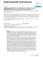
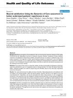
Figure 1 The distribution patterns of mast cells in primary colon cancer, lymph node metastasis and normal regional-draining lymph
node. The tryptase-positive mast cells were stained using an immunohistochemical assay (×400). Higher frequencies of mast cells occurred in
the mucosa adjacent to the colon cancer (MCC
adjacent
, Figure 1B) and in the regional-draining lymph node without metastasis (MCC
lnwm
, Figure
1E) than occurred in the lymph node metastasis (MCC
slnm
and MCC
alnm
, Figure 1C and Figure 1D) and the stroma of the primary colon cancer
(MCC
stroma
, Figure 1A). Ca: cancer tissue; Ly: lymph node; Mu: colon mucosa.
Xia et al. Journal of Translational Medicine 2011, 9:88
/>Page 3 of 6
without metastasis were not correlated with OS
(Table 3).
Multivariate Cox proportional hazards analysis
Multivariate Cox proportional hazards analysis was used
to determine whether the mast cell counts in the primary
tumor, lymph node metastasis and normal regional-drain-
ing lymph node could serve as independent predictors of
OS. Variables included age, gender, location of primary
tumor, pathologic classification, pathologic grade, growth
pattern, tumor invas ive depth and the dis tributions of
mast cells (MCC
stroma
,MCC
adjacent
,MCC
slnm
,MCC
alnm
and MCC
lnwm
). The results show that none of the vari-
ables was associated with OS (Table 4).
Discussion
Multiple studies have analyzed the role of mast cells in
the progression of primary colon cancer. Initial studies
indicatedthatmastcellproperties are independent
prognostic factors [13,14]. However, this conclusion was
questioned by subsequent studies [15-18]. Most of these
studies have significant weaknesses, such as the mixture
of colon with rectal cancers, the mixture of TNM stages,
and small sample sizes [15-19]. This study analyzed 93
stage IIIB colon cancer patients to avoid those short-
comings. The results show that, although the mast cell
count in the normal mucosa adjacent to the primary
colon cancer (MCC
adjacent
) was higher than tha t in the
stroma of the primary colon tumor (MCC
stroma
), neither
MCC
adjacent
nor MCC
stroma
was correlated with the clin-
icopathologic parameters or 5-year survival rate. There-
fore, in this patient population there was no direct
evidence that infiltration of mast cells into primary can-
cer tissue impacted the progression of colon cancer.
Table 2 Correlations between various MCCs and clinicopathologic characteristics
Variable n MCC
stroma*
P MCC
adjacent
P MCC
slnm
P MCC
alnm
P MCC
lnwm
P
<2.6 ≥2.6 <10.6≥10.6 <4.0 ≥4.0 <5.2 ≥5.2 <10.2 ≥10.2
Age 0.243 0.911 0.377 0.121 0.471
<60 43 1825 2122 2221 2518 2320
≥60 50 27 23 25 25 21 29 21 29 23 27
Gender 0.407 0.574 0.726 26 0.250 0.045
Male 58 30 28 30 28 26 32 26 32 24 34
Female 35 15 20 16 19 17 18 20 15 22 13
Location of primary tumor 0.431 0.336 0.094 0.588 0.472
Left 54 28 26 29 25 21 33 28 26 25 29
Right 39 17 22 17 22 22 17 18 21 21 18
Pathologic classification 0.732 0.652 0.900 0.038 0.576
Papillary + tubular 73 36 37 37 36 34 39 32 41 35 38
Mucoid + signet ring 20 9 11 9 11 9 11 14 6 11 9
Pathologic Grade 0.799 0.998 0.991 0.582
G1 2 1 1 1 1 1 1 0 2 0.001 11
G2 69 32 37 34 35 32 37 28 41 32 37
G3 22 12 10 11 11 10 12 18 4 13 9
Growth type 1.000 0.769 0.239 0.769
Pushing 31 15 16 16 15 17 14 18 13 16 15
Infiltrating 62 30 32 30 32 26 36 28 34 0.241 30 32
Invasive depth 0.683 0.293 0.826 0.615
T3 77 38 39 40 37 36 41 39 38 39 38
T4 16 7 9 6 10 7 9 7 9 0.615 7 9
*: MCC
stroma
, the count of tryptase-positive mast cells in the cancer stroma of the primary colon tumor; MCC
adjacent
, the count of tryptase-positive mast cells in
the normal mucosa adjacent to the colon cancer; MCC
slnm
, the count of tryptase-positive mast cells in the stroma of matched lymph node metastasis; MCC
alnm
,
the count of tryptase-positive mast cells in the normal lymph tissue adjacent to the lymph node metastasis; MCC
lnwm
, the count of tryptase-positive mast cells
in the regional-draining lymph node without metastasis.
Table 1 Mast cell counts in colon cancers
Location of mast cells Mast cell count
(median±interquartile range)
MCC
stroma*
2.60 ± 4.80
MCC
adjacent
10.60 ± 8.90
MCC
slnm
4.00 ± 5.90
MCC
alnm
5.20 ± 4.90
MCC
lnwm
10.20 ± 10.00
*: MCC
stroma
, the count of tryptase-positive mast cells in the cancer stroma of
the primary colon tumor; MCC
adjacent
, the count of tryptase-positive mast cells
in the normal mucosa adjacent to the colon cancer; MCC
slnm
, the count of
tryptase-positive mast cells in the stroma of matched lymph node metastasis;
MCC
alnm
, the count of tryptase-positive mast cells in the normal lymph tissue
adjacent to the lymph node metastasis; MCC
lnwm
, the count of tryptase-
positive mast cells in the regional-d raining lymph node without metastasis.
Xia et al. Journal of Translational Medicine 2011, 9:88
/>Page 4 of 6
These results also refute the randomized distribution
model of mast cells in cancer tissues suggested by
Ribatti [20]. The reason that this kind of non-rando-
mized distribution of mast cells would not impact the
progression of colon cancer is unclear, it is possible that
the role of mast cells was outweighed by that of angio-
genesis, w hich is induced by multiple factors, including
mast cells [21-23].
Since IIIB i s a locally advanced stage and the potential
effects of mast cells may be stronger in earlier stages of
colon cancer development such as stage I, stage II and
their function in metastatic disease may show quite dif-
ferent results. We analyzed this in the early research
work and found the consistent result. Paraffin-embedded
speci mens, including tumor tissues and adjacent normal
mucosa tissues obtained from 39 patients with patholo-
gic eva luation-confirmed colon adenomas and 155
patients with colon cancers (the samples from stage I to
IV w ere 38, 38, 38, 41), who underwent radical surgery
orbiopsyduringthesameperiodwereanalyzedusing
the same met hod. Results showed that the majority of
mast cells were located in the normal mucosa adjacent
to the colon cancer too, followed by the invasive margin
and then cancer stroma. The mast cell count in the nor-
mal mucosa adjacent to the colon cancer was associated
with the TNM classification characteristics and hepatic
metastases, although it was not a prognostic factor.
Otherwise,themastcellcountintheinvasivemargin
was associated with neither the clinicopathlogic para-
meters nor overall survival, since the mast cell in the
cancer stroma was rare, we didn’t analyze it.
In addition to infiltrating primary tumors, mast cells
also infiltrate metastases. The role of mast cells in metas-
tasisisstillnotknown.Therefore,thisstudyexamined
the infiltration of mast cells in lymph no de metastasis. In
contrast to the infiltration of mast cells in the primary
tumor, a similar distribution of mast cells occurred both
in the stroma of lymph node metastasis (MCC
slnm
)and
in the lymph tissue adjacent to the metastasis (MCC
alnm
).
Although MCC
alnm
was higher in papillary and tubular
adenoma s than in mucoid and signet ring adenomas, and
although higher MCC
alnm
occurred in lower-grade colon
cancers, neither MCC
slnm
nor MCC
alnm
was correlated
with 5-year survival, which suggests that mast cells are
not involved in lymph node metastasis.
Because mast cells might impact tumor progression by
regulating the immune microenvironment of regional-
draining lymph nodes, this study also examined the
mast cell count in the regional-draining lymph node
without metastasis [24-27]. The results show that the
mast cell count in this lymph node (MCC
lnwm
)was
(10.20 ± 10.00)/HP, significantly higher than MCC
slnm
and MCC
alnm
.However,MCC
lnwm
was not correlated
the 5-year survival, which again fails to support the
hyp othesis that mast cells contribute to the progressi on
of colon cancer by an indirect mechanism.
Furthermore, the 5-year survival rate was 70.9% in our
study, a little higher than an analysis of Surveillance,
Epidemiology, and End Results (SEER) data (64.1%) [28].
Most of the cases were N1 status with 12 or more
lymph nodes examined may help partially explain such a
result. However, our study existed some limitations. For
Table 3 Univariate analysis of factors associated with OS
Variable OS (n = 93)
HR, (95% CI) P
Age (<60 y vs. ≥60 y) 0.635 (0.291-1.386) 0.249
Gender (female vs. male) 1.158 (0.537-2.495) 0.707
Location of primary tumor (right vs. left) 1.915 (0.896-4.093) 0.087
Pathologic classification (mucoid + signet
ring vs. papillary + tubular)
2.325 (1.043-5.183) 0.033
Pathologic grade (G3 vs. G2 + G1) 1.749 (0.785-3.894) 0.165
Growth type (infiltrating vs. pushing) 0.856 (0.392-1.870) 0.696
Invasive depth (T4 vs. T3) 0.853 (0.295-2.466) 0.768
MCC
stroma
*(≥2.6 MC/HP vs. <2.6 MC/HP) 1.224 (0.573-2.615) 0.600
MCC
adjacent
(≥10.6 MC/HP vs. < 10.6
MC/HP)
0.943 (0.443-2.006) 0.878
MCC
slnm
(≥4.0 MC/HP vs. < 4.0 MC/HP) 1.588 (0.727-3.469) 0.241
MCC
alnm
(≥5.2 MC/HP vs. <5.2 MC/HP) 1.045 (0.491-2.223) 0.909
MCC
lnwm
(≥10.2 MC/HP vs. <10.2 MC/HP) 0.779 (0.365-1.665) 0.518
*: MCC
stroma
, the count of tryptase-positive mast cells in the cancer stroma of
the primary colon tumor; MCC
adjacent
, the count of tryptase-positive mast cells
in the normal mucosa adjacent to the colon cancer; MCC
slnm
, the count of
tryptase-positive mast cells in the stroma of matched lymph node metastasis;
MCC
alnm
, the count of tryptase-positive mast cells in the normal lymph tissue
adjacent to the lymph node metastasis; MCC
lnwm
, the count of tryptase-
positive mast cells in the regional-d raining lymph node without metastasis.
Table 4 Multivariate Cox analysis of factors associated
with OS
Variable OS (n = 93)
HR, (95% CI) P
Age (<60 y vs. ≥60 y) 0.497 (0.219-1.127) 0.094
Gender (female vs. male) 1.302 (0.571-2.969) 0.531
Location of primary tumor (right vs. left) 2.220 (0.922-5.345) 0.075
Pathologic classification (mucoid + signet
ring vs. papillary + tubular)
2.514 (0.662-9.537) 0.175
Pathologic grade (G3 vs. G2 + G1) 1.108 (0.300-4.094) 0.877
Growth type (infiltrating vs. pushing) 1.195 (0.489-2.917) 0.696
Invasive depth (T4 vs. T3) 1.456 (0.464-4.569) 0.520
MCC
stroma
*(≥2.6 MC/HP vs. <2.6 MC/HP) 1.180 (0.524-2.659) 0.690
MCC
adjacent
(≥10.6 MC/HP vs. < 10.6 MC/HP) 0.812 (0.372-1.774) 0.602
MCC
slnm
(≥4.0 MC/HP vs. < 4.0 MC/HP) 1.890 (0.748-4.773) 0.178
MCC
alnm
(≥5.2 MC/HP vs. <5.2 MC/HP) 0.916 (0.354-2.367) 0.856
MCC
lnwm
(≥10.2 MC/HP vs. <10.2 MC/HP) 0.729 (0.329-1.614) 0.436
*: MCC
stroma
, the count of tryptase-positive mast cells in the cancer stroma of
the primary colon tumor; MCC
adjacent
, the count of tryptase-positive mast cells
in the normal mucosa adjacent to the colon cancer; MCC
slnm
, the count of
tryptase-positive mast cells in the stroma of matched lymph node metastasis;
MCC
alnm
, the count of tryptase-positive mast cells in the normal lymph tissue
adjacent to the lymph node metastasis; MCC
lnwm
, the count of tryptase-
positive mast cells in the regional-d raining lymph node without metastasis.
Xia et al. Journal of Translational Medicine 2011, 9:88
/>Page 5 of 6
example, only 93 continual colon cancer patients were
collected, sample was not big enough and there may b e
some selection bias thus further research is needed.
Conclusion
By examining the distribution of mast cells in the pri-
mary tumor, in lymph node metastasis and in the normal
regional-draining lymph node in 93 stage IIIB colon can-
cer patients, we found that, although the counts of mast
cells varie d with location, none of the mast cell counts
was correlated with the 5-year survival rate. These data
argue against the hypothesis that mast cells are involved
in the progression of stage IIIB colon cancer.
List of abbreviations used
MCC
adjacent
: the count of tryptase-positive mast cells in the normal mucosa
adjacent to the colon cancer; MCC
alnm
: the count of tryptase-positive mast
cells in the normal lymph tissue adjacent to the lymph node metastasis;
MCC
lnwm
: the count of tryptase-positive mast cells in the regional-draining
lymph node without metastasis; MCC
slnm
: the count of tryptase-positive
mast cells in the stroma of matched lymph node metastasis; MCC
stroma
: the
count of tryptase-positive mast cells in the cancer stroma of the primary
colon tumor. OS: Overall survival;
Acknowledgements
This study was supported by research grants from the National (30972882)
and the Nature Science Foundation of Guangdong Province, China
(9151008901000149).
Author details
1
State Key Laboratory of Oncology in South China, Sun Yat-sen University
Cancer Center, Guangzhou 510060, China.
2
Biotherapy Center, Sun Yat-sen
University Cancer Center, Guangzhou 510060, China.
3
Department of
Colorectal Oncology, Sun Yat-sen University Cancer Center, Guangzhou
510060, China.
4
Department of Pathology, Sun Yat-sen University Cancer
Center, Guangzhou 510060, China.
5
Department of Medical Oncology, The
First People Hospital of Yueyang, Yueyang 414000, China.
Authors’ contributions
WXJ and PZZ performed the case collection. XQ and HJH performed the
immunohistochemical staining. ZJ and ZQ analyzed the results. ZXS
conceived the study, participated in the study design, and coordinated the
writing and helped draft the manuscript. All authors read and approved the
final manuscript.
Competing interests
The authors declare that they have no competing interests.
Received: 2 December 2010 Accepted: 9 June 2011
Published: 9 June 2011
References
1. Ferrone C, Dranoff G: Dual roles for immunity in gastrointestinal cancers.
J Clin Oncol 2010, 28(26):4045-4051.
2. Pagès F, Galon J, Dieu-Nosjean MC, Tartour E, Sautès-Fridman C,
Fridman WH: Immune infiltration in human tumors: a prognostic factor
that should not be ignored. Oncogene 2010, 29(8):1093-1102.
3. Zhou Q, Peng RQ, Wu XJ, Xia Q, Hou JH, Ding Y, Zhou QM, Zhang X,
Pang ZZ, Wan DS, Zeng YX, Zhang XS: The density of macrophages in the
invasive front is inversely correlated to liver metastasis in colon cancer. J
Transl Med 2010, 8:13.
4. Peng RQ, Wu XJ, Ding Y, Li CY, Yu XJ, Zhang X, Pan ZZ, Wan DS, Zheng LM,
Zeng YX, Zhang XS: Co-expression of nuclear and cytoplasmic HMGB1 is
inversely associated with infiltration of CD45RO+ T cells and prognosis in
patients with stage IIIB colon cancer. BMC Cancer 2010, 10:496.
5. Kmieciak M, Gowda M, Graham L, Godder K, Bear HD, Marincola FM,
Manjili MH: Human T cells express CD25 and Foxp3 upon activation and
exhibit effector/memory phenotypes without any regulatory/suppressor
function. J Transl Med 2009, 7:89.
6. Gao YF, Peng RQ, Li J, Ding Y, Zhang X, Wu XJ, Pan ZZ, Wan DS, Zeng YX,
Zhang XS: The paradoxical patterns of expression of indoleamine 2,3-
dioxygenase in colon cancer. J Transl Med 2009, 7:71.
7. Maltby S, Khazaie K, McNagny KM: Mast cells in tumor growth:
angiogenesis, tissue remodelling and immune-modulation. Biochim
Biophys Acta 2009, 1796(1):19-26.
8. Ribatti D, Crivellato E: The controversial role of mast cells in tumor
growth. Int Rev Cell Mol Biol 2009, 275:89-131.
9. Galinsky DS, Nechushtan H: Mast cells and cancer–no longer just basic
science. Crit Rev Oncol Hematol 2008, 68(2):115-130.
10. Groot Kormelink T, Abudukelimu A, Redegeld FA: Mast cells as target in
cancer therapy. Curr Pharm Des 2009, 15(16):1868-1878.
11. Gounaris E, Erdman SE, Restaino C, Gurish MF, Friend DS, Gounari F,
Lee DM, Zhang G, Glickman JN, Shin K, Rao VP, Poutahidis T, Weissleder R,
McNagny KM, Khazaie K: Mast cells are an essential hematopoietic
component for polyp development. Proc Natl Acad Sci USA 2007,
104(50):19977-19982.
12. Gounaris E, Blatner NR, Dennis K, Magnusson F, Gurish MF, Strom TB,
Beckhove P, Gounari F, Khazaie K: T-regulatory cells shift from a protective
anti-inflammatory to a cancer-promoting proinflammatory phenotype in
polyposis. Cancer Res 2009, 69(13):5490-5497.
13. Fisher ER, Paik SM, Rockette H, Jones J, Caplan R, Fisher B: Prognostic
significance of eosinophils and mast cells in rectal cancer: findings from
the National Surgical Adjacent Breast and Bowel Project (protocol R-01).
Hum Pathol 1989, 20(2)
:159-163.
14. Nielsen HJ, Hansen U, Christensen IJ, Reimert CM, Brünner N, Moesgaard F:
Independent prognostic value of eosinophil and mast cell infiltration in
colorectal cancer tissue. J Pathol 1999, 189(4):487-495.
15. Gulubova M, Vlaykova T: Prognostic significance of mast cell number and
microvascular density for the survival of patients with primary colorectal
cancer. J Gastroenterol Hepatol 2009, 24(7):1265-1275.
16. Yodavudh S, Tangjitgamol S, Puangsa-art S: Prognostic significance of
microvessel density and mast cell density for the survival of Thai patients
with primary colorectal cancer. J Med Assoc Thai 2008, 91(5):723-732.
17. Acikalin MF, Oner U, Topçu I, Yaşar B, Kiper H, Colak E: Tumour
angiogenesis and mast cell density in the prognostic assessment of
colorectal carcinomas. Dig Liver Dis 2005, 37(3):162-169.
18. Tan SY, Fan Y, Luo HS, Shen ZX, Guo Y, Zhao LJ: Prognostic significance of
cell infiltrations of immunosurveillance in colorectal cancer. World J
Gastroenterol 2005, 11(8):1210-1214.
19. Kalady MF, Sanchez JA, Manilich E, Hammel J, Casey G, Church JM:
Divergent Oncogenic Changes Influence Survival Differences between
Colon and Rectal Adenocarcinomas. Diseases of the Colon & Rectum 2009,
52(6):1039-1045.
20. Guidolin D, Nico B, Crivellato E, Marzullo A, Vacca A, Ribatti D: Tumoral mast
cells exhibit a common spatial distribution. Cancer Lett 2009, 273(1):80-85.
21. Nechushtan H: The complexity of the complicity of mast cells in cancer.
Int J Biochem Cell Biol 2010, 42(5):551-554.
22. Crivellato E, Nico B, Ribatti D: Mast cell contribution to tumor
angiogenesis: a clinical approach. Eur Cytokine Netw 2009, 20(4):197-206.
23. Crivellato E, Nico B, Ribatti D: Mast cells and tumour angiogenesis: new
insight from experimental carcinogenesis. Cancer Lett 2008, 269(1):1-6.
24. Galli SJ, Grimbaldeston M, Tsai M: Immunomodulatory mast cells: negative, as
well as positive, regulators of immunity. Nat Rev Immunol 2008, 8(6):478-486.
25. Kalesnikoff J, Galli SJ: New developments in mast cell biology. Nat
Immunol 2008, 9(11):478-1223.
26. Cochran AJ, Huang RR, Lee J, Itakura E, Leong SP, Essner R: Tumour-
induced immune modulation of sentinel lymph nodes. Nat Rev Immunol
2006, 6(9):659-670.
27. Preynat-Seauve O, Contassot E, Schuler P, Piguet V, French LE, Huard B:
Extralymphatic tumors prepare draining lymph nodes to invasion via a
T-cell cross-tolerance process. Cancer Res 2007, 67(10):5009-5016.
28. O’Connell JB, Maggard MA, Ko CY: Colon cancer survival rates with the
new American Joint Committee on Cancer sixth edition staging. J Natl
Cancer Inst 2004, 96(19):1420-1425.
doi:10.1186/1479-5876-9-88
Cite this article as: Xia et al.: No relationship between the distribution
of mast cells and the survival of stage IIIB colon cancer patients. Journal
of Translational Medicine 2011 9:88.
Xia et al. Journal of Translational Medicine 2011, 9:88
/>Page 6 of 6