o cáo hóa học:" Serum cytokine profiles in healthy young and elderly population assessed using multiplexed bead-based immunoassays" ppt
Bạn đang xem bản rút gọn của tài liệu. Xem và tải ngay bản đầy đủ của tài liệu tại đây (289.77 KB, 7 trang )
RESEARC H Open Access
Serum cytokine profiles in healthy young and
elderly population assessed using multiplexed
bead-based immunoassays
Hyun Ok Kim
1†
, Han-Soo Kim
1†
, Jong-Chan Youn
2
, Eui-Cheol Shin
3
and Sungha Park
2*
Abstract
Background: Lipid metabolites and cytokines, including chemokines and growth factors, are the key regulators of
immune cell function and differentiation, and thus, dysregulation of these regulators is associated with various
human diseases. However, previous studies demonstrating a positive correlation of cytokine levels with aging may
have been influenced by various environmental factors and underlying diseases. Also, da ta regarding cytokine
profiling in the elderly are limited to a small subset of cytokines.
Methods: We compared the profiles of 22 cytokines, including chemokines and growth factors, in a case-
controlled study group of a gender-matched, healthy cohort of 55 patients over the age of 65 and 55 patients
under the age of 45. Assessment of serum cytokine concentrations was performed using commercially-available
multiplex bead-based sandwich immunoassays.
Results: Soluble CD40 ligand (sCD40L) and transforming growth factor alpha (TGF-a) levels were significantly
higher in the elderly pa tients, whereas granulocyte colony-stimulating factor (G-CSF), granulocyte-monocyte
colony-stimulating factor (GM-CSF), and monocyte chemoattractant protein-1 (MCP-1) levels were significantly
lower in the elderly patients. The partial correlation analysis demonstrating the correlation between cytokine levels
when controlled for gender, systolic blood pressure, total cholesterol, HDL cholesterol, triglyceride, and serum
creatinine levels further demonstrated that G-CSF, GM-CSF, and MCP-1 had significant negative correlations with
age, whereas sCD40L and TGF-a had significant positive correlations.
Conclusions: Future studies will focus on examining the significance of these age-related changes in circulating
cytokines and other biological markers and their potential contribution to the development of different age-
associated diseases.
Background
Aging is accompanied by a decline in immune functions,
referred to as immune aging or immune senescence. Para-
doxically, life-long exposure to environmental factors and
countless interactions with infectious agents leads to a
chronic inflammatory state in older individuals, termed
inflammaging, characterized by an increase in proinflam-
matory mediators present in serum [1,2]. Changes in T-
cell homeostasis with aging are associated with a decline
in immunity and increa sed inflammation. I ncreased
accumulation of regulatory T cells contributes to impaired
CD8 and natural killer cell activities [3,4]. Also, a decrease
in naïve T cells may result in impaired acquired immune
responses, whereas clonal expansion of CD25 null T cells
may result in increased secretion of tumor necrosis factor-
alpha (TNF-a) and interleukin-6 (IL-6), resulting in a
heightened degree of inflammation [5].
Lipid meta bolites and cytokines, including chemokines
and growth factors, are the key regulators of immune cell
function and differentiation. Thus, dysregulation of these
regulators is associated with various human diseases.
Age-associate d elevation of inflammatory factor s includ-
ing TNF-a ,IL-6,prostaglandinE
2
(PGE
2
), and IL-1b
have been described previously [6-8]. This elevation may
be attributable to both the derangement of inflammation
* Correspondence:
† Contributed equally
2
Division of Cardiology, Yonsei Cardiovascular Center, Yonsei University
College of Medicine, Seoul 120-752, Republic of Korea
Full list of author information is available at the end of the article
Kim et al. Journal of Translational Medicine 2011, 9:113
/>© 2011 Kim et al; licensee BioMed Central Ltd. This is an Open Access article distributed under the terms of the Creative Commons
Attribution License ( which permits unrestricted use, distribution, and reproduction in
any medium, provided the original work is properly cited.
regulation and lifelong exposure of the immune system
to environmental risk factors such a s smoking, aging,
hypertension, and diabetes [8-10]. However, previous stu-
dies that demonstrated positive correlations of cytokine
levels with aging were performed in general aging popu-
lations that may have been influenced by various envir-
onmental factors and underlying dise ases. Additionally,
data regarding cytokine profiling in the elderly have been
limited to a small subset of cytokines. In this study, we
compared the profiles of 22 cytokines, chemokines, and
growth factors in a case-controlled study group of a gen-
der-matched, healthy cohort of 55 subjects over the age
of 65 (Median age 68) and 55 subjects under the age o f
45 (median age 34). The levels of the cytokines, chemo-
kines, and growth factors were analyzed using multi-
plexed bead-based immunoassays.
Methods
Subject population
The study group was comprised of 110 healthy subjects
who were enrolled in the Cardiovascular Genome Center
(male:female = 44:66). The Cardiovascular Genome Cen-
ter is a Korean government-sponsored research project
with the objective of determining the genetic factors
associated with the development of c ardiovascular dis-
ease in a large, prospective study group. The study sub-
jects were enrolled in the Cardiovascular Genome Center
cohort as healthy control subjects. The study subjects did
not have any past histories of hypertension, diabetes mel-
litus, cardiovascular disease, cerebrovascular disease, can-
cer, chronic ren al disease, or any chronic inflammatory
conditions. Group 1 consisted of 55 subjects under the
age of 45 and group 2 consisted of 55 subjects over the
age o f 65. The study subjects were not permitted to per-
form strenuous exercise or drink alcoholic beverages
24 h before the laboratory test. The study subjects were
also instructed to avoid eating or drinking anything
except water during the testing period. Written, informed
consent was obtained from all patients and the protocol
was approved by the Institutional Review Board of Yonsei
University College of Medicine (4-2001-0039). Rese arch
was conducted in compliance with the Helsinki
Declaration.
Blood collection
Blood samples were obtained from the forearm of each
subject via the anticubital vein after a minimum of 12
hours of fasting. Samples were collected in EDTA-treated
and plain tubes.
The methods for determining the concentrations of
each lipid parameter were based on an enzymatic method
(Hitachi 7600-110, Hitachi Co., Japan) that analyzed total
cholesterol and triglyceride levels. After pre cipitat ion of
serum chylomicron, LDL, and VLDL with dextran
sulfate-magnesium, the HDL-C remaining in the super-
natant fluid was measured using the enzymatic method
(Hitachi 7600-110). LDL cholesterol levels were calcu-
lated using the Friedewald formula with serum triglycer-
ideconcentrationslessthan4.52mol/L(400mg/mL)
[11].
Anthropometric and blood pressure measurements
The body weight and height of each undressed and
barefoot subject were measured in the morning. After 5
minutes of rest, the brachial blood pressure was mea-
suredfromthedominantarmusinganOMRONHEM
7080 IT while the subject remained seated. The average
of three measurements was recorded for each subject.
Multiplex bead-based immunoassay
Simultaneous assessment of serum concentrations of
epidermal growth factor (EGF), fibroblast growth factor
2 (FGF2), FMS-like tyrosine kinase 3 ligand (Flt-3L),
granulocyte colony-stimulating factor (G-CSF), granulo-
cyte-monocyte colony-stimulating factor (GM-CSF),
interferon-a2(IFN-a2), INF-g, IL-10, IL-15, IL-17, IL-
1b,IL-2,IL-6,IL-8,INF-g inducible protein 10 (IP-10),
monocyte chemoattractant protein-1 (MCP-1), macro-
phageinflammatoryprotein-1b (MIP-1 b), platelet-
derived growth factor-AA (PDGF-AA), soluble CD40
ligand (sCD40L), transforming growth factor alpha
(TGF-a), TNF-a, and vascular endothelial growth factor
(VEGF) was performed using commercially-available
multiplex bead-based sandwich immunoassay kits
(MPXHCYTO-60K-25, Millipore, Billerica, MA) as per
the manufacturer’s instructions. Briefly, plasma samples
(25 μL/well) or standards (25 μL well) were incubated
with 25 μL of the pre-mixed bead sets in pre-wetted 96-
well microtiter plates at 4°C overnight. After washing,
25 μL of the fluorescent detection antibody mixture was
added for 30 min a nd 25 μL of streptavidin-phycoery-
thrin was added to each well for an additional 30 min at
room temperature. A range of 3.2-10,000 pg/mL recom-
binant cytokines was used to establish standard curves
and to maximize the sensitivity and dynamic range of
the assay. Cytokine levels were determined using a
Luminex IS 100 (Luminex, Austin, TX), and the data
were reported as median fluorescent intensities.
Statistical analysis
Results are expressed as means ± standard deviation. In
this study, comparisons of discrete variables were made
using the chi-square method and t-tests were used for
continuous variables. Because the distribution of the
cyt okines was skewed, a lo g transformation of the cyto-
kines was performed for independent t-tests and partial
correlation analyses. For the partial correlation analysis,
the correlation between aging and serum biomarkers
Kim et al. Journal of Translational Medicine 2011, 9:113
/>Page 2 of 7
was assessed while controlling for gender, smoking,
body mass index (BMI), fasting blood glucose (FBG),
systolic blood pressure ( SBP), total cholesterol, HDL
cholesterol (HDL), triglyceride (TG), and serum creati-
nine l evels. A two-tailed value of P < 0.05 was consid-
ered statistically significant. All statistical analyses were
performed using SPSS 13.0 (SPSS Inc., Chicago, IL).
Results
Compared to the younger subjects in group 1, the
elderly subjects in group 2 were associated with signifi-
cantly higher SBP, total cholesterol, TG, serum albumin,
serum blood urea nitrogen (BU N) and serum creatinine
(Table 1). Comparison of the serum concentration of 22
cytokines-chemokines-growth factors demonstrated that
sCD40L (group 2: 20370.6 ± 71 662.0 pg/mL vs group 1:
2205.8 ± 4699.2 pg/mL, P value = 0.016) and T GF-a
(group 2: 4.9 ± 4.8 pg/mL vs group 1: 3.2 ± 4.0 pg/mL,
P value = 0.026) were significantly higher in the elderly
subjects, whereas G-C SF (group 1: 14.7 ± 13.2 pg/mL vs
group 2: 9.9 ± 8.8 pg/mL, P-value = 0.009), GM-CSF
(group 1: 40.9 ± 108.6 pg/mL vs group 2: 20.3 ± 60.4
pg/mL, P value = 0.021) and MCP-1 (group 1: 213.5 ±
100.7 pg/mL vs group 2: 168.0 ± 73.0 pg/mL, P value =
0.027) were significantly lower in the elderly subjects
(Table 2). The serum level of EGF, FGF-2, Flt-3L, INF-
A2, INF-g, IL-10, IL-15, IL-17, IL-1b, IL-2, IL-6, IL-8,
IP-10, MIP-1b,PDGF-AA,TNF-a and VEGF showed
no significant difference (Table 2). The partial correla-
tion analysis demonstrating the correlation between
cytokines-chemokines-growth factors when controlled
for gender, SBP, total cholesterol, HDL, T G and se rum
creatinine demonstrated that G-CSF, GM-CSF and
MCP-1 has a signifi cant negative correlation with age
whereas sCD40L and TGF-a has a significant, positive
correlation (Table 3, Figure 1).
Discussion
To our knowledge, this is the first study that has c om-
pared extensive profiles of cytokines, including chemo-
kines and growth factors, in healthy elderly and young
subjects. As compared to the younger subjects in group
1, the elderly subjects had significantly higher SBP, total
cholesterol, TG, serum albumin, serum blood urea
nitrogen (BUN), and serum creatinine levels (Table 1).
Comparison of the serum concentrations of 22 cyto-
kines, chemokines, and growth factors demonstrated
that sCD40L and TGF-a levels were significantly higher
in the elderly subjects, whereas G-CSF, GM-CSF, and
MCP-1 were significantly lower in the elderly subjects
(Table 2). The serum levels of EGF, FG F-2, Flt-3L, INF-
a2, INF-g, IL-10, IL-15, IL-17, IL-1b, IL-2, IL-6, IL-8,
IP-10, MIP-1b, PDGF-AA, TNF-a,andVEGFshowed
no significant differences between the two groups (Table
2). The partial correlation analysis demonstrating the
correlation between the levels of the cytokines, chemo-
kines, and growth factors when controlled for gender,
SBP, total cholesterol, HDL, TG, and serum creatinine
levels further indicated that G-CSF, GM-CSF, and
MCP-1 had significant negative correlations with age,
whereas sCD40L and TGF-a had significant positive
correlations (Table 3 and Figure 1).
There was a lack of association of IL-6 levels with
aging in the healthy study populations (Table 2), which
is in concordance with previous studies [12,13]. How-
ever, unlike our findin gs that indicated no significant
association of TNF-a, IL-6, and IL-1b levels with age,
some previous studies have indicated that these cytokine
levels are elevated in elderly subjects as compared to
younger subjects [8,14-16]. A likely reason for the dis-
crepancy is that in the previo us studies, the elderly sub-
jects were not controlled fo r associated diseases, such as
hypertension an d diabetes, which could increase inflam-
mation. In a study by Ferrucci et al., controlling for car-
diovascular risk factors attenuated the regression
coefficient between aging and IL-6 [8]. In contrast to
that study, we excluded subjects with previous histories
of hyperte nsion, cardiovascular disease, cerebrovascular
disease, diabetes mellitus, can cer, or chronic renal dis-
ease, which minimized the confounding effects of con-
comitant disease processes that could alter the
inflammatory state of the study patients. Additionally, in
the study by Ferrucci et al., the highest level of IL-6 was
in subjects over the age of 85, whereas the differences in
IL-6 levels between subjects 65-74 years of age and
patients 20-49 years of age was not as large [8]. The
Table 1 Average baseline clinical characteristics of
patients
Group 1
(Age < 45)
Group 2
(Age ≥ 65)
P-value
b
Gender (male:female) 23:32 23:32
Age 34.8 ± 5.7 70.4 ± 4.9 < 0.001
SBP
b
(mmHg) 115 ± 13 135 ± 19 < 0.001
DBP (mmHg) 75 ± 9 77 ± 11 0.383
BMI (kg/m
2
) 22.9 ± 3.6 23.3 ± 3.4 0.526
Smoking (%) 21 (38.2%) 6 (10.9%) 0.001
T chol (mg/dL) 192 ± 30 194 ± 35 0.033
TG (mg/dL) 110 ± 66 162 ± 122 0.007
HDL (mg/dL) 50 ± 12 51 ± 11 0.838
FBG (mg/dL) 84.8 ± 10.4 98.7 ± 38.3 0.011
Albumin (g/dL) 4.7 ± 0.3 4.5 ± 0.3 < 0.001
BUN (mg/dL) 11.7 ± 2.6 15.2 ± 3.8 < 0.001
Cr (mg/dL) 0.68 ± 0.17 0.79 ± 0.23 0.007
a
Differences with P < 0.05 are considered significant.
b
Abbreviations; SBP: Systolic blood pressure, DBP; diastolic blood pressure,
BMI: body mass index, T chol: Total cholesterol, TG: Triglycerid e, HDL: High
density lipoprotein, FBS: Fasting blood glucose, BUN: Blood urea nitrogen, Cr:
Creatinine
Kim et al. Journal of Translational Medicine 2011, 9:113
/>Page 3 of 7
average age of the elderly subjects in this study was 70.4.
Therefore, the lack of a very elderly population may be
another possible explanation for the discrepancy in
results.
This is the first study to demonstrate that sCD40L levels
are significantly associated with aging (Tables 2 and 3 and
Figure 1). The CD40/CD40L system belongs to the tumor
necrosis factor superfamily and is a key pathway that links
inflammation and atherothrombosis [17]. C D40 and
CD40L are expressed in a variety of cell types, including
platelets, vascular smooth muscle cells (VSMC), and
immune cells [17,18]. Increased interaction s between
CD40 and CD40L may result in increased expression of
cell adhesion molecules on endothelial cells and VSMCs,
which subsequently results in increased vascular inflam-
mation. Additionally, sCD40L and CD40 interactions
increase oxidative stress and endothelial dysfunc tion,
which may also contribute to an increase in the inflamma-
tory cascade [17, 19]. Increased secreti on of sCD40L ma y
be one explanation for the increased inflammation asso-
ciated with aging, and may be a pathway that links aging
with an increased risk of atherothrombosis.
TGF-a, a member of the EGF family, is a potent mito-
gen and chemotactic factor [20], and was positively cor-
related with aging (Tables 2 and 3 and Figure 1). TGF-a
binds to the EGF receptor with a high affinity [21] and
is indispensable for the proper development of many tis-
sues and organs, wound healing, bone resorption, and
angiogenesis [22]. TGF-a is implicated in numerous dis-
ease states, including coronary a rtery diseases, cystic
fibrosis, psoriatic lesions, oral leukoplakia, submucosal
fibrosis, Barrett’s esophagus syndro me, and cancer [22].
Recent results also implicate this growth factor in the
development of certain diabetic complications, such a s
atherosclerosis [23]. Though it is unknown whether
TGF-a plays an important role in regulating homeosta-
sis and/or diseases in aged subjects, increased serum
levels of this cytokine in the elderly population may play
Table 2 Serum levels of cytokines, chemokines, and growth factors according to age
Cytokines
(pg/ml)
Group 1
(Age < 45)
Group 2
(Age ≥ 65)
P-value
a
G-CSF 14.7 ± 13.2
b
(0.03-75.8) 9.9 ± 8.8 (0.03-35.2) 0.009
GM-CSF 40.9 ± 108.6 (0.5-728.1) 20.3 ± 60.40 (0.50-415.1) 0.021
MCP 1 213.5 ± 100.7 (27.9-667.8) 168.0 ± 73.0 (39.34-355.9) 0.027
sCD40L 2205.8 ± 4699.2 (268.6-27703.8) 20370.6 ± 71662.0 (115.8-380396.7) 0.016
TGF-a 3.2 ± 4.0 (0.93-26.8) 4.9 ± 4.8 (0.86-20.8) 0.026
EGF 31.3 ± 35.9 (3.2-210.5) 61.0 ± 65.1 (3.20-251.6) 0.073
FGF-2 18.9 ± 11.3 (6.7-65.6) 20.1 ± 13.9 (3.20-72.83) 0.863
Flt-3L 10.2 ± 10.1 (0.84-59.3) 13.2 ± 15.9 (0.03-78.42) 0.759
IFN-a2 21.3 ± 22.6 (2.42-102.2) 33.3 ± 70.2 (2.42-449.2) 0.822
IFN-g 13.1 ± 22.7 (0.14-126.8) 10.3 ± 18.4 (1.09-117.7) 0.948
IL-10 1.32 ± 3.06 (0.01-19.8) 1.58 ± 6.17 (0.01-41.7) 0.325
IL-15 3.04 ± 2.17 (1.25-13.1) 3.49 ± 4.31 (1.32-28.9) 0.668
IL-17 6.53 ± 7.42 (1.58-37.8) 12.2 ± 37.9 (1.43-275.1) 0.640
IL-1b 2.04 ± 4.93 (0.17-24.) 2.52 ± 7.41 (0.17-39.0) 0.645
IL-2 5.13 ± 2.31 (2.88-18.3) 5.58 ± 4.17 (3.06-32.1) 0.601
IL-6 2.91 ± 6.45 (0.16-37.7) 2.57 ± 5.22 (0.16-31.5) 0.750
IL-8 23.9 ± 29.7 (4.2-132.6) 27.6 ± 43.9 (4.76-217.0) 0.995
IP-10 462.2 ± 364.7 (145.3-2152.2) 451.3 ± 256.4 (149.8-1394.8) 0.673
MIP-1b 40.5 ± 38.8 (3.2-227.2) 40.4 ± 33.6 (3.20-231.1) 0.633
PDGF-AA 1528.3 ± 878.8 (140.6-3290.2) 1615.3 ± 1125.0 (55.3-3421.7) 0.485
TNF-a 3.21 ± 4.04 (0.93-26.8) 4.94 ± 4.79 (0.86-20.8) 0.916
VEGF 114.9 ± 147.1 (13.1-864.1) 100.5 ± 75.4 (6.9-329.3) 0.853
a
Differences with P < 0.05 are considered significant.
b
Average concentrations ± standard deviation in pg/mL.
Table 3 Partial correlation between aging and cytokines
controlled for gender, smoking, body mass index, fasting
blood glucose, SBP, total cholesterol, HDL, triglyceride,
and creatinine levels
Correlation coefficient P-value*
EGF 0.078 0.451
G-CSF -0.214 0.037
GM-CSF -0.297 0.003
MCP-1 -0.293 0.004
Soluble CD40L 0.277 0.007
TGFa 0.261 0.011
* P < 0.05 is considered significant.
Kim et al. Journal of Translational Medicine 2011, 9:113
/>Page 4 of 7
a pathophysiological role in vascular remodeling and
atherogenesis.
Monocytes and neutrophils, key components of the
first line of defense, are the first inflammatory cells
recruited t o local tissue sites in response to i nfection or
inflammation. Both G-CSF and GM-CSF are essential
for leukocyte generation from hematopoietic stem cells,
and are important mediators of the host response to
infection. G-CSF and GM-CSF regulate other cell types
in addition to neutrophils, such as monocytes, natural
killer cells, and dendritic cells [24]. L ike most growt h
factors and cytokines, G-CSF modulates cytokine pro-
files that alter the composition and function of immune
cell populations. The serum levels of G-CSF and GM-
CSF are often elevated in response to infection, suggest-
ing that these hematopoietic cytokines play key roles in
immunity [25]. The age-related decrease in circulating
G-CSF and GM-CSF levels seen here may c ontribute to
the impaired inflammatory responses and recruitment of
leukocytes often seen in response to infections in elderly
populations.
MCP-1 (CCL2), a member of the CC chemokine
family, regulates monocyte migration by promoting their
exitfromthebonemarrowinto the circulation or from
circulation to the site of inflammation [26,27]. An ele-
vated baseline level of MCP-1 is associated w ith acute
coronary syndromes [28]. However, the age-related
decline of circulating MCP-1 seen in our study (Tables 2
and 3) i s in sharp contrast to other r eports that showed
increased levels in aged populations [29,30]. This discre-
pancy may be due to the rigid selection criteria imposed
in the current study to exclude patients with histories of
hypertension, diabetes, or other disease-related condi-
tions. The decreased production of G-CSF, GM-CSF, and
possibly MCP-1 in the elderly population may partly
explain the age related reduction of circulating
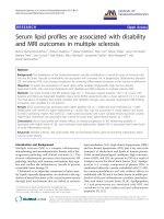
Figure 1 Simple correlation between age and serum biomarkers (sCD40L, G-CSF, GM-CSF, and TGF-a in pg/mL). The × axis is age. The Y
axis consists of log transformed sCD40L, G-CSF, GM-CSF and TGF-a. Simple correlation analysis was performed between age and the cytokines.
Age showed significant positive correlation with log transformed sCD40L (R = 0.257, P = 0.007) and log transformed TGF-a (R = 0.232, P =
0.015), whereas age showed significant negative correlation with log transformed G-CSF (R = -0.232, P = 0.016) and log transformed GM-CSF
(R = -0.249, P = 0.009).
Kim et al. Journal of Translational Medicine 2011, 9:113
/>Page 5 of 7
monocytes and other leukocytes often observed i n aged
populations [31].
One of the limitations of the study is the fact that we
could not mat ch the percentage of smokers in the study
population. However, we tried to minimize the influence
of smoking on the levels of cytokines by controlling for
smoking in the partial correlation analysis.
Conclusions
Aging was associated with significant increases in the
serum concentrations of sCD40L and TGF-a and signif-
icant decreases in the serum concentrations of G-CSF,
GM-CSF, and MCP-1. Future studies will focus on
understanding the significan ce of these age-rel ated
changes in circulating cytokines, chemokines, and other
biological markers a nd their potential contribution to
the development of various age-associated diseases.
Acknowledgements
This study was supported by a grant (2010-0020766) from the Happy Tech.
Program through the National Research Foundation of Korea (NR F) funded
by the Ministry of Education, Science and Technology, Republic of Korea.
Author details
1
Department of Laboratory Medicine and Cell Therapy Center, Yonsei
University College of Medicine, Seoul 120-752, Republic of Korea.
2
Division of
Cardiology, Yonsei Cardiovascular Center, Yonsei University College of
Medicine, Seoul 120-752, Republic of Korea.
3
Laboratory of Immunology and
Infectious Diseases, Graduate School of Medical Science and Engineering,
KAIST, Daejeon 305-732, Republic of Korea.
Authors’ contributions
All authors participated in the study design, result interpretation and in the
writing. HOK and HSK performed the analysis of the data and drafted the
manuscript. JCY and ECS participated in the design of the study and
performed the statistical analysis and SP conceived and designed the
experiments and wrote the paper. All authors read and approved the final
manuscript.
Competing interests
The authors declare that they have no competing interests.
Received: 1 June 2011 Accepted: 20 July 2011 Published: 20 July 2011
References
1. Franceschi C, Capri M, Monti D, Caruso C, Candore G, Vasto S, Oliveri F,
Marchegiani F, Sansoni P, Baggio G, Mari D, Passarino G, De Benedictis G:
Inflammaging and anti-inflammaging: a systemic perspective on aging
and longevity emerged from studies in humans. Mech Ageing Dev 2007,
128:92-105.
2. Park S, Kim HO, Kim HS: Aging associated decline in immunity and
therapeutic strategies to counteract it. Tissue Engin Regen Med 2011,
8:124-132.
3. Trzonkowski P, Szmit E, Mysliwska J, Mysliwski A: CD4
+
CD25
+
T-regulatory
cells inhibit cytotoxic activity of CTL and NK- cells in humans-impact of
immunosenescence. Clin Immunol 2006, 119:307-316.
4. Dejaco C, Duftner C, Schirmer M: Are regulatory T cells linked with aging?
Exp Gerontol 2006, 41:339-345.
5. Zanni F, Vescovini R, Biasini C, Fagnoni F, Zanlari L, Telera A, De Pede P,
Passeri G, Pedrazzoni M, Passeri M, Franceschi C, Sansoni P: Marked
increase with age of type 1 cytokines within memory and effector/
cytotoxic CD8+ T cells in humans: a contribution to the relationship
between inflammation and immunosenescence. Exp Gerontol 2003,
38:981-987.
6. Wick G, Grubeck-Loebenstein B: Primary and secondary alterations of
immune reactivity in the elderly: impact of dietary factors and diseas.
Immunol Rev 1997, 160:171-184.
7. Roubenolf R, Harris TB, Abad LW, Wilson PW, Dallal GE, Dinarello CA:
Monocyte cytokine production in an elderly population: effect of age
and inflammation. J Gerontol A Biol Sci Med Sci 1998, 53:M20-M26.
8. Ferrucci L, Corsi A, Lauretani F, Bandinelli S, Bartali B, Taub DD, Guralnik JM,
Long DL: The origin of age-related proinflammatory state. Blood 2005,
105:2294-2299.
9. Krabbe KS, Bruunsgaard H, Hansen CM, Moller K, Fonsmark L, Qvist J,
Madsen PL, Kronborg G, Andersen HO, Skinhoj P, Pedersen BK: Ageing is
associated with a prolonged fever response in human endotoxemia. Clin
Diagn Lab Immunol 2001, 8:333-338.
10. Woodward M, Rumley A, Lowe GD, Tunstall-Pedoe H: C-reactive protein:
associations with haematological variables, cardiovascular risk factors
and prevalent cardiovascular disease. Br J Haematol 2003, 122:135-141.
11. Friedewald WT, Levy RI, Fredrickson DS: Estimation of the concentration of
low-density lipoprotein cholesterol in plasma, without use of the
preparative ultracentrifuge. Clin Chem 1972, 18:499-502.
12. Beharka AA, Meydani M, Wu D, Leka LS, Meydani A, Meydani SN:
Interleukin-6 production does not increase with age. J Gerontol A Biol Sci
Med Sci 2001, 56A:B81-B88.
13. Ahluwalia N, Mastro AM, Ball R, Miles MP, Rajendra R, Handte G: Cytokine
production by stimuated mononuclear cells did not change with aging
in apparently healthy, well-nourished women. Mech Ageing Rev 2001,
122:1269-1279.
14. Baggio G, Donazzan S, Monti S, Mari D, Martini S, Gabelli C, Dalla Vestra M,
Previato L, Guido M, Pigozzo S, Cortella I, Crepaldi G, Franceschi C:
Lipoprotein(a) and lipoprotein profile in healthy centenarians: a
reappraisal of vascular risk factors. FASEB J 1998, 12:433-437.
15. Goetzl EJ, Huang MC, Kon J, Patel K, Schwartz JB, Fast K, Ferrucci L,
Madara K, Taub DD, Long DL: Gender specificity of altered human
immune cytokine profile in aging. FASEB J 2010, 24:3580-3589.
16. Zhu S, Patel KV, Bandinelli S, Ferrucci , Guralnik JM: Predictors of
interleukin-6 elevation in older adults. J Am Geriatr Soc 2009,
57:1672-1677.
17. Antoniades C, Bakogiannis C, Tousoulis D, Antonopoulos AS, Stefanadis C:
The CD40/CD40 Ligand system: Linking inflammation with
atherothrombosis. J Am Coll Cardiol 2009, 54:669-677.
18. Shonbeck U, Libby P: CD40 signaling and plaque instability. Circ Res 2001,
89:1092-1103.
19. Chen C, Chai H, Wang X, Jiang J, Jamaluddin MS, Liao D, Zhang Y, Wang H,
Bharadwaj U, Zhang S, Li M, Lin P, Yao Q: Soluble CD40 ligand induces
endothelial dysfunction in human and porcine coronary artery
endothelial cells. Blood 2008, 112:3205-3216.
20. Derynck R: Transforming growth factor alpha. Cell 1998, 54:593-595.
21. Pike LJ, Marquardt H, Todaro GJ, Gallis B, Casnellie JE, Bornestein P,
Krebs EG: Transforming growth factor and epidermal growth factor
stimulate the phosphorylation of a synthetic, tyrosine-containing
peptide in a similar manner. J Biol Chem 1982, 257:14628-14631.
22. Booth BW, Smith GH: Roles of transforming growth factor-alpha in
mammary development and disease. Growth Factors 2007, 25:227-235.
23. McClain DA, Paterson AJ, Roos MD, Wei X, Kudlow JE: Glucose and
glucosamine regulate growth factor gene expression in vascular smooth
muscle cells. Proc Natl Acad Sci USA 1992, 89:8150-8154.
24. Buzzeo MP, Yang J, Casella G, Reddy V: Hematopoietic stem cell
mobilization with G-CSF induces innate inflammation yet suppresses
adaptive immune gene expression as revealed by microarray analysis.
Exp Hematol 2007, 35:1456-1465.
25. Baldridge MT, King KY, Goodell MA: Inflammatory signals regulate
hematopoietic stem cells. Trends Immunol 2011, 32:57-65.
26. Serbina NV, Pamer EG: Monocyte emigration from bone marrow during
bacterial infection requires signals mediated by chemokine receptor
CCR2. Nature Immunol 2006, 7:311-317.
27. Tsou CL, Peters W, Si Y, Slaymaker S, Aslanian AM, Weisberg SP, Mack M,
Charo IF: Critical roles for CCR2 and MCP-3 in monocyte mobilization
from bone marrow and recruitment to inflammatory sites. J Clin Invest
2007, 17:902-909.
28. de Lemos JA, Morrow DA, Sabatine MS, Murphy SA, Gibson CM, Antman :
Association between plasma levels of monocyte mhemoattractant
Kim et al. Journal of Translational Medicine 2011, 9:113
/>Page 6 of 7
protein-1 and long-term clinical outcomes in patients with acute
coronary syndromes. Circulation 2003, 107:690-695.
29. Inadera H, Egashira K, Takemoto M, Ouchi Y, Matsushima K: Increase in
circulating levels of monocyte chemoattractant protein-1 with aging. J
Interferon Cytokine Res 1999, 19:1179-1182.
30. Gerli R, Monti D, Bistoni O, Mazzone AM, Peri G, Cossarizza A, Di
Gioacchino M, Cesarotti ME, Doni A, Mantovani A, Franceschi C, Paganelli R:
Chemokines, sTNF-Rs and sCD30 serum levels in healthy aged people
and centenarians. Mech Ageing Dev 2000, 121:37-46.
31. de Martinis M, Modesti M, Ginaldi L: Phenotypic and functional changes
of circulating monocytes and polymorphonuclear leucocytes from
elderly persons. Immunol Cell Biol 2004, 82:415-420.
doi:10.1186/1479-5876-9-113
Cite this article as: Kim et al.: Serum cytokine profiles in healthy young
and elderly population assessed using multiplexed bead-based
immunoassays. Journal of Translational Medicine 2011 9:113.
Submit your next manuscript to BioMed Central
and take full advantage of:
• Convenient online submission
• Thorough peer review
• No space constraints or color figure charges
• Immediate publication on acceptance
• Inclusion in PubMed, CAS, Scopus and Google Scholar
• Research which is freely available for redistribution
Submit your manuscript at
www.biomedcentral.com/submit
Kim et al. Journal of Translational Medicine 2011, 9:113
/>Page 7 of 7